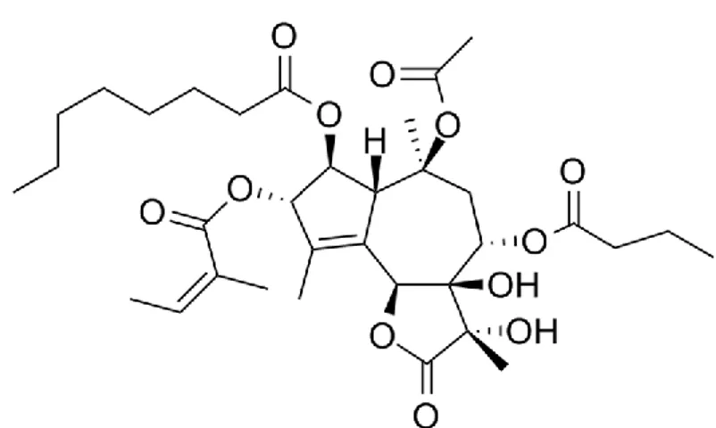
FACOLTÀ DI SCIENZE MATEMATICHE, FISICHE E NATURALI
Corso di Laurea Specialistica in Scienze e Tecnologie Biomolecolari
“Study on the role of protein deiminases in the
nervous system”
CANDIDATA RELATORI
Valentina Millarte Dott.ssa Patrizia Ferretti
Prof.ssa Renata Batistoni
ANNO ACCADEMICO 2010/2011
MARZO 2011
INDEX
INDEX ………..pag 1 ABBREVIATIONS………...pag 5 Summary……….pag7 Introduction ………...pag 9 Acetylation………..pag 10 Deacetylation………..pag 11 Methylation……….pag 11 Demethylation……….pag 112. PADI gene family and citrullination………...pag 12 3. Physiological roles of citrullination ………pag 15
3.1 Nuclear substrates………pag 15 3.2 Cytoskeletal proteins………...pag 16 3.3 Myelin basic protein……….pag 18
3.4 Apoptosis………...pag 18 4. Citrullination and disease……….pag 20
4.1 Rheumatoid arthritis……….pag 20 4.2 Multiple sclerosis………..pag 22 4.3 Alzheimer’s disease………..pag 24 4.4 Psoriasis………....pag 24 4.5 PAD of a pathogen potential virulence factor………..pag 25
4.6 Tumors...pag 25
Objectives………....pag 28 Materials and methods……… ....pag 31
1. Cell cultures and media………..pag 34 1.1 Chicken midbrain cells………...pag 34 1.2 Proliferating medium………..pag 34 1.3 Differentiating medium………..pag 34 1.4 LAN5 cells………..pag 34 2. Cell passaging………..pag 35 3. Treatments………....pag 35 3.1 Cl-amidine………...pag 35 3.2 Thapsigargine………...pag 37 3.3 Pifithrin………....pag 37 4. Chick embryos collection………...pag 38 5. RNA analysis: Extraction and Reverse Transcription………..pag 39 5.1 RNA extraction……….pag 39 5.2 RNA quantification………...pag 40 5.3 Reverse Transcription………....pag 40 6. Real Time PCR……… .pag 40 6.1 Primer designing……….pag 40 6.2 Real time PCR……….pag 41 6.3 Agarose Electrophoresis………..pag 42 7. Growth assays………...pag 42 7.1 Methylene blue assay……….……….pag 43 7.2 Time-lapse analysis after pifithrin treatment………..pag 44 9. Cell death and survival………....pag 45 9. Immunocytochemistry………...pag 45 9.1 Immunofluorscent stainings...……….pag 46
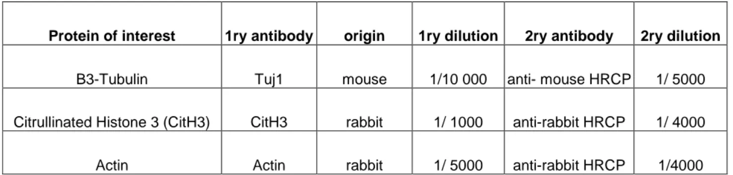
9.2 Summary of Antibodies and conditions ………….………pag 47 10. Western blotting……….pag 49 11.1 Protein extraction……….pag 49 11.2 Bradford standard curve method and protein measurement…….pag 49 11.3 SDS- PAGE Electrophoresis physical bases………...pag 50 11.4 SDS- PAGE electrophoresis protocol…….………....pag 52 11.5 Protein transfer..….………..pag 52
11.6 Blocking………...pag 53 11.7 Ibridization with primary and secondary antibodies …………...pag 53
11.8 Development and detection by Enhanced chemiluminescence ...pag 54 RESULTS………..pag 55 1. Chick embryo development and Pad3 Expression Pattern………...pag 56 2. PAD3 and β3-Tubulin expression in chick E7 cells………...pag 57 3 Methylen Blue assay dose-dependent curves and statistic………..pag 60 3.1 Thapsigargine in Proliferating medium………pag 62 3.1.1 t Test……….pag 62 3.1.2 ANOVA-1………pag 63 3.1.3 Dose dependent curve………...pag 63
3.2 Cl-amidine in Proliferating medium………..….pag 64 3.2.1 t-Test……….pag 64
3.2.2 ANOVA-1………pag 65 3.2.3 Dose dependent curve………..pag 65 3.3 Cl-amidine in Differentiating medium….………..pag 66 3.3.1 t-Test……….pag 66 3.3.2 ANOVA-1………pag 67
3.3.3 Dose dependent curve………….………pag 67 4 Effect of pifithrin on proliferation of chick MBcells…………...…….pag 68 4.1 Proliferating medium………pag 69 4.2 Differentiating medium……….pag 70 5 Cl-amidine prevents cell-death in thapsigargine treated cells…………....pag 70 5.1 Cell death and survival………...pag 70 5.2 Tuj1 expression in control and treated cells………...pag 71 6 PAD3 and CitH3 in immunocytochemistry………...pag 73 6.1 PAD3 and CitH3 in chick in P12……….pag 74 6.2 PAD3 and CitH3 in chick in P12……….pag 75 7 Pad3 transcript with time in culture………...pag 77 8 Western blotting: PAD3 expression in chick cells………pag 78 9 LAN5 immunostainings: PAD3, PAD4 and CitH3………pag 80 10 PCR in LAN5 cells: PADI 1-4 and p21………..pag 82 11 Western blotting: CitH3 amount in LAN5 cells……….pag 84 12 Cl-amidine effect on cell adhesion………..pag 84 CONCLUSION..……….pag 86 FIGURES AND TABLES………....………...pag 89 REFERENCES...pag 93
ABBREVIATIONS
Ab antibody
ACPA anti-citrullinated protein antibodies Arg arginine
bp base-pair
CitH3 citrullinated histone 3 Cl-am chlore-amidine
CNS central nervous system
cPAD chick peptidyl arginin deiminase Cys cysteine
DMSO dimethyl sulfoxide
E11 embryo at day 11 of development E15 embryo at day 15 of development EtBr ethidium bromide
FBS fetal bovine serum
GFAP glial fibrillary acidic protein H1 histone 1
H2A histone 2A H2B histone 2B H3 histone 3 H4 histone 4
HDAC histone deacetylases HLA human leukocyte antigen
HS horse serum Lys lysine
MBP myelin basic protein MS multiple sclerosis
NAWM normal appearing white matter P12 passage 12
P30 passage 30
PAD peptidyl (or protein) arginin deiminase PBS Phosphate Buffered Saline
PFA paraformaldehyde PI isoelectric point
PVDF polyvinylidene fluoride RA rheumatoid arthritis
SAHA suberoylanilide hydroxamic acid SDS Sodium Dodecyl Sulphate TAE Tris-Acetate EDTA
TBST Tris Buffered Saline Tween Thaps thapsigargin
"Study on the role of protein deiminases in the nervous system"
Citrullination (deimination) is the conversion of protein-bound arginine to citrulline. Citrullination is catalyzed by a family of calcium-dependent enzymes, the peptidylarginine deiminases (PADs). In mammals there are five PADs (PAD1, PAD2, PAD3, PAD4 and PAD6), whereas only 3 are found in chicken (PAD1-3). PAD isoenzymes are widely distributed in mammalian tissues and several studies suggest that citrullination occurs in extreme conditions such as during apoptosis, and during differentiation, when there is an increase in intracellular calcium concentration. Citrullination of different PAD target proteins has been associated with certain diseases, such as Alzheimer's disease, multiple sclerosis, rheumatoid arthritis and psoriasis. In humans, citrullination might be an early marker in neurodegenerative diseases.
The role of PADs and citrullination in some human diseases is poorly understood and the physiological roles of PADs have yet to be fully investigated. At present citrullination is believed to play a role in myelin sheath formation and during keratinocytes terminal differentiation. Recent studies in mouse have suggested that PAD4 regulates histone methylation at the p21/WAF1/CIP1 promoters in a p53-dependent manner.
The group of PF at UCL Institute of Child Health in London has identified PAD3 among calcium-dependent molecules differentially regulated in response to spinal cord injury at stages of development permissive (E11) and non-permissive (E15) for regeneration in chick embryos. Consistent with the up-regulation of PAD3 in spinal cords injured at E15, more extensive citrullination was observed after injury at this stage. This paralleled high apoptosis and significant tissues loss in injured E15 spinal cords. Following treatment at the time of injury with a PAD inhibitor, Cl-amidine, the secondary injury response in E15 spinal cord was greatly reduced.
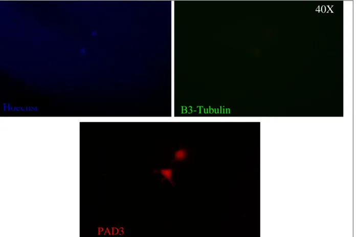
The aim of my project was first to see if it was possible to model the injury response in neural cells in vitro to eventually establish a model for studying PAD, citrullination and injury response in human neural cells and secondly I tried to better characterize expression pattern and possible role(s) of PAD, focusing mainly on PAD3, in neural cell death and survival initially using neural progenitor cells derived from E7 (embryonic day 7) chick midbrain (cMB cells),which were shown to express PAD3 both at the protein and mRNA level and subsequently in a human neural tumor cell line, the neuroblastoma cell line LAN5.
To this purposes we first studied the effects of PAD inhibition, using Cl-amidine, and PAD activation, using thapsigargine to raise intracellular calcium, on expression and cellular localization of PAD and on citrullination of a PAD target, histone 3 (H3) in cMB cells. In some experiments chick midbrain cells from E7 embryos were treated also with the p53 inhibitor, pifithrin, to assess whether p53 is required for PAD activity.
Thapsigargin treatment induced significant cell death, but this was reduced by pretreating the cells with Cl-amidine. Reduction of cell death upon PAD inhibition parallels the in vivo finding in the injured chick spinal cord.
Significantly, PAD3 mainly localized to the nucleus in cMB cells at passage 12 following thapsigargine treatment, whereas it remained largely cytoplasmatic in controls and cells pre-treated with Cl-amidine. Citrullinated H3 (CitH3) was not restricted to the nucleus in thapsigargine-treated cells while it is definitely nuclear in control and Cl-amidine treated cells. These results suggest that the PAD-associated apoptotic effect is likely due to PAD activity in the nucleus and that increased citrullination of H3 results in its export from the nucleus. As cMB cells were found to become senescent and die around passage 30 (P30), we investigated whether expression of PAD3 and CitH3 changed with time in culture. At P30 PAD3 was largely nuclear both in control and treated cells (thapsigargine/Cl-amidine), and CitH3 distribution resembled that observed at P12 following
amidine-treatments at P30, unlike at P12 where PAD3 localized in the nucleus only in thapsigargine-treated cells. Western blotting showed no differences in the amount of CitH3 among the different treatments. These results suggest an increased nuclear activity of PAD3 and increased turnover of CitH3 with aging that may be associated with cellular senescence and death.
As certain tumor cell lines were reported to express PADs, we assessed expression of PAD3 and PAD4, another PAD that can localize to the nucleus, and the response to thapsigargine and Cl-amidine treatment in LAN5 cells. PAD3 and PAD4 appeared to be mainly perinuclear, and their expression and localization was not affected by any drug combination (assessed by immunocytochemistry). Western blotting and immunocytochemistry showed that also CitH3 amount and localization were not affected in treated cells. These results parallel to what observed in P30 chick cells.
In addition to an effect on cell death, PAD activity was found to play a role in cell adhesion. When plated in the presence of Cl-amidine, most cMB cells did not attach to the dish, but were able to do so upon removal of Cl-amidine. Therefore their adhesion ability rather than survival appear to be affected. When cells were grown in neural differentiation medium (serum-free) to study a possible role of PAD in differentiation, such recovery was not observed. However, Given that PAD3 expression was observed both in cell positive and negative for the neuronal marker,
ß3-tubulin, and that in the presence of serum Cl-amidine did not affect expression of ß3-tubulin, as indicated by immunocytochemistry and Western blotting, a role for PAD in neuronal differentiation does not seem to be likely.
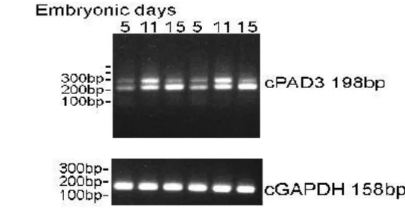
The expression pattern of PAD3 during chick embryonic development was assessed by mRNA analysis and RT-PCR in tissues dissected from embryos in various stage of development.
INTRODUCTION
Epigenetics refers to the several mechanisms modulating changes in gene expression independently of modifications of the primary DNA sequence . Epigenetics regulation of gene expression has been shown to have a critical role in the development of organs and tissues. Several factors contribute to epigenetic regulation of gene expression, such as microRNA, DNA methylation and post-translational modifications of nucleosomal histones (Gyorgy et al., 2006).
Posttranslational modifications of proteins are biological processes that give critical structures and functions to protein after synthesis and they are crucial because may alter physiological and chemical properties, folding, distribution, stability, activity and consequently the function of the target proteins, some of which being involved into diseases. Methylation, acetylation, phosphorylation, ubiquitination and citrullination are examples of posttranslational modifications having a significant role in epigenetics mechanisms control including the modifications of nucleosomal histones.
Nucleosomes are the basic unit of chromatin; they are formed by 150pb DNA wrapped around octamers histones, of which four major have been identified: H2A, H2B, H3 and H4.
H1 is an additional histone protein, distributed in the liker regions between two confinant octamers.
Structural studies of nucleosomes have revealed that histones are arranged as globular core whit the N-terminal tails projecting outward and for this reason the amino acids on the tails are the most easily accessible targets to enzymatic activities.
The major post-translational modifications occurring on nucleosomes are:
ACETYLATION : it is the transfer of acetyl groups to the lysine residues in the tail of nucleosomal histones. It is a reaction catalysed by family of enzymes called histone acetyltransferases (HAT) and functionally correlated with transcriptionally competent chromatin.
DEACETYLATION: it is the removal of acetyl groups, catalysed by a family of histone deacetylases (HDAC) and is functionally correlated with transcriptionally inactive chromatin.
METHYLATION: It is a process of adding one, two or three methyl groups to lysine and arginine on histone tails. Depending on the specific lysine and arginine, methylation may result in gene activation, such as the methylation on Lys 4 residue on histone H3 and on Arg 3 on histone H4 and Arg 17 oh histone H3, or may results in gene repression, such as methylation on Lys 9 and 27 on H3 and on Arg 8 on H3 and Arg3 on H4.
DEMETHYLATION: lysine and arginine methylation are reversible and can be removed by specific deiminases, such as human peptidylarginine deiminase 4 (PAD4) which convert arginine into citrulline. (Gyorgy et al., 2006).
Fig.1: histone modifications regulate chromatin structure and functions. From: “Research Summary “Epigenetic Histone Modifications in Cell differentiation and Cancer” Wang Y. Web site
PAD GENE FAMILY AND CITRULLINATION
Citrullination is the conversion of arginine to citrulline, a reaction of protein-deimination, catalysed by a family of calcium binding enzymes, the peptidylarginine deiminases. Recently, vertebrate PADIs were categorized into five isoenzymes (PAD1-6), based on their amino acid sequences, substrate, specificity and tissue location. Meanwhile, with the development of genomic project and advanced genomic prediction
approach, it was found that only the potential PAD, similar to PAD2 exist in fish ( Danio rerio,
Takifugu rubriepes and Tetraodon nigrouris) and amphibians (Xenopus leavis). Otherwise, there
are three isotypes ( cPad1-3) predicted in birds (Gallus gallus). In mammals, the chromosomal
Fig.2 Phylogenetic relationship of a part of PAD enzymes from representative species. From: ”Transcriptional regulation of peptidylarginine
deiminase expression in human keratinocyte, S.Ying et al”,2008. (A) PAD1 genes published or predicted from genomic researches. Following the
species evolution only one isotype of PAD exists in fishes (zebrafisch, Danio rerio) and amphibians (African clawed frog, Xenopus leavis,), three types in birds (chicken, Gallus gallus), and five types in mammals (mouse, Mus musculus; human, Homo sapiens). (B) Phylogenetic tree of the PADI gene. The unrooted phylogenetic tree was constructed from available PADI genes by ClustalW 1.83 on DDBJ website. Distancese were calculated according to Kimura method as default on the website (http://clustalw.ddbj.nig.acjp/top.j.html). The tree view was generated by TreeView (win32) 1.66 and the bar rapresents one substitution per 10 positions. GeneBank accession numbers of the cDNA sequen ces used in alignments are as follows; zpadi2: NM-200112: fpadi2: NM_001086900, cpadi1-3: XM_425729, XM_425730 and NM_205043, respectively; mPadi1-6: NM_011059, NM_008812, NM_011060, NM_011061 and NM_153106, respectively; hPADI1-6: NM_013358, NM_007365, NM_016233, NM_012387 and NM_207421, respectively. The records of these sequences are predicted using automated genomic tools by NCBI annotation.
localization of all five types of PAD genes has been determinated in mouse (Mus musculus), rat (Rattus norveicus), and human (Homo sapiens). As shown in Fig.2A it seems that the number of PAD isotypes increases by the biological evolution. Furthermore, the phylogenetic tree of the PADI genes constructed using ClustalW from available cDNA sequences data on NCBI, shows that the PAD2 gene is likely to diverge first from the common ancestor, the other PADI gene being derived by duplication during phylogenetic development (Fig2B). This presumption is also consistent with phylogenetic analysis of PAD amino acid sequences. Moreover, PADI2 is expressed in the broadest tissues distribution and this suggests that PAD2 retains an ancestral function and it is also speculated that the other PADI genes have evolved to be expressed in specific tissues and to target
specific substrates.
In humans PADI-1,-3,-4, and 6 are located at a single cluster which spans on about 334,7 kb region on chromosome 1p36.1. PADI-2 is the odd one-out: the largest gene in the family, it is located farther away from the other genes and transcribed in the opposite direction. This peculiar genomic organisation is conserved among species and the localisation, direction of the genes, the intragenic sequence lengths, exon-intron boundaries and the coding sequences are similar between mouse and man. Each of the PADI genes has the same exon/intron structure and the genomic structure of the PAD gene cluster is conserved as well in all mammals. (Ying S. et al.; 2008)
Fig3:genomic localization of PADI1-6:
The five PADI genes clusters are located on Chromosome 1p36.1 The organization and orientation are indicated by the arrows. From:
”Transcriptional regulation of peptidylarginine
deiminase expression in human keratinocyte,
PADI isoenzymes are widely distributed in mammalian tissues:
PAD1 is predominantly expressed in the epidermis and the uterus PAD3 in the hair follicles and CNS
PAD4 in neutrophils, eosinophils and oligodendrocytes. PAD6 has been detected in eggs, ovaries and early embryos
PAD2 is ubiquitous, expressed in skeletal muscles, spleen, brain, secretory glands etc. The proposed catalytic mechanism for these enzymes is showed below:
During the reaction the arginine is linked by the Cys residue of the PAD establishing a tetrahedral adduct with release of ammonia; the adduct is then cleaved by the nucleophilic attack of a water molecule that regenerates the Cys residue and forms the
keto-group.
In this way both ammonia group and the strong basic charge ( pI = 10,76) of arginine are lost and the resulting citrulline is a neutral amino acid that can influence and modify the charge distribution, ionic and hydrogen bond and give to protein a “more opened “ conformation which could have a critical biological function and which is easier attached by proteases.
Fig4: catalytic mode of peptidylarginine deiminase (PAD). From: ”Transcriptional regulation of peptidylarginine deiminase expression in human keratinocyte, Ying et al”, 2008 (A) The chemical reaction of PAD that catalyses the conversion of protein-bound arginine residues to citrulline redisues (protein deimination) in the presence of calcium ion. (B) A model of deimination effect. PAD action results modification which alter the charge of residues from positive to neutral, probably resulting in the unfolding of target proteins. It could induce dissociation of protein A/B complex, or the opposite, enable the association of protein C/D complex.
Several experiments reported in literature suggest that citrullination occurs only in extreme conditions, such as during apoptosis, and during differentiation, when the calcium concentration get higher than in physiological conditions within PADs are inactive.
Citrullination is also involved in pathological conditions such as rheumatoid arthritis and multiple sclerosis.
PHYSIOLOGICAL ROLES OF CITRULLINATION
Nuclear substrates
Nakashima et. al (2002) discovered that PAD4 is the isoenzyme associated with apoptosis; in granulocytes, during programmed cell death the intranuclear calcium concentrations are high enough to active PAD4 to its maximal activity that results in non-specific deimination histones that dramatically reduce their positive charges affecting nucleosomal stability. Citrullination of histones causes the nucleosomes to open up (weak connection among DNA and less basic proteins) and expose DNA to nucleases.
More recent studies (Li P. et al. 2010) show histone Arg methylation and Lys acetylation as modifications which cooperatively regulate the expression of p53-target genes in 293T cells. PAD4 citrullinates histone arginine and momomethyl-arginine residues thereby regulating histone Arg methylation; PAD4 serves as a p53 corepressor to regulate histone Arg methylation at the p53-target genes p21/WAF1/CIP1 promoter. Histone deacetylase (HDAC2) and PAD4 interact with p53 through distinct domains an simultaneously associate with the p21 promoter to repress gene expression.
After DNA damage, PAD4 and HDAC2 dissociate from p53 target gene promoters with a concomitant increase in histone Lys acetylation and Arg methylation at these promoters. p53 is known to recruit corepressors, such as HDAC1 and HDAC2, to regulate gene expression. Because
of PAD4 has not distinguishable motif for DNA binding and the inhibition of PAD4 increased p21 expression, it is assumed thatPAD4 might be targeted to specific gene promoters by transcription factors, such as p53 to modify histones.
The increase in p53-target gene expression after both Cl-amidine and SAHA (HDAC2 inhibitor) treatments suggest that a combination of these two inhibitors may more effectively inhibit cancer cell growth. ( P Li et al., 2010).
Cytoskeletal proteins
In the epidermis keratinocytes coexist in several states of differentiation to maintain the homeostasis of the tissue so as to assure its vital barrier function protecting the body against physical, chemical and biological insults.
In the epidermis, PAD1 has been immunodetected throughout the tissue, PAD2 in the superbasal living keratinocytes, and PAD3 in the granular keratinocytes and in the deeper corneocytes; PAD4 and PAD6 are not expressed.
Cytokeratin is an intermediate filament produced by keratinocytes and determines the consistency of skin, hair and nails. During terminal differentiation keratinocytes travel from the basal layers of the epithelium to the upper areas before undergoing cell death. During this process the intracellular environment is exposed to increasing calcium concentrations that gradually activate PAD1, 2 and 3 (Vossenaar et al, 2003). Three molecules are known to be deiminated in the epidermis: filaggrin and keratins K1 and K10. Citrullination enables these proteins to bind to each
Fig5 : immunoistochemical location of PAD1-3 in human epidermis. Bar=20μM. As shown, PAD1 was detected in the entire epidermis with an increasing intensity gradient from the basal to the granular layer. PAD2 was mainly detected in both the spinous and granular layers with a more intense staining of the latter, whereas PAD3 expression was shown to be restricted to the granular layer and lower stratum corneum.
From: ”Transcriptional regulation of peptidylarginine deiminase expression in human keratinocyte, S.Ying et al”, 2008
other by reducing their isoelectric point. Native cytokeratin and loricrin have very low affinity for each other, since both proteins are strongly basic; citrullinated keratin indeed, binds well to loricrin and to desmoplakin (desmosomal protein that helps the keratin matrix to extend transcellulary). Keratin citrullination has also been implicated in the pathomechanisms of psoriasis.
Vimentin is an intermediate filament expressed by various cells. The head domain of vimentin contains many β-turns that could easily be deiminated. Both phosphorylation and deimination reduces the isoelectric point of the head domain of vimentin; as result, the protein loses its ability to polymerise. During apoptosis when the intracellular calcium concentration is very high, activation of PAD enzymes results in the complete loss of vimentin intermediate filament network through the high rate of head domain deimination. This mechanism could play a role in the morphological changes associated with apoptosis (Asaga et al. 1998)
Another cytoskeletal protein is glial fibrillary acid protein (GFAP), which is specific to astrocytes the major intermediate filament in these cells. GFAP exists in various isoforms, and some of them are citrullinated and present in the brain of adult rats. The physiological role of GFAP deimination is still unknown, however, during kainate acid or hypoxia induced neurodegeneration, both the total amount of GFAP and its level of citrullination increased in the astrocytes.
PAD2 protein is found in vivo in astrocytes, microglial cells and in vitro in oligodendrocytes; normally PAD2 seems to become activated during neurodegeneration (under hypoxic conditions, the amount of PAD2 mRNA is increased).
GFAP deimination is characteristic for certain diseases, such as Alzheimer’s in humans and experimental autoimmune encephalomyelitis in mice (EAE). This raises the possibility that citrullination could be an early marker in neurodegenerative diseases. (Citrullination: A
Myelin basic protein
A key element in myelin sheat formation is the lipid-protein interaction based on ionic interactions between negatively charged ( phosphatidyl-serine and sialic acid containing gangliosides) and the basic proteins (MBP, lipophilin). Any alteration of the isoelectric point of these proteins can cause dramatic changes in the interaction with lipids ( Boggs et al. 1999).
MBP is synthesised in various isoforms that later undergo several posttranslational modifications, such as deimination, deimination, sulphoxyde-formations, methylation and phosphorilation (Pritzker et al., 2000). Citrullination of MBP reduces the interactions with the negatively charged phosphatidylserines due to the loss of basic residues. The ratio of deiminated MBP/ total MBP is crucial in the physiological function of CNS; native MBP contains several arginines (not deiminated) and forms very tight and compact myelin sheaths. These can reorganise only slowly. Citrullinated MBP is not able to form such compact sheaths (Beniac et al, 2000), on the hand, the lipid complex formation is more rapid. The MBP Cit/ total MBP ratio changes amazingly in postnatal life: under 2 years of age nearly all MBP is deiminated. Above 4 years, the ratio is only 18% and this ratio remains constant in adults. Changes of MBP-Cit/ total MBP correlate with the high plasticity of the brain of a young child. Similarly, the caudal areas of CNS with more rudimentary functions contain more citrullinated MBP ( Nicholas et al 2003).
Citrullination of MBP has been implicated in the pathomechanism of multiple sclerosis. (Gyorgy et al. 2006).
Apoptosis
PAD3 is among calcium-dependent molecules differentially regulated in response to spinal cord injury at stages of development permissive (E11) and non-permissive (E15) for regeneration in chick embryos. Consistent with differences in up-regulation of PAD3 in spinal cord injured at E11 and E15, more extensive citrullination was observed at the latter, non regenerative stage; by 24
hours, extensive citrullination as well as apoptosis and significant tissues loss is visible in injured E15 spinal cords (Fig 6 Fig7). The chick spinal cord can regenerate following injury until advanced developmental stages. It is conceivable that changes in stem/progenitor cell plasticity contribute to the loss of this capacity, which occurs around E13: increased proliferation in the grey matter and up-regulation of transitin expression following injury at E11, but not E15, suggested high levels of plasticity within the E11 spinal cord progenitor population that are lost by later stages. (Whalley et al. 2009)
Figure7: Detection of apoptotic nuclei by TUNEL. From: “Changes in response to spinal cord injury with development:vascularisation, hemorrhage and apoptosis”; Whalley et al. 2006
(A-D) and Hoechst nuclear staining (E-F) in spinal cord injured at regeneration competent (E11), and incompetent (E15) stages of development. A) E11 spinal cord injury (indicated by arrow), 24 hours after surgery: TUNEL positive cells (green) are located at the immediate injury site, but do not extend more than 500 mm cranial or caudal to the lesion. B) E11 spinal cord injury 3 days after surgery: no positively labeled cells are present. The arrow points to the site of injury. C) E15 spinal cord injury 24 hours after surgery: many TUNEL positive cells are present, extending for distances of several millimetres along the spinal cord. D) E15 spinal cord injury 4 days post lesion: TUNEL positive cells are still abundant within the cavity (asterisk) of the injured cord, and to a lesser extent in the surrounding regions. E-F) E15 spinal cord injury 24 hours after surgery: examples of pycnotic nuclei (arrows) indicative of apoptotic cell death are shown at a high magnification. Scale bars = 500 µm in A-C, 100 Figure6 : analysis of apoptosis after injury of E15 spinal cord. Changes in progenitors populations and ongoing neurogenesis in the regenerating chick spinal cord; K Whalley et al. 2009A) H&E spinal cord 12 hours after injury and treatment with PBS. A’) Adjacent sections from the same spinal cord stained by TUNEL.
A
A’
Fig6 : analysis of apoptosis after injury of E15 spinal cord. From: “Changes in response to spinal cord injury with development: vascularisation, hemorrhage and apoptosis”; Whalley et al. 2006 A) H&E spinal cord 12 hours after injury and treatment with PBS. A’) Adjacent sections from the same spinal cord stained by TUNEL.
Intracellular calcium increase following neural damage plays a key role in secondary injury response. At non-regenerating stages treatment with calcium chelator after spinal cord injury, reduces apoptosis and cavitation of the chick spinal cord. (Lange et al; 2011 submitted).
Cl-amidine is a specific PAD inhibitor; following Cl-amidine treatments at the time of injury, secondary injury response in E15 spinal cord is greatly reduced with decreased apoptosis and cavity size.
CITRULLINATION AND DISEASE
Citrullination is also associated with several pathological conditions.
Rheumatoid arthritis
Rheumatoid arthritis is a chronic autoimmune disease characterised by symmetric inflammation of the peripheral synovial joints. Initially, the inflamed joints are painful and swollen and later, if not treated , the inflammation may lead to cartilage and bone destruction and could result in disability. The serum proteins and synovial fluid samples of patients with RA contain high concentration of autoantibodies against various targets as collagen type 2, aggrecan, heat shock proteins and some glycoproteins (Firestein, 2003) but these autoantibodies are not highly specific to RA. However, there are two autoantibody families that seem to be highly characteristic of the disease: Rheumatoid Factor (IgM antibody against human IgG Fc portion) and the family of anti-citrullinated protein antibodies (ACPA). Several experiments carried out by Masson-Bessiére et al., have identified the antigen recognised in the joints, by anti-keratin antibodies (AKA), anti-filaggrin antibodies (AFA) and APF (that belong to the ACPA family of antibodies), as deiminated α and β chains of fibrin (Masson-Bessiére et al. 2001). Fibrin plaques are frequently found in the synovial tissue. Under physiological circumstances small amounts of fibrinogen (FBG) and other pro-coagulant proteins
can penetrate the capillary wall and travel to the interstitium, where they can be cleaved to fibrin peptides and fibrin monomers producing local plaques. These microplaques are then broken down by enzymes or may undergo endocytosis by macrophages. There is a sensitive balance between the presence and absence of fibrin molecules in the interstitium (balance of coagulation and fibrinolysis), which can be defective under pathological circumstances such as in RA. Citrullination appears to be the key element in shifting this balance. Polymerised fibrin is degraded by plasmin that cleaves near basic amino acid residues (Lys and Arg). The disappearance of the arginine by citrullination reduces the number of cleavage sites, hence increasing the quantity of polymer fibrin (Sebbag et al., 2004). Citrullination of antithrombin reduces its ability to inhibit thrombin, leading to a higher speed of coagulation (Chang et al., 2005). Citrullination of fibrin also renders the molecule antigenic, recognised by ACPA. The immune response results in increased plasma exudation and endothelial cell contraction. This in turn, increases the amount of FBG and other procoagulant protein in the interstitium (Rubin & Sonderstrup 2004). It has been suggested that smoking, vibration, exposure to mineral dust and injury increase the risck of developing RA (Aho & Heliovaara, 2004; Klareskog et al., 2006; Olsson, et al., 2004). These environmental effects often induce cell death, thus necrosis could possibly play a role in the aetiology of RA (Van Vernooij & Pruijn, 2000). During necrosis the integrity of the cell membrane is lost, and the cytoplasm containing enzymes like PAD, is released from the cell. The high interstitial concentration of Ca2+ can initiate the citrullination process. It has been shown that vimentin can be deiminated under these circumstances (Asaga et al., 1998). Its peptide fragments could be presented to T-helper cells by HLA molecules inducing a specific immune response (Hill et al., 2003). Synovial citrullinated proteins seem to be specific for RA and it has also been demonstrated that PAD2 expression is higher in RA joints then in controls and that both up-regulation of PADI2 gene and activation of PAD2 are important in the pathogenesis of RA (De Rycke et al., 2005). Recent studies suggest that citrullinated collagen types 1 and 2 are also target for ACPA (Burkhar et a., 2005; Suzuki et al.,
2005). Citrullination of a self-antigen breaks immuno tolerance and collagen, as a key joint antigen, contributes to RA pathogenesis, because citrullinated forms of collagen type 2 increases immunogenicity and arthritogenecity. It was also shown that the severity of the arthritis correlates with PAD4 expression (in the infiltrating mononuclear cells) and with the amount of citrullinated collagen. Most importantly, it was also demonstrate that clinical signs of arthritis preceded the presence of citrullinated proteins and PAD4 expression. This strongly suggests that citrullination is more likely a consequence rather than a cause of joint inflammation. However, the pronounced antibody response against citrullinated joint antigens could contribute to the progression of the autoimmune inflammation.
Multiple sclerosis
Multiple sclerosis (MS) is a severe autoimmune disease that affects myelin sheaths in the CNS. The neurons of the CNS gradually lose their myelin sheat synthesised by oligodendroglial cells. As a result, electrical conduction is disturbed. The disease eventually causes paralysis and death. Citrullination plays a key role in the pathogenesis of MS. Current knowledge attests that MS is caused mainly by overcitrullination of the MBP (Moscarello et al., 1994; Vossenaar et al., 2003; Wood et al., 1996). There is an increase in both the overall ratio of MBP-Cit/total MBP and the number citrullines within the MBP-Cit; in MS the brain resembles an ontogenetically earlier state, since young children have similar MBP-Cit/total ratios. Citrullination reduces the positive charge of the protein, lowering its affinity to the negatively charged myelin phospatidyl-serine residues. It has been shown in vitro that the amount of citrulline residues in MBP negatively correlates with its lipid-aggregating ability (Mastronardi et al., 1996; Wood & Moscarello, 1989). Cathepsin D can degrade MBP more easily if it is citrullinated, as MBP-Cit has a more open structure (Pritzker et al., 2000). The protease can release an immunodominant peptide, the 44Phe-Phe89 peptide, and this way an autoimmune response can be elicited (Whitaker at al., 1980). Lymphocytes and other immune
cells infiltrate the nervous tissue and cause local inflammation, oxidative stress and nerve cell death or myelin sheath destruction.
Pritzker et al. proposed a mechanism for the increased citrullination in MS patients, claiming that methylation affects citrullination (Pritzker et al., 2000). MBP is a methylated protein, and if it is not methylated, no myelin sheath is formed. Methylation increases the hydrophobicity of MBP and reduces its ability to become citrullinated, as methylated MBP has a more condense structure then the unmodified version. It could be speculated that a reduced methyltransferase activity is responsible for the enhanced citrullination of MBP in MS. GFAP (glial fibrillary acid protein), an astrocyte-specific intermediate filament, also undergoes deimination. Nicholas et al., suggested that GFAP could be overcitrullinated in MS (Nicholas et al., 2004) and supported the hypothesis that citrullination plays a cardinal role in the development of MS.
As citrullination seems to be a key process in MS, a PAD2 inhibitor therapy trial is currently underway. A cytotoxic, chemotherapeutic taxol derivative, paclitaxel inhibits human PAD2 in vivo and in vitro (Pritzker & Moscarello, 1998). Administering paclitaxel to DM-20 mice, the demyelinisation process slows down and the CNS symptoms abate (Moscarello et al., 2002). Not only enhanced citrullination of myelin basic protein contributes to destabilization of the myelin membranein the CNS of multiple sclerosis (MS) patients: it has been recently report increased citrullination of nucleosomal histones by PAD4 innormal-appearing white matter (NAWM) of MS patients and in animalmodels of demyelination. Histone citrullination was attributableto increased levels and activity of nuclear PAD4. PAD4 translocation into the nucleus is attributable to elevated tumor nercrosis factor- (TNF) proteins which may derive fromastrocytes. In cell cultures of mouse and human oligodendroglial cell lines, PAD4 is predominantly cytosolic but TNF- treatment induces its nuclear translocation. Several experiments carried out on transgenic mice show that high citrullination of histones, consequent to PAD4 nuclear translocation is part of the process that leads to irreversible changes in oligodendrocytes and may contribute to apoptosis of oligodendrocytes in
MS (Increased Citrullination of Histone H3 in Multiple Sclerosis Brain and Animal Models of
Demyelination: A Role for Tumor Necrosis Factor-Induced Peptidylarginine Deiminase 4
Translocation ; Mastronardi et al., 2006).
Alzheimer’s disease
A recent report claims that patients with Alzheimer’s disease (AD) have significantly elevated rate of citrullination in their CNS, mainly in the hippocampus, which is the region of the brain most affected by the disease (Ishigami et al., 2005). Comparison of data from patients with AD with those obtained from control individuals has revealed that it was increased in the patients. One can hypothesise that during neurodegeneration a higher concentration of Ca2+ activates the citrullination process. The predominant over-citrullinated proteins include vimentin, MBP and GFAP.
Psoriasis
This disease is characteristic by an enormous mitotic activity in the human epidermis and the rapid cell proliferation results in abnormal cornification. Typical symptoms are itching, sensitivity of affected skin and the presence of red patches covered with silvery-white scales of dead skin. Citrullination is implicated in the phatomchanism of psoriasis, however the exact mechanism is unknown. Cytokeratin K1 has been shown to contain reduced amount of cytrullyl residues in the psoriatic epidermis (Ishida-Yamamoto et al., 2000). Interestingly, in a recent phase 2 pilot trial the PAD2 inhibitor paclitaxel (also implicated as a therapeutic agent in MS) has been demonstrated to have therapeutic activity in patients with severe psoriasis (Ehrlich et al., 2004).
Peptidylarginine deiminase of a pathogen potential virulence factor
Peptidylarginine deiminases are ancient enzymes that developed early in evolution (Vossenaar et al., 2003). This has been verified by the identification of a prokaryote PAD enzyme in
Porphyromonas gingivalis (McGraw et al., 1999). This enzyme shows little sequence similarly to
the human PAD enzymes, however, it can efficiently deiminate either peptidyl arginine or soluble arginine substrates (in a Ca2+ -independent manner). The enzyme plays a key role in the pathogenesis of periodontitis, a common disease caused by this bacterium (McGraw et al., 1999). During Arg conversion, ammonia is produced that contributes to the neutralisation of the local pH. The enzyme can inactivate anaphylatoxins, produced locally during complement activation, or by the cleavage of C5 by the bacterial RGPs protease (Wingrove et al., 1992). It can inactivate bradikinine and can regulate plasma-outflow (Imamura et al., 1994). It can also inactivate special anti-adhesive molecules produced by the host. For these reasons, bacterial PAD could be regarded as a potential virulence factor.
Tumors
Recently it has been found a relationship between PADI expression and certain tumors. Immunohistochemistry indicates that PAD4 had significant expression in many tumor tissues, especially various adenocarcinoma. Western blotting with anti PAD4 antibody and immunostaining with anti citrulline antibody confirm the expression of the enzyme in these tumor. Mostly, PAD4 seems to contribute to the disrupted apoptosis of tumors by caspase-mediated cleavage of cytokeratin, CK. Another important finding comes from double immunofluorescent labeling which detected co-location of PAD4 with CD34, a cell marker of heamatopoietic progenitor cells (HPC) in bone marrow and other normal tissues, as well as in some fibroblast-like cells at stroma region of tumors tissues. Because there are more CD34+ cells expressing PADI4 in tumour tissues then in normal tissues it is postulated that the development of PADI4-expressing tumour cells may be
associated with the abnormal proliferation of CD34+ stem cells or their progeny. (Chang et Han, 2006; Chang et Fang 2010). A work carried out from Wang et al. in 2010 suggests also an important role for PADI4 in ovarian cancer tumorigenesis that is under the regulation of estrogen. Not only PAD4 but also PAD2 is found in several tumors such as breast (mammary gland) tumor, colorectal tumor, gastrointestinal tumor, glioma, head and neck tumor, leukemia, ovarian tumor, pancreatic tumor, prostate cancer, skin tumor, soft tissue/muscle tissue tumor (data from Genes to System Breast Cancer Database, G2SBCD).
Neuroblastoma is the most common extracranial solid cancer in childhood and the most common cancer in infancy, with an annual incidence of about 650 new cases per year in the US. It is a neuroendocrine tumor, arising from any neural crest element of the sympathetic ganglia of the paraspinal region or the adrenal gland. Neuroblastoma in children under 5 years presents in the abdominal region and in infants under 1 year of age has an high incidence in the thoracic region. The most widely characterized cytogenetic alterations in neuroblastoma tumors, include the loss or rearrangement of the distal portion of the short arm of chromosome 1 (1p31-term) and amplification of the N-myc gene (Thiele, 1998) . Many neuroblastoma cell lines has been created to allow studies on protein expression and mRNA analysis. LAN5 cells belong to such neuroblastoma cell lines and as they comes from embryonic child tumor, I used LAN5 cell line in my project to investigate if there is a relationship between neural embryonic stem/progenitor-derived tumor and PADI expression like what found for PAD4 and CD34+ stem cells (Chang et Han, 2006; Chang et Fang, 2010).
At present it is hypothesised a two-regulation model for PADs: at an extremely high concentration of Ca2+ PAD is fully active and may lose substrate specificity; this could also happen extracellularly, if the concentration of Ca2+ were high enough. There may be a physiological
regulatory mechanism at low concentration of Ca2+ , possibly mediated by other proteins or PAD protein interactions; when PAD is active at a low-level of Ca2+ , it has the higher substrate- and arginine-specificity . At high level of Ca2+ , PAD can deiminate nearly any arginines of any proteins. This could lead to autoimmunity, since the highly variable deiminated proteins (in which arginines are citrullinated), may be recognised as neoantigens by the immune system.
OBJECTIVES
A number of calcium-dependent mediators of apoptosis are known to be activated in response to injury and contribute to secondary injury response. The screening carried out in the chick spinal cord has identified PAD3 as one of these factor (Lange et al; 2011 submitted).
Recent experiments on chick spinal cord show that extensive apoptosis and cavitation, rapid increase in citrullination and PAD3 up-regulation as consequences of treatment with thapsigargine (chemical able to rise cytosolic calcium concentrations). An earlier treatment with Cl-amidine can reduce the effects of thapsigargine with less apoptosis and cavitation then the control and that supports a putative role of PADs and citrullination during apoptosis, as indicated by the results following PAD inhibition. Because of PADs have been implicated in a number of neurodegenerative diseases, the identification of the role of PAD and the possibility of inhibiting its activity pharmacolgically may be important for the development of cures to reduce damage in humans.
As first aim of my project I investigated on the possibility to model the injury response in neural cells in vitro to eventually establish a model for studying PAD, citrullination and injury response in human neural cells. Secondly I tried to better characterize expression pattern and possible role(s) of PAD, focusing mainly on PAD3, during neuronal differentiation first and then in neural cell death and survival; the experiments were carried out initially using neural progenitor cells derived from E7 (embryonic day 7) chick midbrain (cMB cells),which were shown to express PAD3 both at the protein and mRNA level (Lange et al; 2011 submitted), and subsequently in a human neural tumor cell line, the neuroblastoma cell line LAN5. To this purposes my experiments focused on PAD3 protein and transcript, on β3-Tubulin expression and on CitH3 amount ,as histone3 has been shown to be the nuclear target of PAD (Li et al. 2010; Mastronardi et al., 2006).
MATERIALS AND METHODS
MATERIALS AND METHODS
I chose to work in chick cells from embryos at day7 because that is a good stage to collect enough tissue which is easier to dissect then younger embryos but still with a large neural precursor population.
As qualitative approach I set up immunostainings and observed the results on fluorescent microscope, while the quantification of the levels of Cit-H3 and β-3 tubulin has been performed by western blot. In the first experiments I tried to asses a possible relationship between PAD3 activity and neuronal development and differentiation. To this purposes PAD3 protein and β3-Tubulin expression have been studied at the same time by double immunostainings in cells kept in both proliferating and neuronal differentiating medium, while by PCR I analyzed Pad3 transcript in different tissues during chick embryonic development.
The second part of the experiments done, started with the study on the possibility to model the injury response in neural cells in vitro. I first studied the effects of PAD inhibition, using Cl-amidine, and PAD activation, using thapsigargine to raise intracellular calcium, on cell survival, and then on expression and cellular localization of PAD and on citrullination of histone 3 (H3) in cMB cells. In some experiments chick midbrain cells from E7 embryos were treated also with the p53 inhibitor, pifithrin, to assess whether p53 is required for PAD activity.
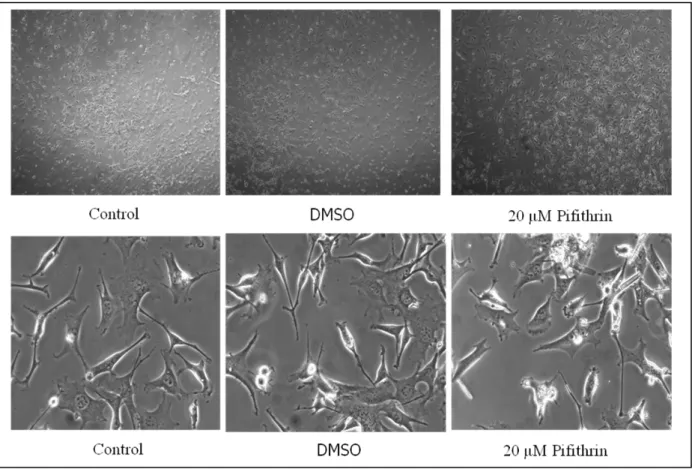
As first step I made dose-dependent curves to choose among different concentrations of Cl-amidine and thapsigargine the highest non-toxic one, and growth-assays for different pifithrin doses. Pifithrin is an inhibitor of p53 which has been used to see if the activity of PAD in chick MB cells could be dependent by p53 interaction, according to what has been shown for PAD4 in mice, or not ( Li et al., 2010).
Once the dose of each chemical has been chosen, I decided to use qualitative and quantitative approaches trying to figure out if those treatments gave differences in PAD3, b-3 tubulin and Cit-H3 amount in chick midbrain cells. As the results obtained excluded a role of PAD3 in cell differentiation, I studied if PAD3 may have an effect during apoptosis and senescence. To investigate whether expression of PAD3 and CitH3 amount may change with time in culture, PAD3 and CitH3 localization and response to the treatment was investigated also in chick cells at passage 30 (P30) as cMB cells were found to become senescent and die around P30.
As certain tumor cell lines were reported to express PADs, I assessed expression of PAD3 and PAD4, another PAD that can localize to the nucleus, and the response to thapsigargine and Cl-amidine treatment in human neuroblastoma cells, LAN5 .
In the experiments carried out I focused the attention only on PAD3 in chick midbrain cells, because this is the only PAD enzyme found in chick nervous system till now , and for PAD3 and PAD4 in LAN5 cells because in human cells these show nuclear localization.
I set up PCR both on chick cells and tissues and on LAN5 cells to check if these enzymes (PAD3 in chick, PADI1-4 in human) are really expressed (as results in previous western blot) and if there is a difference in the pattern of PAD3 expression during development and cell differentiation: to this purpose chick embryos in different stages were collected and dissected to isolate skin and different parts from the nervous system and PCR for Pad3 transcript has been repeated several times, trying to optimize the conditions of reaction for results as clear as possible.
1 Cell cultures and media
1.1 Chicken midbrain cells :
Chick midbrain cells from embryos in day 7 were grown in flasks in proliferating medium at 5% CO2 and 37°C as conditions of incubation. Trying to push these cells into neuronal differentiation a
differentiating medium has been used as well.
1.2 Proliferating medium:
10% FBS ( fetal bovine serum), 5% HS (horse serum), 1% penicillin-streptomicine, 1% glutamine in high glucose DMEM ( Dulbecco’s modified eagle medium).
1.3 Differentiating medium:
1% N2 , 1% penicillin-streptomicin, 1% glutamine in NeurobasalA medium. Cells were kept
differentiating minimum 3 days before doing the western blot analysis on β3-Tubulin amount.
1.4 LAN5 cells:
LAN5 cells are Neuroblastoma cell line characterized by cytogenetic alterations, including the loss or rearrangement of the distal portion of the short arm of chromosome 1 (1p31-term) and amplification of the N-myc gene. LAN5 cells are morphologically small, neuroblastic shaped and grow in cell aggregates. ( Neuroblastoma Cell Lines, Thiele 1998 ). They have been grown in Neuroblastoma proliferating medium at 5% CO2 and 37°C.
Neuroblastoma proliferating medium: 10% FBS, 1% penicillin-streptomicin, 1% glutamine in MEME (minimum essential medium eagle).
2 Cell passaging
Both cell cultures were split in new flasks containing fresh medium every time they reached 70% of confluence. This work has been done under antiseptic condition using laminar flow hood. After aspirating medium and the dead-floating cells with a glass pipette, cells attached on the flask surface has been washed 1 minute in 10x PBS. PBS has been aspirated and 1 ml ( T-25 cm2 flasks) or 2 ml thripsin ( T-75 cm2 flasks) added and left for 1 min at 37° C at 5% CO2 .
Thripsin has been neutralized adding 4 ml ( T-25 cm2 flasks) or 8 ml ( T-75 cm2 flasks) of proliferating medium , and detached cells have been harvested by aspiration of the mixture medium + thripsin. The harvested suspension has been placed in plastics tubes and spin out 5 min at 1000 rpm. Centrifugation share 2 phases: one pellet on the bottom of the tube and the supernatant on the top. Cells were harvested by aspirating the surnatant and resuspending the pellet in 1 ml of proliferative medium. Living cells concentration has been calculate by counting on emocytometer in which we transferred 10 μL of a solution consisting in 10 μL cell suspension and 90 μL trypan blue. About 1x10⁵ cells were plated in T-75 cm2 flask containing10 ml of medium.
3 Treatments
3.1 Cl-amidine
Cl-amidine ( N-α-benzoyl-N5-(2-chloro-1-iminoethyl)-L-ornithine amide ) is a specific irreversible time and concentration dependent inactivator of PAD; it is the most potent PAD inhibitor.
Mechanism of inactivation: binding of calcium to PAD triggers a conformational change that moves a Cys645 into a position that is competent for catalysis; thus, if Cl-amidine reacts with an active site residue, it preferentially inactives the calcium-bound form of the enzyme by > 10-fold. The
Inhibitory properties of Cl-amidine-mediated inhibition : calcium-dependent inactivation and irreversible nature of the enzyme-Cl-amidine complex.
The rate constants for the inactivation process KI, KINACT, KI / KINACT have been calculated using
the following non-linear progression curves:
[ Cit ] = vi ( 1-e Kobs^t ) Kobs
where vi is the initial velocity, Kobs is the apparent pseudo-first-order rate constant for inactivation
and [ Cit ] refers to the concentration of citrullination produced during the time course. By plotting Kobs versus [ Cl-amidine ] values for KINACT ( 2,4 +/- 0,2 min-1 ) and KI ( 180 +/- 33μM ) are
determinated, and the second-order rate constant , results to be KINACT / KI = 13000 M-1 min-1.
(Y. Luo et al.,2006 , Y. Luo et al. 2008” ).
3.2
Figure8: Two potential mechanisms of PAD inactivation. Mechanism 1 involves direct substitution of the halide, whereas mechanism 2 involves the formation of a tetrahedral intermediate, which first evolves into a three-membered sulfonium ring and subsequently rearranges to the thioether with the collapse of the tetrahedral intermediate. The letter mechanism is invoked to account for the poor leaving group potential of chloride or fluoride. From:” Inhibitors and inactivators of protein argine deiminase 4: functional and structural”, Luo . et al.,2006)
3.2 Thapsigargine
Is a non-competitive inhibitor of a class of enzymes known by the acronym SERCA, which stands for sarcoplasmic and endoplasmic reticulum Ca2+ ATPase. It is structurally classified as a sesquiterpene lactone and is a tumor promoter in mammalian cells. Thapsigargine raises cytosolic calcium concentration by blocking the ability of the cell to pump calcium into the sarcoplasmic and endoplasmic reticula which causes these stores to become depleted. Store-depletion can secondarily activate plasma membrane calcium channels, allowing an influx of calcium into the cytosol. Thapsigargine acts through inhibition of the endoplasmic reticulum calcium pump by recognition and interaction with a site found in all of the known intracellular-type calcium pumps (SERCA1, SERCA2a, SERCA2b, SERCA3). ( Lytton et al. 1991).
Figure 9: molecular structure of thapsigargine. From website: http://en.wikipedia.org/wiki/Thapsigargin
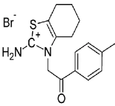
3.3 Pifithrin
Pifithrin (chemical name 2-(2-imino-4,5,6,7-tetrahydrobenzothiazol-3-yl)-1-p-tolyethenone hydrobromide) is a chemical inhibitor of p53. It is soluble in DMSO up to 20mg/mL. It is a reversible inhibitor of p53-mediated apoptosis and p53-dependent gene transcription such as cyclin G, p21/waf1, and mdm2 expression, and for this reason pifithrin is the lead compound for a novel
group of small molecules that are being developed for use as anticancer agents. Pifithrin inhibits p53-dependent apoptosis through an undeterminated mechanism. ( Hoagland et al., 2005).
Figure 10: molecular structure of pifithrin. From website http://en.wikipedia.org/wiki/Pifithrin
4 Chick embryos collection
Fertilized Leghorn eggs (Needle Farm, Cambridge, UK) were incubated at 37°C in a humidified forced flow incubator. Before incubation a small hole was made in the shell to create a window by which check chick embryos growth. All the procedures were approved under the Animals Scientific Procedures Act 1986.
At E3, E5, E7, E11, E13 and E15 stages a pair of fine forceps was used to make a small hole in the chorioallantonic membrane, carefully avoid major blood vessels. A glass hook, prepared by heating and bending a glass pipette, was pushed through the hole and used to lift the embryo gently towards the window. E5, E11 and E15 embryos were transferred in a Petri dish containing 1X PBS. Working under stereomicroscope brain and spinal cord were carefully dissected from E5 embryo usingfine forceps and a needle, and the same protocol has been used to dissect forebrain, midbrain, hindbrain, spinal cord and skin from E11, and E15 embryos. Each sample was quickly stored in eppendorf tube at -80°C until it was needed for protein and mRNA extraction.
The whole E3 and E7 embryos were stored at -80°C without previous dissection.
5 RNA analysis: Extraction and Reverse Transcription
RT-PCR was used to study Pad3 gene expression in chick cells at different developmental stages , Pad3 transcript in chick MBcells with aging, and in LAN5 cells PADI1-4 and p21 expression (keratinocytes used as human positive control).
5.1 RNA extraction
Total mRNA was extracted from each tissue, from LAN5 cells and from chick following the protocol of INVITROGEN life technologies:
1. tissues from E5, E11, E15 and the whole E3 and E7 embryos and the sample pellets from culturing were homogenized/ resuspended in 1 mL of TRIZOL ;
2. incubation of the homogenized samples for 5 minutes at room temperature to permit the complete dissociation of nucleoprotein complex;
3. adding 0,2 ml of chloroform per 1 ml of TRIZOL reagent. Tubes were shacked vigorously by hand for 15 seconds and incubated at 15 room temperature for 2 minutes. 4. centrifugation at 12000 x g for 15 minutes at 4 °C;
5. the aqueous phase was transferred to a fresh tube and mixed with 0,5ml of isopropyl alcohol to precipitate the RNA.
6. incubation of the samples for 10 minutes at 15 to 30°C and centrifugation at 12000 x g for 10 minutes at 4 °C;
7. RNA precipitate formed a gel-like pellet on the side and bottom of the tube. The supernatant was removed and the RNA pellet washed adding 1ml of 75% ethanol.
8. centrifugation at 7500 x g for 5 minutes at 4 °C.
9. drying of RNA pellet and resuspention in 30 μL of milliQ water RNAse free.
5.2 RNA quantification
mRNA quantification was performed using NanoDrop Spectrophotometer (1000) and the ND-1000 software. 2 μL aliquots of RNA were assessed for concentration and ratio (260/280) to check the quality of the samples. Following RNA quantification, aliquots of 1 μg RNA were made, transferred in new microtubes and stored at -80°C.
5.3 Reverse Transcription
For real time PCR, cDNA was produced in 20 μL from 1 μg of total RNA using the following protocol:
1 μg of RNA was mixed with 1 μl of random examer primers.
2. incubation for 10 minutes at 70°C, using program 58 set on the PCR machine.
3. 4 μl MLV-RT buffer, 2 μl dntp (10 microM), 1 μl MLV-RT enzyme, 1 μl RNA inhibitor and water RNAse-free were added to the RNA+ random examer primers samples in 20 μL of final volume.
4. reagents were mixed by vortex;
4 incubation for 1 hour at 42°C first and then for 10 minutes at 95°C 5 the resulting cDNA was stored at -80°C.
6 Real Time PCR
6.1 Primers designing
Chick’s Pad3 primer sequences were designed using Primer3 Output and BLAST softwares, and NCBI and Quantitative PCR Primer Databases.
As primers for human PAD1, PAD2, PAD3, PAD4 , p21 and GHDPH were used sequences published in the recent scientific article (Méchin et al., 2010).
The table below shows the primer sequences we used and the amplicon size.
Gene Specie Forward primer sequence Reverse primer sequence Exon/s Product size
Annealing Temp
PAD1 human 5’–cgccatcctctctgccctcttgcta-3’ 5'–ggtttttctgtccttgtttgtccac–3' 16 3'-UTR 571 bp 58
PAD2 human 5’–atgcaccttcatcgacgacattt–3’ 5’–tttcagcagggacagagtcgag–3’ 16 332 bp 54
PAD3 human 5’–cagagacaggcccctgaacgataa–3’ 5’–aagatggttccgccctgatctaa-3' 16 3'-UTR 483 bp 54
PAD3 human 5’-gcagagtgtgacatcattgacatcc-3’ 5’-gaccgcaccttctcctccag-3’ - 167bp 57
PAD4 human 5’–tcttgtgaatattgtggctccct–3’ 5’–agagcagaactgagtgtgcagtg–3’ 16- 3'-UTR 134 bp 56
GDPH human 5'-ccttcattgacctcaactacatggt-3' 5'-ctaagcagttggtggtgcaggt-3' - 488 bp 60
p21 human 5'-ctgggtgtgagccctgcgtg-3' 5'-tgaggcacagcgagccaacg-3' - 187 bp 57
Pad3 Gallus
gallus 5’ -ctccagcctcgactccttc- 3’ 5’- gcagccagtctgagaagagc- 3’ 11-12 198 bp 55
Gdph Gallus
gallus 5'-ccaggttgtctcctgtgact-3' 5'-cacaacacggttgctatatc-3' 12-13 158 bp 55
6.2 Real Time PCR
For each pair of primers I made a PCR solution consisting in 0,1 µL of Biotaq enzyme, 0,75 µL MgCl, 2 µL Buffer-Taq, 1 µL cDNA, 12,5 µL H20, 1 µL primer F, 1 µL primer R and 2 µL dntp in 20 µL total volume.
As there are differences in GC content, melting temperature Tm, length (number of nucleotides) and specificity on the target sequences between couple of primers, all PCR reactions have been run separately in different eppendorf tubes and the optimal reaction conditions for each kind of PCR have been studied and used.
6.3 Agarose Electrophoresis
Agarose electrophoresis was performed to visualize PCR products. This step allowed to determine if the mRNA previously reverse-transcripted and amplified, of a specific gene, was expressed in the cells of interest and if the resulting PCR products had the correct size.1% Agarose Gel was made in a glass becker by mixing 1,0 g of agarose in 100 ml TAE Buffer 1X (solution of Tris base, Glacial Acetic Acid and EDTA in distilled water). Agarose powder was melted in TEA by warming up the solution from 1 to 2 minutes in a microwave and after a rapid cooling of the solution by placing the becker under cold running water, 5 μL EtBr (Etidium Bromide) were added. The solution was then poured into gel tray pre-assembled into a casting chamber and the casting comb was placed in the appropriate slot. The gel was let sit 30 minutes until it became cool to touch and opaque in appearance and then completely wet with Running Buffer (solution of bromophenol blue orxylen cyanol, sucrose and water). Samples of cDNA kept in ice, were weighted by adding 5 μL Loading Buffer (solution of 30% w/v sucrose, Orange G and distilled water) and then carefully loaded in the wells after removing the comb from the solidified agarose gel. 5 μL Ladder were loaded in the first well. After loading, cDNA were run by the application of a constant current of 120 mV. The gel was placed under UV rays in a UV chamber to visualize PCR product
7 Growth assays
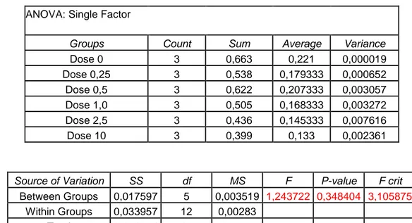
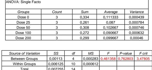
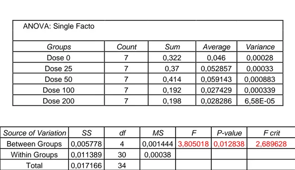
Before using the treatments previously described it has been necessary to choose the highest-non toxic concentration for each one. To this purpose several doses of thapsigargine and Cl-amidine has been tested by methylene blue assay which indicates the effect of each dose on cell survival. The concentration of pifithrin has been chosen by using a different approach, checking not only cell survival (as in methylene blue assay) but also cell proliferation by time-laspe microscopy. On the base of the results found the concentrations chosen were: 20 μM for pifithrin, 10 μM for thapsigargine, and 200μM for Cl-amidine.
7.1 Methylene Blue Assay
The methylene blue assay was adapted from that described in Oliver (1989). After culturing in 96 well plates chick cells from midbrain of embryo in stage 7, thapsigargine and Cl-amidine were added in different concentrations in cell mediums and the sample were stored at 4°C overnight. Three reply have been done for each concentration. The day after, cell mediums were drained using a multichannel pipette and each well fixed with 100μL of 4% PFA in 0,15 sodium chloride for 30 minutes. Fixative was removed by a prorating flick of the wrist into a sink and the surface of the plate inverted and patted dry with a towel. 100μL of methylene blue solution was added to each of the previously fixed wells for 30 minutes. The dye was removed by a prorating flick of the wrist as described before. The plate was placed upside down on a paper towel to remove the methylene blue. Excess methylene blue in each well was washed with 200μL of 0,01 M borate buffer by filling from left to right with a multichannel pipette, before licking into the sink as described previously. The second wash was performed by filling from right to left, to ensure that the washing time per well was the same on average. The first and second washes were repeated. The plate were then immersed