UNIVERSITY OF CATANIA
International PhD in Chemical sciences - XXIV cycle
S
YNTHESIS AND BIO
-
PHARMACOLOGICAL
ACTIVITY OF ANTISENSE
PHOSPHATIDYL
-
OLIGONUCLEOTIDES
Candidate: Dott. VALENTINA GRECO
Supervisor: Prof. Sebastiano Sciuto Coordinator: Prof. Corrado Tringali
Imagination is more important than knowledge….
Albert Einstein
Index
I
NDEX
List of abbreviations...6
Abstract...………...………...8
1. Introduction...10
1.1 Molecular mechanism of antisense oligonucleotides...10
1.2 Chemical modifications of native structure of ODNS – State of Art...13
1.3 Open question on the conjugation of biocompatible units to oligonucleotides...19
2. Aim of the work...21
3. Methods and results...24
3.1 Synthesis of the linker and solid support functionalization……... 28
3.2 Synthesis of 1,2 diacyl-sn-glycero-3-(2-cyanoethyl)-N,N diisopropylphosphoramidites………..30
3.3 Preparation of unprotected nucleoside phosphoramidites... 31
3.4 Synthesis of 5‟-O-(1,2-O-diacyl-sn-glycero-3-phosphoryl)- d(TGGCTTGAAGATGT)...32
3.5 Spectroscopic characterization of 5‟-O-(1,2-O-diacyl-sn-glycero-3-phosphoryl)-d(TGGCTTGAAGATGT)...36
Index
3.6 Melting temperature of “antisense” phosphatidyl-ODN / “sense”-
ODN duplex...43 3.7 Biological assay...46 4. Conclusive remarks………..………….……….50 5. Experimental...53 5.1 Synthesis of silyl-linker………..………53 5.2 Synthesis of 1,2-Diacyl-sn-glycero-3-(2-cianoethyl)-N,N- diisopropyl phosphoramidite...56 5.2.1 [1,2-dimyristoyl-sn-glycero-3-(2-cyanoethyl)-N,N- diisopropylphosphoramidite]………...57 5.2.2 [1,2-dipalmitoyl-sn-glycero-3-(2-cyanoethyl)-N,N- diisopropylphosphoramidite]………...58 5.2.3 [1,2-distearoyl-sn-glycero-3-(2-cyanoethyl)-N,N- diisopropylphosphoramidite].………...59
5.3 Deacylation of N-protected 3‟-deoxynucleoside phosphoramidite ...60
5.3.1 Debenzoylation of 5'-O-(4,4'-dimethoxytrityl)-N6-benzoyl-2'- deoxyadenosine-3'-O-(2-cyanoethyl)-N,N-diisopropylphosphoramidite……….60
5.3.2 Debenzoylation of 5'-O-(4,4'-dimethoxytrityl)-N6-benzoyl-2'- deoxycytidine-3'-O-(2-cyanoethyl)-N,N-diisopropylphosphoramidite……….………...…….61
5.3.3 Deacylation of 5'-O-(4,4'-dimethoxytrityl)-N2-isobutyryl-2'- deoxyguanosine-3'-O-(2-cyanoethyl)-N,N-diisopropylphosphoramidite………….………..…..62
Index
5.5 Characterization of 5‟-O-(1,2-O-diacyl-sn-glicero-3-phosphoryl)-
d(GGCTTCATGACCT)………...66
5.6 Synthesis of unmodified sense and antisense ODNs………...70
5.7 Melting temperature measurement by DSC...72
5.8 Biological assays...73
5.8.1 Evaluation of antisense activity on Neuroblastoma cells ...74
5.8.2 Viability tests (MTT assay)...76
L
IST OF
A
BBREVIATIONS
ACN AcOH AIDS APCI AP-CPG AS-ODNs BOPCl CMV COSY CPG DBU DEPC DMF DMT DNA DSC ESI HDL HOBt HPLC LCAA-CPG LDL Acetonitrile Acetic acidAcquired Immune Deficiency Syndrome Atmospheric-Pressure Chemical Ionization Aminopropyl-CPG
Antisense Oligodeoxynucleotides
Bis(2-oxo-3-oxazolidinyl)-phosphinyl chloride Cytomegalovirus
Correlation Spectroscopy Controlled Pore Glass
1,8-Diazabicycloundec-7-ene Diethylpirocarbonate
Dimethylformamide Dimethoxytrityl Deoxyribonucleic acid
Differential Scanning Calorimetry Electrospray Ionization
High Density Lipoprotein 1-Hydroxy-Benzotriazole
High Performance Liquid Chromatography Long chain amino alkyl- CPG
MeOH MF-ODN mRNA MS MTT NMP NMR PNA PS RNase H RP TBAF TEA THF TLC TOF Tm VEGF Methanol Morpholino Oligodeoxynucleotide Messenger Ribonucleic acid Mass Spectroscopy
3-(4,5-Dimethylthiazol-2-yl)-2,5-diphenyltetrazolium bromide 1-Methyl-2-pyrrolidone Nuclear Magnetic Resonance Peptide Nucleic Acid
Phosphorothioate Ribonuclease H Reverse Phase Tetrabutylammonium fluoride Triethylamine Tetrahydrofuran
Thin Layer Chromatography Time Of Flight
Melting Temperature
A
BSTRACT
Lipid conjugated oligonucleotides are of great interest in the field of antisense oligonucleotides used for functional genomics, gene target validation and therapeutic pourpose. Although various lipid conjugates of oligonucleotides have already been prepared, it was not possible until now to prepare phosphatidyl conjugates owing to an actual difficulty encountered in a direct attachment of the phosphatidyl group to oligonucleotides elongated on the solid phase by standard phosphoramidite chemistry procedures. Now, appropriately exploiting some synthetic opportunities, available in the recent literature for the preparation of oligonucleotides bearing residual base-labile side groups, we designed a synthetic path to obtain 5‟-O-phosphatidyloligonucleotides.
By applying this synthetic route we prepared some phosphatidyltetradecanucleotides all having the antisense sequence against the Vascular Endothelial Growth Factor (VEGF) gene, but differing from each other in their phosphatidyl moiety. This consisting of different fatty acyl residues, such as myristoyl, palmitoyl and stearoyl as well.
The newly synthesized phosphatidyloligonucleotides have been analyzed for their spectroscopic (NMR, MS) and chromatographic (HPLC) properties which confirmed the expected structure. The annealing features of these compounds have been investigated by Differential Scanning Calorimetry analysis .
1,2-O-Abstract
of VEGF at mRNA level in human Neuroblastoma cells. Both the phosphatidyl-tetradecamers were able to inhibit the expression of VEGF mRNA with an effective concentration significantly lower than was found, in parallel experiments, for the corresponding unmodified antisense oligonucleotide.
Introduction
1. I
NTRODUCTION
The backbone for all research within the field of biology lies within the “central dogma” which describe the basic processes occurring in living cell. Genomic DNA is transcribed into messenger RNA (mRNA) in the cell nucleus. The mRNA, which is then transported in the cytoplasm, codes for the subsequent transcription of protein (Fig. 1).
Many diseases are caused by over-expression of certain genes belonging to the same cell or of recombinant genes derived from viral infections in various cell types. Considering that the complete sequence of the human genome has been known since 2001 [1], the knowledge of the coding sequences of many genes would allow the suppression of the gene of interest by a set of gene manipulation techniques.
Antisense oligodeoxynucleotides (AS-ODNs) are molecules whose sequence is complementary to a codifying region of a gene which and so, they are able to inhibit gene expression. Using antisense DNA fragments to block the expression of selected genes, and thereby assess their function, is a powerful and currently used new tool. Conceptual simplicity, the possibility of rational design, relatively inexpensive cost, and developments in the sequencing of human genome have led recently to consider antisense oligonucleotides as potentially useful therapeutic agents for the treatment of viral infections, cancer or inflammatory diseases.
Introduction
antisense technology is relatively straightforward: the use of a sequence, complementary by virtue of Watson-Crick base-pair hybridization, to a specific mRNA can inhibit its expression and then induce a blockade in the transfer of genetic information from DNA to protein. However, although antisense oligonucleotides are commonly in use both in the laboratory and clinic, this theoretical simplicity belies the many questions concerning the molecular mechanisms of action of these compounds. Oligonucleotides are in theory designed to specifically modulate the transfer of the genetic information to protein, but the mechanisms by which an oligonucleotide can induce a biological effect are subtle and complex. Although some of these mechanisms of inhibition have been characterized [3], rigorous proof for others is still frequently lacking. On the basis of mechanism of action, two classes of antisense oligonucleotide can be discerned:
1) RNase H-dependent oligonucleotides, which induce the degradation of mRNA;
2) Steric-blocker oligonucleotides, which physically prevent or inhibit the progression
of splicing or the translational machinery.
The majority of the antisense drugs investigated in the clinic function via an RNase H-dependent mechanism. RNase H is a ubiquitous enzyme that hydrolyzes the RNA strand of an RNA/DNA duplex. Oligonucleotide-assisted RNase H-dependent reduction of targeted RNA expression can be quite efficient, reaching 80–95% down-regulation of protein and mRNA expression. Furthermore, in contrast to the steric-blocker oligonucleotides, RNase H-dependent oligonucleotides can inhibit protein expression when targeted to virtually any region of the mRNA.
Introduction
Fig.1
Fig.1 shows the RNase H-dependent antisense mechanism. Single-stranded oligonucleotides are transported across the plasma membrane (step 1). Once in the cytoplasm, single-stranded oligonucleotides bind to their targeted RNA (step 2). The hetero duplex RNA-DNA blocks the translational machinery (step 3), than the protein expression. Further, RNase H recognizes the oligonucleotide/RNA duplex as a substrate and so cleaves the RNA strand releasing the antisense oligonucleotide (step 4). Although the cleavage of the RNA by RNase H is shown to occur in the nucleus, RNase H is also present in the cytosol, allowing for cleavage to occur in that cellular
Introduction
1.2 Chemical modifications of native structure of ODNs
(
State of Art)
One of the recurring problems encountered for in vivo application of oligonucleotides is the low permeability of cell membranes to these large polyionic molecules [4]. In order to overcome this problem and to enhance resistance to nuclease degradation of these molecules as well, many chemical modifications of the native structure of ODNs have been attempted, although no one of the proposed solution has been shown to be free from drawbacks. Presented below is a brief report of what until now has been done to improve the practical utility of antisense.
As above cited, in 1978 Paul Zamecnik and Mary Stephenson demonstrated the feasibility of using short antisense oligodeoxynucleotides to block the expression of targeted genes within intact cells [2]. This work provided the first evidence that oligonucleotides, although highly negatively charged, could be transported into cells at some finite rate.
Further, John Goodchild [5] in 1998 described electron microscopy studies using radiolabeled derivatives that provided further evidence for intracellular uptake of oligonucleotides.
Following these studies, it was clear that oligonucleotides can be taken up into mammalian cells and can exert inhibitory effects on selected genes.
Over the past decade, substantial development in antisense technology and manufacturing led to the approval of the first antisense drug Fomivirsen (VitraveneTM), for the treatment of Acquired Immune Deficiency Syndrome (AIDS) related Cytomegalovirus (CMV) retinitis [6].
In the mean time, up to 50 new AS-ODNs have entered phase I/II, and in some cases, phase III trials (Table 1).
All these AS-ODNs have a chemical structure different from that of the relevant native ODNs; most of changes having been made in order to
Introduction
enhance the ability of these molecules to permeate cell membrane and their resistance to nucleases as well.
Fig.2
In general, three types of modifications of oligonucleotides (Fig.2) can be distinguished [7]: - Modification of nucleobases. - Modification of sugar moieties
and phosphate backbones. - Conjugation to side moieties.
Product Chemistry Disease Administration Phase
Vitravene™ PS CMV Retinitis Intavitreal On market
Affintak™ PS Cancer Parenteral III
Alicaforsen™ PS Crohn's Disease Parenteral III
ISIS 2302 PS Topical Psoriasis Topical II
ISIS 2302 PS Ulcerative Colitus Enema II
ISIS 2503 PS Cancer pancreatic Parenteral II ISIS 14803 PS-DNA Hepatitis C Parenteral II
ISIS 104838
Chimeric-PS
(2nd generation) Rheumatoids Arthritis Parenteral/Oral II
ISIS 104838 (2nd generation) Chimeric-PS Psoriasis Topical II
OGX-011 N/A Cancer Parenteral I
Genasense™ PS Cancer Intravenous II/III
E2F Decoy N/A Artherosclesis Ex-vivo II/III
Resten-NG (3rd generation) MF Restenosis Intravenous III Heptazyme™ RNA/DNA Hepatitis C Intravenous II
1018-ISS PS Hepatitis B Intravenous II/III
Introduction
- Modifications of nucleobases
Nucleobases of nucleic acids are the recognition sites for the Watson-Crick base pairing rules and so any ODN modification must maintain these specific hydrogen-bonding interactions. Therefore, the aims of nucleobase modifications are rather limited. These modifications should not greatly affect the sugar conformation of heteroduplex, would provide little nuclease resistance, but should generally support an RNase H cleavage mechanism [8-10]. In conclusion, the modification of nucleobases might not be a fruitful approach [8, 11-13].
- Modifications of sugar moieties and phosphate backbones
Chemical modifications of ODNs have been mainly focused on the phosphodiester backbone and the sugar moiety and, based on variations of these modifications, AS-ODNs are classified into three generations [14].
1) “First Generation” Antisense Oligonucleotides
They are oligodeoxynucleotides with modified phosphate group. Phosphorothioate oligodeoxynucleotides (PS-ODNs) are the major representatives of first generation DNA analogs that are the best known and most widely used AS-ODNs to date. In this class of AS-ODNs, one of the non-bridging oxygen atoms in the phosphodiester bond is replaced by sulfur. The major disadvantage of PS-ODNs, however, is their binding to certain proteins, particularly those that interact with polyanions such as heparin-binding proteins [15-22].
2) “Second Generation” Antisense Oligonucleotides
The problems associated with PS-ODNs are to some degree solved in the “second generation” ODNs, as they contain nucleotides with alkyl
Introduction
modifications at the 2‟ position of the ribose. Methyl and 2‟-O-methoxyethyl RNAs are the most important members of this class [12, 13]. AS-ODNs made of these building blocks are less toxic than PS-ODNs and have a slightly enhanced affinity towards their complementary RNAs [20,23]. These desirable properties are, however, counterbalanced by the fact that 2‟- O-alkyl RNA cannot induce RNase H cleavage of the target RNA [24].
For most AS-ODNs approaches, however, the cleavage of the target RNA by RNase H is desired in order to increase antisense potency. Therefore, „chimeric strategy‟ has been developed.
Chimeric ODNs analogs bring together the beneficial properties of the two types of chemistry mentioned so far. In general, they have two segments: one that contains an ODN derivative capable of activating RNase H and another that provides increased binding affinity and fewer side effects.
Accordingly, chimeric ODNs consist of a central stretch of DNA or PS-DNA monomers and modified nucleotides such as 2‟-O-methyl RNA at each end.
3) “Third Generation” Antisense Oligonucleotides
Another group of DNA and RNA analogs have been more recently developed in which phosphate linkages or riboses units have been modified; in some cases, nucleotides with a completely different chemical moiety substituting the furanose ring have been also synthesized [12]. These novel nucleotides have been subsequently grouped under the term „third generation
AS-ODNs‟. Some examples of modified nucleotides belonging to these vast
Introduction
properties and high biological stability, but do not elicit target RNA cleavage by Rnase [25-27].
N(3’)-O(5’) Phosphoroamidates (NPs)
NPs are another example of a modified phosphate backbone, in which the 3‟-hydroxyl group of the 2‟-deoxyribose ring is replaced by a 3‟-amino group. NPs exhibit both a high affinity towards a complementary RNA strand and nuclease resistance [28]. Because phosphoroamidates do not induce RNase H cleavage of the target RNA, they might prove useful for specific applications, where RNA integrity needs to be maintained, like modulation of splicing.
Locked Nucleic Acids (LNAs)
Locked nucleic acid are a ribonucleotide containing a methylene bridge that connects the 2‟-oxygen of the ribose with the 4‟-carbon [29,30]. Introduction of LNA into an ODN induces a conformational change of the ODN-RNA duplex and prevents RNase H cleavage of the target RNA [31]. Full LNA-ODNs were successfully used in vivo to block the translation of the large subunit of RNA polymerase II. These ODNs inhibited tumour growth in a xenograft model with an effective concentration that was five times lower than was found previously for the corresponding PS-DNA.
Morpholino Oligonucleotides (MF-ODNs)
MF-ODNs are nonionic DNA analogs, in which the ribose is replaced by a morpholino moiety and phosphoroamidate bonds. MF-ODNs are promising antisense molecules that possess favourable hybridization, nuclease stability, and toxicity profiles. They do not prevent gene expression by activation of RNase H, rather, they function by translational arrest [32-35].
Introduction
Conjugated Oligonucleotides
In addition to the previously mentioned modifications, various molecular residues have been attached (conjugated) to ODNs in order to improve their pharmacokinetic properties. Some other potential goals include increasing the solubility, enhancing the lipophilicity, and the ability to bind synthetic cleaver intercalaters [36-40]. Although conjugation of various functionalities (e.g. cholesterol, folic acid, long alkyl chain, bleomycins, etc.) to ODNs has been reported to achieve these objectives, the data supporting some of the claims are limited and generalizations are not possible based on the data presently available [41].
The main objective of conjugating a lipid moiety to an oligonucleotide is to increase the hydrophobic character of the latter and its lipid-solubility, then it would pass across the highly lipophilic cell membrane and into the cytosol. Yet, depending on the lipid‟s nature these conjugates may have also some other new biological properties.
Cholesterol is a lipid that has been extensively used in the production of these conjugates. The cholesterol tag can be added at the 3‟ or 5‟ end of an ODN usually using a C4- to C8-linker or a polyethylene glycol linker. Some cholesterol-ODN conjugates in addition to better transduction have shown improved nuclease resistance and anti-viral activity. Conjugation of antisense phosphorothioate-ODNs with cholesterol has yielded compounds with a higher antisense activity than their unconjugated counterparts but this effect resulted not dependent on the oligonucleotide sequence in some cases [42-50].
In addition to cholesterol and other sterols, additional lipid moieties such as alkyl chains, phospholipid-like units, fatty acids and lipid substituted
Introduction
serum lipoproteins LDL and HDL to form complexes that are taken up by the proteins‟ cell receptors facilitating their entry.
Also lipophilic dendrimers have been conjugated to either the 5‟- or 3‟-ends of antisense ODNs to increase their cellular uptake [65, 66]. Increasing in the size of the conjugate‟s dendrimer results in a significant decrease in binding activity as shown by a marked drop in melting temperature.
Despite the significant improvement in the targeting efficiency in vitro of conjugated ODNs ( also called pendant ODNs), only a limited number of
in vivo experiments has been performed. Nevertheless, the value of
conjugation chemistry has been clearly demonstrated both in vitro and in
vivo.
1.3 Open questions on the conjugation of biocompatible units to
oligonucleotides
Current research in oligonucleotides conjugation is especially aimed at conjugating AS-ODNs to biocompatible lipophilic molecular moieties derived from natural compounds; this would provide, at the same time the maximum benefit expected from the antisense activity and the lowest toxicity effects. Indeed, considering that the transport of these molecules into cells has been shown to be facilitate by linking them to lipophilic carriers, antisense oligonucleotides bearing a terminal biodegradable lipophilic group attached through a phosphoester bond could be useful pro-drugs with improved cellular uptake.
At this regard, the phosphatidyl group appears to be one of the most suitable lipophilic groups, on account of its widespread occurrence in the molecular structure of many lipid constituents of cell membranes. But, this group has never been covalently linked to oligonucleotides .
Introduction
In fact, the lability of the carboester bond in the phosphatidyl group, under the strongly basic conditions routinely used in standard phosphoramidite chemistry procedures [67] (see Fig. 3) to remove all protecting groups and to detach oligos from the solid support, has been the major obstacle to overcome until now.
Fig.3 Current protocol for solid phase synthesis of oligonucleotides using phosphoramidite chemistry.
Aim of the work
2. A
IM OF THE WORK
New chemical procedures have been recently reported regarding the synthesis of oligonucleotides. In particular, some of these methodologies do not require the final treatment with ammonium hydroxide (Fig. 3) used in current solid-phase synthesis of oligonucleotides. So, the application of these new procedures would be in principle useful for the preparation of oligonucleotides conjugated to base-labile moieties such as the phosphatidyl one.
Indeed, by exploiting these new synthetic opportunities, it was possible, during my graduate thesis, to obtain, as a first approach, a 5‟-O-phosphatidyl-tetranucleotide.
Of course, the chemical and bio-pharmacological properties of this new class of compounds have not been studied yet. So, if we want to prepare phosphatidyloligonucleotides to be used as antisense, their actual antisense activity should be assessed.
To evaluate the influence of the lipophilic moiety in improving membrane permeability and, consequently, its effect on the antisense activity, it is necessary that a fair number of phosphatidyl-ODNs are available, differing from each other either in their phosphatidyl moiety (consisting of different fatty acids) or in their sequence.
To carry out these kind of studies the coding sequence against which we want exert the antisense activity should be selected. So, to this purpose, we have chosen to prepare some phosphatidyl-tetradecanucleotides all having the
Aim of the work
Factor (VEGF) gene, but differing from each other in their phosphatidyl moiety.
These, whose structures are shown in Fig. 4, are the following:
5‟-O-[1,2-O-dimyristoyl-sn-glycero-3-phosphoryl]-d(TGGCTTGA-AGATGT-3‟) 5‟-O-[1,2-O-dipalmitoyl-sn-glycero-3-phosphoryl]-d(TGGCTTGA-AGATGT-3‟) 5‟-O-[1,2-O-distearoyl-sn-glycero-3-phosphoryl]-d(TGGCTTGAA-GATGT-3‟)
The acyl chains in the phosphatidyl moiety have been selected within those which mostly occur in membrane lipids, choosing them so that we could also study possible relationships between the total number of carbon atom in the acyl chain and chemical and bio-pharmacological properties of these compounds.
As regards the sequence of the ODN moiety, this was chosen targeted to a coding sequence of the VEGF gene. This gene is widely studied because the protein it codifies plays a key role in angiogenesis; then, the relevant mRNA sequence is well known and extensively used in many studies. Several of these studies [68-69] have shown that the better sequence toward which to target antisense activity is contained in the 261-281 region. For this reason the oligonucleotide sequence of the antisense moiety of our phosphatidyl-ODNs was chosen to be complementary to a specific part of this region. Aim of this work was to synthesize for the first time these new phosphatidyl
antisense molecules, to analyze their spectroscopic properties, to evaluate
their ability to give duplexes with the target sequence, and to test the desired biological activity.
Aim of the work
Fig. 4
5’-O-[1,2-O-dimyristoyl -sn-glycero-3-phosphoryl]-d(TGGCTTGAAGATGT)-3’
5’-O-[1,2-O-dipalmitoyl -sn-glycero-3-phosphoryl]-d(TGGCTTGAAGATGT)-3’
5’-O-[1,2-O-distearoyl -sn-glycero-3-phosphoryl]-d(TGGCTTGAAGATGT)-3’ O O O O O H2 C C H2 H2 C C H2 H2 C C H2 H2 C C H2 H2 C C H2 H2 C C H2 C H2 H2 C C H2 H2 C C H 2 H2 C C H2 H 2 C C H2 H2 C C H2 H2 C P O OH O H N O O N O H O H H H H P O O H O H N N N O H2N N O H H H H H O P O O H O H N N N O H 2N N O H H H H H O P O O H O N N H2 O N O H O H H H H P O O O H H N O O N O H O H H H H P O O O H H N O O N O H O H H H H P O O O H H N N N O H2 N N O H H H H HOP O O H O N H N N O N H 2 N O H H H H HO PO O O H N N N N N H 2 O H O H H H H P O O H O N N N N N H2 O H O H H H H P O O H O N N N N N H2 O H O H H H H P O O O H H N O O N O H O H H H H P O O O H N H O O N O H O H H H H H H N N N O H 2 N N O H H H H HO P O O H O H3 C C H3 O O O O O H2 C C H2 H2 C C H2 H2 C C H2 H2 C C H2 H2 C C H2 H2 C C H2 C H2 H2 C C H2 H2 C C H 2 H2 C C H2 H 2 C C H2 H2 C C H2 H2 C P O OH O H N O O N O H O H H H H P O O H O HN N N O H2 N N O H H H H H O P O O H O H N N N O H 2N N O H H H H H O P O O H O N N H2 O N O H O H H H H P O O O H H N O O N O H O H H H H P O O O H H N O O N O H O H H H H P O O O H H N N N O H2N N O H H H H H OP O O HO N H N N O N H 2 N OH H H H HO P O O O H N N N N N H 2 O H O H H H H P O O H O N N N N N H 2 O H O H H H H P O O H O N N N N N H2 O H O H H H H PO O O H H N O O N O H O H H H H P O O O H N H O O N O H O H H H H H H N N N O H2N N O H H H H H OP O O HO H2 C C H2 H3 C C H2 H2 C C H3 O O O O O H2 C C H2 H2 C C H2 H2 C C H2 H2 C C H2 H2 C C H2 H2 C C H2 C H2 H2 C C H 2 H2 C C H2 H 2 C C H2 H2 C C H2 H2 C C H 2 H2 C P O OH O HN O O N O H O H H H H P O O H O H N N N O H2 N N O H H H H H O P O O HO HN N N O H 2N N O H H H H H O P O O H O N N H2 O N O H O H H H H P O O O H H N O O N O H O H H H H P O O O H H N O O N O H O H H H H PO O O H HN N N O H2N N O H H H H H OP O O H O N H N N O N H 2 N O H H H H HOPO O O H N N N N N H 2 O H O H H H H P O O H O N N N N N H 2 O H O H H H H P O O H O N N N N N H2 O H O H H H H P O O O H HN O O N O H O H H H H PO O O H N H O O N O H O H H H H H HN N N O H2N N O H H H H H OP O O H O H2 C C H2 H2 C C H2 H 2 C C H2 H2 C C H3 C H2 H3 C M o l. W t.: 5 02 0, 81 M o l. W t.: 49 64 ,7 1 M o l. W t.: 4 90 8, 6 1 2 3 C14 C16 C18
Methods and Results
3. M
ETHODS AND
R
ESULTS
As a previously mentioned, to achieve the synthesis of the three phosphatidyloligonucleotides, it was necessary to avoid the use of ammonium hydroxide in the final step of the current solid phase synthesis of oligonucleotides. (Fig 3)
However, this step allows us to carry out in a single operation: a) deprotection of nucleobases,
b) removing β-cyanoethyl group from phosphate bridges c) detachment of oligos from the solid support.
To meet this limitation, it was necessary to modify the classical oligonucleotide synthetic pathway taking into account any recent developments in the oligonucleotide synthesis
New opportunities in oligonucleotide synthesis
The use of unprotected nucleobases
For more than two decades, various methods and reactions for the chemical synthesis of oligodeoxynucleotides have been developed [70, 71]. The key reaction in the classical solid phase synthesis of oligonucleotides is the coupling of 3‟-O- phosphoramidite derivatives of nucleosides (appropriately protected at the nucleophilic groups of nucleobases) to the
5‟-Methods and Results
Scheme 1.
It has been recently reported [72] that it is possible to carry out an efficient DNA synthesis without any protection of nucleobases, even in the phosphoramidite chemistry, by using 1-hydroxy-benzotriazole (HOBt) as a promoter for the activation of deoxynucleoside 3‟-O-phosphoramidite building blocks. This procedure, called “activated phosphite method”, is based on the formation of bulky phosphite-type intermediates which can be generated from phosphoramidite derivatives of deoxynucleotides by an alcohol-type activator and would not react with free amino groups of nucleobases (Scheme 2) .
Methods and Results
Such protocols have been used in practice for solid phase synthesis of alkali-labile modified DNAs such as oligonucleotides containing DNA lesions [73]. In our case, the possibility to carry out the oligonucleotide elongation using 3‟-O- phosphoramidite derivatives of nucleosides unprotected at nucleobases would allow us to avoid the ammonia treatment that the use of protected nucleobases would have required.
.
Anchoring by “silyl linkers”
Although the use of the “activated phosphite method” would allow us to avoid the ammonia treatment for removal of the protecting groups of nucleobases, this treatment should have been still necessary for the detachment of the oligonucleotide from the solid support.
In fact, in the current automated solid-phase synthesis of DNA, oligonucleotides are bound to solid support through an ester bond with a succinyl linker; the removing from the support being accomplished by hydrolysis with aqueous ammonia. Therefore, as previously stated, DNA oligomers having base-labile functional derivatives cannot be synthesized using this standard linker. Therefore, we decided to use one of the silyl-linkers recently reported in the literature and, in particular, we looked at the linker [74] reported in scheme 3.
Methods and Results
The use of this linker, would allow us to detach oligonucleotides from the solid support following mild treatment with tetrabutylammonium fluoride (TBAF).
So, at least with respect the two operations for the deprotection of nucleobases and the removal of oligonucleotide from the support, it seemed possible , in principle,to avoid the final ammonia treatment by combined use of the “activated phosphite method” and the just said silyl-type linker
Removing of cyanoethyl groups by the use of strong bases in organic solvents.
At this point the last unresolved problem was the one on how to remove cyanoethyl groups from phosphate bridges without using ammonia.
In this regard, we considered to apply a useful innovation in oligonucleotide synthesis [75] whereby the final deprotection of phosphate group is performed in anhydrous solvent with 1,8-diazabicycloundec-7-ene (DBU) (Scheme 4).
In summary , the opportunities so far mentioned, if properly developed within a synthetic plan, would allow us to carry out the solid phase synthesis of modified oligonucleotides bearing alkali-labile moieties (like as phosphatidyl-ODNs), overcoming the drawbacks in the current phosphoramidite procedure. Scheme 4 . N N N N NH2 O H O H H H H P O NC O N NH2 O N O H O H H H H O O H N N
Methods and Results
3.1 Synthesis of the linker and solid support functionalization
The various silyl-type linkers proposed in recent literature [76-80], are usually anchored to either siliceous (controlled pore glass, CPG) or organopolymeric (high crosslinked polystyrene, HCP) solid supports. Simply for reason of expediency, we chose to use the silyl linker 4 (Scheme 5), whose synthesis and loading to aminopropyl-CPG (AP-CPG) had been previously described by a Japanese research group [74].
Methods and Results
Scheme 5 shows the synthetic route reported in the literature for obtaining the silyl-type linker 4 and anchoring the first nucleoside to solid support. According to this route, triethylammonium 4-(diisopropylsilyl)-benzoate is synthesized from 1,4-dibromobenzene by using stepwise halogen-metal exchange reaction (steps 1 and 2). The benzoate is then converted into the relevant tribromophenyl ester (step 3) by condensation with tribromophenol in the presence of bis(2-oxo-3-oxazolidinyl) phosphinyl chloride (BOPCl). The tribromophenyl ester is converted by treatment with 1,3-dichloro-5,5-dimethylhydantoine into the corresponding chlorosilanedyl derivative (step 4) which is allowed to react with 5‟-O-DMTr-thymidine in the presence of imidazole to give the anchor nucleoside (step 5).
The final amidation of the activated benzoate group by amino groups of long chain aminopropyl controlled pore glass (AP-CPG) furnish the desired support for solid phase elongation of ODNs (step 6). This step allow the Authors to obtain the 5‟-DMT-T loaded AP-CPG with a loading capacity of 20.8µmol/g.
Aiming to improve the yield of this crucial step of the synthesis, we made some changes to the reported protocol. In particular, to favour the nucleophilic acyl substitution reaction we used as a component of the suspending solution the highly polar aprotic solvent dimethylformamide (DMF), and used also, in the presence of a tertiary amine, long chain amino alkyl-CPG (LCAA-CPG) instead AP-CPG. Indeed, suspending LCAA-CPG in a mixture of 50:50 (v/v) CH2Cl2/DMF containing 1 equivalent of
triethylamine per equivalent of active ester, it was possible to obtain, after 30 hours, 5‟-DMT-T-(LCCA-CPG) whose loading capacity proved to be 30.6 µmol/g.
With this support, the elongated ODNs were detached from the solid phase following a treatment with tetrabutylammonium fluoride (TBAF) in neutral conditions.
Methods and Results
3.2 Synthesis of 1,2 diacyl-sn-glycero-3-(2-cyanoethyl)-N,N-
-diisopropylphosphoramidites
To introduce the desired phosphatidyl group at the 5‟-hydroxyl of the elongated olgonucleotide, it was necessary that the relevant diacylglycerophosporoamidite would be available to be used in a final coupling step of the synthesis. To this purpose we prepared each of the required 1,2-O-diacyl-sn-glycero-3-O-phosphoramidites, which are not commercially available, by reaction of 2-cyanoethyl
N,N-diisopropylchlorophosphoramidite with the pertinent diacylglycerol (scheme 6). 1,2-Ditetra, 1,2-dihexa- and 1,2-dioctadecanoylglycerol were used for this purpose having all the S configuration at the stereogenic carbon, so that the resulting phosphoramidites would have had the same stereochemistry at the chiral center as in naturally occurring phospholipids (R). After chromatography purification, each diacylglycerophosphoramidite was characterized on the basis of its spectroscopic ( 1H-13C-NMR) properties.
Methods and Results
3.3 Preparation of unprotected Nucleoside phosphoramidites
Wanting to carry out the synthesis of phosphatidyl-oligonucleotides by applying the phosphite activated method, it was necessary to have N-unprotected deoxynucleoside-3‟-O-phosphoramidites. A convenient method for the synthesis of these building blocks has been reported [81] that makes the commercially available N-protected deoxynucleoside-3‟-O-phosphoramidites reacting with methylamine (Scheme 7).
Following this procedure, we prepared N-unprotected guanosine, cytidine and adenosine phosphoramidites, which were purified and spectroscopically characterized before use (see experimental section).
Methods and Results
3.4
Synthesis of 5’-O-(1,2-O-diacyl-sn-glycero-3-phosphoryl)-
d(TGGCTTGAAGATGT) (1-3)
The title synthesis was carried out on a common DNA synthesizer equipped with a column filled with the previously prepared 5‟-DMT-T-(LCAA-CPG). The utilized protocol was the same as commonly used in solid phase synthesis by standard phosphoramidite chemistry except that HOBt was used as activator and N-unprotected nucleoside 3‟-O-phosphoramidites were used as building blocks.
Of course, no capping step was performed throughout the synthesis.
After the oligonucleotide elongation, a final coupling was performed with the appropriate diacylglycerophoshoroamidite. As in this step we encountered a problem in using acetonitrile to dissolve the relevant diacylglycerophosphoramidite, anhydrous dichloromethane was used as the solvent for this purpose. Furthermore, to ensure the maximum efficiency of this last coupling step, this was repeated twice.
As above mentioned, to preserve the ester bond in the conjugated phosphatidyl moiety, removal of phosphate protecting group (β-cyanoethyl) was accomplished by passing through the reaction column a solution of 1,8-diazabicycloundec-7ene (DBU) in anhydrous acetonitrile. Detachment of phosphatidyloligonucleotides from the solid support was then carried out by flushing the column with a solution of a mixture of tetrabultyl ammonium fluoride / acetic acid (TBAF-AcOH) in anhydrous tetrahydrofuran (THF).
The overall synthetic sequence followed to achieve the solid phase synthesis of each of phosphatidyloligodeoxynucleotides 1-3 (Fig. 4) is depicted in Scheme 8.
Methods and Results
Scheme 8: Synthetic route for preparing 1,2-diacyl-sn-glycero-phosphatidyl-oligonucleotides.
Methods and Results
The high excess of TBAF necessary to achieve complete detachment of the oligos from the reaction column gave rise to serious problems in the subsequent purification steps. After several trials, removing of TBAF excess was accomplished by membrane ultrafiltration.
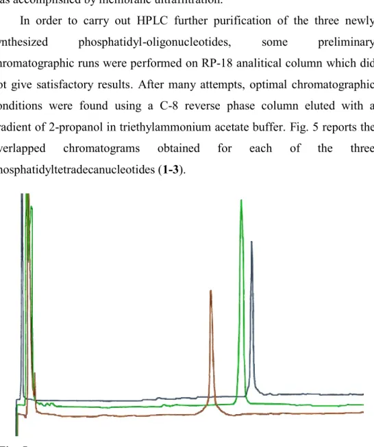
In order to carry out HPLC further purification of the three newly synthesized phosphatidyl-oligonucleotides, some preliminary chromatographic runs were performed on RP-18 analitical column which did not give satisfactory results. After many attempts, optimal chromatographic conditions were found using a C-8 reverse phase column eluted with a gradient of 2-propanol in triethylammonium acetate buffer. Fig. 5 reports the overlapped chromatograms obtained for each of the three phosphatidyltetradecanucleotides (1-3).
Fig. 5: Overlapped chromatograms of compound 1, compound 2 and
Methods and Results
the phosphatidyl moiety. More, it was found a linear correlation between the percentage of 2-propanol needed for elution and the total number of carbon atoms in the hydrophobic tails of the phosphatidyl group.
Final purification of the three phosphatidyloligonucleotides, was then carried out by transferring to a semi-preparative scale the experimental conditions optimized for the analytical chromatographic separation. Unfortunately, it was not possible to apply the standard procedures for the scaling-up of the analytical parameters. In fact, the increased amounts of the phosphatidyl-ODNs which had to be injected into the semi-preparative chromatographic system, due to their strong tendency to self-aggregate, gave rise to a serious broadening of the chromatographic peaks. On the other hand it is known [82] that lipo-ODNs tend not only to aggregate into micelles but also to exhibit a strong interaction with stationary reversed phase, therefore, HPLC purification of these compounds is difficult to achieve. Among other things, the conversion of the optimized analytical parameters into new parameters for the semi-preparative chromatography was also complicated because in the latter we replaced triethylammonium acetate in the eluent solution with ammonium acetate, in order to get the three phosphatidyl-ODNs coupled with counter-ions compatible with the subsequent spectroscopic analyses.
Anyway, after a number of trials, the best semi-preparative chromatographic conditions were found by setting the solvent flow up to the highest value allowed by the maximum pressure limit in the chromatographic apparatus and adequately lowering the concentration of the injected solution of lipo-ODN. In practice, it was necessary to use intermediate conditions between analytical and semi-preparative scale.
After the chromatographic purification, the compounds 1-3 were undertaken to lyophilization in order to remove all residual ammonium acetate and stored for further analyses.
Methods and Results
3.5
Spectroscopic characterization of
5’-O-(1,2-O-diacyl-sn-glycero-3-phosphoryl)-d(TGGCTTGAAGATGT-3’)(1-3)
The compounds 1-3, after purification, were each analyzed for their MS, UV and NMR spectroscopic properties.
Mass Spectra
MALDI-TOF mass spectrometry performed with the use of 3-hydroxypicolinic acid as a matrix, is one of the most commonly used methods [83]to obtain mass spectra of oligonucleotides. So, the three lipo-ODNs were first undertaken to this kind of analysis, but, it was not in ay way possible to detect the expected signals in the relevant spectra. This is probably due to the presence of the conjugated lipophilic moiety in these modified oligonucleotides.
Taking into account that recent literature in this field [84,85] reported mass spectra of modified oligonucleotides obtained by using 2,4,6-trihydroxyacetophenone or 3,4-diaminobenzophenone as matrices, these have been even used but all attempts did not lead to any results.
Recent reports on the application of HPLC-ESI mass spectrometry to oligonucleotides [86] prompted us to try this opportunity too. By applying this method and suitably adapting it to our case, mass spectra of the compounds were finally obtained. The HPLC chromatographic separation before the ESI/MS analysis was performed on a RP-C18 column using a gradient of 2-propanol in ammonium acetate buffer.
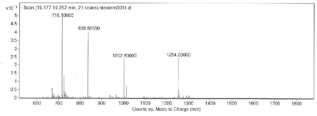
The first compound to be analyzed was 5'-O-[1,2-O-distearoyl-sn-glycero-3-phosphoryl]-d(TGGCTTGAAGATGT-3‟) which gave the
Methods and Results
chromatographic eluent, the adducts no longer appeared in the spectrum but only signals from multi-charged ions were present (Fig. 6). HPLC-ESI/MS analysis of the remaining two lipo-ODNs, performed under these last experimental conditions, gave mass spectra showing signals from the expected multi-charged ions (see Experimental).
Fig. 6 – ESI-MS(-) of compound 3.
UV Spectra
UV spectra of aqueous solutions of each compound (1-3) showed the classical absorption curve of ODNs (max at 260 nm) allowing in principle their quantification. These spectra showed in some cases in the region between 300 and 450 nm absorbance values higher than the expected ones, indicating possible scattering phenomena. These could have been associated to the formation of aggregates and/or supramolecular complexes, about which it would be interesting to carry out further investigation.
However, taking into consideration this fact, when UV spectra were run for quantification purposes, properly diluted solutions had to be prepared for the spectral analyses.
Methods and Results
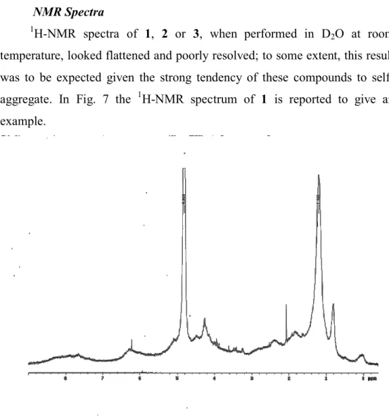
NMR Spectra
1H-NMR spectra of 1, 2 or 3, when performed in D
2O at room
temperature, looked flattened and poorly resolved; to some extent, this result was to be expected given the strong tendency of these compounds to self-aggregate. In Fig. 7 the 1H-NMR spectrum of 1 is reported to give an example.
Fig. 7. 1H-NMR of compound 1 run at 25°C.
When 1H-NMR experiments were carried out at 60 °C all peaks became sharper and spectra compatible with the expected molecular structures were obtained, which were also quite similar to each other. Fig. 8 shows again the
Methods and Results
Fig.8. 1H-NMR of compound 1 run at 60°C.
1H-NMR spectra of compounds 1-3, although sharpened by the increase
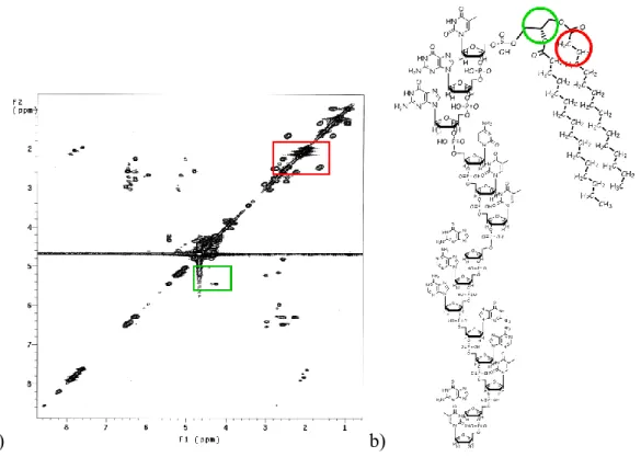
of temperature, were however quite complex ones; so, unequivocal assignments of 1H resonances had to be supported by 1H-1H COSY experiments which were performed, these too, at 60 °C (Fig. 9). Since 1 H-NMR spectra from 1, 2 and 3 were quite similar to each other, we‟ll discuss in detail only the spectrum of compound 1, taken just for example. The overall 1H-NMR data for compounds 1-3 are reported under experimental.
Methods and Results
a) b)
Fig. 9: a) 1H-1H COSY spectrum of compound 1; b) molecular structure of compound 1. Areas with color edge in the graph are used to highlight the regions of the spectrum where diagnostically important cross peaks fail.
In the spectrum of 1 (HOD 4.67 ppm ) methyls of myristoyl chains
appeared as a broad triplet at 0.95, accompanied by two broad signals centered at 1.22 and 1.31 for all - to μ-CH2 of the same alkyl chains, and a
multiplet for the two -CH2 at 1.65. The two -CH2 resonances fell within
partially overlapped multiplets ( 2.17÷3.06) attributable to all H2‟ and H2” of the sugar rings; on the basis of the -CH2/-CH2 correlation peak showed
in the COSY spectrum it was possible to assign the resonance of CH2 at
Methods and Results
a) b)
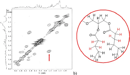
Fig. 10. A enlarged section of 1H-1H NMR COSY spectrum of 1, showing a cross-correlation peak (a) of the -CH2/-CH2 in the acyl chain (b).
Methyls of the thymine nucleobases T12, T14 and T1 gave singlets at 1.95, and 2.06 respectively, while methyls of T5 and T6 gave two partially overlapped singlets at2.12 and 2.13 H6 of tymines appeared as singlets at 7.62, 7.73, 7.81, and two partially overlapped ones at 7.89 and 7.90, for T12, T1, T14, T6 and T5 respectively. These assignments were unequivocally done on the basis of the thymine H6/CH3 cross-peaks clearly observable in the COSY spectrum .
H2 and H8 of adenines, together with H8 of guanines and H6 of cytosine, gave multiplets at 8.10÷8.60, while the signal from the H5 of cytosine was superimposed on multiplets at 6.19÷6.59 from most of H1‟of the deoxyribose rings. Signals relating to the latter protons appeared, in fact, as multiplets at 5,93 (1 H), 6.04 (1 H) and 6.19÷6.59 (12 H). Other signals from the protons allocated on 3‟-carbons of the sugar backbone appeared as multiplets at 4.79 (1 H) and 4.97†5.28 (12 H), while the H3‟resonance of the T14 unit was obscured by the residual HOD signal. The remaining H5‟,
Methods and Results
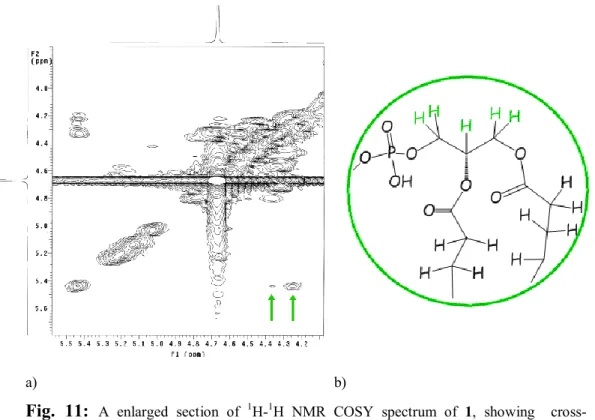
H5” and H4‟ of the deoxyribose rings gave multiplets at 4.15÷4.60 altogether with H3 (2 H) and H1 (2 H) of glycerol, whose H2 appeared as a multiplet at 5.44. On the basis of the H1a/H2, H1b/H2 and H23/H2
cross-peaks in the COSY spectrum it was possible to assign the resonance of H1a of glycerol at 4.35 and H1b and H23 of glycerol at 4.26 ppm. (Fig. 11 a).
a) b)
Fig. 11: A enlarged section of 1H-1H NMR COSY spectrum of 1, showing cross-correlation peaks (a) of the protons of glycerol backbone (b).
The above spectroscopic data, in addition to the results previously obtained from MS analysis, allowed us to unequivocally confirm the expected structure for the new synthesized compounds 1-3.
Methods and Results
3.6 Melting temperatures of ‘antisense’ phosphatidyl-ODN /
‘sense’ ODN duplexes.
Once the main structural properties of the new synthesized phosphatidyl-ODNs were determined, we went to evaluate some of their bio-molecular and biological features as well.
Among these properties, the ability of these antisense phosphatidyl -ODNs to form duplexes with the complementary sense sequence it seemed interesting to be assessed. In general, the higher this ability, the higher should be the antisense activity. As it is known, duplex stability is strictly related to its thermal stability; the latter being currently expressed in terms of melting temperature (Tm) that is in turn deduced from absorbance vs temperature (melting and annealing curves) profiles of oligonucleotides, recorded on a temperature-programmable UV-spectrophotometer.
To this end, first we proceeded to synthesize the complementary native ODN sense sequence (5‟-ACATCTTCAAGCCA-3‟) necessary to obtain the desired duplex melting curves. The synthesis was carried out on solid phase by applying, in this case, the currently used phosphoramidite chemistry (see above Fig. 3).
On the other hand, the not-modified ODN antisense sequence should have been also available because it would be used as a control in parallel annealing experiments. So, after having synthesized the above cited native ODN sense sequence, we used once again the classical phosphoramidite chemistry to synthesize, on solid phase, the not-modified ODN antisense sequence (5‟-TGGCTTGAAGATGT-3‟).
At this point, because of the impossibility to have access to an adequate variable temperature UV spectrophotometer, we decided to determine the desired melting temperatures by another suitable technique. To this end, the
Methods and Results
ability of our phosphatidyl-ODNs to give duplexes with the complementary
sense oligonucleotide was tested by Differential Scanning Calorimetry (DSC)
analysis [87].
It is known in fact that DNA/DNA binding thermodynamic quantities may be determined from DSC measurements on the thermal dissociation or „melting‟ of DNA/DNA duplexes at high temperatures. DSC measurements on the heat absorbed in the transition from a duplex to the single-strand states yield direct determinations of the binding enthalpy that are model-independent and direct determinations of the heat capacity change for the transition. So, we went to determine, from DSC measurements the thermodynamic quantities describing the melting or dissociation of our three antisense phosphatidyl-ODN/sense ODN duplexes and their corresponding antisense ODN/sense ODN duplexes. In particular the determined thermodynamic quantities were: a) the temperature at half the transition peak area (Tm) and b) the heat capacity change accompanying the transition (ΔCp) from the difference in the pre-transitional baselines at Tm.
The following pairs of oligonucleotides, appropriately dissolved 100 mM NaCl buffered solution (pH 7.4), were used for DSC experiments:
(5‟-TGGCTTGAAGATGT-3‟) / (5‟-ACATCTTCAAGCCA-3‟) 5‟-O-[1,2-O-dimyristoyl-sn-glycero-3-phosphoryl]-(5‟-TGGCTTG-AAGATGT-3‟) / (5‟-ACATCTTCAAGCCA-3‟) 5‟-O-[1,2-O-dipalmitoyl-sn-glycero-3-phosphoryl]-(5‟-TGGCTTG-AAGATGT-3‟) / (5‟-ACATCTTCAAGCCA-3‟) 5‟-O-[1,2-O-distearoyl-sn-glycero-3-phosphoryl]-(5‟-TGGCTTGA-AGATGT-3‟) / (5‟-ACATCTTCAAGCCA-3‟)
Methods and Results
repeated scans. In particular, the phosphatidyl-ODN referred to in the figure is the dimyristoyl-glycerophosphoryl-tetradecamer 1.
Fig.12: Overlapped DSC scans of unmodified antisense/sense duplex and re-scan of the same; compound 1/sense duplex and it re-scan.
The temperature of 55,9 °C corresponding to the peak maximum of the
antisense phosphatidyl-ODN 1/sense ODN duplex is a few degrees higher
than that of the peak maximum of the antisense ODN/sense ODN duplex (53,5°C), indicating that the former duplex is thermally stable at least as much as the corresponding antisense ODN/sense ODN duplex is. Also, the reappearance of the transition peaks upon a re-scan of the samples shows that the transitions are reversible and that there is not a significant degradation of the sample after heating to 90°C. Dipalmitoyl-glycerophosphoryl-tetradecamer 2 showed a similar behavior (Tm= 53.2 °C), while the Tm value of the duplex of distearoyl-glycerophosphoryl-tetradecamer 3 was 9.1 °C lower than that of the reference duplex.
Considering that, in the case of phosphatidyl-ODNs 1 and 2, the Tm values obtained for antisense phosphatidyl-ODN/sense ODN duplexes were not very different from the reference value of unmodified antisense ODN/sense ODN duplex, it can be inferred that the duplexes formed by antisense
Methods and Results
phosphatidyl-ODNs 1 and 2 with the sense counterpart have stabilities similar to that of the relevant unmodified (native) duplex.
The result obtained for phosphatidyl-ODN 3 is probably due to the larger size of the stearoyl chain in the phosphatidyl moiety, somewhat disturbing the base pairing near the phosphatidyl.
In all the thermograms obtained for antisense phosphatidyl-ODNs
(1-3)/sense ODN duplexes the observed heat capacity changes (Cp) were
lower than those measured in thermograms from the reference unmodified duplex. In the light of the observed tendency of these molecules to self-assemble, these results may not be surprising; in fact, if certain number of molecules of phosphatidyl-ODNs are involved in the formation of aggregates, they will be subtracted from taking part in duplex formation and therefore, the heat exchanged for the transition duplex/single strand will be lower. Anyway, further investigations are needed to clarify these points.
3.7 Biological assays
Considering that the sequence of the oligonucleotide moiety of phosphatidyl tetradecamers 1-3 had been chosen as to be that of an antisense oligonucleotide from the region 261 to 274 of human VEGF165 coding region,
some preliminary tests were carried out to ascertain the antisense effectiveness of these compounds.
VEGF expresses in a lot of solid tumor cells. Shi et al. [88] has studied the influence of the VEGF AS-ODN on the blood vessel generation behavior and proliferation of renal carcinoma cell line-Caki-1cell, which manifested
Methods and Results
neuroblastoma, We decided to evaluate the effect of VEGF antisense phosphatidyl-ODNs on the expression of VEGF at mRNA level in Human Neuroblastoma cell line SH-SY5Ycells, which could provide experimental foundation for the potential clinical application of the newly synthesized VEGF phosphatidyl-ODNs.
In this regard, wanting to perform the antisense tests in the experimental conditions of maximum activity of VEGF expression by cells, we evaluated the possibility of carrying out the experiments on cell cultures undertaken to the so called “hypoxic conditioning”. It‟s know [90-91] in fact that administration to cells of cobalt chloride (CoCl2) produces hypoxia
which induces in turn VEGF production. For this purpose, the cellular response (in terms of VEGF production) to different concentrations of CoCl2
was investigated. The best results were obtained following administration of 25µM CoCl2 .
At this point, cells of neuroblastoma SH-SY5Y line were incubated, in the presence of 25µM CoCl2, with the two phosphatidyl antisense
5‟-O-[1,2-O-dimyristoyl-sn-glycero-3-phosphoryl]-(TGGCTTGAAGATGT-3‟) and
5‟-O-[1,2-O-dipalmitoyl-sn-glycero-3-phosphoryl]-(TGGCTTGAAGATGT-3‟), both at various concentrations. In parallel experiments, the relevant unmodified antisense ODN was administered as a control. After 60 h incubation, cells were treated with TRIZol™ reagent and mRNA was extracted (see experimental section). Transcription from mRNA to cDNA and PCR were then performed and the amplified DNA was electrophoretically analyzed on agarose gel.
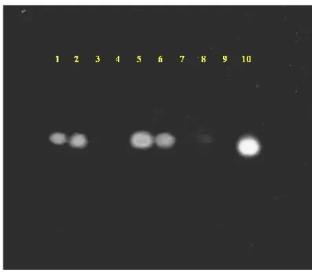
As can be deduced from Fig. 13 reported below, unmodified (native)
antisense ODN, when administered at concentration of 20 µM or higher is
able to lower the cellular VEGF m-RNA concentration, but is not able to do it at 10 µM concentration. Interestingly, both phosphatidyl tetradecamers 1-2
Methods and Results
were able to significantly decrease the cellular VEGF m-RNA already at 10 µM concentration.
Fig. 13. Agarose gel of cDNA from Neuroblastoma cells. - 1: Untreated; 2: 10 µM unmodified ODNs+25 µM CoCl2; 3: 20 µM unmodified ODNs+25 µM CoCl2; 4: 30 µM unmodified ODNs+25 µM CoCl2; 5: 5 µM of 2+25 µM CoCl2; 6: 5 µM of 1+25 µM CoCl2; 7: 10 µM of 2+25 µM CoCl2; 8: 10 µM of 1+25 µM CoCl2; 9: 15 µM of 1+25 µM CoCl2; 10: 25 µM CoCl2.
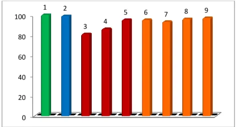
To ascertain the cell viability in the course of our experiments, and in particular to verify possible cell toxicity of our phosphatidyl-ODN, all cell cultures undertaken to the antisense tests were controlled by MTT assay [92]. Further, MTT assay was performed also on cell cultures treated with 10, 20
Methods and Results
From the results of MTT assays (Fig. 14) it was possible to state that phosphatidyl-ODNs used in our antisense tests did not exert any cellular toxicity at the used concentrations.
Fig. 14. Results of MTT assay on Neuroblastoma cells. - 1: Untreated; 2: 25 µM CoCl2; 3: 30 µM unmodified ODNs+25 µM CoCl2; 4: 20 µM unmodified ODNs+25 µM CoCl2; 5: 10 µM unmodified ODNs+25 µM CoCl2; 6: 10 µM of 2+25 µM CoCl2; 7: 15 µM of 1+25 µM CoCl2; 8: 10 µM of 1+25 µM CoCl2; 9: 5 µM of 2+25 µM CoCl2.
Although the preliminary results above described were very encouraging ones, further work is currently underway to better define the molecular aspects underlying the observed data.
0 20 40 60 80 100 1 2 3 4 5 6 7 8 9
Conclusive remarks
4
C
ONCLUSIVE REMARKS
As has been said in the introduction, oligonucleotides bearing a terminal lipophilic and biocompatible group attached possibly through a biodegradable bond could be useful as antisense pro-drugs with improved cellular uptake. Nevertheless, despite various synthetic strategies have been addressed to the lipophilic modification of oligonucleotides, the phosphatidyl group, which is widely distributed among the cell membrane lipids, has never been used for this purpose. This is mainly due to the lability of the ester bond in the phosphatidyl during the treatment for the deprotection of synthetic oligonucleotides.
In the course of this research work, exploiting various synthetic opportunities, and making also some appropriate changes, we succeeded to synthesize for the first time 5‟-O-phosphatidyloligodeoxynucleotides. The versatility of the synthetic route we developed allows the preparation of phosphatidyl-oligonucleotides differing in their phosphatidyl tail and/or in the sequence and the length of the oligonucleotide moiety. We would like to emphasize that the synthesis was designed so that it can be easily carried out by using a common automated DNA synthesizer.
In particular we turned our attention toward the preparation of some phosphatidyl-tetradecadeoxynucleotides all having the antisense sequence against a coding region of the VEGF gene but differing from each other in the fatty acid composition of their phosphatidyl moiety. To this purpose, phosphatidyl-ODNs 1, 2 and 3 were prepared having O-dimyristoyl-,
1,2-Conclusive remarks
The structure of the newly synthesized compounds was then confirmed on the basis of their spectroscopic properties (UV, MS, NMR).
Both UV and NMR measurements have highlighted a strong tendency of these compounds to give supramolecular aggregates whose features (liposomes, micelles, etc.) it has not been possible to establish because of lack of time. Further studies aimed at characterization of these aggregates are now under way.
Thermal melting profiles of different duplexes formed by each of the
antisense phosphatidyl-ODNs (1-3) and the unmodified complementary sense ODN were obtained by DSC in order to evaluate the annealing
properties of these new compounds.
Melting temperatures (Tm) have been extracted and compared to that found in parallel experiment with the relevant native duplex. Phosphatidyl-ODNs 1 and 2 showed Tm very close to that of native duplex, while the Tm value of phosphatidyl-ODN 3 dropped by 9.1 °C above that of the unmodified sequences. This is probably due to the larger size of the stearoyl chain in the phosphatidyl moiety, somewhat disturbing the base pairing near the phosphatidyl. How the length of the acyl chain and/or that of the nucleotide sequence may influence the duplex stability of these phosphatidyl-ODNs are interesting topics that deserve to be investigated in the near future.
Further, the actual effectiveness of our antisense phosphatidyl-ODNs in inhibiting the expression of VEGF mRNA was assessed following administration of phosphatidyl-ODNs 1 and 2 to Neuroblastoma cell lines. As a result, both 1 and 2 were effective in lowering drastically the cellular VEGF m-RNA, and they have done so at concentrations significantly lower than those required to produce the same effect by the unmodified antisense ODN.
Conclusive remarks
As a whole, the results of this work confirm the idea that antisense oligonucleotides with native molecular structure, but covalently linked to a natural lipid moiety such as phosphatidyl, may be subject to an enhanced cellular uptake compared to the non-conjugated ones, such requiring lower concentrations than the latter to exert the same desired biological effect. In other words this means that, if these compounds are used in vivo to modulate gene expression for pharmacological purposes, the possibility that systemic toxicity occurs would be greatly reduced.
Considering that the phosphatidyl-ODNs in the first instance we synthesized all have saturated acyl chains in their phosphatidyl group, it would be interesting to study the influence of the presence of unsaturated acyl chains in the same group on the ability of these compounds to interact with bio-membranes and, ultimately, on their cellular uptake. So, the synthesis of this kind of new molecules and the evaluation of their interaction with membrane models ( mono- and bilayers, LUV, etc.) will be interesting research topics.
Finally, one more consideration.
Albert Einstein is said to have said this sentence: “imagination is more important than knowledge”. So, let me imagine an antisense oligonucleotide that bears two phosphatidyl moieties covalently linked to its 3‟- and 5‟-end; in addition to a more pronounced cellular uptake and an increased nuclease resistance (both to 3‟- and 5‟-exonucleases), a compound of this type may have, especially, a marked tendency to self sick its lipid ends, thus giving rise to a plasmid-like ODN. It is not difficult to imagine the pharmacological potential of a compound of this kind.
Experimental
5
E
XPERIMENTAL
5.1 Synthesis of silyl linker.
Materials
1,4-Dibromobenzene was purchased from Sigma Aldrich and, before using, it was dried under vacuum for 24 h in the presence of P2O5. The
butyl-lithium solution and chlorodiisopropylsilane were obtained from Fluka and Sigma Aldrich respectively.
Solid CO2 was freshly prepared following gas expansion.
2,4,6-Tribromophenol and bis(2-oxo-3-oxazolidinyl) phosphinyl chloride (BOPCl) were purchased from Sigma Aldrich; before using, they were kept under vacuum for 24 h in the presence of P2O5.
1,4-Dichloro-5,5-dimethyl hydantoine was obtained from Fluka and dried as above.
5'-O-(4,4'-Dimethoxytrityl)-2'-deoxythymidine and imidazole were purchased from Sigma Aldrich and Fluka respectively, and they were dried too.
LCAA-CPG (Long Chain Amino Alkyl Controlled Pore Glass) was obtained from Sigma Aldrich.
Synthesis of silyl-linker and anchoring of the first nucleoside to CPG.
To 10 ml of anhydrous THF ( -78 ºC) 1,4-dibromobenzene (0.5 g; 2.12 mmol) was added under Ar and then 0.785 ml (1 equivalent) of butyl-lithium (2.7 M in heptane solution) were added to the solution. The mixture was
Experimental
allowed to react for 5 minutes and then was added with chloro-diisopropyl silane (0.360 ml; 2.12 mmol). After 15 minutes at -78°C, 0.785 ml (1 equivalent) of butyl-lithium (2.7 M solution in heptane) were added to the reaction mixture which, 5 minutes after, was treated with a large excess of solid CO2. One hour later, the mixture was evaporated from the solvent and
was purified by liquid chromatography. The elution conditions were taken from TLC [silica gel, CH2Cl2/MeOH/TEA (95:4:1)], where the product
showed Rf = 0.12. Column chromatography on silica gel was carried out with
a methanol gradient from 4 to 20% in CH2Cl2 in the presence of 1% TEA.
Triethylammonium 4-(diisopropylsilyl)benzoate (250 mg), was then analyzed for its spectroscopic (1H-NMR) properties which confirmed the structure.
1
H NMR (CDCl3): δ 7.89 (d, Jortho = 8 Hz, 2 H, H-2 e H-6), 7.43 (d, Jortho = 8
Hz, 2 H, H-3 e H-5), 3.83 (s, J29Si,H = 6 Hz, 1 H altogether, Si-H), 3.02 (q, J = 7 Hz, 6 H, NCH2CH3), 1.25 (t, J = 7 Hz, 9 H, NCH2CH3), 1.10 (m, 2 H, CH
isopropyl), 0.94 (d, J = 7.5 Hz , 6 H, CH3 of -CH(CH3)2 ), 0.86 (d, J = 7.5
Hz , 6 H, CH3 of -CH(CH3)2).
In the next step triethylammonium 4-(diisopropylsilyl)benzoate (0.250 g; 1.06 mmol) was dissolved in anhydrous pyridine (20 ml) and to the solution 4 equivalent of BOPCl ( 4.24 mmol; 1.079 g) and 2 equivalent of 2,4,6-tribromophenol (2.12 mmol, 0.701 g) were added under Ar. After 1 h the solvent was evaporated and the product was purified by liquid choromatography, using hexane/ethyl acetate/TEA (96:3:1) as the eluent mixture. 419 mg of 2,4,6-tribromophenyl 4-(diisopropylsilyl)benzoate were obtained and analyzed by NMR spectroscopy which confirmed its structure .
1H NMR (CDCl