UNIVERSITÀ DEGLI STUDI DI CATANIA
DOTTORATO IN BIOLOGIA UMANA E BIOINFORMATICA:
BASI CELLULARI E MOLECOLARI DEL FENOTIPO
XXVIII CICLO
DIPARTIMENTO DI SCIENZE BIOMEDICHE E BIOTECNOLOGICHE
Virulence gene expression of three hypervirulent
S. pyogenes M1T1 and membrane
vesicles isolation
TESI DI DOTTORATO
Dott.ssa Valeria Metoldo
Coordinatore: Chiar.mo Prof. MICHELE PURRELLO Tutor: Prof.ssa MARIA SANTAGATI
Table of Contents
1. Introduction p.1 1.1 Streptococcus pyogenes p.3 1.1.1 Classification of GAS p.3 1.1.2 GAS Surface p.5 1.1.3 Streptococcal Superantigens p.7 1.1.4 Evolutionary Origin of Clone M1T1 GAS serotype from itsparental M1 serotype
p.13 1.1.5 Disease model for invasive GAS serotype M1T1 p.16
1.2 Bacterial membrane vesicles p.20
1.2.1 Background and History p.20
1.2.2 What are Membrane Vesicles? p.21
1.2.3 Regulation of Vesicles Biogenesis p.22
1.2.4 Functions of MVs p.24
1.2.5 MVs in Gram positive Bacteria p.27
2. Study aim p.30
3. Materials and Methods p.31
3.1 Bacterial strain and growth conditions p.31
3.2 Dna extraction p.31
3.3 Molecular gene identification p.31
3.4 Rna extraction p.32
3.5 RT and q-RT-PCR p.33
3.6 MVs isolation protocol p.34
3.7 Purification of MVs p.35
3.8 Scanning Electron Microscopy (SEM) analysis p.37 3.9 Transmission Electron Microscopy (TEM) analysis p.37
3.10 Dynamic Light Scattering (DLS) p.37
4. Results p.38
4.1 Molecular identification of emm-typing and MLST determination p.38 4.2 Determination of antibiotic resistance profiles and virulence gene content
p.39 4.3 Gene expression study of some virulence toxins in different growth phases by Real-Time PCR
p.40 4.4 Secretion of MVs from Gram positive bacteria, S. pyogenes p.45
5. Discussion p.47 6. References p.52
1
1. Introduction
The genus Steptococcus [Rosenbach, 1844] consists of catalase-negative, Gram-positive cocci which are arranged in pairs and chains and are usually facultatively anaerobic. Streptococci are a diverse collection of species inhabiting many body sites and they are both commensals and pathogens. In particular, nonpathogenic streptococci, are the most abundant bacterial species at the oropharyngeal level, and they have been found to exert an important role in the protection against pathogenic agents causing inflammation and infections [Tagg JR et al., 2003]. Much attention has recently been devoted to the analysis of the oral microbiota to develop bacteriotherapy focused on prevention and/or treatment of upper respiratory tract infections. In this regard, a key species is Streptococcus salivarius, a lactic acid bacterium that is mainly encountered in the mouths of human beings. It is the first commensal bacterium that appears in the oral cavity of newborns where it colonizes the upper respiratory tract [Aas JA et al., 2005] and persists there as a predominant member of the native microbiota throughout the life of its human and sole natural host [Favier CF et al., 2002]. Many LAB (Lactic Acid Bacteria) strains, including S.salivarius, are prolific producers of bacteriocins, which are an abundant and
diverse group of ribosomally synthesized antimicrobial peptides produced by bacteria that kill or inhibit species closely related to the producer bacterium. Furthermore, according to several studies, large populations of S.salivarius efficiently adhere to oral epithelial cells, especially the papillary surface of the tongue that is a strategic location to carry out a population surveillance within the oral microbiota [Tagg JR et al., 1983; Wescombe PA et al., 2010 ]. The presence of an adhesion system such as pili, fibrils, saliva-binding proteins and host-cell-binding
2 proteins, together with its high competition rate, helps this species to stay in the human mouth [Nobbs AH et al., 2009].
In our laboratory, during my PhD studies, we characterized one strain, S.salivarius 24SMBc, isolated from one healthy child that showed excellent inhibitory activity against S.pneumoniae and S.pyogenes and a potent capacity of adhesion to HEp-2 cells. The main aim of this study was to evaluate the clinical evidence of a probiotic application of S.salivarius 24SMBc for the prevention or reduction of recurrent medium otite (OM) children [Santagati M et al., 2014]. Therefore, this strain was included in a randomized, placebo-controlled, double-blind paediatric trial that involved 100 otitis prone children. This preliminary study showed a reduction of OM episodes in children who received the intranasal administration of this probiotic with respect to children treated with placebo [Santagati M et al., 2014].
The study of S.salivarius 24SMBc ended with the production and marketing of a new medical device, the Rinogermina nasal spray, in collaboration with D.M.G s.r.l Italy. Streptococci, as mentioned above, include both nonpathogenic and pathogenic
bacteria. In particular, Streptococcus pyogenes (group A streptococci, GAS) is an exclusive human bacterial pathogen. The virulence potential of this species is tremendous. Interactions with humans range from asymptomatic carriage over mild and superficial infections of skin and mucosal membranes up to systemic purulent toxic-invasive disease manifestations [Fiedler T et al., 2015]. Simultaneously with the study of S.salivarius 24SMBc, my PhD project focalized on global regulation of virulence expression genes of three hypervirulent strains of Streptococcus pyogenes [Santagati M et al., 2014] and their eventual production of membrane vesicles like new delivery system of virulence-associated components.
3
1.1 Streptococcus pyogenes
Group A streptococcus (GAS), or Streptococcus pyogenes, is a Gram-positive coccus and is an important human pathogen. GAS is usually present in the respiratory tract in 5 to 15% of individuals without signs of disease. As part of our flora, if defenses are compromised, S.pyogenes can generate a variety of suppurative infections. It is the most common cause of bacterial pharyngitis and is the cause of scarlet fever and impetigo. In the past, GAS strains were a common cause of puerperal sepsis or childbed fever. Today, group A streptococcus is responsible for streptococcal toxic shock syndrome and, recently it has gained notoriety as the “flesh-eating” bacterium which invades skin and soft tissues [Cunningham et al., 2000]. It is also responsible for a diverse range of clinical manifestation, such as bacteremia, cellulitis, meningitis, pneumonia and necrotizing fasciitis [Lamagni et al., 2008]. GAS causes an estimated 700 million cases of mild, non invasive infections each year, of which 650,000 progress to severe invasive diseases with 25% mortality [Carapetis J.R et al., 2005]. Epidemiologic studies, in 2008, showed that the resurgence of severe
invasive GAS infections represented a global spread, ushering in a new pandemic, similar to that reported in the earlier part of the 20th century [Aziz et al., 2008].
1.1.1 Classification of GAS
Streptococci are classified on the basis of hemolysis, colony morphology and serologic specificity. They are divided into three groups by the type of hemolysis on blood agar:
β-hemolytic (clear, complete lysis of red cells)
α-hemolytic (incomplete, green hemolysis)
γ hemolytic (no hemolysis).4 GAS belongs to the beta-hemolytic streptococci family and it was defined in 1933 by Rebecca Lancefield as Group A Streptococcus on the basis of its specific cell wall polysaccharides (group A, B, C, F and G) or lipoteichoic acids (group D) [Lancefield R.C, 1933]. The Lancefield classification scheme of serologic typing distinguished the beta-hemolytic streptococci based on their group A carbohydrate, composed of N-acetylglucosamine linked to a rhamnose polymer backbone. A serological differentiation based on the most abundant exposed surface antigen, the M protein, is also extensively used in infection diagnosis and, over the years, the number of serotypes has progressively increased up to 150. Streptococci were also serologically separated into M protein serotypes based on a surface protein. Serotype M1 is among the most frequent serotypes from streptococcal pharyngitis and invasive diseases and the resurgence of severe invasive GAS infections is now correlated with single clone M1T1 GAS serotype [Musser JM et al., 2007]. The M1T1 strains contain the bacteriophage-encoded virulence factors extracellular streptodornase D (Sda1) and exotoxin type A (SpeA). The acquisition of prophages encoding Sda1, SpeA and the recombination of a 36kb chromosomal region encoding an extracellular toxin NAD-glycohydrolase (NADase) and streptolysin O (SLO), were a crucial factor in dissemination, potential virulence and emergence of a very abundant clone serotype M1T1 GAS that are different from M1 GAS [Musser JM et al., 2005] (Fig.1.)
5
Fig.1.Paul Sumby et al. J Infect Dis. 2005
1.1.2 GAS Surface
Many GAS virulence factors are present on the bacterial cell surface, and of particular interest are those involved in colonization and evasion of the host immune responses. Invasive GAS disease requires successful colonization of skin or oropharynx. Initial weak interaction with mucosa, mediated by pili or lipoteichoic acid, is followed by a stronger binding lectine-carbohydrate and protein-protein interactions that confer tissue specificity. Group A Streptococcus has a highly complex and diverse cell surface, rich in antigenic components such as the polysaccharide capsule (C-substance), the cell wall peptidoglycan and many surface proteins including the M protein, fimbrial and fibronectin-binding proteins [Walker MJ et al., 2011].
6 M Protein
The streptococcal M protein is now probably one of the best-defined molecules of the known bacterial virulence determinants. The M protein is a major surface protein and virulence factor of group A streptococci, with more than 150 distinct serotypes identified. The amino-terminal region extends from the surface of the streptococcal wall, while the carboxy-terminal region is within the membrane. The serotype specificity of GAS is determinated by N-terminal region, called the A repeat region, that is highly variable among M serotypes. The B repeat region, the mid-region, varies from serotype to serotype and the carboxy-terminal or C repeat region contains a conserved sequence shared among all of the serotypes [Fischetti et al., 1989]. The M proteins are able to bind serum fibrinogen allowing the bacteria to resist phagocytosis and contain those particular epitopes that mimic human tissues, some of these are the cause of rheumatic fever, leading to an autoimmune carditis. Furthermore, M protein increases bacterial survival in neutrophils, so this protein is essential for full virulence during GAS infection. Epidemiological studies have shown a clear correlation between M-type and type of infection caused, partitioning the group of S.pyogenes into two classes:
Class I (M-types: 1, 3, 5, 6, 14, 18, 19, 24) are found associated with throat infection and rheumatic fever.
Class II (M-types: 49, 57, 59, 60, 61) produce serum opacity factor (SOF)and are associated with pyoderma and acute glomerulonephritis. [Cunningham et al., 2000].
7 The Streptococcal Capsule
The group A streptococcal capsule is composed of a linear polymer of hyaluronic acid containing repeating units of glucuronic acid and N-acetylglucosamine that is produced by enzymes encoded in the highly conserved has ABC hyaluronan synthase operon [PH Weigel. J et al., 1995]. The hasA gene encodes hyaluronate synthase; hasB encodes UDP-glucose dehydrogenase [Crater DL et al., 1993]; and hasC
encodes UDP-glucose pyrophosphorylase. The hyaluronic acid capsule is required for resistance to phagocytosis [Wessels MR et al., 1994] and may be an important adherence factor during host colonization because it is able to bind CD44 on epithelial cells [Wessels MR et al., 1998]. Glucuronic acid and N-acetylglucosamine, nearly identical to the polysaccharides that are in the human host, encourage GAS evasion of the host immune response. Streptococcal isolates have different amounts of hyaluronic acid capsule that could be related to the has operon promotor. In the well-encapsulated strain, the promoter is more active than in a poorly encapsulated strain [Wessels MR et al., 1998]. Furthermore, Levin and Wessels demonstrated that capsule production and virulence is highly influenced by a negative regulator (CsrR) that is part of a two-component regulatory system [Levin J et al., 1998]. More recent work provides definitive evidence that the capsule is a major virulence determinant involved in resistance to phagocytosis in conjunction with the streptococcal M protein.
1.1.3 Streptococcal Superantigens
Streptococcus pyogenes has acquired many virulence determinants that allow it to
survive within the host and they are involved in the pathogenesis of toxic shock, necrotizing fasciitis, and invasion of soft tissue and skin. The superantigens (Sags)
8 are the extracellular pyrogenic exotoxins A, B, C (SpeA, SpeB, SpeC) and newly discovered exotoxins such as exotoxin F (SpeF) and streptococcal superantigen (SSA) [Holm SE et al., 1994]. All of these toxins act as superantigens that interact with the major histocompatibility complex (MHC) class II molecule via high zinc binding and the activation liberates large amounts of interleukins and other inflammatory cytokines such as gamma interferon and tumor necrosis factor. Furthermore, the superantigens are able to bind the beta chain (Vβ) of the T-cell subset without any processing by antigen-presenting cells. The pyrogenic exotoxin speA and spec are encoded by bacteriophages [Cunningham et al., 2000], while speB
is chromosomal and it is a cysteine proteinase that degrades vitronectin, fibronectin, and IL-1 precursor.
Streptolysin O
SLO is part of a family of cholesterol-binding cytotoxins produced by many pathogenic Gram-positive bacteria including Streptococcus pneumoniae (pneumolysin), Listeria monocytogenes (listeriolysin O) and Bacillus anthracis (anthrolysin) [Rest RF et al., 2003]. SLO is a 69 kDa protein that oligomerizes to form large pores in the host cell membrane [Tweten RK et al., 2000], and has several functions in GAS pathogenesis, including the induction of apoptosis in epithelial cells, neutrophils and macrophages [Timmer AM et al., 2009]. SLO, also, prevent direct uptake of GAS into lysosomes and enhances GAS survival in the human host by interfering with both bacterial uptake and intracellular killing of GAS by pharyngeal epithelial cells.
9 Streptococcal inhibitor of complement, SIC
The sic gene, which encodes the Streptococcal inhibitor of complement-mediated lysis (SIC), is a highly polymorphic extracellular protein and putative virulence factor secreted only by M1 and M57 GAS strains. SIC is a secreted 31 kDa protein that is located in the Mga virulence regulon [Akesson P et al., 1996]. Sic was shown to bind C5b67 of the membrane attack complex (MAC), thus impairing terminal complement function. In particular, SIC promotes GAS M1T1 survival and enhance the progression of invasive infection.
C5a peptidase
The endopeptidase C5a is a 130 kDa proteolytic enzyme localized on the surface of GAS strains. The C5a peptidase is encoded by a gene that is regulated by mga in concert with M protein [LaPenta, D et al., 1994]. Peptidase cleaves the complement-derived chemotaxin C5a at its PMN-binding site [Cleary P et al., 1992] and this event inhibits the recruitment of phagocytic cells to the site of infection [Cleary PP et al., 1996]. The identity of the amino acid sequence of the enzyme is greater than
95% among class I and class II M protein serotypes of group A and B streptococci. Consequently, the surface of GAS presents a double barrier to the complement defenses of the host: first, the M protein, discussed above, and second, the streptococcal C5a peptidase inactivates C5a and chemotaxis.
Streptodornase D
One important distinguishing feature of global dissemination of the M1T1 GAS clone, compared to less pathogenic GAS strains, is the acquisition of a prophage (ΦM1T1Z) encoding a potent secreted DNase, Sda1[Aziz RK et al., 2004]. This gene has been shown to promote MIT1 GAS virulence via
10 degradation of NETs. NETs are composed of DNA, granule protease, antimicrobial peptides and histones that are secreted by host neutrophils to capture and eliminate bacteria at the site of infection [Cole JN et al., 2011; Buchanan JT et al., 2006; Walker MJ et al., 2007]. The presence of ΦM1T1Z and a second prophage that encodes the superantigens SpeA, distinguishes the GAS serotype M1T1 from its closely related M1 type.
Streptococcal exotoxin B , SpeB
SpeB, a cysteine proteinase, is the most secreted protein from S.pyogenes. The streptococcal cysteine proteinase is one of the best studied and the earliest identified secreted proteins from S. pyogenes. Much is known about its structure, function, processing, substrate specificity and regulation [Ashbaugh et al., 1998; Ashbaugh and Wessels, 2001]. Despite the large amount of literature on SpeB functions, the role of SpeB in GAS infections is still unknown, but recent evidence has conclusively demonstrated that SpeB is critical for the pathogenesis of severe invasive disease caused by GAS. speB is a chromosomal gene and is highly conserved and found in > 99% of GAS isolates, although there is a significative variation in expression levels among strains [Bohach et al. 1988; Ferretti J et al., 1991]. The gene encodes the 398 amino acid SpeBz (40 kDa) and undergoes conversion to mature 28-kDa SpeBm form by autocatalytic truncation [Liu and Elliott 1965b; Doran et al., 1999]. This enzyme is able to degrade the extracellular matrix, cytokines, chemokines, complement components, immunoglobulins, serum protease inhibitors, and also cleaves and inactivates many surface-associated and extracellular GAS virulence determinants, including M1 protein, [Ringdahl U et al., 2000], different superantigens, [Aziz RK et al., 2004; Kansal RG et al., 2003],
11 streptokinase [Sun H et al., 2005], Sda1 [Aziz RK et al., 2004], and SIC [Pence M A et al., 2010]. Furthermore, SpeBm can cleave the cell wall-anchored C5a peptidase
from the bacterial surface and this causes the inhibition of recruitment of neutrophils to the site of infection [Wexler and Cleary, 1985; Berge and Björck, 1995]. As a consequence, the bacterial properties are altered, which is important for the transition from localized to systemic S.pyogenes infection [Cole et al., 2006]. Regulation of speB expression in GAS is extremely complicated. Maximal expression occurs from
late logarithmic to stationary phase in response to environmental factors, pH and NaCl concentration. Although there are many papers on speB expression, the precise regulatory mechanisms that occur in vivo remain unclear. [Musser et al., 2011]. During the different stages of an infection or response to external stresses from the environment, GAS strains are able to control transcriptional regulation of genes and protein secretion through signal peptidases. The cumulative contribution of the secreted cysteine protease SpeB to the pathogenesis of invasive GAS infection is at present unclear, and studies in various GAS serotypes and animal models have produced varying results. The Streptococcal exotoxin B is involved in bacterial phenotype shift in different ways. For example, in some S.pyogenes strains, deletion of the speB gene leads to a down regulation of gene encoding for the hyaluronic acid capsule, but the underlying molecular mechanism is not well understood [Woischnik et al., 2000]. Some studies have shown that a high production of SpeB is associated
with STSS, whereas others show that low SpeB production is associated with severe infection [Talkington et al., 1993; Kansal et al., 2000]. Musser’s group demonstrated, in different papers that, using speB mutants in animal models, they all showed that SpeB contributes to tissue damage and bacterial dissemination
12 [Lukomski et al., 1997, 1998, 1999 ]. Later, Kotb’s group showed that SpeB is downregulated in invasive M1T1 isolates during infection in mice [Kazmi et al., 2001 ]. Recently, Aziz’s group, in 2004, demonstrated that GAS MIT1 WT, SpeB-positive bacteria undergo a phase–shift to the speB-negative phenotype after infection in mice [Aziz RK et al., 2004]. This phase switch was observed to be the result of mutations in the two-component regulatory system covR/S [Walker MJ et al., 2007]. CovR/S is a global regulator, also called CsrR/S, a two component signal
(TCS) transduction system that has been studied extensively because of its critical role in virulence. Normally, covR/S represses expression of about 15% of GAS genes including many involved in virulence [Federle et al., 1999]. Some studies have revealed a correlation between an invasive phenotype and the up-regulation of some virulent genes, coding proteins such as streptodornase, streptokinase, streptolysin O, streptococcal inhibitor of the complement and the hyaluronic acid capsule synthesis operon [Sumby P et al., 2006]. At the same time, a mutation in the covR/S operon, decreases the expression of speB and consequently prevents the degradation of streptokinase and the M1 protein. The presence of these proteins promotes invasive infection and bacterial dissemination. [Cole et al., 2006]. Furthermore, studies of M1T1 clinical isolates from invasive disease cases, found an inverse relationship between SpeB expression and clinical severity. In particular, SpeB levels were higher in GAS M1T1 serotype isolates from non-severe invasive infections than in isolates from severe cases, such as STSS and necrotizing fasciitis. [Kansal et al., 2000 ]. In conclusion, the precise function of streptococcal exotoxin B is extremely complex and for this reason is still unknown, because different studies have produced contradictory results [Svensson MD et al., 2000; Ashbaugh and Wessels, 2001;
13 Lukomski et al., 1997, 1998, 1999]. Together the data suggest that the effects of SpeB during the interaction between host-bacteria in vivo will depend on the balance between the actions of several regulatory systems that control SpeB synthesis, post-translational modification, and enzymatic activity [Kansal et al., 2000] (Fig.2.).
Fig.2. Model explaining mutation of Streptococcus pyogenes regulatory locus during invasive
infection. Claire Turner & Shiranee Sriskandan .Nature Medicine (2007)
1.1.4 Evolutionary Origin of Clone M1T1 GAS serotype from its
parental M1 Serotype
Genome sequencing, DNA-DNA microarray, PCR profiling, single-nucleotide polymorphism analysis, and several other genetic analysis techniques have highlighted the origin of a new clone, M1T1, genetically distinct from the serotype M1 strains, responsible for most recent human infections. Usually, bacteria can evolve slowly through the accumulation of point mutations or more quickly by horizontal gene transfer events, such as conjugation, transformation and prophage transduction [Ochman H et al., 2000]. Nevertheless, the exact molecular mechanisms that lead to the onset of unusually virulent pathogens are still unclear. Recently,
14 Musser’s group have demonstrated the evolutionary origin of a new clone serotype derived from its parental M1 GAS serotype. In the past, serotype M1 GAS was the pathogen responsible for most invasive diseases, such as STSS, NF, and septicemia, but during the mid-1980s, the frequency of invasive infection caused by serotype M1 GAS suddenly increased [Musser JM et al., 1998]. Musser’s studies have shown that there are two main events that contribute to the evolutionary origin of the hypervirulent clone M1 GAS:
acquisition of two prophage genes: streptococcal pyrogenic exotoxin A (SpeA) and streptodornase D (Sda1)
the recombination of a 36-kb chromosomal region from M12 serotype encoding NADase (extracellular toxin NAD+ - glycohydrolase) and SLO (streptolysin O)
MGAS5005 is genetically representative of new serotype M1T1 strains causing contemporary infections. This clone evolved from a less virulent ancestor, SF370, through a series of recombination events. MGAS5005 was isolated for the first time in 1996 from the cerebrospinal fluid of an infected patient in Ontario, Canada, and has been used in many studies of GAS pathogenesis [Virtaneva K et al., 2003; Voyich JM et al., 2001; Sumby P et al., 2005], while SF370 was isolated in 1985 from the infected wound of a patient [Ferretti JJ et al., 2001; Smoot JC et al., 2002; Beres SB et al., 2002; Stevens DL et al., 2000]. Consistent with the data from the DNA-DNA microarray analysis, the genomes of strains MGAS5005 and SF370 showed a strong similarity.(Fig.3)
15
Fig.3. Atlas comparing the chromosomes of strains SF370 and MGAS5005. Paul Sumby et al. J Infect
Dis. 2005.
Even if MGAS5005 has three genes that encode putative secreted DNases. Two of these genes are associated with prophages (spd3 carried by prophage Φ5005.2 and sdaD2 by prophage Φ5005.3), and one is chromosomally encoded (spd). DNA-DNA
microarray analysis among 30 serotype M1 isolates from 6 countries [Musser et al., 2005] (Fig.4) found a match of core genomes to 93% of the ORFs present in the genome of the reference serotype SF370 strain. [Ferretti JJ et al., 2001]. The prophage regions Φ5005.1 and Φ5005.3 of MGAS5005 strain were organized in ORFs similar to the sequenced genomes of M3 and M18 serotypes [Smoot JC et al., 2002; Beres SB et al., 2002]. Furthermore, the genomes of MGAS5005 and SF370 differ for small insertions and deletions, and many SNPs. Musser et al. showed that the majority of SNPs between these two strains were localized in a 36-kb region that extends from purA to nadC, which is virtual identical to the chromosomal region present in the M12 genome serotype. Although it seems clear that lateral gene transfer was involved in the 36-kb region from M12 serotype, there are no experiments which prove the molecular mechanism [Musser et al., 2005] (Fig.5).
16
Fig.4.Schematic comparing the gene content of 29 serotype M1 group A Streptococcus (GAS) isolates
with that of reference strain SF370. Paul Sumby et al., J Infect Dis. 2005
Fig.5. Open reading frame (ORF) map of a 51-kb region in the genomes of strains MGAS9429
(serotype M12), MGAS5005 (serotype M1), and SF370 (serotype M1). Paul Sumby et al., J Infect Dis. 2005
1.1.5 Disease model for invasive GAS serotype M1T1
In Group A Streptococci thirteen TCSs have been identified, but the best characterized is the CovRS system (also known as CsrRS). This Two Component regulatory System is required for survival of bacteria under
17 environmental stress conditions, such as low pH, iron starvation [Dalton TL et al., 2004; Froehlich B et al., 2009] high concentrations of NaCl, elevated
temperature, and response to antibiotic stress [Sawai J et al., 2007]. CovS is a sensor kinase localized on the membrane surface that is able to change the state of phosphorylation of the regulatory response, CovR [Dalton TL et al., 2004], which is essential for growth under stress conditions [Froehlich B et al., 2009; Sway J et al, 2007]. CovRS is a negative regulatory TCS that directly or indirectly influences expression of 10% to 15% of GAS genes, including several virulence factors [Levin J et al., 1998; Federle et al., 1999; Graham MR et al., 2002 and 2005; Gryllos I et al., 2003; Engleberg NC, et al. 2001]. Mutations in the genes encoding the CovRS system in GAS serotype M1T1 affect the expression of virulence factors that are important determinants of the pathogenesis of these invasive bacterial strains. Phosphorylated CovR seems to negatively regulate many genes, including speB, HasA, ska, Sda1 and sagA [Heath A et al., 1999; Engleberg NC et al., 2001-2004], but the mechanism by which CovR function is not yet fully understood. Sumby et al., using an Affymetrix expression microarray, analyzed nine strains of serotype M1, including six from patients with pharyngitis and three from invasive disease episodes. This study showed a completely different transcriptome cluster based on the analysis of the microarray data (Fig. 6A), which were designated pharyngeal transcriptome profile (PTP) for the six pharyngitis isolated, and invasive transcriptome profile (ITP) for the three invasive isolated [Sumby et al, 2006]. All PTP isolates had wild-type covR/S genes, while all ITP isolates had either a mutated covR or covS gene. The two transcriptomes differ by
18 approximately 10%, and the genes with differences include multiple known and putative virulence-associated genes. Furthermore, GAS isolates with ITP profile were recovered from mice that were subcutaneously with PTP GAS, proving a bacteria shift phenotype in vivo-selected [Kazmi et al., 2001]. The increased virulence of ITP GAS in the bacteremia model was linked to up-regulation of many factors that inhibit PMN function, including capsule, Sic, C5a peptidase, and other important virulence factors, whereas the level of speB mRNA was downregulated. SpeB, as we have already described above, degrades fibronectin, vitronectin, and other host molecules [Aziz RK et al., 2004; Rasmussen M et al., 2002; Kapur V et al., 1993], and also cleaves some bacterial proteins. Thus, decreased SpeB production may play a key role in the increased virulence of ITP GAS by preserving GAS virulence factors [Aziz RK et al., 2004; Raeder R et al., 1998; Wei L et al., 2005]. However, the in vivo
consequences of this in vitro observation are unclear. Taken together, all these data hypothesize that CovS regulates CovR to enhance the repression of one subset of genes speA, hasA and ska, while at the same time reducing the repression of a second subset of genes speB, grab and spd3 (encoding a streptodornase) (Fig. 6B), [Trevino J et al, 2009]. For this reason, it has been proposed that phosphorylated CovR represses promoters of the first gene subset, whereas non-phosphorylated CovR represses the second gene subset [Trevino J et al , 2009; Churchward G et al., 2007]. Kensal et al. reported a similar phenomenon that M1T1 AP (hypervirulent animal-passeged,withSpeB-/ SpeA+/ Sdahighphenotype), which are hardly detected in a WT population (SpeB+/ SpeA-/ Sdalowphenotype), become the bacterial majority community in
19 vivo, maybe because they have a survival advantage due to over expression of
certain virulence genes. In conclusion, the identification of covRS mutations in highly virulent GAS serotypes isolated from human patients with severe disease suggests a key role for survival and persistence in distinct environmental niches [Aziz RK et al., 2010], in contrast to the wild-type phenotype, which is better adapted for the initial stages of the infection. Although it is still unclear how this highly complex system works, it is clear that there is a link between ΔCovRS and the hypervirulent strain. Probably, in the future, we will need detailed structure-function studies and more analyses of the ways that different stimuli can affect CovS and consequently CovR, to better understand the exact molecular mechanism.
20
1.2 Bacterial Membrane Vesicles
1.2.1 Background and history
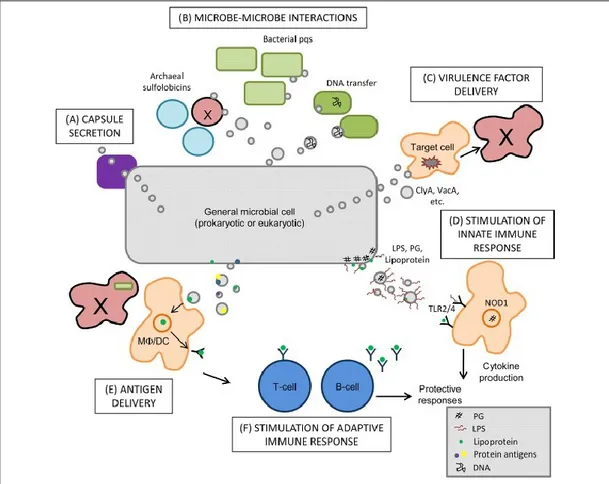
Bacteria have evolved over the millennia to survive in a wide variety of environments. Regardless of living location, the microbes have had to evolve different tools to grow in environments subject to change. One such tool that facilitates microbe-microbe, microbe-host and microbe-environment interactions is the production of membrane vesicles ( MVs). The first discovery of MVs was made over five decades ago, and it is now clear that the production of OM vesicles has been observed for a wide variety of Gram negative bacteria in all stages of growth as well as in a variety of growth conditions, such as infected tissues, with Pseudomonas aeruginosa and Escherichia coli because they are the most studied bacteria model. Vesicles were discovered first by T.J. Beveridge and colleagues, who is still considered the undisputed pioneer of the biological roles of MVs and mechanism of MV formation. The content, composition and purpose of these structures were unknown. For most of the time, the study of MV formation by prokaryotes has focused only on Gram negative bacteria. Recently, attention has turned to determining if MV production is also possible in Gram positive bacteria. Now we know that the production of spherical membranous vesicles is shared among all three branches of the tree of life: eukaryotes, Gram negative and Gram positive bacteria [Lee EY et al., 2009;McBroom AJ et al., 2005; Rivera J et al., 2010], archaea [Ellen AF et al., 2010], fungi [Oliveira DL et al., 2010], and parasites [Silverman JM et al., 2008-2010]. The release of MVs plays an integral role in cell physiology and the pathogenesis of infection. As the OMVs are known to
21 induce the immune system, they have been recognized as promising agents to be used as vaccines. Recently, one successful example is a vaccine for meningitidis caused by Neisseria meningitidis [Findlow et al., 2006; Boutriau et al., 2007; Williams et al., 2007]. Besides direct medical applications, the
study of OMVs is also an opportunity to better understand the physiology of bacteria.
1.2.2 What are MV Vesicles?
Bacterial membrane vesicles are closed spheroid particles produced by both Gram positive and Gram negative bacteria. The outer membrane (OM) vesicles of Gram-negative bacteria have a heterogeneous size from 10 to 300 nm in diameter, while the MVs secreted by Gram-positive bacteria have a smaller diameter between 20 to 150 nm [Dorward and Garon, 1990; Lee et al., 2009; Rivera et al., 2010]. In general, MV vesicles reflect the composition of the outermost portion of the membrane from which they derive, thus the OM vesicles derived from Gram-negative bacteria containing LPS, glycerophospholipids, and OM proteins as well as enclosed periplasmic components [Gankema H et al., 1980; Hoekstra D et al., 1976; Horstman AL et al., 2000; Kadurugamuwa JL et al., 1995; McBroom AJ et al., 2005] (Fig.7).
Just as there are differences in the envelope structure among Gram positive and Gram negative bacteria, MVs are completely different in composition in these two types of microorganism. Gram positive MVs originate from the cytoplasmic membrane and proteomic studies show they are composed mostly of cytosolic protein, cytoplasmic membrane-associated protein, as well as some secreted protein [Gurung et al., 2011].
22
Fig.7. Model of vesicle biogenesis. (LPS) Lipopolysaccharide; (Pp) periplasm; (OM) outer
membrane; (PG) peptidoglycan; (IM) inner membrane; ( Cyt) cytosol. Meta J. Kuehn, 2005. Genes & Development.
1.2.3 Regulation of vesicle Biogenesis
OMVs are secreted by bacteria both in liquid and on solid media, as well as in vivo. The goal of many OMV studies has been to understand how OMVs are formed and how this process is regulated, but is still poorly understood. Initially it was thought that the formation of OMVs was a physical process associated with the turnover of cell OM [Haurat et al., 2011]. Although this idea is still held by some, recent discoveries show that there is likely to be an elaborate mechanism behind the biogenesis of OMVs. There are several reports indicating that vesicles form as a response to membrane stress [McBroom AJ et al., 2007], however, the fact that membrane vesicles are released even when the cells grow without external stress makes it difficult to think that MV biogenesis is exclusively a stress response. For Gram-negative OMVs, several models of biogenesis have been hypothesized based on architectural features of the envelope and vesiculation mutants [Beveridge et al., 1999; Deatherage et al., 2009; Kulp and Kuehn, 2010]. Three mechanisms have been
23 proposed for vesicles biogenesis: (i) the accumulation of misfolded protein in the periplasm, may result in a mechanic force that literally pushes out the outer membrane and eventually leads to the release of OMVs; (ii) some external agents that could intercalate into the outer membrane and as a consequence membrane packaging is disturbed; (iii) some small molecules that normally are secreted through the cell envelope could change the regulation of genes, causing an increment or decrement of levels of OMVs (Fig.8).
In contrast to OMVs, the mechanisms and regulation of MVs of Gram positive bacteria have not yet been completely understood. OMV biogenesis is often considered to be stress regulated, in fact, OMV levels are modulated by altering levels of envelope proteins, temperature, and quorum sensing signals, and antibiotic treatment has been demonstrated to influence several aspects of vesiculation. For
Andrew J.Manning Meta J.Kuehn,2013
Fig.8. Possible Mechanisms of OMV
Induction.Three possib le models to explain how
OMVs form in response to different stimuli. A) The accumulation of misfolded or overexpressed protein in the perip lasm may result in a physical force on the outer membrane, causing the envelope to separate, the outer membrane to bulge, and eventually lead to the release of the OMV. B) External agents that act at the outer membrane could intercalate into the outer membrane, and disturb the packing of the outermost membrane. This disruption may lead to a change in the curvature of the membrane generating a bulge that eventually could bud off completely. C) A naturally secreted small molecule which can pass through the cell envelope could result in a change in the regulation of genes, causing an increase or decrease in levels of OMVs.
24 example, gentamicin and mitomycin affect the secretion and composition of OMVs in Gram-negative bacteria. The level of Shiga toxin-associated OM vesicle production by Shigella dysenteriae increased with mitomycin [Dutta S et al., 2004]. Release of MVs by Pseudomonas aeruginosa increased approximately threefold after exposure of the organism to four times the MIC of gentamicin [Kadurugamuwa JL et al., 1995]. The antibiotic ciprofloxacin interacts with DNA synthesis and leads to an
SOS response, which was found to be involved in increasing OMV levels. Vesiculation was found in both nonpathogenic and pathogenic species, under a range of growth conditions, including in liquid broth and on agar plates in the laboratory [Deatherage BL et al., 2009; Silverman JM et al., 2008], in biofilms [Schooling SR et al.,2006], upon infection with bacteriophages [Loeb MR et al., 1974], and in
pathogenic organisms growing within an animal host [Feldmesser M et al., 2001; Fiocca R et al., 1999, Marsollier L et al., 2007, Necchi V et al., 2007]. In general, pathogenic microbes produce more OMVs than nonpathogenic ones, such as enterotoxigenic E.coli (ETEC) that produces about 10-fold more vesicles than nonpathogenic E.coli [Horstaman and Kuehn, 2002].
1.2.4 Function of MVs
Several studies have shown many different functions for bacterial membrane vesicles. The main idea is that vesicles are necessary for the survival of the bacteria that produce them. MV release is essential for promoting interactions between microbial cells and between eukaryotic host cells and microbes, including communication, release of antigens, and secretion of virulence factors (Fig. 9).
25
Fig. 9. Biological impact of MV release. Brooke L. Deatherage, and Brad T. Cookson Infect. Immun.
2012.
OMVs produced from one bacterium can kill other competing microbes in the same niche, and in this way bacteria destroy coinfectors to prevent competition for limitated nutrients. The predatory nature of MVs was described in P.aeruginosa that has the ability to kill other Gram negative as well as Gram positive bacteria [Li et al., 2008]. In a recent study, Vasilyeva et al found that Lysobacter spp. XL1 secreted bacteriolytic enzymes in OMVs [Vasilyeva NV et al., 2008]. This activity of OMVs shows that they might be capable of distinguishing between self and non-self cells in a mixed community, thus vesiculating bacteria may have a survival advantage in mixed-population infections thanks to their capacity to eliminate competing bacterial strains. Furthermore, OMVs play a role in biofilm formation and maintenance. Biofilm formation is characterized by the expression of genes responsible for
26 exopolysaccharide production and co-aggregation of cells. Analyses of OM vesicle components have demonstrated that vesicles contain a wide variety of virulence factors. These virulence factors include protein adhesins, toxins, enzymes and antigens such as lipopolysaccharide (LPS). Kadurugamuwa and Beveridge showed that MVs liberated from Pseudomonas aeruginosa contain many virulence factors and are important in disease pathogenesis. Factors packaged into P.aeruginosa MVs include phospholipase C, proteases, alkaline phosphatases, and hemolysins. Although to date the real benefit to transport toxin and virulence factors by MVs is still unclear, Beveridge proposed two advantages: (i) the virulence toxin inside the MVs could be more concentrated and more focused to target cells; (ii) virulence factors transported by MVs may protect from degradation and recognition by host factors or microorganism. In addition, vesicles can facilitate the horizontal transfer between bacteria and this contributes to genetic diversity and bacterial survival. For example, M.catarrhalis which carry β-lactamase within MVs, is able to transfer resistance-antibiotic genes to S.pneumoniae and H.influenzae and to promote survival of these bacteria in the presence of antibiotic amoxicillin [Shaar et al., 2001b]. Recently, Fulsundar et al., have shown that OMVs from Acinetobacter baylyi were found to transfer small DNA fragments to E. coli [Fulsundar et al.,
2014]. Vesicle surface factors can mediate adhesion with eukaryotes and, as a consequence, vesicle materials can be internalized. The presence of bacterial membrane vesicles during infection processes has been observed in many human samples and infected tissue. Several reports of clinical isolates of H.pylori show OMVs in contact with epithelial cells. [Fiocca et al., 1999; Keenan et al., 2000]. In
27 studies of S.aureus and B.anthracis, MVs were found to be lethal to host cells [Gurung et al.,2011; Rivera et al.; 2010].
The mechanism of adherence and internalization is still the subject of many studies, however, OMV adherence has been studied in molecular detail for heat-labile enterotoxin (LT) of enterotoxigen E.coli [Horstman and Kuehn, 2002]. It is clear that vesicles contain compounds that are recognized by eukaryotic cells in the innate and acquired immune response pathways. Alaniz et al., demonstrated that OMVs from Salmonella enteric serovar Typhimurium are potent stimulators of proinflammatory
cytokine secretion. Salmonella OM vesicles determine the increased expression of tumor necrosis factor alpha and interleukin-12. The ability of bacterial membrane vesicles to trigger inflammatory response to pathogens has led to the development of immunogenic vaccines. Many studies have focused on investigating the potential of OMVs as vaccines for pathogens including Neisseria meningitides. A study carried out in Cuba showed that the OMV vaccine had a promising efficacy of 83-94% [Sierra GV et al., 1991]. More recently, a vaccine containing three N.meningitides surface antigens was developed in order to provide broad protection and minimize the risk of escape through mutations. New bacterial and viral disease are emerging, and at the same time there is a decline in the efficacy of antibiotics. For this reason, the application of OMVs holds some promise in this context.
1.2.5 MVs in Gram positive bacteria
Despite the fact that the discovery of membrane vesicles in Gram-negative bacteria goes back at least 40 years, MVs have been overlooked in Gram-positive bacteria. This lapse might be attributed to the very different composition of the cell wall between these bacteria.
28 Recently, it has been demonstrated that MVs can be produced by Gram-positive bacteria, such as S.aureus and B.anthracis [Lee et al., 2009; Rivera J et al., 2010]. Similarly, MVs shed from both strains are bilayer spherical vesicles, but the size is smaller than OMVs from Gram-negative bacteria. For the first time, Lee and colleagues showed that MVs from S.aureus contain many virulence factors and toxins, such as adhesins, proteolysin, coagulase and many other related enzymes that are implicated in infections of humans [Lee et al., 2009]. A few years later, Gurung et al. demonstrated that S.aureus secretes membrane vesicles into the extracellular
milieu during in vivo infection and the protein A, one of the toxins from S.aureus, can be efficiently delivered by intact MVs, whereas the protein A from lysed MVs is unable to entry the cytosol of the host cell [Gurung M et al., 2011]. Streptococcus pneumoniae also produces membrane vesicles, although these vesicle are
biologically and biochemically different from the plasma membrane from which they derive [Olaya-Abril A et al., 2014]. In particular, MVs are more enriched in lipoproteins and transmembrane proteins than in the plasma membrane. Furthermore, MVs do not have lipoteichoic acid, a typical molecule on the surface of Gram-positive bacteria. Proteomic analysis showed that MVs have a different fatty acid
29 composition: short-chain saturated fatty acids, conferring much more fluidity than the plasma membrane, consist of large amount of long-chain fatty acids. This might be, at least in part, an explanation for the biogenesis of membrane vesicles from the very hard cell wall of Gram-positive bacteria [Mercier R et al., 2012]. More recently, a large amount of lipoproteins organized in vesicles was isolated from Streptococcus pyogenes [Biagini et al., 2015]. Proteomic analysis of the vesicles revealed that they
were composed of phospatidylglycerol and lipoproteins. Bacterial lipoproteins are possible vaccine candidates because they are a major class of cell-surface proteins that are sensed by Toll-like receptors. Although, MVs shed by this bacterium are composed of lipoproteins, those structures do no act in a pathogen-associated molecular pattern [Biagini et al., 2015]. Taken together, these resultst seem to indicate that MVs serve as a transport system for virulence-associated components. During my PhD project, I tried to check, for the first time, if Streptococcus pyogenes can also be a producer of membrane vesicles and whether this could explain the hyper virulence of some Streptococcus strains due to the delivery of some toxins within MVs.
30
2. Study Aim
During my PhD at the Microbial Molecular Antibiotic Resistance (MMAR) laboratory my research line focused on molecular characterization and evaluation of genetic expression in different growth phases of three hyper virulent strains of S.pyogenes, isolated in different Italian hospitals, that were responsible for serious
cases of acute necrotizing pneumonia that led to the rapid death of patients due to respiratory failure, sepsis and necrotizing hemorrhagic pneumonia [Santagati M, 2014]. Simultaneously to the molecular characterization of clinical isolates, we assessed a production of membrane vesicles (MVs) by Streptococcus pyogenes during growth, hypothesizing that these structures could vehicle some toxins to
explain the hyper virulence of our sample used in this study. In this study, three strains of S.pyogenes were included:
1. RM1, isolated in Umberto I Hospital, Rome
2. RMG1, isolated in Agostino Gemelli Hospital, Rome
3. CT1, isolated in Vittorio Emanuele Hospital, Catania
The phases of the trial plan were:
I. Molecular identification of emm-typing and MLST determination
II. Determination of the antibiotic resistance profile and virulence gene
content by PCR and sequencing
III. Gene expression study of some virulence toxins in different growth phases
by Real-Time PCR
31
3. Materials and Methods
3.1 Bacterial strain and growth condition. All GAS serotype M1T1 strains were previously isolated from different patients with an invasive GAS infection [Santagati M, 2014]. One clinical strain (CT1) was isolated from the emergency department (ED) of Vittorio Emanuele Hospital in Catania, Italy, in July 2012. RM1 was isolated from the ED of the University of Rome Medical Center. RMG1 came to the ED of the Catholic University Medical Center in Rome in February 2012. All of these strains were tested for susceptibility to antibiotics and for the presence of virulence
genes by the PCR method [Santagati M, 2014].
We also used two representative GAS M1 serotypes, SF370 [Ferretti, J. 1991] and serotype M1 strain MGAS5005 were obtained from the American Type Culture Collection (ATTC No.BAA947) [Graham MR, 2002]. All of these strains were grown in vitro in Brain Heart Infusion broth with 0.2% yeast extract at 37°C with 5% CO2.
3.2 DNA extraction. For each sample, all colonies grown on Columbia Agar Base, plus 5% horse blood were collected and washed with 1 ml of a solution of 0.9 % w/v NaCl. Then, microbial DNA was extracted and purified through a QIAcube Extractor using the QIAamp DNA Mini kit (Qiagen, Limburgo, NL).The DNA concentration (absorbance at 260 nm; A260) and the purity (A260/ A280) were calculated using a BioPhotometer D30 (Eppendorf, Hamburg, Germany).
3.3 Molecular gene identification. The molecular identification of strains, emm-type and virulence genes was performed by PCR assays. emm typing was performed by PCR using protocols and the database of the Centers for Disease Control and Prevention (Atlanta, GA, USA;
www.cdc.gov/ncidod/biotech/strep/M-32 ProteinGene_typing.htm). Strains were tested for multiple virulence genes (Table 1) by using the PCR assay [Santagati M, 2014].
Tab.1: Primers used for identification of virulence genes by PCR for S.pyogenes isolates [Santagati
M, 2014].
3.4 RNA extraction. Total RNA was isolated from GAS grown to the mid-exponential phase (optical density at 600nm [OD600] of 0.4) and to stationary phase (optical density at 600nm [OD600] of 1.0). Cells were harvested by centrifugation and the bacterial pellet was re-suspended in 100 µl of diethylpyrocarbonate (DEPC) treated water. 100 µl of lysozyme 20mg/ml (Sigma) was added and incubation continued for 30 min at 37°C. After incubation we added 1ml of TRIZOL Reagent (Life Technologies) and incubation continued for 30 min at RT. After incubation 0.2 ml of chloroform was added per 1 ml of TRIZOL Reagent. The samples were mixed vigorously and then centrifuged at 12,000 × g for 15 min at 4°C. The RNA was precipitated from the aqueous phase by mixing with 0.5 ml of isopropanol. The samples were incubated at room temperature for 10 min and centrifuged at 12,000 ×
33 g for 10 min at 4°C. The pellet was air dried and dissolved in DEPC treated water. Contaminating genomic DNA was removed from each RNA sample using Turbo DNase (Ambion) and verified by PCR. The RNA was quantitated by A260 measurement.
3.5 RT-PCR and qRT-PCR. 10 µg of total RNA was converted into complementary DNA using hex nucleotide primers ImPRO-II Reverse Transcriptase Kit (Promega) according to the manufacturer’s instructions. Quantitative real-time PCR assays were performed using QuantiFast Probe Master mix in a Rotor-Gene instrument (QUIAGEN). Transcription of the gyrase subunit A (gyrA) was not affected under a variety of in vitro experimental conditions hence, gyrA expression was used to normalize in vitro TaqMan data. TaqMan primers and probes for genes of interest are shown in Table 2. Primers and probe (0.25 μM forward and reverse primers and 0.1 μM probe) was prepared, then 0.5 μL of each primer and 0.2 μL of probe were used in a final PCR reaction volume of 20 μL, containing 10 μL of 2X QuantiFast Probe PCR Master Mix (Qiagen, Germany), and 7.8 μL water and 2 μL of the genomic cDNA was added. The thermal profiling of the reaction included an initial denaturation step at 95ºC for 4 min followed by 40 cycles of annealing-extension step at 60ºC for 30 s. The performance of primer and probe sets was tested using theRotor-Gene Q system (Qiagen, Germany), and raw data were analyzed by the Rotor-Gene Q Software 2.1.0.9. All experiments were performed in triplicate on two separate days. Expression analyses were performed using the relative expression software tool REST2009 (Relative Expression Software Tool).
34
Tab.2: Primers/Probes used for TaqMan assay
3.6 MV Isolation protocol. MVs were isolated from supernatant culture following the method of Way et al. [Wai S.N, 2003]. Briefly, S.pyogenes RM1 strain was
GENE PROBE/PRIMERS SEQUENCE FLUOROFOR
gyrA
gyrA-Tq ACCACTGAGCCATACGAACCAT FAM
gyrA-up Tq TCGTCGTATTCTCTATGG
gyrA-rew Tq CAAGCATATGGCGATAAC
covS
covS-Tq TGCCATACGGTCAGCCTCAT FAM
covS-up Tq GGCATATTGGTCTCTTACA
covS-rew Tq GTACGCGAATCATGTCTA
covR
covR-Tq CAACATTAGTCTCAACGGCTTCATCAT FAM covR-up Tq CGTGAATATGATTTGCTTAA
covR-rew Tq CGGAGATAACGAATATAGAC
speB
speB- Tq ATTCTAGGATACTCTACCAGCGGAT HEX
speB-up Tq GAAGCAATGTTTTCTTTACC
speB-rew Tq GAGGATTTGTTATCGTTTCA
sagA
sagA-Tq TACAGCAGCAACAGCAGCCT FAM
sagA-up Tq GTAGCTGAAACAACTCAA
sagA-rew Tq CCAGTAGCAATTGAGAAG
slo
slo-Tq CCTTGTTACTGCTAATGCTGAATCG FAM
slo-up Tq GCAGCTCTTATCATTGGTA
slo-rew Tq GTCGTTGTGGTTTCTGTA
sic
sic-Tq TAGACCAGCCATATTGAGACCAGA FAM
sic-up Tq CTGGAGATGGTTTGTCTA
sic-rew Tq GGCCATTCTTCTTTATCG
hasA
has-Tq AGCACAGACCTATCCGTTATCAGAA FAM
has-up Tq GATGCCGAGTCATTATTAG
has-rew Tq CGACAAATATCCACTTCTC
ska
ska- Tq AACAGTCAAGTCGGTCCAGC FAM
ska-up Tq ACTGTGTTTGCATTAAC
ska-rew Tq CAGGTTGTGATGTTAGATC
sda1
Sda1- Tq AGAGCCACTGAATCCGACTACAAG FAM
Sda1-up Tq GTGGGTGGTATTCTTATTTC
Sda1-rew Tq NCTTCTTCTTAAGCTATCG
speA
speA- Tq CCTCCGTAGATACATGGACTCCTT FAM
speA-up Tq TCACGATTTTAATGTTTCA
35 grown in 1 L BHI-medium and cultivated overnight (Optical Density, OD600 0.9/1.0). After removal of the bacterial cells by centrifugation at 3500 rpm for 10 min at 4°, the supernatant fraction was sequentially filtered through 0.45 µm and 0.22 µm vacuum filters (PVDF Millipore) to remove residual bacteria and cellular debris. Preparations of membrane-derived vesicles were checked for absence of bacterial contamination by cultivating small aliquots on blood agar.
The filtered supernatant was then concentrated by ultrafiltration with Stirred Cell Model 8200 (Amicon) using a 100 kDa Ultrafiltration Disc PLHK (Millipore), and was centrifuged at 150,000 x g for 2h at 4° (Beckman L8-70M Ultracentrifuge, SW28 rotor). The pellet was washed with phosphate-buffered saline (PBS), centrifuged again (150,000 x g for 2h at 4°), and resuspended in 200µl of PBS.
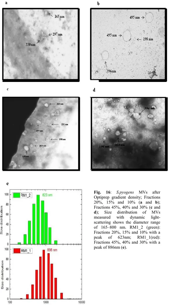
3.7 Purification of MVs. Fractionation of MV preparations was performed by density gradient centrifugation essentially as described by H.Chutkan [H.Chutkan, 2013]. OptiPrep (60% stock, Sigma-Aldrich) solution was added to the pellet containing MVs at a ratio of 1:3 (by volume) to adjust the vesicle preparation to 45% OptiPrep. 2 mL of vesicles were pipetted into 45% OptiPrep on the bottom of a 12.5-mL Ultraclear centrifuge tube, and then different Optiprep/HEPES layers were sequentially added as follows: 2 ml 45%, 2 ml 40%, 2 ml 30%, 2 ml 20%, 1ml 15%, and 2 ml 10%. Gradients were centrifuged at 120,000 x g for 4h at 4°C in an SW 41 Ti rotor (Beckman Instruments Inc.), and fractions of equal volumes (2/1 ml) were removed sequentially from the top. The MV fraction of 10%, 15% and 20% and those of 30%, 40% and 45% were polled together and was added tenfold the sample volume of DPBSS. MV fractions were centrifuged at 38,400 x g for 4h at 4°C to
36 remove the OptiPrep Solution. Vesicle pellets were resuspended in 200µl of DPBSS (Fig.10).
Fig.10. OMV preparation workflow. (1) Cultivation in liquid media (2) Removal of intact bacteria (3)
Concentration of the culture filtrate (4) Purification. Klimentová J, 2014. Microbiological Research.
Fig.11. Schematic of ultracentrifuge tubes loaded with the vesicle preparation in the 45% OptiPrep at
37
3.8 Scanning Electron Microscopy (SEM) analysis.
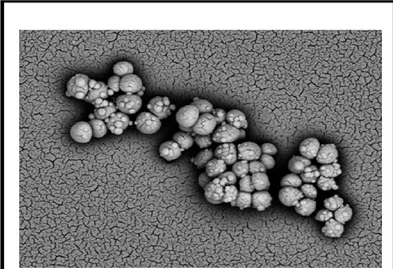
Bacteria for scanning electron microscopy were washed in 0.1 M sodium cacodylate buffer, applied to a poly L-lysine-coated coverslip, fixed with 2.5% glutaraldehyde, dehydrated, sputter coated and viewed on a Philips XL 30 ESEM at 30 kV.
3.9 Transmission Electron Microscopy (TEM) analysis.
The membrane vesicle pellets (20 µl) were fixed in 60 µl of 3% formaldehyde at 4°C. A drop (5 µl ) of the above suspension was layered on a formvar-coated copper grid (Electron Microscopy Sciences, Fort Washington, PA) and allowed to dry for 20 min to absorb exosomes. The grids were rinsed side down 2x2 min in PBS, fixed with 2% glutaraldehyde in PBS for 5 min at room temperature, rinsed 2x2 min with water and therefore were negatively stained with 4% uranyl acetate for 5 min and allowed to air dry. The observations were carried out using a Hitachi H-7000 transmission electron microscope (Hitachi High-Technologies Europe GmbH, Krefeld, Germany).
3.10 Dynamic Light Scattering (DLS). DLS measurements were performed with Zetasizer Nano ZS (Malvern, Herrenberg, Germany). Solvent-resistant micro cuvettes (ZEN0040, Malvern, Herrenberg, Germany) were used for experiments with a sample volume of 150µL. The count rates obtained were then corrected for the attenuator used. For each sample, two measurements were averaged [Palmieri, 2014]
38
4. Results
4.1 Molecular identification of emm-typing and MLST determination
The emm-typing determination of all isolates included in our study was conducted by PCR using protocols and the database of the Centers for Disease Control and Prevention (CDC). All three GAS strains were emm-type 1 and the analysis of allelic
profiles obtained showed that strains RMG1 and CT1 belong to ST28 sequence
typing, while strain RM1 is a new single locus variant (slv) of ST28, which was
deposited by us on the MLST database and called "ST648" [Santagati M, 2014].
Furthermore, determination of the clonal complex, which was performed using the
algorithm eBURST, showed that all strains with ST28/ST648 belong to the Clonal
Complex 28 (CC28)[Santagati M, 2014].
39
4.2 Determination of antibiotic resistance profiles and virulence gene content
All isolates were susceptible to erythromycin, tetracycline, amoxicillin, penicillin, and clindamycin by Etest (bioMérieux, Marcy l’Etoile, France). Results were interpreted according to the European Committee on Antimicrobial Susceptibility Testing breakpoints (www.eucast.org/clinical_ breakpoints) [Santagati M, 2014]. The virulence profile of our isolates was determined by PCR assay and all primers used were designed with the VectorNTI program (Invitrogen, Carlsbad, CA, USA) and are described in Tab.1.
To realize our aim, we introduced two different control strains into the study: S.pyogenes SF370, belonging to the M1-Type, (GenBank No.AE004092) and the
hypervirulent MGAS5005 strain (GenBank No. CP000017.2) with serotype M1T1 responsible for serious invasive infections.
The results obtained showed that the three clinical isolates, RM1, RMG1 and CT1 were identical, in terms of the spe genotype (speA+, speB+, speC–, speG+, speI–, speJ+, smeZ+), to the reference strain MGAS5005 M1T1 and [Santagati, 2014] they
were also positive for sagA and sda1. All the three GAS strains tested demonstrated the absence of ssa, prf and sof; while the SF370 strain was negative for speA and sda1 and positive for speC (Tab.3).
Strains emm-type CC ST speA speB speC speG speJ speI sagA sagBC smeZ slo ssa sda1 pam sof prtF
SF370 M1 28 28
5005 M1
RM1 M1 28 648
RMG1 M1 28 28
CT1 M1 28 28
40
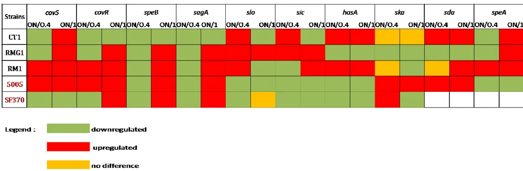
4.3 Gene expression study of some virulence toxins in different growth phases by Real-Time PCR
The expression of virulence genes in GAS is controlled by covRS and changes
during growth-phase. To investigate gene expression levels of most virulence toxins
implicated in GAS infections, we chose three growth phases:OD 0.4 and 1.0 at A600
nm and O/N time points, which coincide with the exponential phase (A600 nm = 0.4)
and the stationary phase (A600 nm = 1.0 & over night,O/N). To check for variation in
individual RNA isolations, total RNA was isolated from triplicate cultures at each
time point.
To determine the virulence expression profile of our three clinical isolates, we used
to different reference strains: SF370 which represents the model of invasive but not
aggressive GAS infection; and MGAS5005 strain, a serotype M1T1 organism that is
genetically representative of contemporary isolates and that has been used
extensively in pathogenesis research.
Our results showed that covS and covR, a sensor kinase and intracellular regulator of
the CovRS system, were downregulated in exponential phase in CT1, RMG1 and
SF370 S.pyogenes and upregulated in OD:1 phase with exception of SF370 in which
expression levels of covS remain low during the stationary growth phase, while those
of covR increase. Instead, levels of covS/R transcripts were upregulated both in the
model of GAS invasive infections, MGAS5005, which in one of the strains, RM1,
which was the clinical isolate that caused a more aggressive infection. RM1 and
MGAS5005 strains showed the same behavior with an upregulation of this locus
41
The transcription of speB and sagA, according to other papers [Chuan, Chiang-Ni et
al., 2009], was restricted to the stationary phase for all isolates, except for CT1
which always presents low levels of speB gene.
In contrast, expression of slo was restricted to the exponential phase for all clinical
isolates, but not for MGAS5005 and SF370, which downregulated both slo and sic
genes. High levels of sic gene were observed in CT1 and RMG1 strains during their
exponential phase; during the stationary phase just in CT1 GAS strain a lower
expression of sic was observed, which remained constant for RMG1 in both phases.
RM1 increased the expression of sic gene in the stationary phase with an opposite
behavior to the slo gene.
The hasA gene encodes hyaluronate synthase, which is required for resistance to phagocytosis, and was upregulated during all growth phases just for CT1 and RM1 strains, for the other strains this gene was downregulated. Regarding ska gene, we cannot describe significant changes in expression levels of our clinical isolates. An important characteristic of the hypervirulent globally disseminated MIT1 clone of GAS is the presence of two prophage-encoded genes, sda and speA. We observed high levels of sda transcripts in all growth phases and in all our samples of M1T1 GAS, except for RMG1; while the transcription of speA was restricted for most of the stationary phase, but not for MGAS5005, which unexpectedly expressed a low level of speA gene.
According to our analyses, none of the clinical isolates has an expression profile of virulence determinants comparable to the two different models of GAS strains, SF370 and MGAS5005. In fact, levels of two regulated genes (covS/covR) along
42 with genes encoding the toxins speB and sagA, greatly increase during their stationary phases for all of our clinical isolates. (fig. 14; Tab. 4)
43
44
Tab. 4: Analysis of GAS in vitro transcript levels.Aliquots were removed for RNA extraction at equivalent OD600 values: 0.4; 1.0 and O/N.
Relative abundance of CovR/S regulated transcripts determined by TaqMan assays. Data shown are fold changes in transcript (O/N culture strain relative to 0.4 culture and O/N relative to 1.0 culture) normalized to gyrA transcript amounts for selected genes.