Università degli Studi di Catania
Scuola Superiore di Catania
International PhD
in
Nanoscience
XXV cycle
Memory, metals and neurotrophins
Alessio Travaglia
Coordinator of PhD
Tutor
Abstract
1.
Introduction
1.1 Memory Consolidation
1.1.1 Synaptic Consolidation and Long-Term Potentiation
1.1.2 System Consolidation
1.1.3 Memory Consolidation: Molecular Perspective
1.2 Neurotrophins
1.2.1 Neurotrophins Signaling
1.2.2 Neurotrophins and Transcription Factors 1.2.3 Neurotrophins and Alzheimer’s Disease
1.2.4 Neurotrophins-based therapy and smart drug delivery
1.3 Metals and Memory
1.3.1 Metals and Metallostasis 1.3.2 Zinc and Memory
1.3.3 Copper and Memory
1.3.4 Alteration of Metallostasis and Neurodegeneration
1.4.1 Binding details of copper(II) and zinc(II) ions with neurotrophins
2.
Aims of the work
3
Materials and Methods
4.
Results and discussion
4.1 Characterization of NGF(1-14) and its metal complexes
4.1.1 Metal-hosting capability of NGF N-terminus peptide
fragments and effect of Cu2+ and Zn2+ on peptide conformation
4.1.2 Speciation, stability constants and coordination modes of the zinc(II) complexes with AcNGF(1-14) and NGF(1-14)
4.1.3 Speciation, stability constants and coordination modes of the copper(II) complexes with AcNGF(1-14) and NGF(1-14)
4.1.4 Proliferative effects of NGF and its N-terminus peptide fragments on SHSY5Y cells
4.2 Characterization of BDNF(1-12) and its metal complexes
4.2.1 Protonation constants and conformational features of BDNF(1-12), BDNF(1-12)D3N and Ac-BDNF(1-12)
4.2.2 Speciation, stability constants and coordination modes of copper(II) complexes with BDNF(1-12), BDNF(1-12)D3N and Ac-BDNF(1-12)
4.2.3 Speciation, stability constants and coordination modes of zinc(II) complexes with BDNF(1-12), BDNF(1-12)D3N and Ac-BDNF(1-12)
4.2.4 Proliferative effects of BDNF protein and its N-terminus peptide fragments on SHSY5Y cells
4.3 In vitro biochemical characterization of NGF(1-14) signaling
4.3.1 NGF(1-14) does not induce and does not maintain the differentiation of PC12 cells
4.3.2 NGF(1-14) affects the proliferation rate of PC12 cells
4.3.3 NGF(1-14) triggers the phosphorylation of TrkA and induces the phosphorylation of CREB at Ser-133.
4.4 In vivo biochemical characterization of BDNF signaling
4.4.1 MeCP2 is induced in many brain regions after learning 4.4.2 Distribution and regulation of MeCP2 in the hippocampus 4.4.3 BDNF signaling plays an essential role in MeCP2
upregulation.
4.5 Characterization of lipid/neurotrophins nanoplatforms
4.5.1 Association of NGF(1-14) to lipid vesicles
4.5.2 Adsorption processes onto planar silica surfaces: QCM-D
4.5.3 Biocompatibility assay of NGF/lipid adlayers with neuroblastoma SHSY5Y cells
4.5.4 Adsorption processes of NGF/lipid adlayers in silica
nanoparticles
5.
Concluding remarks
Acknowledgements
References
Annex 1: List of publications and communications
Abstract
In the last decades, one of the main interests of neuroscientists has been to unravel the molecular mechanisms of memory.
Neurotrophins are proteins involved in development and survival of neurons as well as they are active player in memory formation and synaptic plasticity. d-block biometals, especially copper and zinc, are emerging as crucial player in the physiology of the brain.
As matter of fact, there is a significant overlap between brain areas in which the highest concentration of metals have been measured and those where the neurotrophins exert their biological activity. Metal ions can directly modulate their activities, through conformational changes, and/or indirectly by activating their downstream signaling in a neurotrophin-independent mode. Despite the importance of these modulations, there is the lack of experimental data regarding the coordination features of metal ions complexes with neurotrophins.
The N-terminal domain of neurotrophins is critical for the binding selectivity and activation of their receptors. We synthesized the N-terminus peptide fragments encompassing the human neurotrophins, characterized their copper(II) and zinc(II) complexes by means of potentiometric, spectroscopic (UV/Vis, CD, NMR and EPR) techniques and DFT calculations, tested the metal-driven biological effect. The coordination features of acetylated as well as single point mutated peptides have been also studied to prove the involvement of each donor group. The functional interaction of biometals with neurotrophins and related peptides has been tested by biological assay on SHSY5Y neuroblastoma cell, providing evidence of the correlation between biological activity and coordination environment.
Our biochemical characterizations of the neurotrophins signaling, both in vitro and in vivo, have shed some light on the possible use of neurotrophins and neurotrophins-like peptides in neurological disorders. Indeed, the use of neurotrophins in the early stages of neurodegenerative diseases has recently gained attention. However, there are limits to such therapy, e.g. insufficient permeability of the blood-brain barrier and inappropriate
activation of receptors that trigger side effects. The use of peptidomimetic combined with systems that guarantee their delivery might allow to overcome these restrictions. In view of application as functional nanoplatforms for smart drug delivery, supported lipid bilayers formed by neurotrophin peptidomimetics/small unilamellar vescicles adsorption on silica (both planar model and nanoparticles) have been characterized.
In conclusion, the interaction of metals and neurotrophins might represent a crossroad for neuronal physiology. Better understanding of metal ion-driven neurotrophins signal transduction and intercellular signaling, as well as vice versa, the role of neurotrophins in the control of metal ions homeostasis, could disclose helpful information and it is therefore strongly raising as one of the most critical step in the study of neurodegenerative diseases as well in the physiological mechanisms of memory.
1. Introduction
1.1 Memory Consolidation
Memory is the active process by which incoming information are elaborated, encoded, stored, and subsequently recalled.1-2
The brain is the most astonishing, complex and less understood organ in the human body. It is made up of about 100 billion neurons (or nerve cells), each of which may be connected to up to 10,000 other neurons through special junctions called synapses.3-5 Whenever something is learned, circuits of neurons in the brain are created, altered, strengthened or weakened. 6-8 Thus, in physiological terms, memory is a set of encoded neural connections built up in the brain as a consequence of stimuli.2,4 The sensory system collects outputs, from the outside world, which are converted in chemical and physical signals (encoding phase). Then, the memory traces have to be stored in specific brain regions to be maintained over time (storage phase). Last, the acquired information has to be recalled if required. 9-10
Memory is usually divided into short- and long-term. 1-2,5,11 Short-term memory (STM) is the ability to hold and to recall small quantities of information for a short period of time, usually considered in the order of seconds. STM is due to transient patterns of neuronal connection, involving the paretial and the frontal lobe, especially the dorsolateral prefrontal cortex.5 Long-term memories (LTM), as the name suggest, allow storing unlimited quantities of information for long-lasting duration, sometimes a whole life span. However, the difference between STM and LTM is not only the duration of the memory traces, but also the different biochemical mechanisms of encoding/storage.2 Only a long-term memory triggers changes in neural pathways, necessary to store the information, that can be recalled even years later. It is believed that LTM are maintained through stable and permanent changes in neural connections widely spread throughout the brain.1,5 The hippocampus is a brain region considered essential to learn new information.2,12 Indeed, although it does not seem to store information itself, new memories are unable to be stored into LTM without the hippocampus.13-14 The hippocampus seems to act as temporary
from short-term to long-term memory. 2,11,15 Furthermore, it is involved in changing neural connections for a period of three months or more after the initial learning.2,11,15
Memory consolidation is defined as the process that stabilizes a memory trace after its acquisition. Indeed, intriguingly, after the initial acquisition phase, the new learned information is in a labile form, sensitive to disruption. 2,16-17 Memory consolidation is distinguished into two specific processes, synaptic consolidation, which occurs within the first few hours after learning, and system consolidation, which lasts over a period of weeks to years. 2
1.1.1 Synaptic Consolidation and Long-Term Potentiation
Synaptic consolidation has been observed across all species and occurs within the first few hours after learning. Long term potentiation (LTP) is one of the best understood forms of synaptic plasticity and synaptic consolidation.12,18-19
The most widely studied type of LTP is the N-methyl-D-aspartate receptor (NMDAR)-dependent LTP at the synapses between Schaffer collaterals and commissural neurons in area CA1 of the adult hippocampus 12,18 Much is known, at molecular level, about this process. Glutamate molecules, released into the synaptic cleft, bind NMDA receptor. Only in the presence of a coincident event of depolarization, Mg(II) ion is expelled from the NMDAR channel. When the channel is no more blocked by Mg(II) ion, the binding of glutamate molecules can open the NMDAR channel, allowing an influx of calcium(II) ions into the cell.18,20 The calcium influx trigger the activation of protein kinases, such as Ca2+/calmodulin-dependent protein kinase II (CaMKII) and protein kinase C (PKC).21-22 In turn, these proteins, as well the extracellular signal-regulated kinase (ERK), activate transcription factors that trigger the synthesis of new proteins that maintain the LTP. 23-25
1.1.2 System Consolidation
System Consolidation is a slow and dynamic process in which memory traces are “moved” from the regions in which are first encoded to the cortex, in a permanent form of storage.2-3
It is believed that memory is retained in the hippocampus for up to one week after initial learning. Then, the memory traces are slowly transferred to the neo-cortex, where it is permanently stored.
In this view, the neurogenesis in the adult hippocampus may facilitate to delete the old memories, creating new “space” for the incoming new memories.26-27
1.1.3 Memory Consolidation: Molecular Perspective
Neuroscientists have been prompted to go in deep on the details of long term memory formation since the discovery that, at the beginning of the consolidation process, memory formation can be prevented/disrupted by interferences.3,17,28-29 These include seizure, trauma, neuronal inactivation and brain lesions, but also by new additional learning, inhibition of transcription or specific transcription factors, inhibition of protein synthesis or selective blockade of specific molecular pathways.29-30 From the first discovery, a plenty of papers have shed light on the molecular mechanisms of memory consolidation, providing evidence on the role of specific biochemical events involved in long-term memory formation.7,10,31-33
What are the transcription factors involved in memory formation? What are they target genes and how they allow complex cognitive responses? Or, in other words, what are the proteins required for memory formation? In which brain regions are they necessary? What is the time course of transcription and protein synthesis requirements? Much has been done, but many points have not been completely unraveled.
Least but not last, an increasing number of evidences suggest a potential role of transition metal ions in neuromodulation/neurotransmission. The brain is an organ with extraordinary complexity and unique chemical composition and its inorganic chemistry is inherently rich and remains an open frontier to study. What metal ions are present in the brain? How are they distributed? Do they have a function role in memory formation?
Finally, besides mere knowledge of what transcription factors and proteins are involved in memory formation, it would be interesting to evaluate their interactions/regulations. In this context, metal ions might directly modulate the activities of biological macromolecules, likely through conformational changes, and/or indirectly by activating their downstream signaling.
1.
2 NeurotrophinsNeurotrophins (NTs) are secreted proteins discovered because of their essential role in development, differentiation, and maintenance of specific populations of neurons.34-36 Then, in the last decade, it has been reported that neurotrophins and their receptor match all the characteristics that a protein must have to play a consistent role in memory consolidation and synaptic plasticity:
1) Their expression, regulation and secretion are activity dependent.
2) They are expressed in the right place/time where memory consolidation takes place, and their receptors are localized in the same area.
3) As a consequence of its activity, they modulate the neuronal function, such as membrane excitability, neuronal morphology and connectivity.
Thus, besides their well-established actions in survival and development, today is known that they are involved in the wiring regulation of the central and peripheral nervous system during development, 37-38 can modulate synaptic plasticity39-42 and cognitive functions, 39-41,43-44 as well they seems involved in the etiology/exacerbation of neurological disease, such as Alzheimer‟s Disease.
The neurotrophins family comprises nerve growth factor (NGF), brain-derived neurotrophic factor (BDNF), neurotrophin-3 (NT-3) and neurotrophin-4 (NT-4). The NTs are structurally and functionally related proteins, which exert their biological action as non-covalent dimmers.45 They share a sequence homology close to 50% and a structural homology of about 60%. A single monomer of each NT has a core consisting of two pairs of antiparallel β-strands with well conserved sequences among the neurotrophins family members, and responsible for the dimer assembly. Whereas, the N- and C-termini, as well as loops 2 and 3, determine sequence diversity 46-48
Discovered by Rita Levi-Montalcini and Stanley Cohen almost half century ago, NGF is the founding and best-characterized member of the neurotrophins family.35 All the
neurotrophins are synthesized as precursor (pro-NTs) and cleaved by some proteases (plasmin, furin, MMP7 and MMP3)49-50 to produce the C-terminal mature and active forms, which exert their biological action as non-covalent homodimer. Neurotrophins driven cell signaling operate through the interaction with two distinct classes of receptors: p75 and Trks (tropomyosin receptor kinase) receptors, thereby activating signal transduction cascades that trigger distinct biological responses.47,51-53 In a simplified view, p75 transmits apoptotic signals, whereas Trks receptors trigger pro-survival outcome (Fig. 1).
Figure 1. Schematic representation of neurotrophins receptors and their response upon activation
1.2.1 Neurotrophins Signaling
Low-affinity neurotrophins receptor, p75, is known as the common neurotrophins receptor, because of its ability to recognize all the NTs, even though with different kinetic constants. The receptor p75 plays a variety of roles in neurotrophins signaling, including the enhancement of Trk activity as well as Trk-independent signaling. Pathways that are independent from Trk activation may, under appropriate conditions, lead to an apoptotic cell death.51-53
includes Fas receptor, tumor necrosis factor receptor, and other receptors.
The intracellular domain of p75NTR lacks any intrinsic catalytic, thus the p75 signaling is believed to be the consequence of association/dissociation of various intracellular adaptor binding proteins.51-53
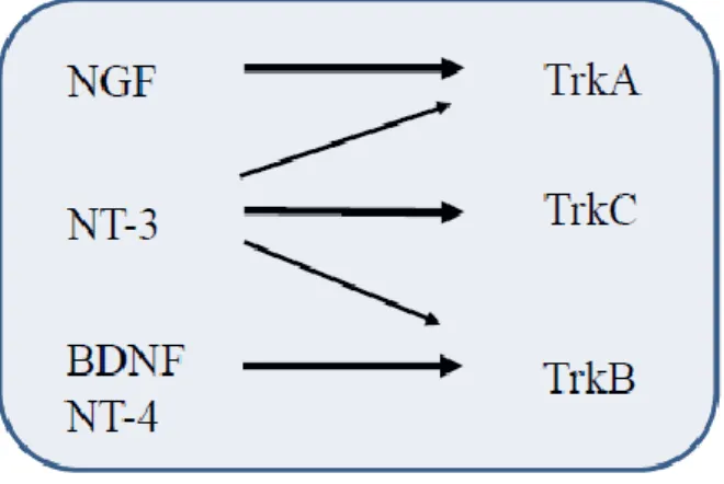
The Trk receptor belongs to a family of three different members: TrkA, TrkB and TrkC. The Trk receptor family shows a high degree of specificity (Fig 2) i.e. NGF binds TrkA; TrkB is the receptor for BDNF and NT-4, whereas NT-3 binds to TrkC. The latter one seems to be promiscuous, since it can also activate TrkA and TrkB.51-53
Figure 2. Schematic representation of interaction between neurotrophins and their cognate Trk receptor
The binding of NGF to TrkA induces receptor dimerization and the consequent trans-autophosphorylation of tyrosine residue in the intracellular domain of the TrkA. This event represents the beginning of a downstream signal, which involves several kinase cascades (Fig 3). The main signal transduction pathways due to TrkA activation include:
a) The classic MAPK cascade, involving activation of the G- protein Ras, and the kinases Raf and MEK, abbreviated as the reaction series Ras/Raf/MEK/MAPK. The major role of the Ras-MAPK pathway is the neurite outgrowth.
b) the phosphatidylinositol-3-kinase (PI3 kinase)/Akt pathway. The PI3K-Akt pathway is a key regulator of the anti-apoptotic/pro-survival response in neurons
c) the phospholipase C gamma (PLCγ) pathway. PLC activation leads to PI3 hydrolysis, with formation of IP3 (inositol trisphosphate) and DAG (diacylglycerol). Consequentially, IP3 induces the release of Ca2+ by stores, resulting in the activation of various Ca2+ regulated enzymes. This pathway may play a role in Ca2+-mediated cellular degeneration in aging and disease by affecting oxidative stress systems.51,54
P
P
P
Gene Expression
Neurotrophin
RSK
MAPK
MEK
AKT
[Ca
2+]
CaMKK
CaMKIV
IP3/DAG
PI3K
PLCg
RAS
Transcriprion
Factors
(e.g. CREB)
Trk receptor
The overall picture of neurotrophins-mediated signaling cascade is tangled by the activity of pro-forms, Indeed, pro-Neurotrophins have been initially considered the precursor of the mature (and active form) of the neurotrophins, thus molecules without any biological activity
Now it is recognized that pro-neurotrophins mediate cellular apoptosis 55 and inhibit neurite growth in many cell lines and primary neurons 56 through p75 receptor, even in the presence of TrkA, with the involvement of sortilin receptor, a member of Vsp10p-domain receptor. 57
In summary, a strict, not fully understood, regulation of p75 and TrkA (and sortilin) pathways by NGF (and its precursor) is emerging as responsible for modulation of many different aspects of neuronal life, like neurite outgrowth and differentiation, pro-survival signals or pro-apoptotic outcome, and molecular mechanisms involved in physiological as well pathological conditions.51,58-59
1.2.2 Neurotrophins and Transcription Factors
Neurotrophins, through their downstream signaling that raise intracellular cAMP and Ca(II) ions concentration, ultimately trigger the activation of transcription factors.
Eric Kandel, Nobel Prize in 2000, has described the process of memory storage as a “dialogue between genes and synapses”.4 The genetic information carried by all organisms is expressed in any biological function via a series of processes that begins with DNA transcription, a process through which the information encoded in the DNA is copied into a molecule of RNA (which is then translated to a specific protein).
Beside to be the first regulatory step in gene expression, transcription is a molecular requisite for long-term synaptic plasticity and long-term memory formation.9,24,60 Indeed, memory consolidation can be altered/prevented by agents able to interfere with protein transcription.23,25,61-63 Thus, researchers have been focused on the identification of families of transcription factors required to mediate memory formation and to elucidate how they exert this function.24-25,64 These include cAMP response element binding protein (CREB), CCAAT enhancer binding protein (C/EBP), methyl-CpG-binding protein 2 (MeCP2) activating protein 1 (AP-1), early growth response factor (Egr), and Rel/nuclear factor κB (Rel/NFκB).24-25,64
Among them, the transcription factor cAMP response element-binding protein (CREB) is probably the most studied. CREB is a 43 kDa protein widely expressed in the brain, especially in the regions essential for encoding learning and memory, including the hippocampus and cortex.65-66 Neurotrophins, as previously mentioned, through their downstream signaling that raise intracellular cAMP and Ca(II) ions concentration, 67-68 trigger the phosphorylation and activation of CREB at Ser-133. The phosphorylated form of CREB (pCREB) binds specific DNA sequences, thus enhancing the expression of genes that results in synapse-specific structural changes.66,69-70 CREB signaling is reputed essential for neuronal changes that mediate many important functions in the nervous system, including neurogenesis and neuronal survival, development, and differentiation, synaptic plasticity, and the conversion from short- to long-term memory.42 Also, CREB
signaling has been recently involved in several neurodegenerative disease, such as Alzheimer‟s Disease.66,69-71
A transcription factor recently connected with the neurotrophins signaling and cognitive function is the methyl-CpG-binding protein 2 (MeCP2). MeCP2 has been first discovered because of its ability to bind methylated DNA. It is expressed throughout the body and is particularly abundant in the brain, where it seems to be essential for the normal function of neurons. 72-75 It has gained popularity and the attention of scientific community because its mutation cause the autism spectrum disorder Rett Syndrome (RTT).76-77 This disease is mainly found in girls with a prevalence of around 1 in every 10,000. Strikingly, there is not sign of disease at the born, but after 6-18 months, RTT patients lose purposeful use of their hands and instead develop stereotypic hand movements. The onset of mental deterioration is accompanied by loss of motor coordination and deceleration of head growth.78-79 The exact function of this protein is not known. It seems to act as a transcriptional repressor, thus preventing gene expression when is not needed. However, MeCP2, in addition to its long-term gene-silencing role, recent works point out that MeCP2 can also activate the gene expression, though it is still debated.75,78
Among the target genes of MeCP2, that encoding for BDNF is the most extensively studied, since the pivotal role played by this neurotrophin in neuronal survival, dendritic arborization, circuit formation and function of connected neurons.44 MeCP2has been reported to specifically bind BDNF promoter III and functions as a negative regulator of BDNF expression. In response to membrane depolarization and consequent calcium influx into the neurons, MeCP2 becomes phosphorylated and is release from the BDNF promoter, thereby permitting BDNF transcription. 78,80-85 Also, despite the evidence that MeCP2 plays an important role in neurons, recent works have raised questions about what is the primary cell types (e.g. neuron, astrocytes or microglia) which lead to the manifestation of RTT phenotypes, despite the low abundance of MeCP2 in non-neuron cell types.80-81
1.2.3 Neurotrophins and Alzheimer’s Disease
Neurodegeneration is the progressive loss of structure and/or function of neurons, and increasing the likelihood of death. 86-87 The increase of elderly population due to life span prolongation has unfortunately led to a high variety of age-related neurologic diseases particularly related to accumulation of misfolded proteins. Alzheimer's disease (AD) is the most common form of dementia in the elderly, the fourth cause of death in the western world (after heart disease, cancer and stroke); AD affects 24 million of patients and it is expected to double by 2020.88
The pathologic hallmark of AD is the formation of intracellular neuronal tangles and the deposition of amyloid plaques in the neural parenchyma. 89-90 Amyloid β (Aβ), a small peptide featured by 39 to 43 amino acids, is the principal component of plaques that originate from the type-I membrane protein, Amyloid Precursor Protein (APP), after cleavage by β- and γ-secretases. 91-92 According the „amyloid cascade hypothesis‟, the excessive production and accumulation of Aβ triggers the pathogenesis of AD. 89 Indeed, plaques and tangles are present mainly in brain regions involved in learning, memory, and emotional behavior such as the entorhinal cortex, hippocampus, basal forebrain and amygdale. However, the amyloid cascade hypothesis is not completely convincing and accepted and the exact nature of the toxic species is debated. 93 It seems to be related to soluble oligomers rather than to plaques, 90 whereas monomers have been found to be neuroprotective. 94-95
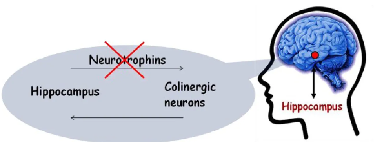
A large number of efforts have been made to contrast the deranged effects of neurodegeneration and to identify therapeutic interventions to prevent the progression of age-related cognitive decline.92-93,96-97 In this respect, NTs and their receptors have gained most attention as potential targets. The homeostasis of different neurotrophins is indeed altered in specific brain areas during the pathogenesis of AD 98-99 In the central nervous system, the highest levels of NGF are present in the hippocampus, the brain area involved in learning and memory, and in the areas containing basal forebrain cholinergic neurons (BFCNs) 100; all these findings led to the formulation of the “NGF hypothesis of AD” (Fig. 4). 101-103
Figure 4. The Neurotrophins hypothesis of Alzheimer’s Disease suggests that an alteration in
NFG/BDNF signaling is related with the etiology/exacerbation of neurodegenerative disease.
It is worth noting that hippocampus is innervated by BFCNs. In turn, BFCNs are supplied with NGF by hippocampal neurons; namely NGF is taken up by the terminals of cholinergic neurons and retrogradely transported back to the cell bodies in the basal forebrain. It is accepted that NGF is essential to maintain the phenotype and the synaptic activity of BFCNs, as well as to prevent their age-related atrophy.104 The degeneration and atrophy of BFCNs has been linked with memory impairment and cognitive dysfunction in AD. 105-106
Impaired axonal transport has been suggested to decrease the level of NGF in BFCNs during the progression of AD, 107 although this model is not completely accepted. 108 Moreover, in support of the neurotrophins hypothesis of AD progression, a marked change in the expression levels of the neurotrophins receptors occurs during ageing, and TrkA expression has been reported to be downregulated in the AD parietal cortex and BFCNs. 109-114 At the same time, there is a progressive upregulation of p75 levels, 115 that indicates a disequilibrium in the TrkA/p75 ratio and consequently in the NGF-mediated signaling. This could play a relevant role in the etiology of AD 110-111,116. Deficits in TrkA receptor phosphorylation signaling contribute to age-related default in long-term potentiation (LTP)
117-119, one of the major cellular mechanisms involved in learning and memory (see paragraph 1.1.1). This can be likely the molecular mechanism for memory impairment in AD.
Contrasting NGF levels have been reported in AD. 111,120-123 Nonetheless, pro-NGF levels double in frontal and occipital cortex and in hippocampus of AD patients, 124 suggesting that defective cleavage of pro-NGF to mature NGF may occur in this disease. 49,125 Pro-NGF is, indeed, the predominant form of Pro-NGF in the brain of AD patients and increases with the progression of disease. 124,126 Intracerebral administration of pro-NGF has been found to causes AD-like learning impairments in mice, supporting the above mentioned relationship. 127 Intriguingly, intrahippocampal injection of Aβ oligomers elicits the upregulation of pro-NGF in rat. 128 The unbalance of NGF processing can therefore be suggested as the onset condition of AD neurodegeneration, but direct, still unexplored, effects of pro-NGF cannot be excluded.
More recently, BDNF is also emerging as a crucial player in the CNS. Its role is highlighted by the fact that hippocampus is the brain area with the highest BDNF expression. The BDNF mRNA expression is 50-fold higher than that of NGF. 129 With few exceptions, 130 the scientific community agrees that the reduced brain level of BDNF and its mRNA could contribute to the progressive atrophy and death of specific neuronal populations in the AD-affected brain. 99,122-123,131 Analogously to NGF, BDNF exists in the brain also as pro-BDNF but, differently from pro-NGF, pro-BDNF level decreases in AD brain. The reduced level of BDNF can, therefore, be correlated with the significant downregulation of the precursor. 132-133 A reduction of both pro-BDNF and mature BDNF levels has been linked with loss of cognitive functions. 134 The study of correlations of NGF processing with AD, or other age-related neurodegenerative diseases could open new perspectives for the particularly demanding challenge of definite therapeutic interventions.
Finally, an intriguing cross-talk between the “amyloid hypotheses” and the “NGF hypotheses” of AD is recently emerging. It has been found that an interruption of NGF signaling can activate the amyloidogenic pathway, inducing cell death through a not fully understood mechanism. 135 The block of NGF signaling has been shown to up-regulate APP, β- and γ-secretases, with deposition increase of amyloid plaques. 136 Similarly,
transgenic mice expressing recombinant antibodies able to neutralize up to 50% of endogenous NGF have been reported to develop AD-like features. 137 In addition, these AD-like features have been shown to be due to an imbalance of the TrkA/p75 ratio and signaling. 116 Indeed, the APP processing is differentially regulated by TrkA and p75 receptors, which reduce and activate the cleavage of APP, respectively.138
Further interest in the role of these receptors in the etiology of AD can be attributed to the finding that Aβ can activate TrkA 139 as well as p75 140 receptors and a downstream cascade which leads to neuronal death. Aβ(1-42) doubles the level of p75 in SH-SY5Y human neuroblastoma cells and in the hippocampus of AD-transgenic mice141, also inducing significant cell death in hippocampal neurons, whereas the same effect has not been observed in p75-deficient neurons. 142 A dose-dependent effect of Aβ has also been observed: i) high concentrations of Aβ determine apoptosis (10-100 µM) 143 or prevent NGF-induced activation of NF-kappaB (800nM) 144 ii) low concentrations of Aβ (25 nM), in contrast, have been reported to be pro-survival, inducing cellular effects similar to those triggered by NGF. 143 In addition, low levels of Aβ upregulate the BDNF mRNA at short times (3-5 h) but dramatically reduce it at longer times (24 or 48 h). One could speculate that aggregation of Aβ occurs 145 and cytotoxic activity of soluble oligomers of Aβ(1-42) will take place. 115
1.2.4 Neurotrophins-based therapy and smart drug delivery
The results presented in the previous paragraph suggest the possibility to develop a neurotrophins-based therapy for neurological disease. However, there are some hurdles in the use of neurotrophins as drugs, due to their poor pharmacological properties, such as the low blood–brain barrier permeability and a wide range of adverse side effects, due to the activation of p75 receptor.120,146-147
Considerable efforts have been made to obtain agents that can mimic or antagonize the NGF activities, with the aim:
i) to cross the Blood-Brain Barrier and achieve adequate concentrations in the target regions of the brain
ii) to elicit certain but not necessarily all the signaling cascades triggered by NGF
Intracerebral injections of NGF, implantation of viral vectors and genetically modified cells/devices able to release NGF are invasive methodologies to be applicable. 148-151 On the other hand, intranasal delivery and topical application of NGF on the ocular surface are non-invasive approaches that need further investigations. 120 Micro- and nano-particles made of different polymeric materials, able to storage and release NGF, have been developed. 152-156 The encapsulation of NGF into liposomes has been reported to prevent enzymatic degradation, whereas the functionalization of the liposome is a good strategy to promote its permeability across the blood-brain barrier. 157
Innovative strategies for immobilization and release of biomolecules are highly challenging. One intriguing approach has been recently proposed for ligand cell targeting. It is based on the multifunctional character of a nanoplatform, so-called „protocell‟, consisting of hybrid materials, made of silica nanoparticles wrapped by fluid-phase lipid bilayers. 158-159 Indeed, lipid bilayers are able to mimic cellular membranes, due to their electric charge, thickness, and permeability. Moreover, they offer the advantage to anchor ligand molecules in a not rigid manner, representing a breakthrough of traditional rigid gel-phase liposome vesicle.160 The non-covalent bond between ligand and drug carrier is a
fundamental pre-requisite to decrease the overall concentration of ligands – thus avoiding adverse biochemical signaling to the cell - but, at the same time, to allow a local enhancement of ligand in proximity of specific cells receptors. 158-159 Also, it has to be mentioned that the overall ligand concentration is only one of the crucial parameters for an efficient drug activity. A suitable ligand-receptors interaction on the target cell is critically driven by local concentration gradients as well as conformational states.
Some of the methodologies described can potentially guide the neurotrophins across the Blood-Brain Barrier. However, encapsulated or not, neurotrophins elicits both TrkA and p75 receptors, the latter responsible of side effects. To overcome this second issue, considerable efforts have turned to design small molecules able to interact selectively with the TrkA receptor mimicking or antagonizing the NGF activities. 146-147 To this aim, important clues derive from the comparison of the crystal structures of neurotrophins 45,161-169
The Neurotrophins-Trkr interface consists of two patches: the first involves the β-sheet of the core of the homodimeric active form; the second appear to be formed by discontinuous stretches of amino acid residues distributed throughout the primary sequence of the molecule. 45 Comparing the structure of the neurotrophins with their cognate Trks receptor, it has been observed that the former domain constitute a conserved binding motif for all neurotrophins family member.45,162-163 Strikingly, the latter contain residues not conserved among the neurotrophins family, suggesting that these patch can guide the specificity for a peculiar Trk receptor.
Among the non conserved residues, many biochemical studies have been focused on the N-terminus tail, unraveling its importance.
It has been reported that a truncated form of NGF, which lacks the first 9 residues of NGF, displayed more than 300-fold lower affinity for TrkA than the whole NGF, and 50-fold lower capacity in eliciting TrkA phosphorylation, without affecting folding, stability or conformation of the molecule.170-171 Among the residues of the N-terminal domain, the His-4 has been reported to be critical. Diethyl pyrocarbonate modification of His-4 and His-8 in a NGF double mutant abolished neuritogenesis, binding to both receptors, and
phosphorylation of TrkA in PC12 cells. NGF(H4D) mutant showed a 1000-fold lower binding affinity for TrkA (compared with wild type NGF), at least 10-fold less efficacy in the TrkA autophosphorylation, and 30-fold less capacity in PC12 cell differentiation.172-173
These studies have been confirmed by computational calculations, which identified the residues lying at the ligand-receptor interface. These include His4, Glu11, Trp 21, Arg 59, and Arg103 of NGF, and Glu 295, Phe 303, Arg347, Asn 349, and Gln 350. 174-175 In particular, the His 4 and Glu 11 residues provide highly persistent H-bond interactions with the receptor, as well as Ile 6 and Phe 7 residues stabilize the binding of the N-term of NGF to TrkA through hydrophobic interactions. 174-175
1.3 Metals and Memory
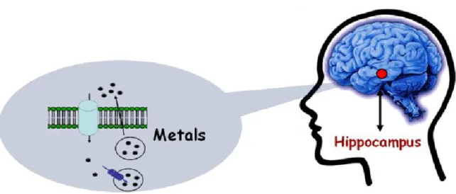
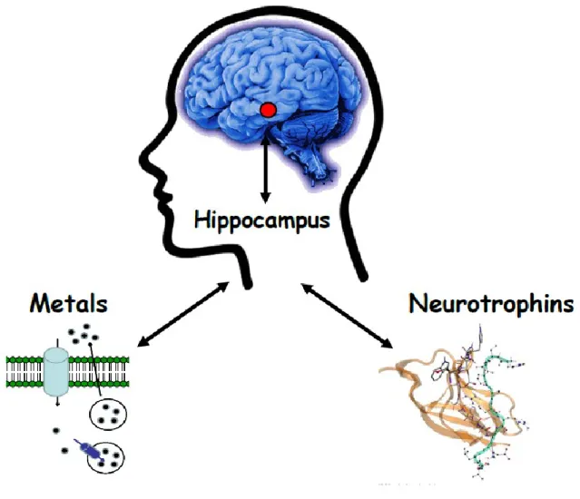
As previously mentioned, the brain is an organ with astonishing complexity and unique chemical composition. The role of s-block metals (Na+, K+, Ca2+) in the brain has been studied in deep. Less is known about the role of transition metal ions. Actually, although they are often called “trace metals”, their concentration (above all copper and zinc) is 10000-fold higher than common neurotransmitters and neuropeptides. The abundance in the synaptic cleft provide a clue of their functional role in the brain and, perhaps, in memory consolidation (Fig. 5).
Figure 5. Metals in the brain. Emerging evidences suggest that transition metal ions are
released in the synaptic cleft and might take part in neuromodulation/neurotransmission.
1.3.1 Metals and Metallostasis
Transition metal ions are essential catalytic and/or structural elements of many enzymes, transcription factors and/or other regulator agents, as well as emerging modulators if neuronal functions.176 However, neurons (as well as all the other cells) need to maintain
the optimum of concentration, lower and higher values are often detrimental to cells life. Thus, a highly orchestrated and regulated mechanism is required to guarantee metallostasis (metal ions homeostasis). 178
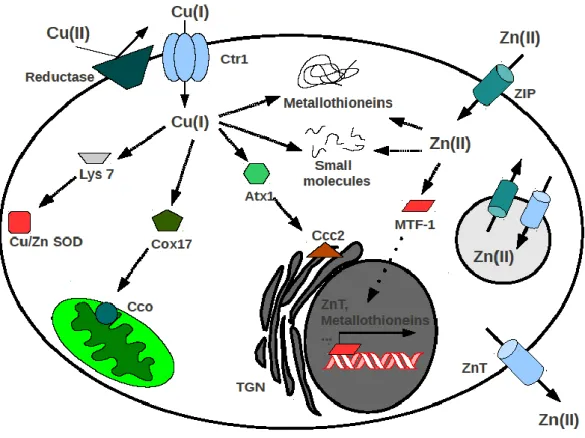
Figure 6. Schematic model of copper and zinc homeostasis. Extracellular Cu2+ is reduced by
plasma membrane reductases, then Cu+ enters the cell via Ctr1 (copper transporter-1). Cu+ can
be funneled to its ultimate intracellular destinations through specific protein-based pathways. Lys7 is the copper metallochaperone for Cu/Zn superoxide dismutase (Cu/Zn SOD), Cox17 loads Cu+ into the cytochrome c oxidase (Cco), Atx1 shuttles Cu+ to the Ccc2 P-type ATPase transporter
localized on post-Golgi network (TGN). ZIP proteins (zrt/irt-like proteins) are influx transporters that mediate Zn2+ uptake from the extracellular environment, or intracellular
vesicles, into the cytoplasm. Otherwise, ZnT carriers (zinc transporter protein) are efflux transporter able to reduce the cytoplasmatic Zn by promoting zinc movement directly out the cell or into intracellular compartment. Metal transcription factor-1 (MTF-1) binds Zn2+ and
translocates to the nucleus to upregulate metallostasis-involved proteins. Besides to be shuttled to various proteins and subcellular organelles, zinc as well as copper ions are buffered by metallothioneins and small molecules (e.g glutathione, ascorbic acid).[from 179 ]
Through a complex network of molecular interactions, metallostasis acts to balance the intracellular metal uptake, trafficking, storage, speciation and signaling (Fig.6). These intricate biochemical pathways allow the storage and delivery of metals to their proteic partner. Also, the strict balance of metallostasis is controlled by metals themselves, through transcription factors and post-transcriptional regulatory mechanisms.
A picture of zinc(II) and copper(II) ion cellular homeostasis is summarized in the following.
The ZnT (Zinc Transporter) 180-181 and the ZIP (Zinc regulated metal transporter, Iron-regulated metal transporter-like protein) families,182 including 10 and 15 members, respectively, consist of multipass transmembrane proteins involved in zinc(II) ion homeostasis. 183 Zip proteins are influx transporters that mediate Zn2+ uptake from the extracellular environment, or intracellular vesicles, into the cytoplasm. ZnT carriers are efflux transporters that reduce the cytoplasmatic Zn2+ by promoting metal ion movement directly out the cell or into intracellular compartment.
It is worth to note that both ZnTs and Zips exhibit unique tissue- and subcellular- specific expression. For instance, ZnT1 is a cellular zinc exporter and is predominantly expressed on the plasma membrane of cells and seems to play a key role in early embryonic development and a protective role in the nervous system. 184
ZnT3 transporter is particularly abundant in regions that have high levels of labile, chelatable Zn2+ and it has been suggested that it accumulates zinc in synaptic vesicles and it has a neuromodulatory role in synaptic transmission. It has been linked to degenerative neurological diseases such as Alzheimer‟s.185-186
Once inside the cell, besides to be shuttled to various subcellular organelles, zinc ions are bound by metallothioneins (MTs), a class of cystein-rich cytosolic protein with high affinity for metals. Although a single essential function of MT has not been demonstrated , MTs have been proposed as playing an important role in controlling whole body zinc metabolism.187
The expression of several genes, involved in zinc metabolism, is regulated by zinc itself. Indeed, zinc can be bound by specific transcription factor, such us the metal response element-binding transcription factor-1 (MTF-1). MTF-1, after the binding with zinc, translocates to the nucleus to bind the DNA in the metal response element (MRE), activating the expression of genes as ZnT-1, Metallothioneins and gamma glutamylcysteine synthetase (an enzyme that is essential for glutathione synthesis).188-190
The uptake of copper(II) ion has been well studied in yeast and, more recently, in human.191 Cu+ is transported across the plasma membrane by the high-affinity transporter Ctr1 (Copper transporter 1), after to be reduced by plasma membrane reductases, then Cu+ is buffered by metallothioneins or, through a variety of protein-based pathways, delivered by specific copper chaperone proteins to the mitochondria (for insertion into cytochrome c oxidase), to cytosolic enzymes (e.g Cu/Zn superoxide dismutase) and to post Golgi compartments (Fig. 6). 192-193
1.3.2 Zinc and Memory
Zinc is the second most abundant transition metal in the body. The brain is the organ with the highest zinc concentration, but it is not homogeneously distributed and it is mostly present in the hippocampus, amygdala and neocortex.194
Over 90% of the Zn2+ found in the brain and the body have been classified as static, playing structural roles in transcription factors and enzyme, whereas histochemically reactive zinc, revealed by Timm‟s sulfide-silver staining method, represent the other 10% of zinc(II) ions. The latter predominantly exists in presynaptic vesicles and increasing evidences have suggested it plays a role in neuromodulation and synaptic plasticity.195-197
In the extracellular space, the basal Zn2+ concentrations have been estimated to be very low (10-8 M) 198 though Zn2+ concentration has been reported to increase in both extracellular and intracellular compartments by excitation of zincergic neurons. 199 Studies with live hippocampal slices have supported the idea that Zn2+ is co-released with glutamate from
zincergic presynaptic mossy fiber boutons,200-202 Strikingly, it has been measured concentration values that are up to 10000-fold higher than common organic neurotransmitters. 194-195,203 For instance, Zn2+ concentrations can reach up to 300 μM in the mossy fiber boutons of hippocampal neurons that extend from the dentate gyrus to neurons in the CA3 fields.185,195-196 Similar input-output systems in the cortex, amygdala, and olfactory bulb also require such “zinc containing neurons”. 194-195 From a cognitive perspective, the abundance of vesicular zinc in synapses of the cortex, hippocampus, and amygdala suggests a possible role in learning and memory.
However, despite indications of synaptic Zn2+ release by many laboratories, the precise roles for these mobile zinc stores remain controversial. 204
Many studies have been focused on the effect of zinc on Long-Term Potentiation 205-206 as well on the interactions between Zn2+ and NMDA receptors (NMDAR), shedding light on the potential role(s) for synaptic Zn2+. Zinc is a known antagonist of NMDARs, thus it has been proposed that Zn2+ can modulate the activity of this receptor, shaping the NMDAR response at these synapses. NMDARs, in turn, can reduce the neurotoxicity of excess Zn2+ stores by blocking their entry into postsynaptic dendrites. 207
Among multiple pathways, AMPA and NMDA receptors, as well voltage-dependent calcium channels (VDCC) seems the channels involved in zinc-mediated synaptic activities.208-211 Indeed, it is worth to note that Zn2+ released into the synaptic cleft has the unique ability to potentially enter the postsynaptic neuron, a phenomenon not observed with other neurotransmitters or neuromodulators. However, it is unknown whether the moderate increase in Zn2+ in the cytosolic compartment affects memory processing in the hippocampus.212 Also, no organelle-based storage pool for labile intracellular Zn2+ has been identified, though metallothioneins have been proposed to buffer the zinc content. 213
Although it is likely that many zinc-binding proteins and transporters act in concert to modulate transient changes in cytosolic zinc ion concentration, zinc uptake into synaptic vesicles requires the zinc transporter protein ZnT-3.
Supporting the role of Zn2+ signal in learning and memory, it has been reported that ZnT-3 KO mice exhibit age-dependent deficits in learning and memory. Intriguingly, these deficits have been linked with reduced activation of the Erk1/2 MAPK in hippocampal mossy fiber terminals, as well as of the BDNF pathway.214-216
1.3.3 Copper and Memory
Copper is the third-most abundant transition metal in the brain, after zinc and iron. The copper concentration in the human brain has been estimated to be on the order of 0,1 mM. However, copper is distributed unevenly within brain tissue.217-219 Experiments in rats showed that copper is particularly abundant in the medial geniculate nucleus (the center processing visual information), superior colliculus (a component of the midbrain associated with motor functions) and periaqueductal grey (the neural region responsible for physiological responses to stress and panic). 220-222 Copper concentration is also high in the lateral amygdala (groups of nuclei involved in memory and emotional reactions) and in the dorsomedial aspect of the diencephalon (the part of the forebrain containing thalamus, hypothalamus and the posterior portion of the pituitary gland). Strikingly, copper distribution within the brain varies between species. For example, human hippocampus has the highest copper concentration compared to other regions, providing clue of a higher metabolic demands or a specific – still unraveled- role of this metal in this brain region. What is more, copper levels change during brain development, increasing with age. 223-224 Rat neonatal brain has low copper levels, which rapidly increase during the first two weeks after birth, especially between the postnatal day 7 and 14, when the striatum, thalamus, and superior colliculus show marked and preferential accumulation of copper. Increase in copper concentration have been also observed in patients with neurological symptoms related to dementia; in Alzheimer‟s-like dementia copper has been reported to be 2-fold higher than in age matched controls225-226
The next important frontier of copper neurobiology is to provide mechanistic understanding of copper metabolism in the central nervous system and its regulation in different cell types as well as the entire brain.
It is been proposed a direct link between copper homeostasis and neuronal activation in central nervous system. Indeed, postsynaptic NMDA neuritis have been recently reported to release copper(II) ions upon NMDA activation. 227 It has been observed a mechanism that correlates Ca2+ entry (through NMDA receptor) and ATPase 7A translocation toward the synaptic cleft. Thus, after NMDA receptor activation, copper (II) ions are releases in the synaptic cleft, where copper(II) can act as a switch, blocking NMDA receptor and exerting a protective action against NMDA-mediated excitotoxicity and abnormal influx of calcium.228 In this context, lack of secretable copper in Menke‟s disease might results in a decreased ability of the neuron to modulate NMDA receptor activation, suggesting a role for Menkes ATPase in memory and learning.227-228
It has also been reported that copper ions are able to activate the phosphoinositide 3-kinase (PI3K)/Akt signaling cascade, a biochemical pathway known to be antiapoptotic and cytoprotective, deeply involved in the regulation of gene expression and, intriguingly, overlapping with neurotrophins signaling. 51,229-232 Exposure to copper(II) salts elicits an activation of Akt in various cell types. 229-232 Akt has a key role in cell signaling, integrating incoming messages about cell growth, differentiation, apoptosis and metabolism. The PI3K/Akt cascade, in addition to its role in regulating apoptosis and proliferation, is a major mediator in insulin signaling. This pathway act through the involvement of Glycogen synthase kinase-3 (GSK-3), an in vivo substrates of Akt phosphorylated upon exposure of cells to metals, as well as the FoxO-family transcription factors. 233
Intriguingly, a similar effect on the activation of Akt has been observed in several cell types exposed to Zn2+, but this metal elicits the same effect due to copper with approximately 10-fold higher dose, perhaps due to a different strength of the complex formed.
Many researchers are investigating the use of ionophores/metal complexes able to modulate this pathway, as well regulate the metal ion homeostasis. 234 For instance, it has been proposed that the mechanism of action of clioquinol-copper complexes is through the activation of the PI3K/Akt pathway and subsequent phosphorylation of JNK and
1.3.4 Alteration of Metallostasis and Neurodegeneration
Homeostasis of metals needs to be controlled spatiotemporally for proper brain functions, and their dyshomeostasis is associated with neurological diseases. 199
As, previously mentioned (see paragraph 1.3.1), modulators of metallostasis are metal chaperones, metal transporters, metalloproteins, small molecules and metal transcription factors. 194,236 All together, they can partially correct metal ion dys-homeostasis (e.g., sequestering the metal ion in cytoplasmic vesicles or increasing its efflux/influx) that contributes to a broad range of human diseases, mostly linked to ageing. 237 Normal ageing and, particularly neurodegenerative diseases, have been reported to be also characterized by metal homeostatic machinery alteration, although it is not well understood why this occurs. 238 This represents one of the major challenges in understanding the molecular basis of neurodegeneration.
Indeed, the AD metal hypothesis that involves transition metal ions as prominent factors has been proposed in the development and exacerbation of Neurodegeneration. 239 This hypothesis emerged from the observation that metal ions are particularly abundant in AD plaques. 240 The binding of zinc(II) and copper(II) to Aβ can induce changes in the secondary structure, 241 which in turn can favor the aggregation of amyloidogenic peptide in vitro,242 with pathological consequences such as oxidative stress in AD brains. 243 Otherwise, metal chelators have been found to be able to dissolve Aβ aggregates in AD post-mortem brains. 244 An early diagnosis of AD can be particularly helpful for more efficacious therapeutic approaches. Squitti et al have reported that the serum copper(II) level, not bound to ceruloplasmin, is higher in AD patients, an observation that could be indicative about the pathophysiology of the AD brain condition, 245-246 leading to the hypothesis that an association between copper metabolism deregulation and unfavorable evolution of Alzheimer‟s disease occurs. 247 This approach could not be reliable because the alteration of metallostasis is reflected in the intricate network of metal transporters and chaperones, 178 which have been found to be deregulated in AD.
However, there are few evidences of the molecular mechanisms involved in metallostasis and there is the lack of knowledge of the chemical species involved in metal
dys/homeostasis, e.g. if the deregulated species in pathologic condition are metal-transporters, -chaperones, or -transcription factors.
Of note, several zinc(II) ion transporters have been linked with protein aggregation, amyloid plaque formation, and the early progression of Alzheimer disease. 248-249 Both ZnTs and ZIPs exhibit unique tissue-specific expression. ZnT3, the main zinc transporter in the brain, 250 accumulates zinc in synaptic vesicles, and it has been suggested to play a neuromodulatory role in synaptic transmission as well as in neurodegenerative diseases such as AD. 185-186 This is indeed strongly supported by the evidence that double transgenic hAPP(+)/ZnT3-null mice display reduced levels of synaptic zinc(II) ion, and, in turn, a reduced plaque formation.251 At the same time, the role of Cu+ chaperone ATPase7b has been correlated with the AD pathology. In fact, a genetic experiment has been carried out to investigate how this metal ion might modulate Aβ-dependent pathologies in vivo; the transgenic (Tg) CRND8 line of TgAPP mice in conjunction with a mutant allele of the CuATPase7b copper transporter showed a Cu+ accumulation, exhibiting a reduced number of amyloid plaques and diminished plasma Aβ levels. 252
1.4 Metals and Neurotrophins
The above findings are drawing a new perspective in which metallostasis seems to play fundamental roles in neuromodulation/neurotrasmission. This is particularly relevant for glutamatergic synapses in the hippocampus, the brain area involved in learning and memory and the place of early occurrence of Aβ amyloid plaques, as well brain areas in which the highest concentration of Zn2+ and Cu2+ have been measured and where and neurotrophins exert their activity (Fig.7).
Figure 7. Metal ions can modulate the activities of the neurotrophins, likely through
conformational changes, and/or indirectly by activating their downstream signaling in a neurotrophin-independent mode.
Four neutrophic factors, BDNF, CNTF (Ciliary neurotrophic factor), PEDF (Pigment epithelium-derived factor), GDNF (Glial cell-derived neurotrophic factor), have been reported to increase the intracellular Zn2+ level in RPE (retinal pigment epithelium) cells, modulating the expression of zinc(II) transporters which increase metal ion uptake. 253 The expression of the influx transporter ZIP2 is regulated by all four neurotrophic factors, whereas other zinc(II) transporters are regulated in a selective mode. In particular, CNTF and PEDF decrease the expression of ZIP4 and ZIP14, CNTF and GDNF decrease ZnT6, whereas PEDF and GDNF promote the expression of ZnT2, and only PEDF stimulates the expression of ZnT. 253 Although these data are referred to the regulation of zinc(II) transporters in retinal cells, one could reasonably speculate that similar mechanisms control the metal homeostasis in other cells.
On the other hand, it has been reported that metal ions affect neurotrophin activities. High concentrations of Zn2+ and Cu2+ have been reported to inhibit the effects of NGF as well as BDNF, NT-3 and NT-4/5 in vitro. Namely, these metal ions have been shown: i) to block the NGF-mediated neurite outgrowth in chick dorsal root ganglia (DRG); ii) to decrease the cell viability: 254 and iii) to counteract the NGF-mediated protection from oxidative stress in pheochromocytoma (PC12) cells. 255 These effects have been attributed to metal-induced conformational changes, which can alter the NGF binding to TrkA receptor, and, as a consequence, the activation of its downstream pathways. 256 Otherwise, recent contributions have singled out the presence of Zn2+ as a key factor for the protective activity of NGF, 257 showing that zinc(II) ion loading on native NGF increases its ability to trigger TF1 cell proliferation and mediates PC12 cell survival. Zinc(II) and copper(II) ions have also been found to antagonize p75-driven apoptosis in chick neural retina. 258 Previous studies demonstrated that NGF-mediated apoptosis, due to the p75 receptor pathway, occurs in the normal development of chick neural retina 58,259 In these cells, p75 is the only receptor available to interact with NGF, as TrkA has not been detected with chemical cross-linking and immunoprecipitation. 258 Interestingly, 100 μM of zinc(II) or copper(II) ions, but not other cations, block the NGF binding to p75, attenuating its pro-apoptotic signaling cascade in chick embryonic cell cultures. Therefore, metal ions have been proposed to cause an apoptotic outcome, avoiding the activation of the TrkA mediated signal and, at the same time, they show an anti apoptotic effect, interrupting p75
cascades. 254-256,258,260
Until recently, Trk receptors have been reported to activate only by the binding of a specific neurotrophin, whereas zinc(II) ion transactivates TrkB. 261 This is characterized by a ligand-independent and indirect activation of a receptor. In response to neural activity, endogenous zinc stored in secretory vesicles has been shown to be released at the mossy fiber CA3 pyramidal synapses, which are the most Zn2+ enriched in the brain. Herein, zinc(II) ions are able to transactivate TrkB in vivo, with a mechanism distinct from BDNF-induced activation. This activation process has also been observed in BDNF null mutant (−/−) neurons, namely those that do not express BDNF. 261 Zinc(II) ions act as trans-synaptic messengers. They enter posttrans-synaptic neurons through voltage-gated calcium channel (VGCCs) and N-Methyl- D-Aspartate receptor (NMDA) receptors. Once inside postsynaptic terminals, Zn2+ activates Src family kinase (SFK), then TrkB receptor is activated through Src kinase-mediated phosphorylation at specific tyrosine residues. The activation of TrkB in a neurotrophin-independent manner seems to play a role in long-term potentiation (LTP). Indeed, exogenous zinc(II) ion has been found to potentiate the efficacy of the hippocampal mossy fiber -CA3 pyramid synapse, whereas the LTP is impaired chelating zinc by mean of EDTA. 262 Moreover, the treatment with BDNF in the presence of Zn2+ (10-300 μM) has been reported to slightly inhibit TrkB phosphorylation. 263 This has been correlated to Zn2+ induction of metalloproteinases (MMPs) activity and pro-BDNF processing. Similar results have been reported on the Cu2+ ability to activate TrkB in a metalloproteinase-assisted manner. 264 Thus, Zn2+and Cu2+ are able to decrease the intracellular level of pro-BDNF and to increase the level of pro- and mature BDNF in the cell culture medium.
Levels of BDNF and NGF have been shown to decrease in zinc(II) deficient mice, 265 hence zinc(II) dietary supplementation seems to replenish the NGF 266 as well as the BDNF level. 267 Moreover, chronic treatments with zinc(II) ion induced the upregulation of BDNF mRNA in the cortex and hippocampus, 268 strongly suggesting a more direct role of this metal ion in the neurotrophins expression.
The neuronal survival and differentiation during NGF signal transduction has been recently correlated with copper(II) ion, in particular it has been reported that NGF is able to
increase the cellular level of this metal ion that is required for optimum neurite outgrowth of PC12, 269 highlighting the role of this biometal in signal transduction.
1.4.1 Binding details of copper(II) and zinc(II) ions with neurotrophins
The above findings suggest that zinc(II) and copper(II) ions may be required in different but not yet fully understood aspects of neurotrophins activities that mediate neuronal differentiation and protection. This stresses the need for major insight into the molecular details of the interaction of Cu2+ and Zn2+ with neurotrophins, shedding light on the intricate mechanisms involving biometals.
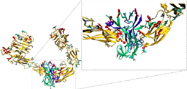
Pattison and Dunn have provided the first proof of the direct interaction between metal ions and NGF, 270 showing that zinc(II) ion is specifically bound to native 7S NGF (e.g., an oligomeric protein composed of 5 non-covalently linked subunits: 2α, 1β, 2γ). The active NGF has also been found to bind zinc(II) ions in the native protein, with an uncommon Zn2+ to NGF molar ratio of 1:14.6 (determined by means of flameless atomic absorption spectroscopy measurements). This presence of Zn2+ in the native molecule contributes to significant conformational changes and increased biological activity of the protein.257 It is interesting to underline the high occurrence of histidine residues in TrkA (14 His) and in NGF (4His), facing the extracellular space (Fig. 8 and Fig 9). Of great interest is the close proximity of negatively charged residues, such as Asp and Glu, to the His residues (Fig 9). These are structural requisites for the binding of metal ions, as zinc(II) and copper(II) ions.
The crystal structure of NGF reveals potential metal ion binding sites, with the involvement of His84 and Asp105. 161 According to Holland et al. a knot motif confers structural stability and maintains the position of these two residues, preserving the metal ion binding site.161,260
Figure 8. Representation of the extracellular domain of the NGF/TrkA dimer. NGF chains are
shown in green and blue, D1-D5 TrkA sub-units are shown in yellow. His residues are shown in red according to the vdw radii. [From 179]
Figure 9. Representation of the extracellular domain of the NGF/TrkA dimer. The negative
charged residues, Asp and Glu, are shown in cyan and gray respectively. Both of them are close to His residues, shown in red. [From 179]
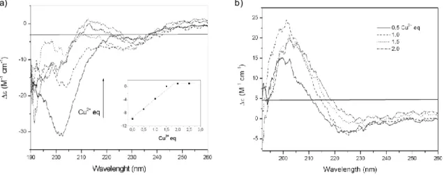
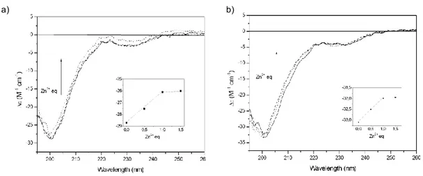
To the best of our knowledge, the coordination features of metal-ion complexes with NGF have been determined only by theoretical methods. 255-256,271 The computational results suggested a model in which Zn2+ alters the conformation of NGF by forming a pentacoordinate complex. This metal ion has been found to bind to the imidazole nitrogen atoms of His4 and His8 of one monomer and to the imidazole nitrogen atom of His84 and both oxygen atoms of Asp105 of the other unit of the NGF dimer; 255-256 the same coordination environment has been proposed for Cu2+. 271 As a consequence of the conformational change resulting from metal chelation, no cross-link between primary amino groups of Ser1 of a NGF unit and Lys115 of another unit takes place, unlike for the active dimeric form of NGF in the absence of metal ions. Thus, it has been suggested that the binding of metals is responsible for the alteration of NGF conformation and, in turn, for the inhibition of its biological activities. 256 Also, computational approaches have been carried out to investigate the interaction of NGF with other transition metal ions. 260,271 It is interesting to note that copper(II) ion has been reported to be the only metal ion able to bind the imidazole nitrogen stronger than a proton at pH values higher than 6-6,5. At lower pH values, the imidazole is unable to coordinate Cu2+. 271 Moreover, it has been proposed that NGF might interact with its receptors as a complex with zinc(II) ion.
2.
Aims of the work
On the basis of the literature data above reported, we have investigated the issue of the interactions of the metal ions with the neurotrophins.
To the best of our knowledge, the coordination features of metal ions complexes with neurotrophins have been determined only by theoretical methods and only for NGF. No experimental data inherent the coordination features been reported, neither one of the hypotheses according to which Zn2+ and Cu2+ could have different binding environments, or Ser1 α-amino group could be involved in coordination have been supported.
Crystallographic, biochemical and computational studies converge to suggest that the N-terminal residues are crucial for specific activities of the neurotrophins. However, nobody has tested if just this small sequence is able to mimic the whole protein or at least some of its biochemical signaling. Among them, the activation of transcription factors is particularly attractive, because they are involved in gene expression but also in memory consolidation.
The use of neurotrophins in the early stages of neurodegenerative diseases has gained attention. However, there are limits to such therapy, e.g. insufficient permeability of the blood-brain barrier and activation of receptors that trigger adverse side effects. The use of peptidomimetic combined with appropriate systems that guarantee their delivery might allow to overcome these restrictions.
Briefly, the work has been summarized in sub-aims:
1) Synthesis of the peptide fragment encompassing the sequence 1-14 of the human NGF amino-terminal domain (NGF(1-14)) , blocked at the C-terminus, and characterization of its Cu2+ and Zn2+ complexes by means of potentiometric and spectroscopic (UV-vis, CD, NMR and EPR) techniques. N-terminus acetylated form of NGF(1-14) has also investigated to evaluate the involvement of Ser1 α-amino group in the metal ions coordination. The functional interaction of Cu2+ and Zn2+ ions with NGF peptides and with
![Figure 15 . A schematic representation of the Zn 2+ coordination environment in the[ZnL]](https://thumb-eu.123doks.com/thumbv2/123dokorg/4516962.34763/60.892.140.783.730.991/figure-schematic-representation-zn-coordination-environment-znl.webp)
