XXXII PhD course in Applied Biology and Experimental Medicine Department of Chemical, Biological and Pharmaceutical Sciences
University of Messina
THE IN VIVO PHARMACOLOGICAL MANIPULATION OF THE NRF2 PATHWAY AND ITS THERAPEUTIC SIGNIFICANCE IN BRAIN DISEASES, WITH A
POSSIBILE IMPLICATION IN H2S-REGULATED LIPIDIC METABOLISM
Dr. Giovanna Casili
Relators:
Prof. Emanuela Esposito
Prof. Salvatore Cuzzocrea
Prof. Csaba Szabo
This work was performed under the supervision of:
Prof. Emanuela Esposito and Prof. Salvatore Cuzzocrea
at the University of Messina, Department of Chemical, Biological and
Pharmaceutical Sciences
and
Prof. Csaba Szabo
Table of contents
PART I ... 11
ABSTRACT ... 11
1.0 CHAPTER ONE: The role of Nrf2 pathway on oxidative stress regulation ... 13
1. 1 Oxidative stress... 13
1.1.2 Pathophysiology of oxidative stress ... 14
1.2 The Nrf2-Keap1 pathway ... 15
1.2.1 Component structures and functions ... 17
1.2.2 Mechanisms and regulation of ARE-dependent gene expression ... 19
1.3 The Nrf2 regulation: Keap1-dependent regulation ... 20
1.3.1 Identification of Keap1 as an inhibitor of Nrf2 ... 20
1.3.2. Ubiquitination and proteasomal degradation of Nrf2 ... 20
1.3.3 The cysteines of Keap1 as sensors of oxidative stress ... 22
1.3.4 Autophagic degradation of Keap1 ... 24
1.4 Keap1-independent Regulation of Nrf2 ... 25
1.4.1 Transcription regulation and autoregulation ... 25
1.4.2 Post-transcriptional regulation: microRNAs ... 26
1.4.3 Post-translational modification: phosphorylation/acetylation ... 27
1.4.4 Nrf2 cysteine modifications ... 27
1.4.5 Cross-talk between Nrf2 and other pathways ... 28
1.5 The role of Nrf2 in diseases ... 28
1.5.1 Nrf2 in cardiovascular disease ... 29 1.5.2 Nrf2 in liver disease ... 29 1.5.3 Nrf2 in kidney disease ... 30 1.5.4 Nrf2 in diabetes ... 31 1.5.5 Nrf2 in cancer ... 31 1.5.6 Nrf2 in neurodegenerative disease ... 31
2.1 Fumaric acid esters ... 33
2.1.1 Different FAE-formulations ... 33
2.1.2 Pharmacokinetics and pharmacodynamics of FAE ... 34
2.2 Dimethyl fumarate (DMF) ... 34
2.2.1 Tolerability and safety of DMF... 35
4
2.2.3 Mechanism of action ... 36
... 36
2.3 Aim of thesis PART I ... 37
3.0 CHAPTER THREE: Dimethyl Fumarate attenuates neuroinflammation and neurobehavioral deficits induced by experimental traumatic brain injury. ... 38
3.1 Introduction ... 38
3.2 Materials and Methods ... 40
3.2.1 Animals ... 40
3.2.2 Controlled cortical impact (CCI) experimental TBI... 40
3.2.3 Experimental groups ... 41
3.2.4 Tissue processing and histology ... 42
3.2.5 Stereological assessments of lesion volume and neuronal loss: Crystal violet... 43
3.2.6 Fluoro‐Jade C (FJC) staining ... 43
3.2.7 TUNEL staining ... 44
3.2.8 Behavioral testing ... 44
3.2.9 Western blot analysis ... 47
3.2.10 Immunohistochemical analysis ... 48
3.2.11 Myeloperoxidase activity (MPO) ... 49
3.2.12 Materials ... 49
3.2.13 Statistical analysis ... 49
3.3 Results ... 50
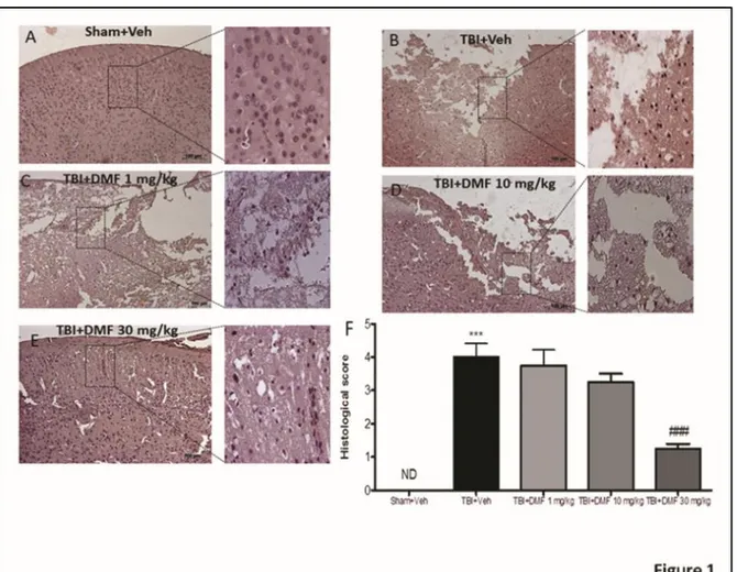
3.3.1 DMF treatment reduced the severity of brain trauma and the infarct outcome .... 50
Figure 3.1: Effects of DMF treatment on histological alterations after TBI ... 51
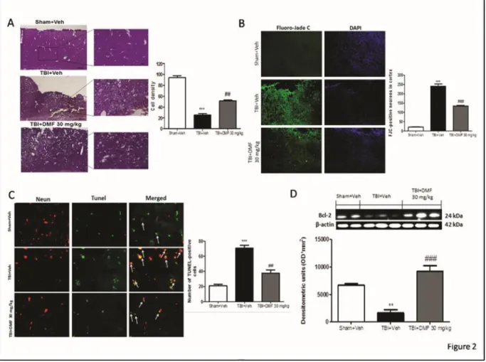
3.3.2 Protective effects of DMF treatment to reduce acute neural injury and degeneration and to modulate apoptosis‐induced cell death through Bcl‐2 ... 51
Figure 3.2: Effects of DMF treatment on infarct area and volumes following TBI ... 53
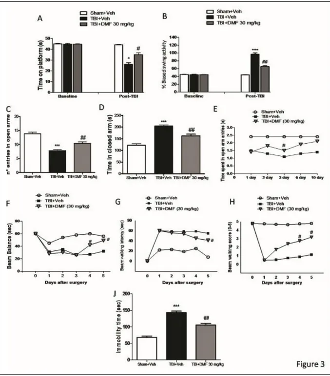
3.3.3 Effects of DMF treatment on recovery and improvement in behavioral function . 53 Figure 3.3: Effects of DMF treatment on behavioral and neurological function ... 56
3.3.4 Effects of DMF treatment on antioxidant response activation ... 57
Figure 3.4: Effects of DMF treatment on antioxidant response activation ... 58
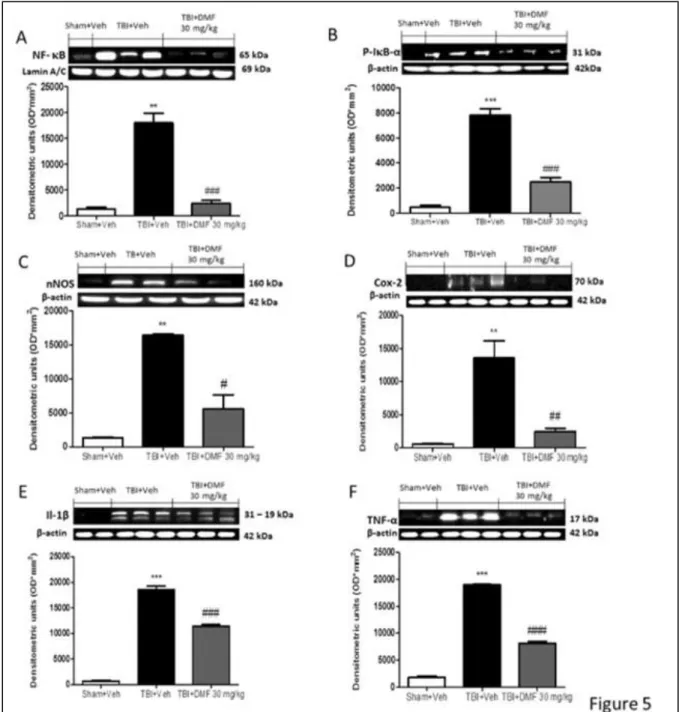
3.3.5 The anti‐inflammatory effects of DMF treatment following TBI ... 58
Figure 3.5: Effects DMF treatment on inflammatory pathway ... 60
3.3.6 The role of DMF treatment on myeloperoxidase (MPO)‐mediated oxidative stress, microglia and astrocytes activation ... 61
Figure 3.6: Effects of DMF treatment on neutrophil accumulation and microglia
activation ... 62
3.4 Discussion ... 63
3.5 Conclusions ... 68
4.0 CHAPTER FOUR: Dimethyl fumarate alleviates the nitroglycerin (NTG)-induced migraine in mice ... 70
4.1 Introduction ... 70
4.2 Materials and methods... 73
4.2.2 Migraine induction and DMF administration ... 73
4.2.3 Experimental groups ... 73
4.2.4 Behavioral testing ... 74
4.3 Results ... 77
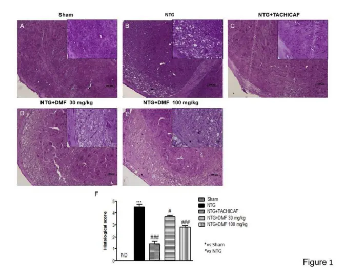
4.3.1 DMF treatment restored the NTG-induced damage in trigeminal nucleus... 77
Figure 4.1: Effects of DMF treatment on NTG-induced damage ... 78
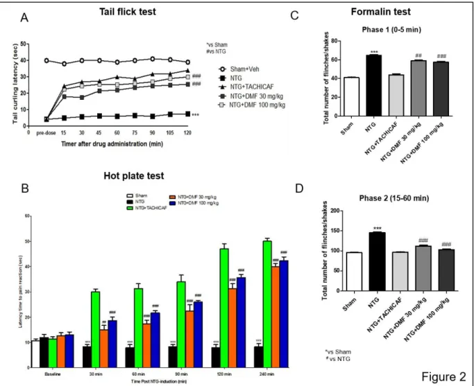
4.3.2 The protective effects of DMF to reduce NTG-induced hyperalgesia ... 78
Figure 4.2: Effects of DMF treatment on NTG-induced hyperalgesia ... 80
4.3.3 The role of DMF in the comorbidity between migraine and anxiety ... 80
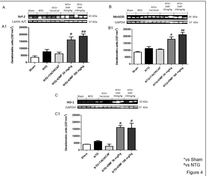
4.3.4 The role of DMF on antioxidant system in NTG-induced migraine ... 82
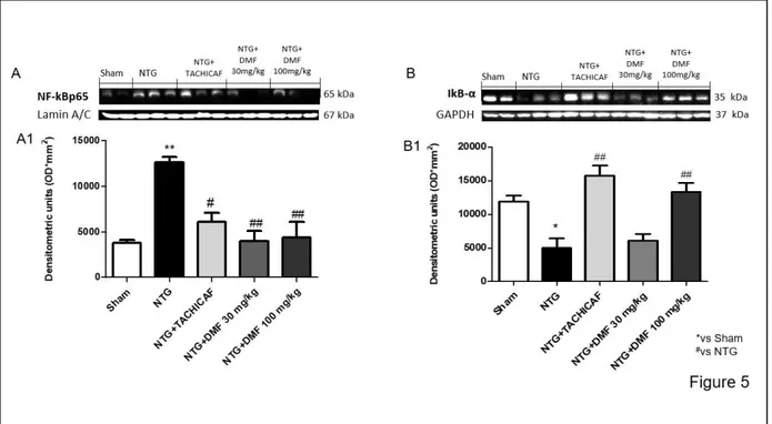
4.3.5 The effects of DMF on NF-κB inflammatory pathway in NTG-induced migraine .... 83
4.3.6 Statistical analysis ... 86
4.4 Discussion ... 86
4.5 Conclusions ... 90
PART II ... 92
ABSTRACT ... 92
5.0 CHAPTER FIVE: The role of H2S on adipogenesis ... 94
5.1 Hydrogen sulfide ... 94
5.1.1 Physical and chemical properties of H2S ... 94
5.1.2 H2S reaction with oxidants ... 95
5.1.3 Endogenous production of H2S ... 95
Figure 5.1: Enzymatic synthesis of H2S ... 97
5.1.4 Exogenous production of H2S ... 97
5.1.5 H2S Metabolism ... 97
Figure 5.2: Mitochondrial oxidant production ... 98
6
5.2.1 Antioxidant roles of H2S in the cardiovascular system ... 99
5.2.2 Antioxidant roles of H2S in nervous system ... 100
5.3 Adipose tissue ... 101
5.3.1 The anatomy of multi-deposit of adipose tissue ... 103
Figure 5.3: WAT and BAT ... 104
5.3.2 Adipose cell differentiation ... 104
5.3.3 Transdifferentiation Process ... 106
5.3.4 Adipokines and Cytokines ... 108
5.4 Obesity as adipose tissue disfunction ... 109
Figure 5.5 : Differences in adipose tissue function and body fat distribution between MHO and metabolically unhealthy obese individuals. ... 110
5.4.1 Obesity and oxidative stress ... 111
5.5 Regulation of H2S in adipose tissue ... 111
5.5.1 Role of H2S in the regulation of lipolysis ... 112
5.5.2 Effect of H2S on adipose tissue insulin sensitivity and glucose uptake ... 112
5.5.3 Role of H2S in obesity ... 114
5.6 The Nrf2 regulation on lipid metabolism ... 114
5.7 Aim of the thesis PART II ... 115
6.1 Introduction ... 117
6.2 Material and methods ... 119
6.2.1 Cell Culture and Differentiation ... 119
6.2.2 Cell viability and toxicity assay ... 119
6.2.4 Oil Red O Staining ... 120
6.2.5 Detection of H2S with 7-Azido-4-methylcoumarin (AzMC) probe ... 120
6.2.6 Western Blot analysis ... 121
6.2.7 shRNA transfection ... 121
6.2.8 Transcription factors (TFs) Activation Profiling Array ... 121
6.2.9 Materials ... 122
6.2.10 Statistical analysis ... 122
6.3 Results ... 122
6.3.1 Adipogenesis process in 3T3L1 cells ... 122
Figure 6.1: Adipogenesis process in 3T3L1 cells ... 123
6.3.2 Effects of H2S donor and 3-MST inhibitor on differentiation of 3T3L1 preadipocytes into mature adipocytes ... 123
Figure 6.2: Effects of GYY4137 and HMPSNE on adipogenesis. ... 124
6.3.3 Effects of H2S presence or absence on cell viability and toxicity ... 124
Figure 6.3: Cell viability and toxicity of GYY4137 and HMPSNE treated adipocytes ... 126
6.3.4 Effects of GYY4137 and HMPSNE treatment on H2S release ... 126
Figure 6.4: Detection of H2S released by adipocytes using AzMC probe ... 126
6.3.5 Effects of GYY4137 treatment on H2S synthesis and degradation... 126
Figure 6.5: GYY4137 treatment on H2S synthesis and catabolism ... 127
6.3.6 Effects of HMPSNE treatment on H2S synthesis and degradation ... 128
Figure 6.6: HMPSNE treatment on H2S synthesis and catabolism ... 128
6.3.7 Effects of GYY4137 or HMPSNE treatments on 3-MST knockdown adipocytes .... 129
Figure 6.7: GYY4137 and HMPSNE treatments on 3-MST knockdown adipocytes ... 130
6.3.8 Nrf2 involvement on adipogenesis ... 130
Figure 6.8: Role of Nrf2 transcriptional expression on adipogenesis ... 131
6.4 Discussion and Conclusions ... 131
PART I
ABSTRACT
Nuclear factor erythroid 2-related factor 2 (Nrf2), a redox-sensitive transcription factor, plays a critical role in the regulation of cellular defence and contributes to a number of cellular processes. Nrf2 is regulated through an interplay of complex transcriptional and post- translational mechanisms that modulates its activity during cellular perturbations or other biological processes thereby ensuring cellular homeostasis is maintained through the orchestration of adaptive responses. Therefore, an ability to modulate the activity of the Nrf2 pathway holds promise as a therapeutic strategy in certain disease settings.
In this thesis, the role of Nrf2 was evaluated in a mouse model of traumatic brain injury (TBI). TBI is a serious neuropathology that causes secondary injury mechanisms, including dynamic interplay between ischemic, inflammatory and cytotoxic processes. Fumaric acid esters (FAEs) showed beneficial effects in preclinical models of neuroinflammation and toxic oxidative stress, so the aim of the present work was to evaluate the potential beneficial effects of dimethyl fumarate (DMF), the most pharmacologically effective molecules among the FAEs, in a mouse model of TBI induced by controlled cortical impact (CCI). Mice were orally administered with DMF at the doses of 1, 10 and 30 mg/Kg, 1h and 4h after CCI. We performed histological, molecular, and immunohistochemistry analysis on the traumatic penumbral areas of the brain 24 hours after CCI. DMF treatment notably reduced histological damage and behavioral impairments, reducing neurodegeneration as evidenced by assessments of neuronal loss, Fluoro‐jade C and TUNEL staining; also, treatment with DMF blocked apoptosis process increasing B‐cell lymphoma 2 (Bcl‐2) expression in injured cortex. Furthermore, DMF treatment up‐regulated antioxidant Kelch‐ like ECH‐associated protein 1/ Nuclear factor erythroid 2‐ related factor (Keap‐1/Nrf2) pathway, inducing activation of manganese superoxide dismutase (Mn‐SOD) and heme‐ oxygenase‐1 (HO‐1) and reducing 4‐hydroxy‐2‐
nonenal (4‐HNE) staining. Also, regulating NF‐κB pathway, DMF treatment decreased the severity of inflammation through a modulation of neuronal nitrite oxide synthase (nNOS), interleukin 1 (Il‐1β), tumor necrosis factor (TNF‐α), cyclooxygenase 2 (COX‐2) and myeloperoxidase (MPO) activity, reducing ionized calcium‐binding adapter molecule 1 (Iba‐1) and glial fibrillary acidic protein (GFAP) expression.
Moreover, the role of Nrf2 pathway, was discusses in a mouse model of nitroglycerin -induced migraine (NTG). Oxidative stress and inflammatory pathways are involved in NTG and endogenous antioxidant defense system has a role in the prevention of hyperalgesia in migraine. In this study, we aimed to evaluate the role of DMF in regulating the hypersensitivity in a mouse model of NTG-induced migraine.
Mice were orally administered with DMF at the doses of 10, 30 and 100 mg/kg, 5 minutes after NTG intraperitoneal injections. We performed histological and molecular analysis on the whole brain and behavioral tests after 4 h by NTG-migraine induction. The expression of nuclear factor kappa-light-chain-enhancer of activated B cells (NF-κB) subunit p65, nuclear factor of kappa light polypeptide gene enhancer in B-cells inhibitor alpha (IκBα), inducible nitrite oxide synthase (iNOS), cyclooxygenase 2 (COX-2), Nrf2, manganese superoxide dismutase (Mn- SOD) and heme-oxygenase-1 (HO-1) were detected by Western blot. Tail flick, hot plate, formalin and photophobia tests were used to evaluate neuropathic pain and migraine-related light sensitivity. DMF treatment notably reduced histological damage as showed by cresyl violet staining; also, regulating both NF-κB and Nrf2 pathway, DMF treatment decreased the severity of inflammation and increased the protective antioxidant action. Moreover, the headache-associated neuropathic pain was significantly reduced.
These results provide the evidence that DMF restores neurological in damaged-brain and it has modulating effect on central sensitization, suggesting a new insight into the potential therapeutical application of DMF on Nrf2 pathway modulation.
1.0 CHAPTER ONE: The role of Nrf2 pathway on oxidative stress regulation
1. 1 Oxidative stress
The origin of the basic principle of stress and stress responses dates back to Selye in 1936 and the concept of oxidative stress has been introduced for research in redox biology and medicine in 1985, in an introductory book entitled ‘Oxidative Stress’, presenting the knowledge on pro- oxidants and antioxidants and their endogenous and exogenous sources and metabolic sinks [1]. Oxidative stress is considered as an imbalance between production of free radicals and reactive metabolites or reactive oxygen species (ROS) and their elimination by through protective mechanisms, including antioxidants. This imbalance leads to damage of cells and important biomolecules, having a potential adverse impact on the whole organism [2]. Cells are constantly being exposed to oxidative stress, which result from various chemical and oxidative reactions such as those involving environmental toxicants, mutagens and carcinogens. These insults disrupt cellular homeostasis and function and, not surprisingly, have been associated with disease pathogenesis [3].
ROS are a group of molecules and free radicals (atoms or molecules with an unpaired valence electron) derived from oxygen. They can be of exogenous or endogenous origin. The latter is a result of the cell’s own metabolism and play important roles in the stimulation of signaling pathways in plant and animal cells in response to changes in intra- and extra-cellular environmental conditions [4]. During endogenous metabolic reactions, aerobic cells produce ROS, such as superoxide anion (O2-), hydrogen peroxide (H2O2), hydroxyl radical (OH•), and
1.1.2 Pathophysiology of oxidative stress
The terms reactive oxygen species (ROS) and reactive nitrogen species (RNS) refer to reactive radical and non-radical derivatives of oxygen and nitrogen, respectively [6]. There are endogenous and exogenous sources of ROS/RNS:
•Endogenous sources of ROS/RNS include: nicotinamide adenine dinucleotide phosphate (NADPH) oxidase, myeloperoxidase (MPO), lipoxygenase, and angiotensin II [7]. NADPH oxidase is the prevalent source of the radical superoxide anion (O2•) which is formed by the one-electron reduction of molecular oxygen, with electrons supplied by NADPH, during cellular respiration. Most of the O2• is dismutated into the hydrogen peroxide (H2O2) by
superoxide dismutase (SOD). H2O2 is not a free radical because it has no unpaired electrons,
but it is able to form the highly reactive ROS hydroxyl ion (OH•) through the Fenton or Haber– Weiss reaction. Hydroxyl radicals are extremely reactive and react especially with phospholipids in cell membranes and proteins. Finally, O2 may react with NO to form another
relatively reactive molecule, peroxynitrite (ONOO−) [8].
•Exogenous sources of ROS/RNS are air and water pollution, tobacco, alcohol, heavy or transition metals, drugs, industrial solvents, cooking (eg, smoked meat, waste oil, and fat) and radiation, which inside the body are metabolized into free radicals [9].
ROS can be divided into two groups: free radicals and non radicals. Molecules containing one or more unpaired electrons and thus giving reactivity to the molecule are called free radicals, while the sharing of unpaired electrons by two free radicals produce non radical forms. Under normal conditions, ROS production and ROS elimination are balanced; while low ROS levels are related with physiological conditions, an increased production of ROS levels is generally associated with pathological conditions; particularly, when ROS levels increase beyond the threshold of this buffering capacity, these reactive species trigger uncontrolled reactions with non-target intracellular compounds, thus oxidizing nucleic acids, proteins, cellular membrane,
and other lipids, given rise to oxidative stress and cellular damage [10]. ROS also affects the expression of several genes by upregulation of redox-sensitive transcription factors and chromatin remodeling, via alteration in histone acetylation/deacetylation. Regulation of redox state is critical for cell viability and organ functions [11].
Oxidative stress plays a role in inflammation, accelerates aging and contributes in variety of degenerative conditions: cardiovascular diseases, atherosclerosis, cancer, cataract, central nervous system disorders, Parkinson's disease, Alzheimer's disease, inflammatory bowel disease, rheumatoid arthritis, diabetes, respiratory diseases, autoimmune diseases, liver diseases, kidney diseases, skin conditions [12].
1.2 The Nrf2-Keap1 pathway
Mammalian cells have evolved complex signaling pathways and defense systems that function synergistically in order to reduce the deleterious effects of such intrinsically and extrinsically generated insults. At the center of the biological response to oxidative stress is the Kelch-like ECH-associated protein 1 (Keap1) - nuclear factor erythroid 2-related factor 2 (Nrf2)- antioxidant response elements (ARE) pathway, which regulates the transcription of many several antioxidant genes, preserving cellular homeostasis and detoxification genes, to avoid cellular damage [13].
Nrf2 is a redox-sensitive transcription factor belonging to the cap'n'collar (CNC) subclass of the basic leucine zipper region containing the protein family. Nrf2 binds to a specific DNA site, the antioxidant response element (ARE), regulating transcription of an array of detoxifying or antioxidant enzymes (gamma-glutamylcysteine synthetase, superoxide dismutase, catalase, glutathione reductase, thioredoxin reductase, peroxiredoxins, and glutathione S-transferase) [14]. This pathway is a redox sensitive transcription factor that induces the expression of a
variety of genes that protect against the deleterious effects of oxidative and chemical stress, thus ensuring normal cellular functions are maintained or restored.
The Keap1–Nrf2 system is a major oxidative stress response pathway [15]. Nrf2 is a basic leucine zipper (bZIP) transcription factor and its heterodimer with small Maf proteins controls the expression of anti-oxidant proteins that protect against oxidative damage triggered by injury and inflammation. Keap1 is an adaptor protein of Cullin-3-based ubiquitin ligase. The mechanism proposed for the Keap1-mediated activation of Nrf2 is the “hinge and latch model”, based on the finding that Keap1 homodimer binds to a single Nrf2 molecule through two distinct binding sites (DLG and ETGE motifs), within the Neh2 domain of Nrf2 [16]. The Keap1 forms a homodimer that recognizes ETGE and DLGex motifs of one Nrf2 molecule through the same binding pocket within the DC (double glycine repeat and C-terminal) domain located at the bottom surface of Keap1 and this binding causes Nrf2 ubiquitination [17]. Binding of the high-affinity ETGE motif and low-high-affinity DLG motif provides a hinge and latch, which facilitate optimal positioning of the lysine residues between the two motifs for ubiquitin conjugation [18]. Under normal conditions, the levels of Nrf2 protein are kept low by the E3 ubiquitin ligase Keap1 (Kelch ECH-associating protein 1), which ubiquitinates Nrf2 in the cytoplasm and targets it for degradation by the 26 S proteasome [19]. This constitutive degradation of Nrf2 allows for only the basal expression of its target stress response genes as part of a housekeeping function. However, under conditions of oxidative stress, or in the presence of electrophilic xenobiotics, the activity of Keap1 decreases and Nrf2 can accumulate in the nucleus where it activates the inducible high expression of its target genes.
In this way, Keap1 functions as a critical sensor of cellular stress and its high redox sensitivity is determined by a number of cysteine residues that are distributed throughout the Keap1 protein and are vulnerable to oxidation or to covalent modification by electrophiles. The Keap1–Nrf2
system protects cellular proteins and DNA from oxidative damage caused by reactive oxygen species and electrophiles, implementing a critical role for cellular detoxification. Consequently, the Keap1–Nrf2 system is an important therapeutic target in cancer and neurodegenerative conditions, as well as many autoimmune and inflammatory diseases [20], [21], [14], [2]. Critical for such intervention strategies is a deep understanding of the structure and function of these proteins. Progress in this respect has relied on the definition of the domain architecture of the Keap1 and Nrf2 proteins and the identification of the specific regions mediating their interactions [22].
1.2.1 Component structures and functions
Nuclear factor erythroid 2-related factor 2 (Nrf2)
The Nrf2 protein in humans is 605 amino acids long and contains seven highly conserved regions known as [Nrf2–ECH (erythroid cell-derived protein with CNC homology) homology] (Neh) domains. Six functional Neh units were identified in Nrf2, each well conserved in the Nrf2 molecules of various species. Neh1 contains the CNC–bZIP domain, which mediates heterodimerization with Maf [23]. The Neh2 domain contains the two degrons that are specifically bound by Keap1, commonly known as the DLG and ETGE motifs after their sequence conservation in the single-letter amino acid code [24]. The Neh3 domain functions as a co-activator that facilitates the transcription of the ARE-dependent genes, while Neh4 and Neh5 domains act together with CBP [CREB cyclic AMP-response element binding protein (CREB) binding protein] and synergistically contribute to the strong transcriptional activation exerted by Nrf2 [25] . Neh6 is the domain that regulates the Keap1-independent regulation of Nrf2 pathway [26]. Neh7 is the domain that facilities retinoid X receptor alpha (RXRα)- mediated Nrf2 repression through a direct interaction between the two proteins [27].
The Nrf2 protein also contains nuclear import/localization signals (NLSs) and nuclear export signals (NESs) which regulate Nrf2 shuttling in and out of the nucleus. Also, human Nrf2 contain six cysteine residues; two of the cysteines (Cys183 and Cys506) and two other key amino acid residues (Ser40 and Tyr568) may also regulate Nrf2 localization and transactivation of target genes through oxidation and phosphorylation, respectively. Nrf2 is truly a master regulator of the antioxidant response [28].
Kelch-like ECH-associated protein 1 (Keap1)
Keap1 is a 69-kDa protein that shares some homology with actin-binding Kelch protein and serves as a negative regulator of Nrf2. The human Keap1 protein sequence contains 627 amino acid residues organized into five domains: i) the N -terminal region (NTR), ii) the Broad complex, Tramtrack, and Bric-a-Brac (BTB), iii) the linker intervening region (IVR), iv) the Kelch domain, and v) the C -terminal region (CTR). BTB domain that attached to act in binding proteins is responsible for homodimerization and interaction with Cullin (Cul3) based ubiquitin E3 ligase complex for Nrf2 ubiquitination. IVR containing cysteine residues, sensitive to oxidation and nuclear export signal (NES) motif. Kelch domain has six kelch repeats (KR1- KR6) and possessing multiple protein contact sites that mediate association of Keap1 with Nrf2 (the Kelch domain interacts with Neh2 domain of Nrf2) and cytoskeleton proteins actin and/or myosin [23].
Human Keap1 contains 27 cysteine residues and seven of them (i.e., Cys151, Cys257, Cys273, Cys288, Cys297, Cys434, and Cys613) are highly reactive towards ROS and electrophiles and probably they are involved in redox sensing [29].
Antioxidant response element (ARE)
The induction of many cytoprotective enzymes in response to reactive oxidative stress is regulated primarily at the transcriptional level. This transcriptional response is mediated by a cis-acting element termed ARE [30], initially found in the promoters of genes encoding the two
major detoxication enzymes, GSTA2 (glutathione S-transferase A2) and NQO1 (NADPH: quinone oxidoreductase 1). ARE possesses structural and biological features that characterize its unique responsiveness to oxidative stress [31], but also it is activated in response to chemical compounds with the capacity to either undergo redox cycling or be metabolically transformed to a reactive or electrophilic intermediate. The alteration of the cellular redox status due to elevated levels of ROS and electrophilic species and/or a reduced antioxidant capacity (e.g. glutathione) appears to be an important signal for triggering the transcriptional response. The consensus sequence of ARE cannot be represented as a single sequence as AREs required for some genes are distinctly different for others. Under oxidative stress conditions, stabilized Nrf2 translocates to the nucleus, forms a heterodimer with Maf, and activates ARE-dependent gene expression. Bach1 (BTB and CNC homology-1) is another negative regulator of certain ARE- dependent genes; it associates with ARE and forms a dimer with Maf protein, preventing Nrf2 from binding to DNA under normal physiological conditions [32].
1.2.2 Mechanisms and regulation of ARE-dependent gene expression
The discovery of the ARE lead to the identification of Nrf2 as the transcription factor capable of both binding to this DNA sequence and inducing the expression of cell defence genes and resulted in a burst of research into this pathway as a potential therapeutic target. The vast number of both natural and synthetic compounds able to induce Nrf2 can be divided into at least 10 groups [33]. Numerous Nrf2 inducers, mostly plant-derived compounds with chemopreventive properties are currently in clinical trials for a variety of diseases. What is evident, however, is that Nrf2 plays a major role in health and disease and it is not surprising that Nrf2 is considered as a potential therapeutic target. The development of a number of Nrf2 inducers as possible pharmacological agents without a complete knowledge of the workings of this pathway and its regulation heightens the need to further our understanding and to determine whether activation of Nrf2 would be beneficial in both the short- and long-term.
1.3 The Nrf2 regulation: Keap1-dependent regulation 1.3.1 Identification of Keap1 as an inhibitor of Nrf2
Several models of Nrf2 regulation have been proposed and can be divided into Keap1- dependent and Keap1–independent regulation of Nrf2. The identification of an inhibitor of Nrf2 derived observing that the deletion of the Neh2 region of the Nrf2 protein resulted in a marked increase in Nrf2 activity in erythroblasts and led to the proposal that this region was responsible for the negative regulation of Nrf2 via an interaction with a repressor protein [34]. Later studies further confirmed this idea by cloning the rat homologue of Keap1 by purifying Nrf2- interacting proteins [35]. Following these discoveries, it was seen that two Keap1 molecules are able to bind to one Nrf2 molecule and that the BTB domain is responsible for the homodimerization of Keap1 and the subsequent inhibition of Nrf2 [36]. When transfected into cells it was observed that Nrf2 would accumulate in the nucleus, however when co-transfected with Keap1 the two would co-localise in the cytoplasm. Moreover, in the presence of both Keap1 and a panel of electrophiles, Nrf2 again localizes in the nucleus, suggesting that Keap1 sequesters Nrf2 in an inactive form in the cytoplasm until faced with an oxidative or electrophilic insult, when Nrf2 is freed and translocates to the nucleus [37]. The thesis that Keap1 is vital in the regulation of the Nrf2 pathway in in vivo is highlighted by the observation that Keap1-deficient mice (Keap1−/−) do not survive longer than 3 weeks postnatally due to hyperkeratosis of the digestive system resulting in ulceration of the stomach [38].
It was quickly determined that Keap1 function is not simply binding Nrf2 and sequestering it in the cytosol, thus preventing its translocation to the nucleus, but it also provided a functional role in this pathway.
1.3.2. Ubiquitination and proteasomal degradation of Nrf2
Under basal conditions, Keap1 leads Nrf2 to ubiquitination and proteasomal degradation. In the absence of oxidative stress, Nrf2 is sequestered in the cytosol by the Keap1 homodimer which
acts as a substrate adaptor for the ubiquitination of Nrf2 in a cullin-3 (Cul3) dependent manner [17]. Keap1 acts as a substrate adaptor for Cul3-dependent E3 ubiquitin ligase complex, thereby bringing together Nrf2 and ROC1/RBX1 (Ring-box protein 1), a ring-box protein which recruits a ubiquitin charged E2 molecule; thus, the ubiquitin molecule is conjugated to one of the lysine residues located on Neh2 domain of Nrf2, facilitating the Nrf2 proteasomal degradation and high turnover of Nrf2 protein [39]. Nrf2 binds the DGR site of each Keap1 subunit via 2 distinct binding motifs in its Neh2 domain, one high affinity, 79ETGE82, and one low affinity, 29DLG31 [16]. When bound at both sites, Nrf2 is perfectly positioned to bear poly-ubiquitination via the Cul3 E3 ligase and is consequently degraded by the 26S proteasome, assuring low basal levels of Nrf2 in the cell.
The changes in Nrf2 half-life associated with oxidative stress and specific electrophiles varies due to the dramatic differences in detectable basal levels of the protein [40]; also, conflicting evidence is on the mechanism by which Nrf2 becomes free in the cell, because some studies suggested that dissociation of the Keap1–Nrf2 complex is caused from electrophiles [41], while others supported the thesis that the electrophiles cause the dissociation of Cul3 from Keap1 thus preventing the proteasomal degradation of Nrf2 [42].
Furthermore, there are those who believe that Nrf2 is primarily a nuclear protein and this nuclear localization is responsible for the basal expression of cell defence genes and that the degradation of Nrf2 via Keap1 is downstream of Nrf2 transcriptional activity [43], whereas this would explain how Nrf2 is capable of regulating the basal expression of genes despite being constantly degraded via Keap1, this idea requires the nucleo-cytoplasmic shuttling of Keap1 [44]. In any case, the concordant thesis is that Keap1 is responsible for the proteasomal degradation of Nrf2 and that the half-life of Nrf2 doesn’t depend on the redox state of the cell and that this is at least partially via the interaction with the redox-sensitive Keap1 [45].
Furthermore, following studies showing the involvement of a Keap1-Cul3-Rbx1 complex in directing the proteasome-mediated degradation of Nrf2, demonstrated that, in the presence of several Nrf2 inducers including tBHQ, sulforaphane, eicosapentaenoic acid (EPA) and N- iodoacetyl-Nbiotinylhexylenediamine (IAB), there was a dissociation of Keap1 from Cul3, preventing the ubiquitination and subsequent degradation of Nrf2 [46]. The dissociation of Keap1 and Cul3 was shown to be dependent on the presence of C151 within the BTB domain of Keap1, as C151 is surrounded by four positively charged amino acids (K131, R135 and K150 and H154), theoretically making it highly reactive towards inducers. The modification of C151 by electrophilic Nrf2 inducers may provoke Cul3 dissociation [47].
1.3.3 The cysteines of Keap1 as sensors of oxidative stress
The discovery of Keap1 as a negative regulator of Nrf2 made it possible to test the idea that cysteine residues of Keap1 serve as the sensors for inducers [48]. Several studies have demonstrated the covalent modification of multiple cysteine residues by Nrf2 inducers, which is hypothesized to alter the Keap1 conformation, thus preventing its association with Nrf2, allowing Nrf2 to translocate to the nucleus [49]. As mentioned above, Keap1 is a cysteine-rich protein and the 27 cysteine residues in the human protein are all reactive to varying degrees. The modification of a subset of cysteine residues in Keap1 by Nrf2 inducers with similar structures supports the hypothesis of a “cysteine code” and may underlie the ability of Nrf2 to respond to such a diverse array of compounds [50]. The high frequency of cysteine residues in the Keap1 sequence confers it with a relative high reactivity to electrophilic inducers; particularly, the most reactive Keap1 cysteines residues modified identified are: C257, C273, C288 and C297, within the IVR domain, and a fifth one, C613 in the C-terminal region of murine Keap1 [51].
A later study showed that by modifying the conditions for incubation and sample processing another residue, C151, could be also detected as the most reactive cysteine of the four (C38,
C151, C368 and C489) readily modified cysteine residues in Keap1 by sulforaphane [52]. In oxidative stress conditions or in the presence of electrophiles, it is proposed that subsets of the cysteine residues in Keap1 are modified, contributing to a conformational change in the protein which results in the release of Nrf2 from the low affinity binding site, disturbing the transfer of ubiquitin. In this way, Keap1 molecules become saturated with Nrf2, that is no longer targeted for degradation and newly synthesized, free Nrf2 accumulates in the cytosol.
The modifications of the cysteine residues confer a suppression of Keap1 and a subsequent upregulation of Nrf2, thus providing vital information on the regulation of the Nrf2 pathway. Additionally, although it is plausible that a certain electrophile both modifies a subset of cysteine residues in Keap1 and induces Nrf2, a substantial number of other factors may contribute to this, such as changes in redox state of the cell, changes in transcription and translation and alterations in protein degradation [44].
The precise mechanism through which cysteine modifications in Keap1 lead to Nrf2 activation is not known, but the two prevailing but not mutually exclusive models are (1): the “Hinge and Latch” model, in which Keap1 modifications in thiol residues residing in the IVR of Keap1 disrupt the interaction with Nrf2 causing a misalignment of the lysine residues within Nrf2 that can no longer be polyubiquitinylated and (2) the model in which thiol modification causes dissociation of Cul3 from Keap1 [25]. In both models, the inducer-modified and Nrf2-bound Keap1 is inactivated and, consequently, newly synthesized Nrf2 proteins bypass Keap1 and translocate into the nucleus, bind to the ARE and drive the expression of Nrf2 target genes; also, in addition, proteins such as p21 and p62 can bind to Nrf2 or Keap1 thereby disrupting the interaction between Nrf2 and Keap1[53].
1.3.3.1 Hinge and Latch Model
The ETGE motif within the Neh2 domain of Nrf2 provides a binding site for the beta helix formed by the Kelch repeats of Keap1 and, although a binding occurs in the absence of this
motif, it is insufficient to facilitate the ubiquitination of Nrf2, suggesting the need for a second association to initiate ubiquitination, who sees the involvement of DLG motif [54].
The ETGE and DLG motifs have different affinities for Nrf2 due to different electrostatic interactions. The ETGE motif has 13 electrostatic interactions with Keap1, while DLG has 8 interactions with Keap1; without the binding of both motifs with Keap1, no ubiquitination or degradation of Nrf2 is observed [55].
In the presence of inducers, a conformational change of Keap1 inhibits binding via the DLG motif (the latch) but does not affect binding via the ETGE motif (the hinge) of Nrf2. This prevent the ubiquitination of Nrf2, which is no longer degraded. Keap1 becomes saturated with Nrf2 and newly synthesized Nrf2 proteins are free to accumulate in the nucleus, bind to small Maf proteins and transactivate ARE regulated genes and destabilize Keap1-Nrf2 interaction.
1.3.4 Autophagic degradation of Keap1
The proteasome is the machinery used by the cells to carry out the degradation of specific proteins targeted for destruction for reasons such as misfolding. Autophagy process was generally regarded to be a bulk-degradation pathway for the recycling of a multitude of non- specific cellular organelles and proteins. However, there is growing evidence supporting the notion that the latter pathway is capable of degrading specific targeted proteins [56]. The substrate adaptor Sequestosome1 (p62) acts as a scaffold protein in various signaling pathways targeting specific proteins for degradation via the autophagic pathway [57].
Evidence suggests that p62 has a role in regulating Keap1 degradation via autophagy, altering the ability of the cell to respond to various stresses via this pathway [58]; particularly, it was observed that the ectopic expression of p62 resulted in lower levels of Keap1 protein and increased expression of Nrf2 protein [59]. The suggested model, under conditions of stress, offers a conformational change in Keap1 releases Nrf2 from the low affinity binding site and
p62 takes advantage of this empty site and binds to Keap1 via an STGE motif, a sequence similar to Nrf2s ETGE motif and also to LC3 which is associated with the autophagosome membrane, providing a link between the Keap1–Nrf2 complex and autophagic degradation [60].
1.4 Keap1-independent Regulation of Nrf2
Despite the aforementioned support for the regulation of Nrf2 via Keap1, there is also a mounting evidence showing that Nrf2 can be regulated independently of Keap1. The expression level and function of a protein can be controlled by regulation at various levels, including: transcriptional, post-transcriptional, protein abundance, post-translational modification and subcellular localization. The phosphorylation of Nrf2 by several signal transduction pathways, the involvement of epigenetic factors such as microRNAs or the interaction of Nrf2 with other proteins may also play a role in Nrf2 activation [61].
1.4.1 Transcription regulation and autoregulation
The core DNA sequences, ARE and XRE (xenobiotic-responsive element), are found in the promoter region of many cell defense genes known to be regulated by Nrf2. Nrf2 binds to the ARE to up-regulate gene expression while the XRE is activated by the transcription factor AHR (aryl hydrocarbon receptor). Ligands which activate AHR cause its heterodimerization with ARNT (AHR nuclear translocator) and this complex activates the XRE to promote the activation of the antioxidant pathway via the ARE [62]. XRE and ARE elements are found in proximity within the promoters of several Nrf2 regulated genes as well as Nrf2 itself; the Nrf2 promoter region contains one XRE-like element at position −712 and the Nrf2 mRNA initiation site contains two XRE-like elements at position +755 and +850 [63]. The presence of these DNA binding sites (ARE/XRE) within the promoter region of Nrf2 suggests the ability of Nrf2 to regulate its own transcription.
Since Nrf2 has been implicated in the pathogenesis of various diseases, it is plausible that the Nrf2 gene contains polymorphisms that may predispose individuals to certain health problems. A study by Yamamoto et al. identified the presence of three single nucleotide and one triplet repeat polymorphism in the regulatory region of the Nrf2 gene [64]. In this study, no link was established between the frequency of the polymorphism and the pathogenesis of diseases, but then multiple SNPs in the Nrf2 gene were identified, observing a correlation between the −617 SNP and the susceptibility to acute lung injury (ALI) implicating Nrf2 in the development of this disease [65]. What remains to be determined is what effects these polymorphisms have on the expression levels and activity of Nrf2 and whether this information can be used to tailor drug regimes to individuals and increase drug safety.
1.4.2 Post-transcriptional regulation: microRNAs
MicroRNAs (miRs) are short, single-stranded non-coding RNAs of approximately 21–23 nucleotides in length, transcribed from genetic loci by RNA polymerase II and processed before being exported from the nucleus as short hairpin loops for maturation and cleavage. Upon maturation, these microRNAs form a complex called the RNA-induced silencing complex (RISC) which binds to target mRNAs at the 3′UTR region and exerts its function through mRNA degradation or protein translation inhibition to inhibit protein expression [66]. Recent studies revealed important roles of miRNAs (miRs) in the control of Nrf2 activity through direct targeting of the Nrf2 mRNA and of mRNAs encoding proteins that control the level and activity of Nrf2 [67]. Interestingly, an abnormal expression of miR-144 has been associated with the sickle cell disease (SCD) and this implicates a role for Nrf2 in this disease [68]; recently, the genome-wide miRNA microarray and primary erythroid progenitor data supported a miR- 144/NRF2-mediated mechanism of γ-globin gene regulation in SCD [69].
A similar relationship was seen between Nrf2 and miR-28 in breast epithelial cells, where MiR- 28 was shown to regulate Nrf2 by binding facilitating the degradation of Nrf2 mRNA as well as promoting the degradation of Nrf2 protein; furthermore, MiR-28 had no effect on either Keap1 protein expression or the Keap1/Nrf2 interaction highlighting a mechanism by which Nrf2 is regulated independently of Keap1 [70]. The actual mechanism by which micro-RNAs regulate Nrf2 and other proteins requires further elucidations.
1.4.3 Post-translational modification: phosphorylation/acetylation
Nrf2 contains many serine, threonine and tyrosine residues, which may provide sites for phosphorylation by different kinases and a number of different pathways have been explored including mitogen-activated protein kinase cascades (MAPK), the phosphatidylinositol 3- kinase (PI3K/AKT) pathway, protein kinase C (PKC), GSK3β and ERK pathways [71]. There have been several studies suggesting that phosphorylation of Nrf2 may contribute to its nuclear exclusion and degradation. The majority of known Nrf2 activators also activate other kinase pathways for example, tBHQ activates the PI3K/Akt pathway while BHA activates the MAP kinases [72]. The identification of GSK3β as a key regulator of Nrf2 stability has provided insight to the activation of Nrf2 by phosphorylation and it may act as a “common downstream effector” for many Nrf2 inducers [71]. The molecular mechanism underlying their roles in the activation of these kinase pathways remains poorly understood as activation of specific pathways is dependent on a number of factors such as the chemical characteristics of the inducing agent, the cell type used, and the sequence of the ARE, ultimately adding to the complexity of the regulation of Nrf2 by the previously described signaling pathways.
1.4.4 Nrf2 cysteine modifications
Although much attention has been focused on the modification of reactive cysteine residues in Keap1, the modification of cysteines in Nrf2 is another possible mechanism for its regulation. Under the conditions of oxidative stress or in the presence of electrophiles, it is possible that
modification at Cys-183 prevents the binding of exportin Crm1 to Neh5 domain resulting in nuclear accumulation Nrf2 [73]. Mutations at Cys119, Cys235, and Cys506 reduced the binding of Nrf2 to endogenous ARE demonstrating multiple and critical roles of Nrf2 cysteine residues for inducer-sensing and Keap1-independent ubiquitination/degradation of Nrf2 as well as transactivation by Nrf2 [74].
1.4.5 Cross-talk between Nrf2 and other pathways
A further Nrf2 independent regulation is through its interaction with other proteins. This could be as a result of direct binding with Nrf2 or through competing with its negative regulator Keap1. Many proteins compete with Keap1 including fetal Alz-50 clone, prothymosin and caveolin-1 (Cav-1). Cav-1 is a scaffold protein functions in signal transduction and uptake of lipophilic compounds and the knockdown of Cav-1 using siRNA resulted in Nrf2-Keap1 dissociation; further analysis showed that the Nrf2-Keap1 association was increased following mutation of the Cav-1 binding motif on Nrf2 [75].
NF-κB, another transcription factor important in regulating cellular homeostasis, has been shown to cross-talk with Nrf2, negatively regulating its activity. The p65 subunit of NF-κB binds to Keap1 to enhance ubiquitination of Nrf2, thus affecting the ability of Nrf2 to regulate the expression of downstream ARE-dependent genes [76].
1.5 The role of Nrf2 in diseases
Nrf2 is referred to as the "master regulator" of the antioxidant response, modulating the expression of hundreds of genes that control different processes such as immune and inflammatory responses, tissue remodeling and fibrosis, carcinogenesis and metastasis, and even cognitive dysfunction and addictive behavior [77]. Thus, the dysregulation of Nrf2- regulated genes provides a logical explanation for the connections, both direct and indirect, between oxidative stress and at least 200 human diseases involving these various physiological
processes. Harnessing the beneficial effects of pharmacological activation of Nrf2 remains an important aspect of Nrf2-based chemoprevention and of intervention in other chronic diseases, such as neurodegeneration, diabetes, cardiovascular disease, and chronic kidney and liver disease [78]. However, studies have increasingly revealed that Nrf2 is already high in certain cancer and disease stages, indicating that pharmacological agents designed to mitigate the potentially harmful or transformative effects associated with prolonged activation of Nrf2 should also be considered.
1.5.1 Nrf2 in cardiovascular disease
Cardiovascular disease (CVD) is the main cause of death worldwide and it covers a wide array of disorders. Several risk profiles are involved in CVD where ROS is a central mediator and it can cause apoptosis, increasing adhesion molecules and cytokines that enhance monocyte adhesion [79]. Oxidative stress is involved in mitochondrial dysfunction, which is related to bioenergetic defects and an alteration in mitochondrial dynamics. This provokes transcription impairment and cell damage. Blockage of the mitochondrial electron transfer in complex III leads to the release of electrons which reduce molecular oxygen to superoxide (O2•) and increases intracellular ROS production [80]. A series of studies reported that interventions against endoplasmic reticulum (ER) stress and Nrf2 activation reduce myocardial infarct size and cardiac hypertrophy in animals exposed to I/R injury and pressure overload [81]. Oxidative stress is strongly implicated in the development of cardiac dysfunction and myocardial apoptosis contributes to the pathogenesis of heart failure; furthermore, Nrf2 activation in ischemia and I/R injury is being considered protective towards cardiomyocytes [82]. Nrf2 has been reported to operate downstream of NADPH oxidase-4, regulating GSH redox process in cardiomyocytes and to protect the heart in chronic hypertension [84].
Early studies noted that Nrf2 KO mice were more susceptible to acetaminophen-induced liver injury [83]. Studies have shown the induction of Nrf2 and regulated genes by acetaminophen and its reactive metabolite within the non-toxic and toxic dose ranges. Other compounds have also been associated with enhanced hepatotoxicity in Nrf2 KO animals [84]. Other small molecule inducers of Nrf2 have been shown to protect against the oxidative and electrophilic stress associated with various drug-induced liver injuries [85]. Again, a previous study found that Nrf2 activation prevents alcohol-induced oxidative stress and accumulation of free fatty acids in liver by increasing genes involved in antioxidant defense and by decreasing genes involved in lipogenesis [86]. These findings together suggest the possibility of using Nrf2- inducing agents as an adjuvant/co-treatment to prevent drug toxicities in the liver.
1.5.3 Nrf2 in kidney disease
Kidney disease affects ~ 10% of the population worldwide and oxidative stress is a major driver of various kidney problems, promoting the progression from acute to chronic injury [87]. Yoh et al., demonstrated that aged female Nrf2 KO mice developed lupus-like autoimmune nephritis, suggesting that Nrf2 regulates homeostasis in the aging kidney [90]; in addition, Nrf2 KO mice exposed to streptozotocin showed enhanced susceptibility to hyperglycemia-induced diabetic nephropathy [88]. The role of Nrf2 in conferring protection against kidney injury has been highlighted in animal models of renal ischaemic-reperfusion and the pre-induction of Nrf2 in these models ameliorated the deleterious effects of the nephrotoxins [89]. These studies together highlighted an important role for Nrf2 in the kidney and identify the transcription factor as a promising target for treating numerous renal diseases. Recently, Nrf2 was identified as a key molecule involved in protection against renal damage associated with hemolysis, opening novel therapeutic approaches to prevent renal damage in patients with severe hemolytic crisis [89].
1.5.4 Nrf2 in diabetes
Dysregulations of Nrf2 have also been demonstrated in type I and II diabetes. Increased oxidative stress is a typical feature of diabetes that leads to cellular dysfunction and metabolic changes in many tissues [90]. In diabetes, oxidative stress impairs glucose uptake in muscles and fats and decreases insulin secretion from pancreatic cells; numerous evidences have recently elucidated the role of Keap1-Nrf2 system in metabolic and energy-balance regulation [91]. Recently, Qin et al. showed the properties of antioxidant Ginsenoside Rg1 (GS Rg1) administration to reduce inflammation state in streptozotocin (STZ)-induced diabetic rats. The underlying mechanisms may be involved in AMPK/Nrf2/HO-1 pathway [92].
1.5.5 Nrf2 in cancer
There is an increasing body of evidence pointing toward the role of Nrf2 in cancer initiation and progression; in normal and premalignant tissues, Nrf2 holds an important role in cytoprotection and cancer prevention. Comprehensive genomic analyses have found somatic mutations and other alterations in the Keap1 or Nrf2 genes and in well-known tumor suppressor genes or oncogenes, in various types of cancer [93]. Several studies have shown that Nrf2- Keap1 pathway protects against oxidative stress, chemotherapeutic agents and radiotherapy in cancer [94]. However, Jeong et al. [95] found that Keap1/Nrf2 mutations increase radio resistance and predict local tumor recurrence in patients undergoing radiotherapy. The discovery of the dual role of Nrf2-Keap1 pathway enabled scientists to understand Nrf2 signaling in cancer and to develop pharmacological compounds targeting Nrf2 for the prevention and treatment of cancer.
1.5.6 Nrf2 in neurodegenerative disease
The role of Nrf2 in neurodegenerative diseases is complex. During neurodegeneration, Nrf2 can be activated or suppressed depending on the affected cell type and stage of disease. The target genes of Nrf2 have been involved in the regulation of glutathione (GSH), antioxidant
proteins/enzymes, drug-metabolising enzymes or drug transporters, proteasome subunits, pentose phosphate pathway enzymes and enzymes involved in nucleotide synthesis [96]. The pathological processes generate ROS, which in turn, cause oxidative stress and damage to lipids, proteins and DNA. These pathophysiological events encompass a wide variety of neurodegenerative diseases, including Alzheimer’s disease (AD), Parkinson’s disease (PD), Huntington’s disease, ischaemia and stroke.
Alzheimer’s disease (AD) is a progressive neurodegenerative disease characterised by loss of neuronal integrity, memory impairment and cognitive decline. Studies have shown that the expression of Nrf2 target genes were increased in AD patients [97]. One study provided a strong evidence that direct Keap1-Nrf2 disruptors can specifically target the defects in Nrf2 activity observed in AD, showing a link between Nrf2 and AD-mediated cognitive decline [98]. The neuroprotective role of Nrf2 in AD had been mainly proposed through GSK-3β in the regulation of the Nrf2 pathway [99].
Parkinson’s disease (PD) is a type of movement disorder and it occurs when neurons in the brain do not produce enough dopamine. A link has been revealed between the transcription factor Nrf2 and PD at genetic level, showing that a functional haplotype in the human NFE2L2 gene promoter of Nfr2, with slightly increased transcriptional activity, is associated with decreased risk and delayed age of onset of PD [100]. Furthermore, the localization of Nrf2 was founded in PD brain in addition to cytoplasm, observed in neurons, collectively suggesting that the Nrf2–ARE signal has been activated in PD [101].
2.0 CHAPTER TWO: Dimethyl fumarate (DMF) on Nrf2-pathway regulation 2.1 Fumaric acid esters
Fumaric acid esters (FAE), also known as fumarates, are ester derivatives of fumaric acid, an intermediate in the citric acid cycle, which is a basic cellular process that generates energy in the mitochondria substances, exerting various activities on cutaneous cells and cytokine networks. FAE have been used individually in patients with psoriasis since the late 1950s, but in the 1990s, a mixture of dimethylfumarate (DMF) and three salts of monoethylfumarate (MEF), Fumaderm®, gained approval for the oral treatment of moderate-to-severe plaque-type psoriasis in Germany. FAEs are used to treat adults with moderate to severe psoriasis and also have been reported in treating some patients with systemic lupus and severe disseminated granuloma annulare [102].
The mechanisms of action by which FAE improve psoriasis are not yet completely understood. FAE are thought to elicit their effects through multiple immunomodulating effects [103]. Recent experimental studies have described various immunomodulatory, anti-inflammatory, and anti-oxidative properties of FAE. Given the broad range of FAE’s effects, FAE are now being applied for diseases other than psoriasis [104]. Favorable effects of off-label use of FAE have been reported for several inflammatory and granulomatous skin diseases, including sarcoidosis, cheilitis granulomatosa, granuloma annulare, necrobiosis lipoidica, and lupus erythematosus [105]. In addition, FAE have been proven to be effective in multiple sclerosis and FAE became approved as treatment for multiple sclerosis by the FDA and the European Medicines Agency in 2013 [106]. Furthermore, FAE are being evaluated for various other diseases, including Huntington disease, myocardial infarction, and asthma.
2.1.1 Different FAE-formulations
Today there are several different FAE-formulations in use. The only licensed FAE-formulation for psoriasis treatment to date is Fumaderm, which is approved for use in Germany. Fumaderm
is a mixture of DMF and the calcium-, magnesium- and zinc-salt of MEF. Two strengths of tablets are available: Fumaderm initial 105 mg tablets containing 30 mg of DMF and 75 mg of MEF-salts; and Fumaderm 215 mg tablets, which contain 120 mg DMF and 95 mg of MEF- salts. DMF is thought to be the active FAE component in Fumaderm treatment. A double-blind study comparing DMF monotherapy with combination therapy of DMF and MEF salts showed no statistically significant differences in efficacy between the two FAE formulations [107]. A FAE-formulation with delayed-release DMF (BG-12, also known as Tecfidera, Biogen Idec, Cambridge, MA, USA) was approved for the treatment of relapsing multiple sclerosis in 2013 by the FDA following two successful Phase III studies [108].
2.1.2 Pharmacokinetics and pharmacodynamics of FAE
FAE seem to deplete glutathione in circulating immune cells, which induces the expression of the anti-inflammatory protein heme oxygenase 1 (HO-1); in turn, this results in inhibition of pro-inflammatory cytokine production of TNF-α, interleukin (IL)-12, and IL-23, which could explain the beneficial response by FAE in psoriasis treatment [109]. The mechanisms of action of FAE have also been ascribed to other immunomodulating effects, including inhibiting the maturation of dendritic cells, inducing T-cell apoptosis, differentiation of T-helper 2 and T- helper 17 cells and interfering with leukocyte extravasation by reduction of endothelial adhesion molecule expression [110]. Moreover, FAE are capable of inhibiting keratinocyte proliferation and inhibiting angiogenesis by reducing vascular endothelial growth factor receptor-2 expression [111].
2.2 Dimethyl fumarate (DMF)
Dimethyl fumarate (DMF) is derived from the simple organic acid fumaric acid which is named after the earth smoke plant (Fumaria officinalis). Dimethyl fumarate (DMF) was licensed in 2013 as an oral first-line therapy for relapsing-remitting (RR) multiple sclerosis patients. It has a strong efficacy with neuroprotective and immunomodulatory effects and a favourable benefit-
risk profile. Following oral administration, DMF undergoes rapid enzymatic hydrolysis in the alkaline milieu of the small intestine, where abundant esterases rapidly metabolize orally- ingested DMF into its primary, active metabolite monomethyl fumarate (MMF). Although DMF does not show any binding affinity to serum proteins, MMF is 50% bound to plasma proteins [112]. Systemic MMF concentrations in the circulation peak between 2–2.5 hours after ingestion, while, in clinical practice, systemic MMF peaks may be delayed for several hours if DMF is ingested with high calorific, fat-rich meals. Importantly, orally ingested DMF is not readily found in the systemic circulation and less than 0.1% of its initial dosage can be detected in the urine ; also, there was no evidence of accumulation after multiple doses (e.g. 240 mg delayed-release DMF three times daily for 2 days) with MMF concentrations below detectable limits at the end of treatment day [113].
For optimized pharmacokinetics with predominant release in the small intestine, DMF was formulated as an oral delayed release preparation which was initially named BG12 as the study compound. Given its pharmacokinetic profile, the compounds needs application every 12 hours (i.e. twice daily) with a dosage of 2 × 240 mg used in clinical practice. Lower dosages and application frequencies were initially tested, but did not yield sufficient effects [114].
2.2.1 Tolerability and safety of DMF
Supportive evidence for the safety profile of DMF was provided by a number of other studies with FAE preparations (including DMF/MEF). The major adverse effects that patients treated with DMF can develop are diarrhoea, abdominal pain and flushing, most of them were mild in severity. Gastritis, erosive gastritis, gastric ulcer and gastroduodenitis are most likely to occur during the first few weeks of therapy [115]. Treatment with FAEs, including dimethyl fumarate, may decrease lymphocyte and leukocyte counts, becoming a risk for progressive multifocal leukoencephalopathy (PML) [116].
2.2.2 Dosage and administration of DMF
The recommended starting dosage of DMF is 120 mg orally twice a day for 7 days. The dosage is then increased to the recommended maintenance dosage of 240 mg orally twice a day. DMF can be taken with or without food, however, administration with food may reduce the incidence of flushing [108].
2.2.3 Mechanism of action
Numerous scientific studies are still underway aimed at clarifying the ultimate mechanism of DMF action. Like other first-line immunomodulators, DMF may not only exert a single mechanism of action but is rather characterized by pleiotropic biological effects. The first in vitro studies with high concentrations of FAEs pointed at proapoptotic effects on T-cells, with a dose-dependent ability to promote so called T-helper cell type 2 (Th2) immune responses characterized by the production of interleukin-(IL) 4, or IL-5 [117]. More recent in vivo and in vitro studies further elaborated on the effect of DMF on dendritic cells, showing a prevailing effect on Th2-cell-promoting, so called type II, dendritic cells, these studies provided further insights on the cascades of cellular events that may follow exposure to DMF. Beyond its effects on T-cells and dendritic cells, DMF may also target several other immunologically active cell types: neutrophils, macrophages, monocytes, endothelial cells or keratinocytes [118]. In human studies with lower numbers of MS patients, recent analyses focused on immunophenotyping of peripheral blood mononuclear cells under DMF therapy and proved prevailing effects on CD8- positive T-cells and also regulatory T-cells independent from lymphopenia.
Although, the exact mechanism of action of DMF is unknown, the drug and its metabolite are thought to work by activation of Nrf2-pathway, which is involved in the cellular response to oxidative stress. Protection of the neuron from oxidative metabolic stress may be provided by increased levels of antioxidant proteins. In addition, DMF may have anti-inflammatory effects because of its blockade of nuclear factor kappa-B and its ability to prevent release of
proinflammatory cytokines, lymphocyte activation, and differentiation of antigen-presenting cells [119]. Other experiments suggest that hydroxycarboxylic acid receptor 2 (HCAR2) activation, rather than Nrf2 activation, may be partially responsible for the beneficial action of DMF in PD and MS models [120]. HCAR2 is a G protein–coupled receptor whose ligands are hydroxyl-carboxylic acids produced from energy metabolism in order to sense cellular metabolic status; also, HCAR2 is expressed in immune cells and other cell types. HCAR2 is a major therapeutic target of DMF in neurodegenerative disorders [121].
2.3 Aim of the thesis PART I
On the basis that:
• The Nrf2 pathway plays a critical role in maintaining homeostasis under conditions of cellular stress
• The Nrf2 pathway is involved in brain health and disease, with implication of cerebral inflammation
• The Nrf2 regulation DMF-mediated could be a useful therapeutic tool to counteract brain diseases
The key aim of this thesis was to better define the chemical and molecular mechanisms that are required for the pharmacological manipulation of Nrf2 and the likely therapeutic significance of modulating the activity of this important pathway. In this first part, this thesis examined the responsiveness of the Nrf2 pathway to DMF treatment on two mouse models, respectively of traumatic brain injury (TBI) and nitroglycerin (NTG) induced-migraine, in order to understand the translational relevance of the in vivo findings.
3.0 CHAPTER THREE: Dimethyl Fumarate attenuates neuroinflammation and neurobehavioral deficits induced by experimental traumatic brain injury.
3.1 Introduction
Traumatic brain injury (TBI) is the third cause of mortality and disability [122], clinically considered a “silent epidemic” because the derived problems are not immediately visible and the common underestimation of the actual incidence of this pathology causes that society is unaware of the impact of TBI [123]. In past decades, the research of TBI made great progress in clarifying the pathophysiological mechanisms, so now it is known that TBI is a heterogeneous disorder with several shades [124]. These consist of a primary insult resulting from the direct biomechanical forces and a secondary insult that plays an important role in the brain damage and death following TBI. Instead, the secondary injury is caused by an incompletely understood and complex cascade of physiologic and biochemical factors continuing for hours to days post‐injury, leading to catabolic intracellular processes that involve overproduction of free radicals, activation of cell death signaling pathways and up‐ regulation of inflammatory mediators [125]. The role of secondary brain injury is decisive in determining outcome from severe head injury [126]. Large lines of evidences demonstrate that oxidative stress plays a sensitive role in secondary injury after TBI [127]. For this reason, the oxidative stress was extensively investigated as major contributors to the pathophysiology of secondary TBI damage [128]. The most common free radical generated almost immediately following TBI is superoxide anion (O _.), which promotes the formation of other reactive oxygen species/reactive nitrogen species (ROS/RNS) leading to lipid peroxidation. In post‐ TBI pathophysiologic context, a remarkable importance is attributed to peroxynitrite, produced by coupling NO with superoxide [129, 130]. Recent studies have suggested that Nrf2, a pleiotropic transcription factor, is a master regulator of anti‐oxidative defense responses for protecting cells from cytotoxic/oxidative damage in neuronal environment [131]. Furthermore, the role of Nrf‐
2 in protection against TBI‐induced secondary brain injury, inducing antioxidant and detoxifying enzymes and by regulating inflammatory cytokines, is subject of great scientific interest [132].
Moreover, TBI triggers a series of inflammatory processes that, via nuclear factor kappa‐ light‐ chain‐enhancer of activated B cells (NF‐κB), contribute to neuronal damage and failure of functional recovery, through a complex cascade of post traumatic events that can be divided into a hemodynamic, metabolic, neuroendocrine and immune responses leading to a multifocal pathophysiologic process [133]. This vicious cycle of inflammation‐ damage‐inflammation is driven by infiltrating inflammatory cells like activated microglia, neutrophils and macrophages that produce multiple pro‐inflammatory mediators, such as cytokines, chemokines, neuronal nitric oxide synthase (nNOS) and cyclooxygenase (COX)‐2 and can be additional sources of O2_. [134]. The understanding of oxidative stress mechanisms and the development of anti‐ inflammatory strategies are of primary interest to optimize TBI treatment and may provide useful therapeutic strategies for neurodegenerative diseases. Concerning this aspect, in the last few years, in vitro and in vivo studies have explored the therapeutic potential of a class of molecules, fumaric acid esters (FAEs), for the treatment of inflammatory and oxidative diseases. Dimethyl fumarate (DMF), the most pharmacologically effective molecule among the FAEs, is an oral therapeutic agent for the treatment of relapsing‐remitting Multiple Sclerosis (MS) patients [114]. More importantly, DMF was discovered to impact the anti‐oxidative stress cell machinery promoting the transcription of genes downstream to the activation of Nrf‐2. Furthermore, it was ascertained that DMF can alleviate early brain injury and secondary cognitive deficits after experimental subarachnoid hemorrhage modulating Nrf‐2 pathway [135]. Previously, it has been suggested that the anti‐ inflammatory activities of DMF consists of the NF‐κB inhibition and reduction of pro‐inflammatory responses [136]. Effective TBI treatments remain a major unmet clinical need to limit secondary injury and enhance neural
repair [137]. Given that pathological processes involved in secondary brain injury are complex and interrelated, is desirable to find a possible target which can interrupt multi‐ pathological mechanisms modulating Nrf‐2 pathway, thus providing a valid clinical support therapy.
3.2 Materials and Methods 3.2.1 Animals
Male CD1 mice (25 to 30 g, Envigo, Italy), aged between 10 and 12 weeks, were used for all studies. Mice were housed in individual cages (five per cage) and maintained under a 12:12 hour light/dark cycle at 21 ± 1°C and 50 ± 5% humidity. Standard laboratory diet and tap water were available ad libitum. The University of Messina Review Board for the care of animals approved the study. Animal care was in compliance with Italian regulations on protection of animals used for experimental and other scientific purposes (Ministerial Decree 16192) as well as with the Council Regulation (EEC) (Official Journal of the European Union L 358/1 12/18/1986).
3.2.2 Controlled cortical impact (CCI) experimental TBI
TBI was induced in mice by a controlled cortical impact (CCI) by using the controlled impactor device Impact OneTM Stereotaxic impactor for CCI (Leica, Milan, Italy) as previously described [138]. A craniotomy was induced in the right hemisphere, with a Micro motor hand piece and drill (UGO Basile SRL, Comerio Varese, Italy), among the sagittal suture and the coronal ridge. The bone flap wasremoved and the craniotomy enlarged additionally with cranial rongeurs (New Adalat Garh, Roras Road, Pakistan). A cortical contusion was made on the exposed cortex using the controlled impactor device Impact OneTM Stereotaxic impactor for CCI (Leica, Milan, Italy). Concisely, the impacting shaft was extended and the impact tip was lowered over the craniotomy site until it touched the dura mater. Subsequently, the rod was retracted and the impact tip was advanced to produce a brain injury of moderate severity for mice (tip diameter: 4 mm; cortical contusion depth: 3 mm; impact velocity: 1.5 m/sec).