• XLII/1 • pp. 43–47 • 2004
VANESA ESPURZ, ALEJANDRO PÉREZ-PÉREZ, DANIEL TURBÓN
AN APPROACH TO THE STUDY
OF POST-DEPOSITIONAL PROCESSES AFFECTING
INTER-PROXIMAL WEAR FACETS AND BUCCAL
ENAMEL SURFACES IN HOMINID TEETH
ABSTRACT: The reconstruction the diet and dietary related habits of our ancestors is very informative about their evolutionary behaviour and adaptability. One method to infer diet from skeletal remains is to study the dental microwear pattern of teeth, since these constitute the most abundant remains in the fossil record. By using dental enamel microwear as a direct evidence for food consumption, we can obtain direct evidence of hominid ecology and adaptation to fluctuating environments. During mastication, food repeatedly contacts with dental enamel and abrasive particles, such as phytoliths present in plant tissues, are bound to scratch and rub the enamel surface of teeth. Many fossil teeth generally show poor enamel preservation caused by post-mortem damage, not informative of diet. Therefore, knowledge of ante-mortem buccal microwear variability as well as post-mortem enamel damage is required to make dietary inferences on fossil human populations. The absence of striation-like features on the inter-proximal facets, as opposed to the fine micro-striations generally present on the buccal tooth surfaces, may be considered as indicative of lack of post-depositional processes affecting fossil teeth. However, experimental studies on enamel damage have shown that taphonomic processes rarely produce micro-striations on the enamel. Since taphonomic processes are expected to affect in the same manner to all enamel surfaces in the isolated teeth, a clear microwear distinction between buccal and interdental surfaces would be indicative of no post-mortem damage. Thereby, the comparison of occlusal-to-buccal and interproximal-to-buccal enamel surfaces stands as the best method to discriminate post-mortem damage on tooth enamel surfaces for dietary analysis in ancient primate populations.
KEYWORDS: Buccal microwear – Inter-proximal facets – Enamel – Post-mortem damage
INTRODUCTION
Biological adaptations of primate populations, including population size, social organization, reproductive behaviour, or even physical adaptations to locomotion are directly or indirectly related to diet. Thus, the reconstruction of dietary related behaviour is very informative of population evolution and adaptability. Dental microwear analyses, together with other techniques, such as stable isotope and trace elements analyses, provide direct evidence of hominid ecology that can lead to
paleoecological hypotheses on human evolution and dispersion. Research has demonstrated that different kind of foods produce specific abrasion models on teeth (Teaford 1994), so that it is possible to infer modern human, and other primate, populations diet from the analysis of these models (Pérez-Pérez et al. 1994). The objective of our investigation lies in the classification of microwear pattern variability in hominids. Most dental microwear research has centered on buccal and vestibular tooth surfaces. The microwear pattern observed on the enamel of these surfaces is evidently directly related to diet and dietary related
habits, both in primate species and hominid populations (Grine 1987; Pérez-Pérez, Lalueza 1991, 1992; Lalueza, Pérez-Pérez 1993; Lalueza et al. 1994; Lalueza et al. 1996; Ungar, Teaford 1996; Ungar 1998; King et al. 1999).
Taking into account that the great majority of fossil hominid specimens consist on dental pieces, teeth are the major source for hominid evolutionary inferences. Preservation of teeth is not always the best, though. In a great number of cases, taphonomic processes produce dental enamel damage. Consequently, research on enamel microwear patterns needs to discriminate between abrasion caused by food-to-enamel contact during mastication, generally referred to as ante-mortem or dietary-related microwear, and post-mortem erosion due to taphonomic processes affecting the tooth surface after deposition. Although apparently it seems easy to discriminate well preserved enamel surfaces from eroded ones (Teaford 1988, King et al. 1999, Pérez-Pérez et al. 1999), mild
post-mortem erosion on enamel surfaces might only show up at
great magnification with Scanning Electron Microscopy (SEM). It is very important that only the enamel patches lacking post-mortem erosion be analysed for making dietary inferences, since only well-preserved enamel surfaces can inform us about hominid diet (Teaford 1988). The variability of post-mortem damage is enormous (see Pérez-Pérez et al. 2003). Fortunately, experimental erosion tests (Gordon 1983, Maas 1991), simulating taphonomic processes, both physical and chemical, have shown that post-depositional processes tend to erase microwear features rather than to add new microstriations to the enamel surfaces (Teaford 1988, King et al. 1999, Maas 1991, Ungar 1994a, 1994b). This paper analyses the differences in microwear abrasion on inter-proximal wear surfaces in a sample of fossil hominid teeth from Europe to further clarify the distinction between well-preserved and eroded enamel surfaces.
MATERIALS AND METHODS
The sample studied consists of human fossil teeth from Europe and the Near East, spanning from the Middle Pleistocene to the Upper Paleolithic (Galbany et al. 2004). Only isolated molar and premolar teeth with visible interproximal wear facets were studied, though not all of them showed well preserved surfaces to take a microphotograph. The specimens studied include teeth from Gran Dolina (Atapuerca, Spain, Falguères et al. 2001), Sima de los Huesos (Atapuerca, Spain, Falguères
et al. 2001), Montmaurin (France, Wolpoff 1999),
Malarnaud (France, Wolpoff 1999), Saint Césaire (France, Wolpoff 1999), and Rond du Barry (France, De Bayle, Heim 1989). Isolated teeth showing inter-proximal wear facets were moulded with President Microsystems™ (Coltènes) and EpoTek 301™ (QDA) resin and SEM micrographs were obtained. Comparative observations of buccal enamel surfaces versus interdental wear facets were
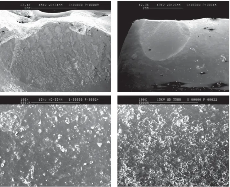
made to further characterise well-preserved enamel on the buccal surface (Figure 1). The interproximal facets, either mesial or distal, of all the isolated teeth were analysed and SEM images were obtained at 15x and 100x magnification. At the same time, the occlusal third of the buccal surfaces of all the studied teeth were also observed obtaining SEM images at 100x magnification (Figure 2a). All the SEM micrographs were obtained at the Serveis Cientificotècnics of the University of Barcelona.
RESULTS AND DISCUSSION
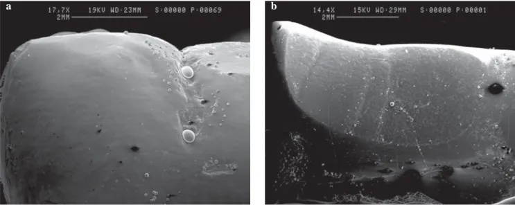
A clear distinction between post-mortem slightly eroded buccal enamel surface and well preserved interproximal facets (without ante-mortem striations) can be observed (Figure 1). Figure 1a shows a fairly smooth enamel surface, with some degree of perykimata, which is frequently observed when post-mortem damage affects the tooth surface. A well preserved inter-proximal surface shows no striations nor enamel polishing. Sometimes the so-called
subvertical grooves can be observed on the inter-proximal
wear facets, probably indicative of strong chewing forces due to occlusal tooth-to-tooth percussion. Figure 2 shows a group of well-preserved enamel surfaces. Figure 2a indicates the best enamel surface to observe buccal microwear, the occlusal third of the tooth crown, avoiding the occlusal rim and the cemento-enamel junction. Figures
2a, b, and c were taken at 100× magnification and show
typical microwear striations. Figure 3 shows well-preserved interproximal facets without ante-mortem damage. All these wear facets (Figures 3c, d) consist of highly pitted surfaces formed by tooth plucking during mastication.
As also shown in previous analyses (Pérez-Pérez et al. 1999, 2003) well preserved enamel on the buccal surface of teeth always shows a clear microwear pattern consisting of striations of various lengths and orientations, with a mainly vertical orientation (Figures 2b, c, d). Post-mortem eroded enamel generally showed either perykimata (Figure
1a), enamel polishing, prism exposure, parallel striations,
pitted surfaces, or a combination of pitting and striations. Such unpreserved surfaces can be clearly discriminated (see Pérez-Pérez et al. 2003). Inter-proximal facets most frequently show a highly pitted topography without the characteristic buccal microwear pattern (Figures 3c, d). Some of the inter-proximal facets may show subvertical grooves (Figure 1b), a frequent trait in Neandertal teeth that could be caused by strong tooth-to-tooth chewing forces (Villa, Giacobini 1995). A reduced number of teeth also showed a parallel striation pattern on the inter-proximal wear facets, in addition to their normal pitted topography. These teeth also tend to show generalized
post-mortem damage in other tooth surfaces, which is highly
indicative of post-mortem abrasion after fossil deposition. However, an ante-mortem microwear like pattern was never observed on any inter-proximal facet.
FIGURE 1. a: V236 Sima de los Huesos AT-561, M1 LR, buccal surface at 17.7×; b: ID237 Sima de los Huesos AT-273, M2 LL interproximal facet at 14.4×.
FIGURE 2. a: V253 Sima de los Huesos AT-588, M2 UR buccal surface at 17.0×; b: V208 Montmaurin, M1 LL buccal surface at 100×; c: V234 Sima de los Huesos AT-817, M2 UR buccal surface at 100×; d: V235 – Sima de los Huesos AT-271, M2 UR buccal surface at 101×.
a b
c d
FIGURE 3. a: ID208 Montmaurin, M1 UR interproximal facet at 15.1×; b: ID235 Sima de los Huesos AT-271, M2 UR interproximal facet at 17.8×; c: ID07 Gran Dolina, M1 UR interdistal facet at 100×; d: ID201 Rond du Barry, Pm4 LL proximal facet at 100×.
From the observations made, we propose that to discriminate between damaged enamel and intact surfaces, two assumptions need to be met: 1) microwear features (either striations or pits) need to be clearly visible; and 2) significantly distinct patterns of microwear need to be discriminated among occlusal, buccal, and inter-proximal tooth surfaces. Pits are rare on buccal surfaces, where striations tend to be vertically oriented; pit-to-striation ratios on the occlusal surfaces vary between primate species; and no pits or striations are present on inter-proximal facets, in which tooth-to-tooth impact produces a characteristic abrasion pattern. Inter-proximal wear facets are not exposed to food during mastication (except for pathological conditions), and tooth-to-tooth percussion causes prism plucking that results in a highly prism-sized pitted surface lacking any type of striations (Puech 1984). The absence of buccal/vestibular like microwear on the interdental facets may be considered indicative of lack of
post-depositional processes affecting dietary related microwear (Ungar, Teaford 1996). Since taphonomic processes are likely to affect in the same manner all enamel surfaces in the isolated teeth, a clear microwear distinction between buccal, occlusal and interproximal surfaces would be indicative of no post-mortem damage. Further experimental research is needed to fully characterise the effect of taphonomic processes on tooth enamel. However, clear differences exist between ante-mortem microwear, related to dietary behaviour, and post-mortem damage on the buccal surface, formed after deposition. The buccal microwear pattern reflects the abrasive capability of ingested food items, as well as technological, cultural and climatic determinants (Pérez-Pérez et al. 2003), but an accurate selection of well-preserved surfaces is required to minimize observational errors.
ACKNOWLEDGEMENTS
This project was funded by a L. S. B. Leakey Foundation grant. All images were obtained at the Serveis
Cientificotècnics of the University of Barcelona.
REFERENCES
DE BAYLE DE HERMENS R., HEIM J. L., 1989: Découverte d'un crâne humain dans une sépulture secondaire du Magdalénien I de la grotte de Rond du Barry, Polignac, Haute-Loire. Paléontologie humaine – C. R. Acad. Sci. Paris 309: 1349– 1352.
FALGUÈRES C., BAHAINM J. J., YOKOYAMJA Y., ARSUAGA J. L., BERMÚDEZ DE CASTRO J. M., CARBONELL E., BISCHOFF J. L., DOLO J. M., 2001: Datation par RPE et U-TH des sites pléistocènes d'Atapuerca: Sima de los Huesos, Trinchera Dolina et Trinchera Galería. Bilan géochronologique. L'Anthropologie 105: 71–81.
GALVANY J., MARTÍNEZ L. M., HIRALDO O., ESPURZ V., ESTEBARANZ F., SOUSA M., MARTÍNEZ H., MEDINA A. M., FARRÉS M., BONNIN A., BERNIS C., TURBÓN D., PÉREZ-PÉREZ A., 2004: Catálogo de dientes de Homínidos de la Universidad de Barcelona (Teeth). Universitat de Barcelona, Barcelona. 193 pp.
GORDON K. D., 1983. Taphonomy of dental microwear: can fossil microwear be studied productively? Amer. J. of Phys. Anthrop. 60: 200–200.
GRINE F. E., 1987: Quantitative analysis of occlusal microwear in Australopithecus and Paranthropus. Scanning Microscopy 1: 647–656.
KING T., ANDREWS P., BOZ B., 1999: The effect of taphonomic processes on dental microwear. Amer. J. of Phys. Anthrop. 108: 359–373.
LALUEZA C., PÉREZ-PÉREZ A., 1993: The diet of the Neanderthal child Gibraltar 2 (Devil's Tower) through the study of the vestibular striation pattern. J. of Hum. Evol. 24: 29–41. LALUEZA C., PÉREZ-PÉREZ A., JUAN J., 1994: Dietary
information through the examination of plant phytoliths on the enamel surface of human dentition. J. of Archaeological Science 21: 29–34.
LALUEZA C., PÉREZ-PÉREZ A., TURBÓN D., 1996: Dietary inferences through buccal microwear analysis of Middle and Upper Pleistocence human fossils. Amer. J. of Phys. Anthrop. 100, 3: 367–387.
MAAS M. C., 1991: Enamel structure and microwear: an experimental study of the response of enamel to shearing force. Amer. J. of Phys. Anthrop. 85: 31–50.
PÉREZ-PÉREZ A., LALUEZA C. 1991. Human paleoecology revealed from bones. J. of Human Ecology 2: 391–398. PÉREZ-PÉREZ A., LALUEZA C., 1992: Dietary reconstruction
from historical information and trace elements analysis in a medieval population from Catalonia (Spain). Internat. J. of Anthropology 7, 1: 51–57.
PÉREZ-PÉREZ A., LALUEZA C., TURBÓN D., 1994: Intraindividual and intragroup variability of buccal tooth striation pattern. Amer. J. of Phys. Anthrop. 94, 2: 175–187. PÉREZ-PÉREZ A., BERMÚDEZ DE CASTRO J. M., ARSUAGA J. L., 1999: Non-occlusal microwear analysis of 300,000 year-old Homo heilderbergensis teeth from Sima de los Huesos (Sierra de Atapuerca, Spain): implications of intrapopulational
variability for dietary analysis of Hominid fossil remains. Amer. J. of Phys. Anthrop. 108: 433–457.
PÉREZ-PÉREZ A., ESPURZ V., BERMÚDEZ DE CASTRO J. M., DE LUMLEY M. A., TURBÓN D., 2003: Non-occlusal dental microwear variability in a sample of Middle and Late Pleistocene human populations from Europe and the Near East. J. of Hum. Evol. 44, 4: 497–513.
PUECH P. F., 1984: Acidic food choice in Homo habilis at Olduvai. Curr. Anthrop. 25: 349–350.
TEAFORD M. F., 1988: Scanning electron microscopy diagnosis of wear pattern versus artefacts. Scanning Microscopy 2: 1167– 1175.
TEAFORD M. F., 1994: Dental microwear and dental function. Evol. Anthrop. 3, 1: 17–31.
UNGAR P. S., 1994a: Incisor microwear in Sumatran anthropoid primates. Amer. J. of Phys. Anthrop. 94: 339–363.
UNGAR P. S., 1994b: Patterns of ingestive behavior and anterior tooth use different in sympatric anthropoid primates. Amer. J. of Phys. Anthrop. 95: 197–219.
UNGAR P. S., TEAFORD M. F., 1996: Preliminary examination of non-occlusal dental microwear in anthropoids: implications for the study of fossil primates. Amer. J. of Phys. Anthrop. 100: 101–113.
UNGAR P. S., 1998: Dental allometry, morphology and wear as evidence for diet in fossil primates. Evol. Anthrop. 6: 205– 217.
VILLA G. B., GIACOBINI G., 1995: Subvertical grooves of interproximal facets in Neanderthal posterior teeth. Amer. J. of Phys. Anthrop. 96: 51–62.
WOLPOFF M. H., 1999: Paleoanthropology. McGraw-Hill, Boston. pp: 594–691.
Vanesa Espurz Alejandro Pérez-Pérez Daniel Turbón
Secc. Antropologia, Dept. Biologia Animal Universitat de Barcelona
Av. Diagonal 645 08028 Barcelona, Spain E-mail: [email protected]