Original article
Scoring ultrasound synovitis in
rheumatoid arthritis: a
EULAR-OMERACT ultrasound taskforce-Part 2:
reliability and application to multiple
joints of a standardised
consensus-based scoring system
Lene Terslev,1 Esperanza Naredo,2 Philippe Aegerter,3 Richard J Wakefield,4 Marina Backhaus,5 Peter Balint,6 George A W Bruyn,7 Annamaria Iagnocco,8 Sandrine Jousse-Joulin,9 Wolfgang A Schmidt,10 Marcin Szkudlarek,11 Philip G Conaghan,4 Emilio Filippucci,12 Maria Antonietta D'Agostino13,14 To cite: terslev l, naredo e,
aegerter P, et al. Scoring ultrasound synovitis in rheumatoid arthritis: a eUlar-OMeract ultrasound taskforce-Part 2: reliability and application to multiple joints of a standardised consensus-based scoring system. RMD Open 2017;3:e000427. doi:10.1136/ rmdopen-2016-000427 ►additional material is published online only. to view please visit the journal online (http:// dx. doi. org/ 10. 1136/ rmdopen- 2016- 000427). received 23 December 2016 revised 9 May 2017 accepted 16 May 2017
For numbered affiliations see end of article.
Correspondence to Professor Maria antonietta D'agostino, rheumatology Department, Versailles Saint Quentin en Yvelines University, ambroise Paré Hospital, aPHP, 9 avenue charles de gaulle, 92100 Boulogne-Billancourt, France; maria- antonietta. dagostino@ apr. aphp. fr ►http:// dx. doi. org/ 10. 1136/ rmdopen- 2016- 000428
AbstrAct
Objectives to test the reliability of new ultrasound (US)
definitions and quantification of synovial hypertrophy (SH) and power Doppler (PD) signal, separately and in combination, in a range of joints in patients with rheumatoid arthritis (ra) using the european league against rheumatisms–Outcomes Measures in rheumatology (eUlar-OMeract) combined score for PD and SH.
Methods a stepwise approach was used: (1) scoring
static images of metacarpophalangeal (McP) joints in a web-based exercise and subsequently when scanning patients; (2) scoring static images of wrist, proximal interphalangeal joints, knee and metatarsophalangeal joints in a web-based exercise and subsequently when scanning patients using different acquisitions (standardised vs usual practice). For reliability, kappa coefficients (κ) were used.
Results Scoring McP joints in static images showed
substantial intraobserver variability but good to excellent interobserver reliability. in patients, intraobserver reliability was the same for the two acquisition methods. interobserver reliability for SH (κ=0.87) and PD (κ=0.79) and the eUlar-OMeract combined score (κ=0.86) were better when using a ‘standardised’ scan. For the other joints, the intraobserver reliability was excellent in static images for all scores (κ=0.8–0.97) and the interobserver reliability marginally lower. When using standardised scanning in patients, the intraobserver was good (κ=0.64 for SH and the eUlar-OMeract combined score, 0.66 for PD) and the interobserver reliability was also good especially for PD (κ range=0.41–0.92).
Conclusion the eUlar-OMeract score demonstrated
moderate-good reliability in McP joints using a standardised scan and is equally applicable in non-McP joints. this scoring system should underpin improved reliability and consequently the responsiveness of US in ra clinical trials.
IntroductIon
Growing data suggest that ultrasound (US) is a valuable tool for assessing and classifying joint involvement and measuring disease activity based on the detection and scoring of synovitis in patients with rheumatoid arthritis (RA).1 The benefit of US in the evaluation and monitoring of patients with RA is mainly based on its greater sensitivity in detecting synovitis compared with clinical examination.2–4 Colour Doppler (CD) and power Doppler (PD) modes are able to detect pathological synovial blood flow, which reflects the inflammatory activity in the joint5–7 and has predictive value in rela-tion to radiographic progression of structural damage8–10 and in relation to disease flare.11–13 In addition, US-detected synovitis aids more
Key messages
What is already known about this subject? ► no consensus existed until now on a single
ultrasound (US) scoring system for rheumatoid arthritis (ra) clinical trials.
What does this study add?
► a consensus-based US scoring system has been validated in multiple joints and has been shown to be highly reliable.
How might this impact on clinical practice? ► this highly reliable consensus-based scoring system
should improve responsiveness and increase the uptake of US in ra clinical trials.
accurate early diagnosis of RA to enable earlier treat-ment.14 15
As RA clinical trials need objective and feasible methods for assessing inflammation response and with clinical practice focusing on tight control of disease activity, it has become imperative to improve the reliability of US in quantifying synovitis. Many scoring systems have been proposed, however, a recent literature review highlighted the lack of an expert-derived consensus.16
The Outcomes Measures in Rheumatology (OMERACT) US Working Group in collaboration with an US working party of the European League Against Rheumatisms (EULAR) conducted a series of US studies in order to understand possible reasons for a low agree-ment in detecting synovitis and to develop and validate an expert-derived consensus for scoring synovitis. The validation process, outlined in supplementary figure 1 in the supplementary online material, was carried out in a multistep approach (four steps) from 2005 to 2014. The first two steps are described in a companion paper17 in
which exercises in static images and in clinical setting revealed that the causes for the inconsistencies and the hampered reliability in scoring synovitis among rheuma-tologists from different European countries were related to several sources of variability such as the perception and weighting of the different US components (ie, synovial hypertrophy (SH), Doppler activity and also effu-sion) used for describing and grading the inflammatory process17 as well as the differences in the US acquisition
technique. Based on these discrepancies, the elementary components were redefined by Delphi consensus. It was agreed: (1) not to include effusion as an inflammatory component, as it was considered to be an inconsistent finding, frequently detected in healthy subjects or in inac-tive RA joints;18 19 (2) to redefine synovitis based on SH
and Doppler only and (3) to score them semiquantita-tively (0–3) both separately and in combination using the novel EULAR-OMERACT combined score.17 These steps
were performed using metacarpophalangeal (MCP) joints as a model.
Having established these basic steps,17 the group
moved to the second part of the validation process which is presented in this paper. The objectives were: (1) to evaluate the reliability of the EULAR-OMERACT combined score for grading synovitis in MCP joints, as well as the definition and quantification of SH and PD individually; (2) to test the reliability of a standardised consensus-based acquisition method compared with a ‘usual practice’ scanning method and (3) to evaluate the reliability of the new definitions for SH, PD and the EULAR-OMERACT combined score in non-MCP joints.
Methods
Twelve US-experienced rheumatologists, who partici-pated in the first part of the standardisation process,17
participated in the following steps: (1) testing the validity of the new proposed definitions for scoring SH and PD
separately and in combination (the EULAR-OMERACT PDUS score) on static images of MCP joints; (2) applying the same definitions and scoring systems in a real-time patient-based reliability exercise, by comparing a consensus-based scanning acquisition method previously obtained17 to a ‘usual practice’ scanning method and (3)
testing the reliability of the new definitions and of the EULAR-OMERACT combined score in non-MCP joints (wrist, proximal interphalangeal (PIP), knee and meta-tarsophalangeal (MTP)) in both reading static images and scanning patients.
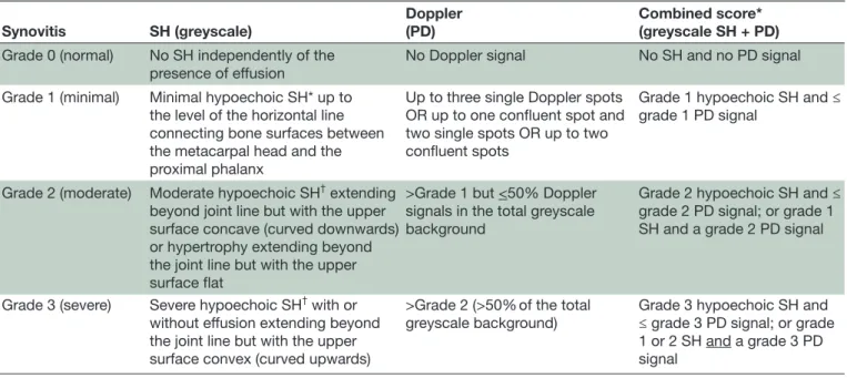
In all the reliability exercises, the participants used both a semiquantitative (SQ) (0–3) and a binary score (yes/no). The definitions and the scoring systems used are presented in table 1.
All patients participating in the reliability exercises fulfilled the American College of Rheumatology clas-sification criteria for RA20 and were attending the
rheumatology department of Ambroise Paré hospital in Boulogne-Billancourt (France).
Patients were selected based on the absence of joint deformities and the willingness to take part. The studies were conducted in accordance with the Declaration of Helsinki and each participant gave written informed consent.
step 1. Web-based exercise
A set of high-quality US images of synovitis of MCP joints were selected from an anonymised register of patients with RA by two independent ultrasonographers (MADA and EN) in order to ensure inclusion of a broad range of synovitis severity. A random selection of images was shown twice in order to assess the intra-reader reliability.
step 2. Patients-based exercise: scanning patients according to a different scanning approach
The experts performed a bilateral US scanning of the second–fifth MCP joints in eight different patients. The dorsal aspect of the joints was examined twice in two rounds over 2 days. In the first round, using a ‘stan-dardised acquisition method’, the US examinations were performed using a longitudinal dorsal scan on the middle of the joint, first, in GS and then PD, to detect joint morphological abnormalities and synovial flow respectively. In the second round, a ‘usual practice (free) acquisition approach’ of the dorsal side of the MCP joint was used. In the standardised scan, the maximal grading was to be assessed in the midline. In the ‘usual prac-tice’ scan method, the examiner recorded the maximal grading from any area of the joint.
step 3. testing the new definitions and the reliability of the euLAr-oMerAct combined score and of sh and Pd individually in non-McP joints
A set of high-quality US images of synovitis of wrist, PIP, knees and MTP joints from patients with RA was evalu-ated using images from the same register and applying the same approach as described in step 1.
After the exercise on static images, the experts performed bilateral US scanning of the wrist, PIP,2–5 knee
and MTP1–5 joints in six different patients twice in two
rounds over 2 days (first day wrist and PIP joints, second day knee and MTP joints), using predefined joint posi-tions as follows:
► Wrist joints (ie, radiocarpal and midcarpal joints were evaluated as a single site): palms facing down and wrist positioned flat on the examining table, as neutral as possible but relaxed; shoulder and elbow relaxed; elbow rested on the table. Scanning at the level of the radio-lunate joint.
► PIP joints: palms facing down and wrist positioned flat on the examining table, as neutral as possible but relaxed, scanning on the dorsal midline aspect. ► Knee joints (ie, suprapatellar and parapatellar
recesses were scored as a single site): knee 30° flexed and scanning on suprapatellar midline for the suprapatellar recess; knee extended and scanning the parapatellar areas using the retinacula as a landmark for the parapatellar medial and lateral recesses. Doppler signal was recorded only in the medial and lateral parapatellar recesses.
► MTP joints: foot placed resting (with knee 30° flexed) over its plantar aspect. Scanning recorded on the dorsal midline aspect.
For all examinations, identical ESAOTE Technos MPX (Genoa, Italy) US machines with an 8–14 MHz linear array transducer were used with identical PD settings (frequency of 10.1 MHz, pulse repetition frequency of 750 Hz and Doppler gain of 50–53 dB). Each patient was assigned to one machine and the sonographers then rotated from one machine to the next in a predefined
sequence with 10 min allocated for scanning and
recording the findings on a standard score sheet. Partic-ipants were blinded to the patients’ clinical details (ie, presence or not of active disease).17
stAtIstIcAL AnALysIs
The intraobserver and interobserver reliability of scoring static and dynamic images were assessed according to weighted Kappa coefficients (κ) relying on abso-lute differences and in order to take into account the magnitude of discrepancy between categories. Intraob-server coefficients were evaluated on pairs of measures performed by the same sonographer at each site, while interobserver coefficients were exclusively based on the first measure of those pairs. Interobserver reliability was studied by calculating the mean κ for all pairs (ie, Light’s κ).21 Kappa values were evaluated according to
Landis and Koch.22 Percentage of observed agreement
(ie, percentage of observations that obtained the same score) and prevalence of the observed lesions were also calculated. Statistical analysis was performed using the R software (http://www. r- project. org/).
resuLts
step 1. testing the definition and reliability of the EULAr-OMErAct combined score on static images
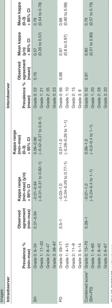
Thirty-six images of MCP joints were scored. Table 2
shows the observed agreement, prevalence and κ values results. The agreement was good for the novel definitions of synovitis components (SH and PD) both separately and in combination (EULAR-OMERACT combined score) with the best obtained for PD alone.
Table 1 Definitions of severity grades (0–3) for each elementary component and for the EULAR-OMERACT combined score
Synovitis SH (greyscale)
Doppler (PD)
Combined score* (greyscale SH + PD)
Grade 0 (normal) No SH independently of the
presence of effusion No Doppler signal No SH and no PD signal Grade 1 (minimal) Minimal hypoechoic SH* up to
the level of the horizontal line connecting bone surfaces between the metacarpal head and the proximal phalanx
Up to three single Doppler spots OR up to one confluent spot and two single spots OR up to two confluent spots
Grade 1 hypoechoic SH and ≤ grade 1 PD signal
Grade 2 (moderate) Moderate hypoechoic SH† extending
beyond joint line but with the upper surface concave (curved downwards) or hypertrophy extending beyond the joint line but with the upper surface flat
>Grade 1 but <50% Doppler signals in the total greyscale background
Grade 2 hypoechoic SH and ≤ grade 2 PD signal; or grade 1 SH and a grade 2 PD signal
Grade 3 (severe) Severe hypoechoic SH† with or
without effusion extending beyond the joint line but with the upper surface convex (curved upwards)
>Grade 2 (>50% of the total
greyscale background) Grade 3 hypoechoic SH and ≤ grade 3 PD signal; or grade 1 or 2 SH and a grade 3 PD signal
*EULAR-OMERACT combined score. †Independently of the presence of effusion.
EULAR-OMERACT, European League Against Rheumatisms–Outcomes Measures in Rheumatology; PD, power Doppler; SH, synovial hypertrophy.
Table 2
Pr
evalence of observed lesions, observed agr
eement, intraobserver and inter
observer r
eliabilities when scoring synovitis in metacarpophalangeal joints on static
images Intraobserver Inter observer Pr evalence % (min–max) Observed agr eement (min–max)
Kappa range (min–max) (y/n) + 95% CI Kappa range (min–max) (0–3) + 95% CI
Pr evalence % (mean) Observed agr eement (mean)
Mean kappa (y/n) + 95% CI Mean kappa (0–3) + 95% CI
SH Grade 0: 9–34 0.31–0.94 −0.01–0.94 (−0.31–0.31 to 0.82–1) 0.06–0.96 (−0.42–0.27 to 0.9–1) Grade 0: 22 0.76 0.57 (0.35 to 0.57) 0.78 (0.54 to 0.78) Grade 1: 11–33 Grade 1: 21 Grade 2: 8–47 Grade 2: 35 Grade 3: 38–67 Grade 3: 23 PD Grade 0: 59–71 0.5–1 −0.03–1.0 (−0.34–0.29 to 0.77–1) 0.01–1.0 (−0.28–0.26 to 1–1) Grade 0: 66 0.88 0.97 (0.8 to 0.97) 0.98 (0.80 to 0.98) Grade 1: 4–15 Grade 1: 10 Grade 2: 11–9 Grade 2: 15 Grade 3: 5–14 Grade 3: 9 Combined scor e* (SH+PD) Grade 0: 13–70 0.39–1 −0.01–1.0 (−0.24–0.4 to 1–1) 0.06–1.0 (−0.0–0.4 to 1–1) Grade 0: 32 0.81 0.80 (0.51 to 0.80) 0.79 (0.57 to 0.79) Grade 1: 4–60 Grade 1: 20 Grade 2: 8–45 Grade 2: 28 Grade 3: 6–67 Grade 3: 20
*EULAR-OMERACT combined scor
e.
EULAR-OMERACT
, Eur
opean League Against Rheumatisms–Outcomes Measur
es in Rheumatology; PD, power Doppler; SH, synovial hypertr
ophy
Surprisingly, the intraobserver reliability showed a great variability between the 12 sonographers for all parameters. Similar results were seen for the binary score. The interobserver reliability was good to excellent for the SQ score of SH, PD and the EULAR-OMERACT combined score. For the binary score, the reliability was good to excellent for PD and the EULAR-OMERACT combined score, but only moderate for SH (table 2). When comparing the interobserver reliability for the SQ score with the binary score for PD and the EULAR-OMERACT combined score, reliability showed almost identical κ values—the highest κ values were seen for the PD score (SQ PD score: κ=0. 98 and binary PD score: κ=0.97). For SH, the binary score was considerably lower (κ=0.57) than the SQ score (κ=0.78).
step 2. testing the definition and reliability of the euLAr-oMerAct combined score in patients
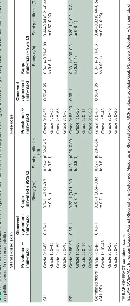
No major differences were recorded in the intraobserver reliability when scanning in a patient-based exercise for the ‘standardised scan’ and ‘usual practice scan’ (slightly better for the standardised scan) for all synovitis components (SH and PD) and the EULAR-OMERACT combined score, and for both binary and SQ grading (table 3).
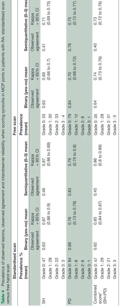
However, interobserver reliability for both SQ and binary scores for all components was better when using the standardised scan approach (table 4). The κ values were good for SQ PD (κ=0.79) but excellent for SH and the EULAR-OMERACT combined score (κ=0.87 and 0.86, respectively). The SQ score performed slightly better than the binary score for PD (SQ score: κ=0.79;
binary κ=0.76) and the EULAR-OMERACT combined
score (SQ score: κ=0.86; binary κ=0.85).
Only the PD grading for both the binary score and the SQ score had better interobserver reliability in static images than when scanning patients with a standardised scan (tables 2 and 4).
step 3. testing the definition and reliability of the euLAr-oMerAct combined score in other joints
In the web-based exercise on static images, 100 images of wrist, PIP, knee and MTP joints were included repre-senting a broad range of different degrees of synovitis.
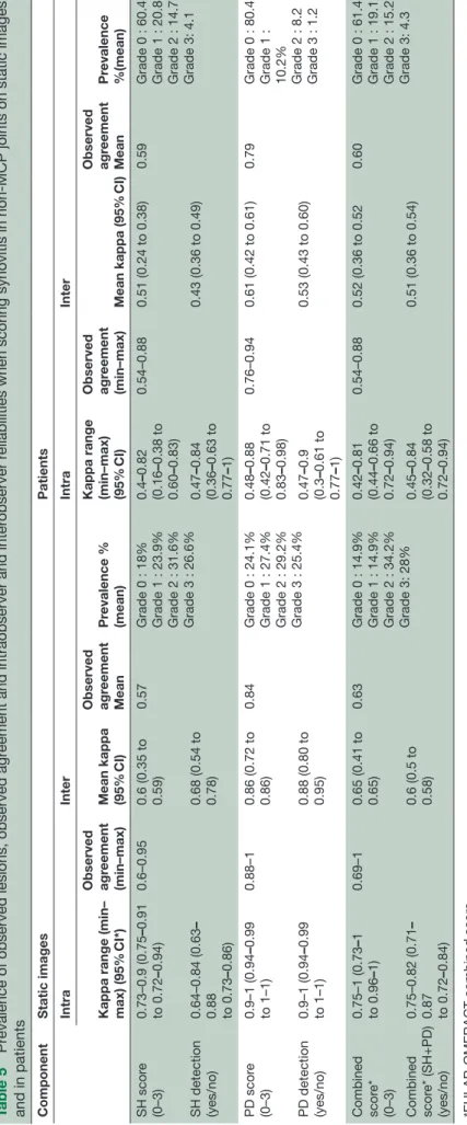
Table 5 shows the observed agreement, prevalence and reliability of the different degrees of SH, PD and the EULAR-OMERACT combined score. When scoring static images, the intraobserver reliability was good to excellent for SH and PD (better for SQ grading than binary) and excellent for the EULAR-OMERACT combined P score (κ=0.84). The interobserver reliability was good for all components (better for binary score than SQ grading) and best for PD (binary=0.88 and SQ=0.86). Table 6 shows the inter-reader reliability for the synovitis components and the EULAR-OMERACT combined score according to the different joints. The inter-reader reliability for the EULAR-OMERACT combined score in the wrist was κ=0.61, for the PIPs κ=0.75, for knees κ=0.55 and for the MTPs κ=0.58.
When evaluating the EULAR-OMERACT combined score in patients, the intraobserver reliability was good with almost identical values for the binary and SQ scores for all single components and in combination, ranging from 0.64 to 0.66 (table 5). The interobserver reliability was moderate to good for all components (0.43–0.61) and best for the SQ PD score (0.61) (table 5).
Supplementary figure 2 (online file) shows image examples on the EULAR-OMERACT combined score applied to PIP, MTP, knee and wrist joints.
Following the results of this multistep project, the group agreed on the following procedures for scoring synovitis by US: (1) The presence of a hypoechoic SH is mandatory for defining the presence of an US-detected synovitis and for grading Doppler activity. (2) Grading synovitis, at joint level, should be performed by using the SQ EULAR-OMERACT score (based on the combined presence of both GS SH and Doppler (table 1)).
(3) If different areas of severity are present in the same joint, the final severity grade is given by the area with the maximum of severity. (4) The acquisition and grading of synovitis by US should be performed by using a dorsal approach. (5) A standardised scan, with the position of the probe in the midline, should be recommended in the case of multicentre clinical trials using US, although it might underestimate the real inflammatory activity of the joint.
dIscussIon
Over the last 10 years, the EULAR-OMERACT US group has worked on standardising the US detection, acquisi-tion and grading of synovitis in patients with RA using a stepwise approach. In the first step, the group developed: (1) new definitions of the elementary components and a novel scoring system based on the grade of severity of SH and PD both separately and in combination: the EULAR-OMERACT combined score; and (2) a standardised image acquisition technique.17 In the second part of
this multistep validation process, the reliability of these new definitions and of the scoring system for SH and PD separately, and EULAR-OMERACT combined was tested in static images, then in patients on MCP and non-MCP joints. The participation of the same multinational team in every step of the validation process added value to the consistency of the results.
The new definitions for grading SH and PD inde-pendently and combined (EULAR-OMERACT combined score) considerably improved the reliability when scoring both static images and patients. In these studies, PD was chosen as the optimal Doppler modality for the partic-ular US machines used for depicting inflammation, but PD may be substituted by CD in the presented scoring system when working with machines where CD is more sensitive than PD.23 The interobserver reliability for the
EULAR-OMERACT combined score in static images was good to excellent. In patients, the intraobserver reliability of the EULAR-OMERACT combined score showed some
Table 3
Pr
evalence of observed lesions, observed agr
eement and intraobserver r
eliability when scoring synovitis in MCP joints in patients with RA: standar
dised scan acquisition versus fr ee scan acquisition Standar dised scan Fr ee scan Pr evalence % (min–max) Observed agr eement (min–max)
Kappa (min–max) + 95% CI
Pr evalence % (min–max) Observed agr eement (min–max)
Kappa (min–max) + 95% CI
Binary (y/n) Semiquantitative (0–3) Binary (y/n) Semiquantitative (0–3) SH Grade 0: 0–80 0.40–1 0.0–1 (−0.21–0.3 to 0.6–1) 0.34–1 (0.32–0.45 to 0.9–1) Grade 0: 0–55 0.50–0.95 0.0–1 (−0.01–0.03 to 0.6–1) 0.44 –0.94 (0.21–0.44 to 0.87–0.97) Grade 1: 5–45 Grade 1: 5–55 Grade 2: 0–50 Grade 2: 5–65 Grade 3: 0–15 Grade 3: 0–5 PD Grade 0: 55–95 0.65–1 0.22–1 (0.21–0.3 to 0.8–1) 0.26–1 (0.25–0.29 to 0.8–1) Grade 0: 55–90 0.65–1 0.38–1 (0.36–0.3 to 0.81–1) 0.29–1 (−0.21–0.3 to 0.6–1) Grade 1: 0–30 Grade 1: 0–25 Grade 2: 0–10 Grade 2: 0–15 Grade 3: 0–5 Grade 3: 0–20 Combined scor e* (SH+PD) Grade 0: 0–80 0.40–1 0.39–1 (0.34–0.43 to 0.7–1) 0.32–1 (0.29–0.34 to 0.8–1) Grade 0: 0–60 0.50–0.95 0.0–1 (−0.11–0.3 to 0.92–1) 0.40–0.93 (0.40–0.52 to 0.76–0.95) Grade 1: 10–45 Grade 1: 5–43 Grade 2: 0–50 Grade 2: 0–70 Grade 3: 0–15 Grade 3: 0–20
*EULAR-OMERACT combined scor
e.
EULAR-OMERACT
, Eur
opean League Against Rheumatisms–Outcomes Measur
es in Rheumatology; MCP
, metacarpophalangeal; PD, power Doppler; RA, rheumatoid
arthritis; SH, synovial hypertr
ophy
Table 4
Pr
evalence of observed lesions, observed agr
eement and inter
observer r
eliability when scoring synovitis in MCP joints in patients with RA: standar
dised scan versus fr ee hand scan Standar dised scan Fr ee scan Pr evalence % (mean)
Binary (yes–no) mean
Semiquantitative (0–3) mean
Pr
evalence
(mean)
Binary (yes–no) mean
Semiquantitative (0–3) mean Observed agr eement Kappa + 95% CI Observed agr eement Kappa + 95% CI Observed agr eement Kappa + 95% CI Observed agr eement Kappa + 95% CI SH Grade 0: 47 0.63 0.87 (0.86 to 0.9) 0.46 0.87 (0.86 to 0.89) Grade 0: 33 0.63 0.69 (0.66 to 0.7) 0.41 0.71 (0.69 to 0.73) Grade 1: 28 Grade 1: 30 Grade 2: 22 Grade 2: 33 Grade 3: 3 Grade 3 : 4 PD Grade 0: 84 0.88 0.76 (0.73 to 0.79) 0.83 0.79 (0.76 to 0.8) Grade 0: 79 0.84 0.70 (0.68 to 0.72) 0.78 0.75 (0.72 to 0.77) Grade 1: 8 Grade 1: 12 Grade 2: 6 Grade 2: 8 Grade 3: 1 Grade 3: 1 Combined scor e* (SH+PD) Grade 0: 47 0.62 0.85 (0.84 to 0.87) 0.45 0.86 (0.8 to 0.89) Grade 0: 35 0.64 0.74 (0.73 to 0.76) 0.40 0.73 (0.72 to 0.76) Grade 1: 29 Grade 1: 28 Grade 2: 21 Grade 2: 31 Grade 3: 3 Grade 3 : 5 MCP
, metacarpophalangeal; PD, power Doppler; RA, rheumatoid arthritis; SH, synovial hypertr
ophy
.
*EULAR-OMERACT combined scor
e.
EULAR-OMERACT
, Eur
opean League Against Rheumatisms–Outcomes Measur
Table 5
Pr
evalence of observed lesions, observed agr
eement and intraobserver and inter
observer r
eliabilities when scoring synovitis in non-MCP joints on static images
and in patients Component
Static images Patients Intra Inter Intra Inter
Kappa range (min– max)
(95% CI*) Observed agr eement (min–max) Mean kappa (95% CI) Observed agr eement Mean Pr evalence % (mean)
Kappa range (min–max) (95%
CI) Observed agr eement (min–max) Mean kappa (95% CI) Observed agr eement Mean Pr evalence %(mean) SH scor e (0–3) 0.73–0.9 (0.75 –0.91 to 0.72 –0.94) 0.6–0.95 0.6 (0.35 to 0.59) 0.57
Grade 0 : 18% Grade 1 : 23.9% Grade 2 : 31.6% Grade 3 : 26.6%
0.4 –0.82 (0.16 –0.38 to 0.60 –0.83) 0.54–0.88 0.51 (0.24 to 0.38) 0.59
Grade 0 : 60.4 Grade 1 : 20.8 Grade 2 : 14.7 Grade 3: 4.1
SH detection (yes/no) 0.64–0.84 (0.63 – 0.88 to 0.73 –0.86) 0.68 (0.54 to 0.78) 0.47 –0.84 (0.36 –0.63 to 0.77 –1) 0.43 (0.36 to 0.49) PD scor e (0–3) 0.9–1 (0.94 –0.99 to 1–1) 0.88–1 0.86 (0.72 to 0.86) 0.84
Grade 0 : 24.1% Grade 1 : 27.4% Grade 2 : 29.2% Grade 3 : 25.4%
0.48 –0.88 (0.42 –0.71 to 0.83 –0.98) 0.76–0.94 0.61 (0.42 to 0.61) 0.79
Grade 0 : 80.4 Grade 1 : 10.2% Grade 2 : 8.2 Grade 3 : 1.2
PD detection (yes/no) 0.9–1 (0.94 –0.99 to 1–1) 0.88 (0.80 to 0.95) 0.47 –0.9 (0.3 –0.61 to 0.77 –1) 0.53 (0.43 to 0.60) Combined scor e* (0–3) 0.75–1 (0.73 –1 to 0.96 –1) 0.69–1 0.65 (0.41 to 0.65) 0.63
Grade 0 : 14.9% Grade 1 : 14.9% Grade 2 : 34.2% Grade 3: 28%
0.42 –0.81 (0.44 –0.66 to 0.72 –0.94) 0.54–0.88 0.52 (0.36 to 0.52 0.60
Grade 0 : 61.4 Grade 1 : 19.1 Grade 2 : 15.2 Grade 3: 4.3
Combined scor e* (SH+PD) (yes/no) 0.75–0.82 (0.71 – 0.87 to 0.72 –0.84) 0.6 (0.5 to 0.58) 0.45 –0.84 (0.32 –0.58 to 0.72 –0.94) 0.51 (0.36 to 0.54)
*EULAR-OMERACT combined scor
e.
PD, power Doppler signal; SH, synovial hypertr
ophy
variability, probably due to the initial difficulty to apply the new definition in ‘real life’ scanning. However, the interobserver reliability was good to excellent.
The number of patients involved in the two patient-based reliability exercises can be seen as a limitation as they are in the lower range of the sample sizes usually used in imaging studies.24 However, as several joints were
scanned in each patient (two times 8 joints in the first exercise and 22 joints per patient in the second exer-cise), which in these exercises are seen as independent contributors, and as several examiners participated, the results can be seen as robust enough for supporting the reliability of the scoring.
By using a stepwise process involving discussion and agreement, we were able to evaluate the real impact of the scanning technique. A standardised approach with the probe in a longitudinal plane on the dorsal aspect and in the midline of the joint was found to improve the reliability as compared with a ‘usual practice’ scanning approach. This provides further evidence supporting the concept that guidelines for image acquisition are needed and that the dorsal aspect of the joint with the probe in the midline is recommended to improve reliability when assessing small joints25 in multicentre trials though a
free scan may detect more accurately the real amount of inflammation in a joint (as stated in the procedures for scanning (3)).
In the first steps of the validation process, the MCP joint was used as a model for evaluating the scoring systems. However, as the final goal of this process was to produce a generalisable instrument to be used in global assessment of disease activity, other joint areas were subsequently incorporated.26–28 The grading of SH and PD
inde-pendently and the combined EULAR-OMERACT score were therefore evaluated in other commonly affected joints such as wrist, PIP, knee and MTP joints. Both intra-reader and inter-intra-reader reliabilities were good in static images but lower in patients compared with the reli-ability in MCP joints and though a standardised scanning approach was applied in these joints, further training in these joint regions may improve the reliability.
Regarding the US assessment of synovitis, it is important to emphasise that although a binary scoring system may be more reliable than a SQ grading, it is optimal only in monitoring patients for whom synovitis completely disap-pears during treatment. A binary score does not have the sensitivity to detect partial improvement and relies on the ability of the treatment to leave no residual inflam-mation, making it unsuitable for monitoring treatment effects in longstanding RA, where a complete disap-pearance of the SH can be hampered by the copresence of osteoarthritis or when complete remission has not been obtained. Considering that a number of studies have reported the presence of minimal abnormalities in both GS and Doppler in healthy controls,18 19 29 the
development of standardised recommendations for scoring synovitis is a major step forward in the develop-ment of the concept of a minimal detectable US synovitis
Table 6
Inter
observer r
eliability r
esults for detection and scoring of synovitis components and for the combined PD US scor
e accor
ding to the dif
fer
ent joints in static
images and in patients
Static images Patients W rist PIP Knee MTP W rist PIP Knee MTP Mean kappa Mean kappa Mean kappa Mean kappa Mean observed agr
eement
Mean kappa Mean observed agr
eement
Mean kappa Mean observed agr
eement
Mean kappa Mean observed agr
eement Mean kappa SH scor e (0–3) 0.51 0.69 0.60 0.54 0.5 0.60 0.7 0.52 0.51 0.5 0.76 0.50 SH detection (yes/no) 0.35 0.74 0.81 0.64 0.23 0.2 0.42 0.34 PD scor e (0–3) 0.84 0.89 0.85 0.86 0.82 0.67 0.95 0.1 0.53 0.49 0.96 0.54 PD detection (yes/no) 0.91 0.92 0.83 0.89 0.42 0.1 0.41 0.48 Combined scor e* (0–3) 0.61 0.75 0.55 0.58 0.87 0.67 0.7 0.53 0.67 0.54 0.72 0.50 Combined scor e* (SH+PD) (yes/no) 0.61 NA 0.62 NA 0.43 0.3 0.41 0.39
*EULAR-OMERACT combined scor
e.
MTP
, metatarsophalangeal; NA, non-applicable; PD, power Doppler signal; PIP
, pr
oximal interphalangeal; SH, synovial hypertr
ophy
and defining the threshold of normality. The use of a consensus process, based on the analysis of disagreement and exploration of factors affecting reliability, represents a major advantage of this programme of work. The inclu-sion of clear definitions of each synovial component and the application of a standardised scanning approach has ensured a highly robust process.
Though the current study did not address possible intermachine variability. This may be a problem in clin-ical practice best solved by using the best equipment giving the patient optimal evaluation.23 In multicentre
trials, the quality of the machines may be different but is almost equivalent as a prerequisite. In addition, the patient is examined always with the same equipment and same settings minimising the variability.
In our study, the reliability of the EULAR-OMERACT combined score was comparable to that of the elementary synovitis components. This has important implications in multicentre studies as both components can be equally reliable in monitoring RA depending on the joint size (ie, Doppler mode is less sensitive for deep anatomic areas) and the Doppler sensitivity of the US machine.23 Furthermore, the participation of several sonographers from different countries confirms the applicability of the proposed scoring system to multicentre clinical trials and in daily practice.
In conclusion, using an expert-derived consensus process, the EULAR-OMERACT group have developed a standardised EULAR-OMERACT combined scoring system taking both PD and SH components into account in the evaluation of synovitis of multiple RA joints, which is highly reliable when applied in scanning patients. The reliability was further improved when a standardised scanning procedure was used. The application of the proposed EULAR-OMERACT combined score and the new definition of synovitis based on the presence of SH and PD, as well as a standardised scanning approach for synovitis in RA, will ensure a greater degree of homoge-neity and comparability in future US studies and facilitate the development of a Global EULAR-OMERACT Syno-vitis Scoring system at patient level for monitoring RA activity in clinical trials and routine care. The group is currently working on establishing an optimal reduced joint set for scoring synovitis in patients with RA using the EULAR-OMERACT combined score.
Author affiliations
1rheumatology department, centre for rheumatology and Spine Diseases, rigshospitalet-glostrup, copenhagen, Denmark
2rheumatology and Joint and Bone research Unit, Hospital Universitario Fundacion Jimenez Diaz, Madrid, Spain
3Public Health Department, aPHP, Hôpital ambroise Paré, Boulogne-Billancourt; inSerM U1173, Versailles-Saint-Quentin University, Montigny le Bretonneaux, France
4leeds institute of rheumatic and Musculoskeletal Medicine, University of leeds and niHr leeds Musculoskeletal Biomedical research Unit, leeds, UK 5Department of internal Medicine, rheumatology and clinical immunology, Park-Klinik Weissensee, Berlin, germany
6national institute of rheumatology and Physiotherapy, Budapest, Hungary 7Department of rheumatology, Mc groep Hospitals, lelystad, the netherlands
8rheumatology Unit, Università di torino, torino, italy 9Department of rheumatology, cHrU de Brest, Brest, France
10Medical centre for rheumatology, immanuel Krankenhaus, Buch, Berlin, germany 11Department of rheumatology, Zealand's University Hospital at Kàge, copenaghen, Denmark
12clinica reumatologica, Università Politecnica delle Marche, Jesi, ancona, italy 13rheumatology Department, aPHP, ambroise Paré Hospital, Boulogne-Billancourt, France
14inSerM U1173, laboratoire d’excellence inFlaMeX, UFr Simone Veil, Versailles-Saint-Quentin University, Montigny le Bretonneaux, France
Contributors MaDa designed the study. all authors contributed to the acquisition of data and have read and revised the manuscript. Pa and MaDa performed all statistical analysis and interpretation of data. lt and MaDa drafted the manuscript. Funding Pgc is supported in part by the national institute for Health research (niHr) leeds Musculoskeletal Biomedical research Unit.
Disclaimer the views expressed are those of the author(s) and not necessarily those of the nHS, the niHr nor the Department of Health.
Competing interests none declared.
Patient consent Detail has been removed from this case description/these case descriptions to ensure anonymity. the editors and reviewers have seen the detailed information available and are satisfied that the information backs up the case the authors are making.
Provenance and peer review not commissioned; externally peer reviewed. Data sharing statement there are no unpublished data available.
Open Access this is an Open access article distributed in accordance with the creative commons attribution non commercial (cc BY-nc 4.0) license, which permits others to distribute, remix, adapt, build upon this work non-commercially, and license their derivative works on different terms, provided the original work is properly cited and the use is non-commercial. See: http:// creativecommons. org/ licenses/ by- nc/ 4. 0/
© article author(s) (or their employer(s) unless otherwise stated in the text of the article) 2017. all rights reserved. no commercial use is permitted unless otherwise expressly granted.
RefeRences
1. Colebatch AN, Edwards CJ, Østergaard M, et al. EULAR recommendations for the use of imaging of the joints in the clinical management of rheumatoid arthritis. Ann Rheum Dis
2013;72:804–14.
2. Backhaus M, Burmester GR, Sandrock D, et al. Prospective two year follow up study comparing novel and conventional imaging procedures in patients with arthritic finger joints. Ann Rheum Dis
2002;61:895–904.
3. Wakefield RJ, Green MJ, Marzo-Ortega H, et al. Should oligoarthritis be reclassified? ultrasound reveals a high prevalence of subclinical disease. Ann Rheum Dis 2004;63:382–5.
4. Naredo E, Bonilla G, Gamero F, et al. Assessment of inflammatory activity in rheumatoid arthritis: a comparative study of clinical evaluation with grey scale and power doppler ultrasonography. Ann Rheum Dis 2005;64:375–81.
5. Szkudlarek M, Court-Payen M, Strandberg C, et al. Power Doppler ultrasonography for assessment of synovitis in the metacarpophalangeal joints of patients with rheumatoid arthritis: a comparison with dynamic magnetic resonance imaging. Arthritis Rheum 2001;44:2018–23.
6. Terslev L, Torp-Pedersen S, Savnik A, et al. Doppler ultrasound and magnetic resonance imaging of synovial inflammation of the hand in rheumatoid arthritis: a comparative study. Arthritis Rheum
2003;48:2434–41.
7. Andersen M, Ellegaard K, Hebsgaard JB, et al. Ultrasound colour Doppler is associated with synovial pathology in biopsies from hand joints in rheumatoid arthritis patients: a cross-sectional study. Ann Rheum Dis 2014;73:678–83.
8. Naredo E, Möller I, Cruz A, et al. Power Doppler ultrasonographic monitoring of response to anti-tumor necrosis factor therapy in patients with rheumatoid arthritis. Arthritis Rheum 2008;58:2248–56. 9. Brown AK, Conaghan PG, Karim Z, et al. An explanation for the
apparent dissociation between clinical remission and continued structural deterioration in rheumatoid arthritis. Arthritis Rheum
10. Døhn UM, Ejbjerg B, Boonen A, et al. No overall progression and occasional repair of erosions despite persistent inflammation in adalimumab-treated rheumatoid arthritis patients: results from a longitudinal comparative MRI, ultrasonography, CT and radiography study. Ann Rheum Dis 2011;70:252–8.
11. Scirè CA, Montecucco C, Codullo V, et al. Ultrasonographic evaluation of joint involvement in early rheumatoid arthritis in clinical remission: power doppler signal predicts short-term relapse.
Rheumatology 2009;48:1092–7.
12. Saleem B, Brown AK, Quinn M, et al. Can flare be predicted in DMARD treated RA patients in remission, and is it important? A cohort study. Ann Rheum Dis 2012;71:1316–21.
13. Foltz V, Gandjbakhch F, Etchepare F, et al. Power Doppler ultrasound, but not low-field magnetic resonance imaging, predicts relapse and radiographic disease progression in rheumatoid arthritis patients with low levels of disease activity. Arthritis Rheum 2012;64:67–76. 14. Minowa K, Ogasawara M, Murayama G, et al. Predictive grade of
ultrasound synovitis for diagnosing rheumatoid arthritis in clinical practice and the possible difference between patients with and without seropositivity. Mod Rheumatol 2016;26:1–6.
15. Nakagomi D, Ikeda K, Okubo A, et al. Ultrasound can improve the accuracy of the 2010 American College of Rheumatology/European League against rheumatism classification criteria for rheumatoid arthritis to predict the requirement for methotrexate treatment.
Arthritis Rheum 2013;65:890–8.
16. Mandl P, Naredo E, Wakefield RJ, et al. A systematic literature review analysis of ultrasound joint count and scoring systems to assess synovitis in rheumatoid arthritis according to the OMERACT filter. J Rheumatol 2011;38:2055–62.
17. D’Agostino MA, Terslev L, Aegerter P, et al. EULAR-OMERACT Ultrasound Taskforce: development of a standardized synovitis Scoring System in rheumatoid Arthritis. (accompanying paper) Submitted.
18. Witt M, Mueller F, Nigg A, et al. Relevance of grade 1 gray-scale ultrasound findings in wrists and small joints to the assessment of subclinical synovitis in rheumatoid arthritis. Arthritis Rheum
2013;65:1694–701.
19. Padovano I, Costantino F, Breban M, et al. Prevalence of ultrasound synovial inflammatory findings in healthy subjects. Ann Rheum Dis. In Press. 2016;75:1819–23.
20. Arnett FC, Edworthy SM, Bloch DA, et al. The American Rheumatism Association 1987 revised criteria for the classification of Rheumatoid arthritis. Arthritis Rheum 1988;31:315–24.
21. Light RJ. Measures of response agreement for qualitative data: some generalizations and alternatives. Psychol Bull
1971;76:365–77.
22. Landis JR, Koch GG. An application of hierarchical kappa-type statistics in the assessment of majority agreement among multiple observers. Biometrics 1977;33:363–74.
23. Torp-Pedersen S, Christensen R, Szkudlarek M, et al. Power and color Doppler ultrasound settings for inflammatory flow: impact on scoring of disease activity in patients with rheumatoid arthritis.
Arthritis Rheumatol 2015;67:386–95.
24. Obuchowski NA. How many observers are needed in clinical studies of medical imaging? AJR Am J Roentgenol
2004;182:867–9.
25. Witt MN, Mueller F, Weinert P, et al. Ultrasound of synovitis in rheumatoid arthritis: advantages of the dorsal over the palmar approach to finger joints. J Rheumatol 2014;41:422–8. 26. Backhaus M, Ohrndorf S, Kellner H, et al. Evaluation of a novel
7-joint ultrasound score in daily rheumatologic practice: a pilot project. Arthritis Rheum 2009;61:1194–201.
27. Naredo E, Rodríguez M, Campos C, et al. Validity, reproducibility, and responsiveness of a twelve-joint simplified power doppler ultrasonographic assessment of joint inflammation in rheumatoid arthritis. Arthritis Rheum 2008;59:515–22.
28. Perricone C, Ceccarelli F, Modesti M, et al. The 6-joint ultrasonographic assessment: a valid, sensitive-to-change and feasible method for evaluating joint inflammation in RA.
Rheumatology 2012;51:866–73.
29. Millot F, Clavel G, Etchepare F, et al. Investigators of the french early Arthritis Cohort ESPOIR. musculoskeletal ultrasonography in healthy subjects and ultrasound criteria for early arthritis (the ESPOIR cohort). J Rheumatol 2011;38:613–20.