UNIVERSITY OF SASSARI
International PhD School in Biomolecular and
Biotechnological Sciences
Curriculum Biochemistry, Physiology and Molecular Biology Cycle XXVIII
Director: Professor Leonardo A. Sechi
Impact of the diabetic environment on the fetal
vasculature: role of microRNA-101 and Enhancer
of Zeste Homologue 2
Supervisor:
PhD
Candidate:
Prof. Gianfranco Pintus Dr. Mariantonella Vardeu
Co-Supervisor:
Thesis Summary ... 6
Acronyms and Abbreviations ... 8
Chapter I – Introduction ... 11
1.1 Gestational diabetes mellitus (GDM) ... 12
1.1.1 Definition and pathophysiology ... 12
1.1.2 GDM and long-term maternal outcomes ... 13
1.1.3 Fetal programming and GDM long-term outcomes for the offspring ... 14
1.1.4 Diagnosis and screening ... 15
1.1.5 Treatment of GDM ... 17
1.2 Maternal hyperglycemia and fetal endothelial dysfunction ... 19
1.2.1 Biochemical and cellular signaling underlying diabetic vascular complications ... 19
1.2.2 “Metabolic memory” and GDM vascular complications ... 21
1.2.3 Models of metabolic memory ... 22
1.3 Role of epigenetic factors in GDM vascular complications ... 24
1.3.1 Epigenetics and transcriptional regulation of gene expression ... 24
1.3.2 Transmission of epigenetic modifications ... 26
1.3.3 Role for microRNAs in epigenetic regulation ... 27
1.3.4 MiRNAs as novel regulators of endothelial function ... 29
1.3.5 Enhancer of Zeste Homologue 2 and its role in angiogenesis ... 30
1.3.6 Enhancer of Zeste Homologue 2 and its novel isoform: EZH2-β ... 31
1.4 Diagnostic and therapeutic potential of miRNAs in GDM vascular complications ... 34
1.4.1 MiRNAs with diagnostic potential for vascular complications ... 34
1.4.2 MiRNAs as therapeutic targets for angiogenesis ... 35
Chapter II - Aim of the Study ... 37
Aim of the study ... 38
3.1 Study subjects and ethics ... 40
3.2 Isolation and culture of Human Umbilical Vein Endothelial cells (HUVECs)40 3.3 Immunocytochemistry ... 41
3.4 Experiments concerning the exposure of cells to high glucose ... 42
3.5 MiRNA and siRNA transient HUVECs transfection ... 42
3.6 HUVECs adenoviral transduction experiments ... 43
3.7 Apoptosis assay ... 43
3.8 BrdU proliferation assay ... 44
3.9 In vitro scratch assay ... 45
3.10 Tube formation assay on Matrigel ... 45
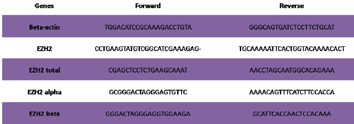
3.11 Total RNA isolation for mRNA and miRNA analysis ... 46
3.12 Quantitative Reverse Transcription Polymerase Chain Reaction (qRT-PCR) for mRNA ... 46
3.13 Quantitative Reverse Transcription Polymerase Chain Reaction (qRT-PCR) for microRNA ... 47
3.14 Western blot experiments ... 47
3.15 Chromatin immunoprecipitation (ChIP) ... 48
3.16 Molecular cloning of mir-101 promoter and activity assessment by luciferase reporter assay ... 49
3.17 Statistical analysis ... 50
Chapter IV - Results ... 51
4.1 HUVECs isolation and characterization ... 52
4.2 Gestational diabetes mellitus induces phenotypic alterations in HUVECs .. 53
4.3 MiR-101 is upregulated in HUVECs exposed to diabetic uterine environment ... 55
4.4 Effect of miR-101 dysregulation on GDM-HUVEC phenotype ... 56
4.5 Enhancer of Zeste Homologue 2 is downregulated in GDM-HUVECs ... 59
4.7 GDM reduces EZH2 and H3K27m3 occupancy at the miR-101 locus
transcriptional regulatory region... 64
4.8 EZH2β inhibits miR-101 transcription in HUVECs ... 65
4.9 In vitro exposure of healthy HUVECs to high concentrations of glucose lowers EZH2 binding to the miR-101 promoter and increases miR-101 expression ... 66
4.10 Effect of EZH2 overexpression on phenotypic alterations in GDM-HUVECs ... 67
Chapter V – Discussion and Conclusions ... 69
Chapter VI – References ... 73
Thesis Summary
Gestational diabetes mellitus (GDM) is a specific form of diabetes affecting about 7% of all pregnancies 1. GDM usually reverses after delivery, but altered glucose
homeostasis harms the mother. Moreover, since it translates into fetal hyperglycemia, GDM can affect the healthy development of the fetus, with consequences that can be experienced in utero and/or at different stages of post-natal life. In particular, the child from a GDM pregnancy has higher chance of developing type 2 diabetes (T2D) and cardiovascular diseases (CVD) 2–4. Several clinical trials and epidemiological studies in
patients with either type 1 or 2 diabetes show that exposure to prolonged hyperglycemia leaves a long-lasting impression on vascular cells leading to the development of vascular complications which persist after glycemic control is achieved. The same studies also suggested that a good glycemic control can delay the onset of vascular complications in diabetic subjects 5.
The term “epigenetic” refers to heritable changes in the cellular phenotype that do not involve changes in the genotype. There are different kind of epigenetic modifications including methylation and acetylation of histones, and DNA methylation, and these modifications influence gene expression 6,7. Although the precise
mechanistic insight is still unclear, epigenetic regulation might be involved in the long-lasting effect of diabetes on the vascular system 8.
In this study, we focused our attention on the methyltransferase Enhancer of Zeste Homologue 2 (EZH2), that is part of the Polycomb Repressor Complex 2 (PRC2) and plays an essential role in the epigenetic maintenance of the trimethylation of lysine 27 on histone3 (H3K27me3). It has been recently shown that alternative splicing of the EZH2 locus yields two major transcriptional variants: EZH2α and EZH2β 9.The two
isoforms can each not only regulate distinct targets, but also share gene expression networks, making the regulation of transcription based on the deposition of the H3K27 histone mark more complex than previously anticipated 9.
MicroRNAs (miRNAs) are single-stranded, non-coding RNAs of 19-25 nucleotides that normally bind the 3’-untranslated region of target mRNAs and contribute to the post-transcriptional regulation of gene expression. A link has been shown between EZH2 and some miRNAs 10,11. In particular, miR-101 reportedly affects endothelial
function and angiogenesis by targeting and inhibiting EZH2 under normoglycemic conditions 12.
The aim of our study was to investigate the impact of GDM on fetal endothelial biology. To this end, we isolated human umbilical vein endothelial cells (HUVECs) from the umbilical cords of GDM and control healthy pregnancies. These cells represent a good cell model allowing for non-invasive analyses of the effect of the uterine environment on the fetal endothelium. The first objective was to investigate the impact of GDM on the functional capacity of HUVECs, with particular focus on apoptosis, migration, proliferation and tube formation. Next, we evaluated the expression of EZH2 (both α and β isoforms) and miR-101 to see whether they affect endothelial function in diabetic conditions. We first found that cultured GDM-HUVECs demonstrated decreased survival and functional capabilities in comparison with HUVECs obtained from healthy non-diabetic mothers. Additionally, we have identified a novel underlying mechanism involving miR-101 and EZH2 in GDM-elicited endothelial dysfunction. In particular, we have found that in HUVECs, EZH2 is both a target of, and a transcriptional repressor for, miR-101. Notably we found that among the two recently identified EZH2 isoforms, EZH2β is the only one which is both a target of GDM and a mediator of some of the GDM-related damaging effects on HUVECs.
Acronyms and Abbreviations
ACOG American Congress of Obstetricians and Gynecologist
Ad Adenovirus
ADA American Diabetes Association AGE Advanced glycation end product BrDU 5-bromo-2’deoxy-uridine
CD-144 Cluster of differentiation 144 CD-31 Cluster of differentiation 31 ChIP Chromatin immunoprecipitation CVD Cardiovascular disease
DCCT Diabetes Control and Complications Trial DGCR8 Di George syndrome critical region
DM Diabetes mellitus
DMEs DNA demethylase
DNMTs DNA methyltransferases ECM Extra cellular matrix ECs Endothelial cells
EDIC Epidemiology of Diabetic Complications and Interventions Trial EDTA Ethylenediaminetetraacetic acid
EED Embryonic ectoderm development EGTA Ethylene glycol tetraacetic acid eNOS Endothelial nitric oxide synthase
ES Endocrine Society
EZH2 Enhancer of zeste homologue 2
FCS Fetal calf serum
FFAs Free fatty acids
GAPDH Glyceraldehyde-3-phosphate dehydrogenase GDM Gestational diabetes mellitus
H3K27me3 Trimethylation of lysine 27 on histone H3 HATs Histone acetyltransferases
HDACs Histone deacetylases
HMTs Histone methyltransferases HPL Human placental lactogen HRP Horse radish peroxidase
HUVECs Human umbilical vein endothelial cells
IADPSG International Association of Diabetes and Pregnancy Study Groups
JHDM2A JmjC domain containing histone demethylase 2A KDMs Histone demethylases
LSD1 Lysine demethylase 1
MAPK Mitogen-activated protein kinase MBPs Methylated DNA-binding proteins MCP-1 Monocyte chemoattractant protein-1
miRNA microRNA
MOI Multiplicity of infection
NBCS Newborn calf serum
NF-kB Nuclear factor kappa B
NG Normal D-glucose
NO Nitric oxide
NR Nuclear receptor
OGGT Oral glucose tolerance test PARP Poly-ADP-ribose polymerase PBS Phosphate-buffered saline
PcG Polycomb-Group
PFA Paraformaldehyde
PKC Protein kinase C
PRC1 Polycomb repressor complex 1 PRC2 Polycomb repressor complex 2 Pre-miRNA Precursor miRNA
Pri-miRNA Primary miRNA
qPCR Quantitative polymerase chain reaction
SD Standard deviation
SDS Sodium dodecyl sulfate SEM Standard error of the mean
STZ Streptozotocin
T1D Type 1 diabetes
T2D Type 2 diabetes
TNF-α Tumor necrosis factor-α
TRBP Transactivation response RNA binding protein UKPDS United Kingdom Prospective Diabetes Study VCAM-1 Vascular cell adhesion molecule-1
vWF Von Willebrand Factor WHO World Health Organization
1.1 Gestational diabetes mellitus (GDM)
1.1.1 Definition and pathophysiology
For many years, gestational diabetes mellitus (GDM) was defined as any degree of glucose intolerance that was first recognized during pregnancy 13, regardless of
whether the condition may have antedated the pregnancy or persisted after the childbirth. This definition facilitated a uniform strategy for detection and classification of GDM, but it was limited by imprecision. The ongoing epidemic of obesity and diabetes has led to more type 2 diabetes (T2D) in women of childbearing age, resulting in an increased number of pregnant women with undiagnosed T2D 14. Therefore, GDM
classification has been recently revised and consists of a carbohydrate intolerance of any degree diagnosed in the second or third trimester of pregnancy, that is not clearly overt diabetes 15. Risk factors for gestational diabetes include certain ethnicities,
obesity, a personal history of GDM, a family history of T2D, maternal age, polycystic ovary syndrome, a sedentary lifestyle and exposure to toxic factors 16. According to the
American Diabetes Association (ADA), GDM complicates approximately 7% of all pregnancies, whereas its total incidence is estimated to be up to 17.8% depending upon the diagnostic criteria used and the demographic characteristics of the population
1. The greatest rises in prevalence are predicted to occur in India, China, Latin America
and the Middle East 17.
GDM is closely associated with T2D, as they share many key pathophysiological characteristics; namely a relative insufficiency of pancreatic β-cell function and insulin resistance. During pregnancy, insulin resistance is increased due to the production of placental hormones, such as estrogens, cortisol and human placental lactogen (HPL), and cytokines including tumor necrosis factor α (TNF-α), which antagonize insulin action 18. This increase is additional to the insulin resistance that occurs due to genetic
susceptibility and/or a suboptimal lifestyle. In the event there is adequate release of insulin by the maternal pancreas, normoglycemia is preserved and the embryo is not affected. However, when insulin release is insufficient, hyperglycemia occurs and an excess amount of glucose is transferred via the placenta to the embryo while maternal insulin cannot pass the placental barrier causing fetal hyperglycemia. As a consequence the fetal pancreas produces more insulin to counter the increased glucose levels leading to hyperinsulinemia and excessive fetal growth (macrosomia) 19.
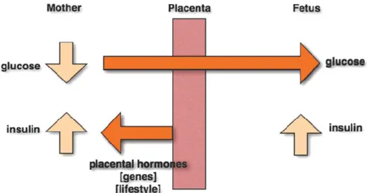
Figure 1.1: Pathophysiology of gestational diabetes mellitus
During pregnancy placental hormones, genetic susceptibility and/or a suboptimal lifestyle lead to an increase of maternal insulin resistance and hyperglycemia. Maternal glucose can then transfer to the fetus through the fetoplacental circulation, while maternal insulin is unable to pass the placental barrier. Maternal hyperglycemia consequently stimulates fetal hyperinsulinemia to counter the extra glucose. The high insulin level in the fetus stimulates growth which results in fetal macrosomia 19. The schematic is adapted from 20
GDM usually reverses after delivery, but altered glucose homeostasis harms the mother and can lead to health consequences in the fetus. The short-term complications for a mother with GDM include pre-eclampsia, polyhydramnios and caesarean section. Similarly, the main short-term fetal and neonatal complications include perinatal mortality, macrosomia, obstetric trauma, hypoglycemia, hyperbilirubinemia, hypocalcemia and cardiac dysfunction 21,22. Additionally, GDM is associated with
long-term complications for both the mother and child, including T2D and cardiovascular diseases (CVD) 23.
1.1.2 GDM and long-term maternal outcomes
It is well established that GDM is associated with an increased risk of developing T2D postpartum, occurring as a consequence of insulin resistance developed during pregnancy 24. For the majority of women who go on to develop T2D, GDM does resolve
immediately postpartum, with the loss of placental hormones and the reduction in circulating cytokines, but their glucose tolerance worsens with time, such that after 5 years 13% of such woman have developed T2D 25. In an Australian retrospective study
the risk of developing diabetes increased with time of follow-up and was 9.6 times greater for patients with GDM 26.
GDM is also a risk factor for CVD events. In a large population-based study in Ontario, Canada, women who had GDM in pregnancy compared with a matched control group were at higher risk of CVD events with an odds ratio of 1.71 (95% CI 1.08 - 2.69) 27. When adjusted for the risk of development of T2D, this ratio was no longer
significant because the considerable part of increased CVD risk was attributable to progression to T2D. Therefore, a diagnosis of GDM for each woman provides an opportunity for intervention to reduce her risk of future T2D and cardiovascular complications.
1.1.3 Fetal programming and GDM long-term outcomes for the offspring
Our knowledge of the role of maternal diabetes in fetal programming started in 1952 with the “Pedersen hypothesis”. Pedersen proposed that elevated maternal glucose crossed the placenta resulting in hyperinsulinemia and excessive fetal growth, thus leading to fetal macrosomia 19. In 1980, Freinkel expanded upon the Pedersenhypothesis and proposed that excesses of mixed “fuels” including glucose, amino acids, lipids, ketones, and possibly altered concentrations of other nutrients, induce “teratogenesis”. Importantly, Freinkel suggested that the teratogenic effects resulting from exposure to elevated levels of maternal mixed nutrients, and the timing of such exposures, might have profound and long-term consequences for the fetus 28. Some
years later epidemiological studies on birth and death records by David Barker and colleagues led to the idea that babies who are born lighter and survived infancy might have an increased risk of developing coronary heart disease later in life. This led to the “Barker hypothesis” or “fetal programming hypothesis” which states that adverse intrauterine environments will have long-term effects on adult health which includes an increase in incidence of T2D, hypertension and coronary heart disease 29. The Dutch
“Hunger Winter” is one of the earliest and best examples of “fetal programming” during World War II, when shortages in food supply caused famine in western Netherland’s population. The pregnancies during that period were affected by “foetal starvation” which ultimately had long-term effects on the health of the newborn 30. What is unique
about the Dutch Famine as an experimental study on the effects of maternal malnutrition, is that the population was strictly circumscribed in time and place and the
sudden onset and relief of the famine was imposed on a previously well-nourished population 31.
This concept of fetal programming of adult metabolic diseases is well supported by several studies and thus now widely accepted. Discordant human siblings born before and after the development of GDM provided compelling evidence that these later life phenotypic consequences are caused by the diabetic uterine environment 2. In
longitudinal studies of the Pima Indians from Arizona, a population that has an extremely high prevalence of obesity and T2D, diabetes during pregnancy does increase the incidence of T2D in the offspring, principally at a young age 2,4. Moreover
an association was found between maternal diabetes and age at offspring’s diagnosis of T2D in the multiethnic SEARCH for diabetes in youth study 3. This evidence supports
the concept that exposure to hyperglycemia during fetal development increases the subsequent risk of diabetes and cardiovascular complications later in life. The molecular mechanisms underlying this developmental programming of metabolic and cardiovascular diseases are still largely unclear. However, in addition to genetics, epigenetic mechanisms appear to play a key role 32.
1.1.4 Diagnosis and screening
Even though GDM is a common disorder in pregnancy, it has been difficult to compare its frequency among various populations and estimate its global incidence, due to the lack of uniform diagnostic criteria 33.Over the last few decades, different
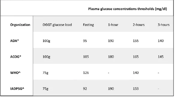
diagnostic criteria have been applied according to different rationales as presented in Table I 34. All these criteria are based on fasting or non-fasting maternal glucose
concentrations, most usually by means of an oral glucose tolerance test (OGTT) 35–37.
As the most important issue in GDM is the development of embryonic, neonatal or maternal complications, maternal glucose concentrations need to be used to predict the development of complications with the highest possible sensitivity and specificity. However, the adoption of a low glucose threshold results in overdiagnosis, as many women who are not likely to develop complications will be diagnosed as having GDM. By contrast, the adoption of a high threshold results in underdiagnosis, as numerous women who would go on to develop complications will be diagnosed as not having GDM. Even the adoption of an intermediate threshold will result in a lower but serious risk for over- and underdiagnosis. Thus, the definition of GDM by means of maternal glucose concentrations constitutes a difficult challenge.
The Hyperglycemia and Adverse Pregnancy Outcomes (HAPO) study attempted to tackle this challenge by assessing 25,505 pregnant women from 15 countries during the third trimester of gestation 38. The study pointed out that there is no specific
threshold for maternal serum glucose over which the chance of developing maternal or neonatal complications increases significantly. By contrast, the correlation between the degree of glycemia and the probability of adverse outcomes (e.g. neonatal birth weight >90th percentile, primary caesarean section, neonatal clinical hypoglycemia, cord-blood
C-peptide concentrations >90th percentile) is linear 38.
Based on the data from the HAPO study, in 2010 the International Association of Diabetes and Pregnancy Study Groups (IADPSG) decided to set the threshold at an odds ratio of 1.75 for developing specific complications, specifically an increase in neonatal fat mass, fetal macrosomia and cord C-peptide concentrations >90 percentile, compared with mean glucose concentrations. This odds ratio corresponds to a fasting plasma glucose of 92 mg/dl, or 180 mg/dl and 153 mg/dl after an OGTT with 75 g of glucose at 1 and 2 hours, respectively (Table I) 39. In other words, a diagnosis of GDM
set by the IADPSG criteria means that the specific woman has a 75% increased odds (odds ratio 1.75) of developing complications as compared to a woman with mean glucose concentrations. According to the same criteria, if a woman has a fasting glucose of more than 126 mg/dl, a random glucose of more than 200 mg/dl or a glycosylated hemoglobin (HbA1c) of more than 6.5%, then a diagnosis of pre-existing diabetes mellitus (DM) has to be made 40. The IADPSG criteria have a number of
advantages. They represent a unique opportunity for uniformity in GDM diagnosis, are simple and can be compared with similar criteria for DM diagnosis in the non-pregnant state. On the other hand, they have been developed based on observational studies of GDM complications and cannot necessarily be used as treatment thresholds. In addition, they increase the GDM prevalence from 5-6% to 16%, being more inclusive than older criteria 37,41.
Table I: GDM diagnostic threshold values from various organizations
*Diagnose GDM if two or more glucose values equal or exceed the threshold values, ^Diagnose GDM if one or more glucose values equal or exceed the threshold values. ADA (American Diabetes Association), ACOG (American Congress of Obstetricians and Gynecologist, WHO (World Health Organization), IADPSG (International Association of the Diabetes and Pregnancy Study Group.
Regarding screening for GDM, the latest guidelines drawn up by the Endocrine Society (ES) are the first to recommend universal testing for overt DM in early pregnancy. A fasting plasma glucose, HbA1c or a random plasma glucose at the first prenatal visit (before 13 weeks of pregnancy or as soon as possible thereafter) is recommended for those women not known to already have DM. In the event of overt DM, a second test is recommended on another day to confirm the diagnosis. In all pregnant women in whom no overt DM or GDM has been diagnosed, a 2-hour 75-g OGTT should be performed at 24 to 28 weeks of pregnancy 42.
1.1.5 Treatment of GDM
The beneficial effect of early diagnosis and treatment of GDM or mild carbohydrate intolerance during pregnancy has been recently demonstrated by two large randomized clinical trials. The Australian Carbohydrate Intolerance Study in Pregnant Women, established that GDM treatment in the form of dietary advice, blood glucose monitoring, and insulin therapy, as required for glycemic control, reduces serious perinatal complications and may also improve health-related quality of life 21. Also, the American
Maternal-Fetal Medicine Units Network study provided further compelling evidence that among women who have GDM and normal fasting glucose levels, treatment that includes dietary intervention and insulin therapy, as necessary, reduces rates of fetal overgrowth, cesarean delivery, and preeclampsia 43. Accordingly, the primary
intervention recommended to women diagnosed with GDM is dietary counseling in combination with physical activity and self-monitoring of blood glucose 44,45. If these
measures are insufficient in terms of achieving optimal glycemic control, subcutaneous insulin administration is the therapy of choice as insulin does not cross the placenta and is therefore considered harmless to the fetus. However insulin is relatively expensive and difficult to administer, it requires education to ensure safe administration and moreover it is associated with an increased risk of hypoglycemia and weight gain. The use of safe and effective oral anti-hyperglycemic agents may therefore offer advantages over insulin but has not yet been formally approved for GDM therapy in all countries 46. In this context, two large randomized controlled trials were performed to
test the effectiveness and safety of metformin 47 and glyburide, a sulphonylurea that
acts as an insulin secretagogue. 48 From these studies, the authors concluded that both
metformin and glyburide, alone or with supplemental insulin, were not associated with increased perinatal complications as compared to insulin. More studies show that both metformin and glyburide have been increasingly and safely used in the treatment of GDM 49. However, both medications can cross the placenta and given the growing
evidence of epigenetic fetal programming in utero, the administration of drugs potentially affecting fetal metabolism is of major concern. As long term follow-up data on both mother and offspring are lacking, oral anti-hyperglycemic agents should be used with caution.
1.2 Maternal hyperglycemia and fetal endothelial dysfunction
1.2.1 Biochemical and cellular signaling underlying diabetic vascular
complications
Hyperglycemia has been shown to be the major risk factor for the development of diabetic complications due to its long-term deleterious effects on various tissues and a particular subset of cell types. Indeed most cells are able to reduce the transport of glucose inside the cell when they are exposed to hyperglycemia, so that their internal glucose concentration remains constant. In contrast, the cells damaged by hyperglycemia are those that cannot do this efficiently 50,51. Thus, diabetes selectively
damages cells, like endothelial cells, mesangial cells, neurons and Schwann cells in peripheral nerves, whose glucose transport rate does not decline rapidly as a result of hyperglycemia, leading to high glucose concentrations inside the cell.
High glucose can lead to the activation of several cellular pathways, including increased oxidative stress 52–54, enhanced flux into the polyol and hexosamine
pathways 55, activation of Protein Kinase C (PKC) 56 and increased formation of
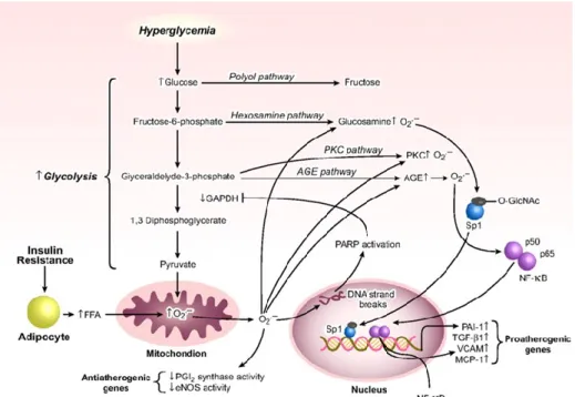
advanced glycation end products (AGEs) 57 as shown in Figure 1.2. Studies in
mesangial, endothelial, and other cell types have associated hyperglycemic activation of these pathways to increased mitochondrial superoxide anion formation (O2-) and
associated oxidative stress 58, which can ultimately lead to increased formation of extra
cellular matrix (ECM) proteins in the kidney, contributing to renal dysfunction 59,60.
In addition, high glucose can activate the pro-inflammatory transcription factor NF-kB, resulting in increased inflammatory gene expression in part through oxidative stress, AGEs, PKC, and MAPKs 61,62. Inflammation can also lead to the acceleration of
diabetic complications due to detrimental effects on vascular cells and insulin resistance in T2D. Furthermore, insulin resistance has been attributed to increased free fatty acids which, in conjunction with hyperinsulinemia, lead to dyslipidemia 63.
Therefore, hyperglycemia and dyslipidemia can alter the expression of inflammatory and other pathological genes and proteins related to the development of endothelial dysfunction 64, which precedes the pathogenesis of vascular complications in diabetes.
Similar to other forms of diabetes, GDM is associated with structural and functional alterations in various tissues including endothelial dysfunction. It is believed that a dysfunctional endothelium could lead to impaired vasodilation, increased leukocyte adhesion to the vascular wall and platelet aggregation, and impaired angiogenic
properties. Endothelial dysfunction in GDM is likely to be at least partially a consequence of hyperglycemia which induces changes in the proliferation and inflammatory responses of endothelial cells (ECs) in vitro. Decreased proliferation of human umbilical vein endothelial cells (HUVECs) from normal pregnancies exposed to elevated glucose have been extensively studied and attributed to increased cell death
65. Moreover HUVECs isolated from gestational diabetes pregnancies presented
enhanced monocyte adhesion and altered L-arginine transport and nitric oxide (NO) synthesis probably due to an altered uptake and metabolism of adenosine 66,67.
Fetal endothelial dysfunction is involved in the abnormal vascular development caused by GDM, and it might be responsible for the increased cardiovascular risk and incidence of T2D observed later in life in the offspring from GDM mothers 68,69.
The mechanisms that link hyperglycemia to fetal endothelial dysfunction and cardiovascular complications in adulthood is still largely unknown and different explanations have been proposed including the existence of a “metabolic memory”.
Figure 1.2: Biochemical and cellular signaling activated by hyperglycemia
Insulin resistance and hyperglycemia increase mitochondrial O2- production which activates atherogenic signaling pathways. Overproduction of mitochondrial O2-, results in DNA strand breaks and activation of poly-ADP-ribose polymerase (PARP). PARP ribosylates and inactivates glyceraldehyde-3-phosphate dehydrogenase (GAPDH). The upstream metabolites of the interrupted glycolytic pathway are processed through polyol, hexosamine, PKC, or AGEs pathways. Activation of transcription factor NF-κB, downstream of the AGE pathway, induces transactivation of vascular cell adhesion molecule-1 (VCAM-1) and monocyte chemoattractant
protein-1 (MCP-1). Increased flux of FFAs, released from insulin-resistant adipocytes, also activates hexosamine, PKC, and AGE pathways. In addition, FFA-induced excess mitochondrial O2- production inactivates anti-atherogenic enzymes such as prostacyclin (PGI2) synthase and
eNOS. The illustration is adapted from 70.
1.2.2 “Metabolic memory” and GDM vascular complications
The results of more than one clinical trial on T1D and T2D patients have demonstrated that the development of diabetic complications persisted even after the achievement of glycemic control, suggesting the existence of a “metabolic memory” dependent from prior exposure to hyperglycemia. T1D patients in the Diabetes Control and Complications Trial (DCCT) under intensive insulin therapy were found to have delayed progression of nephropathy, retinopathy, and neuropathy compared with patients under conventional therapy 71. The large benefits of the intensive therapy led
to the premature termination of the DCCT so that all patients could be placed on intensive insulin therapy and followed long-term under the subsequent Epidemiology of Diabetic Complications and Interventions Trial (EDIC). In line with the DCCT findings, long-term follow-up in the EDIC trial demonstrated that patients who were originally in the intensive treatment group during the DCCT and continued on intensive therapy for the EDIC trial had significantly slower progression of key diabetic microvascular complications in comparison to patients who were in the conventional treatment group during DCCT, even though they were placed on intensive therapy 5. More recently,
patients in the continuous intensive treatment group since the beginning of the trials were found to also have better outcomes for macrovascular complications including stroke, nonfatal heart attack, or death by cardiovascular disease 5, as well as
decreased progression of coronary artery calcification and intima-media thickness, both of which are associated with atherosclerosis 8.
Long-lasting benefits of glycemic control have also been seen in T2D patients. In this context the United Kingdom Prospective Diabetes Study (UKPDS) found that lower fasting plasma glucose at the time of diagnosis correlated with decreased cardiovascular risk 72,73. In addition, the Steno-2 Study found a decreased risk of
cardiovascular events and death following intensive multifactorial therapy including tight glycemic control 74.
Overall, the findings from these major clinical trials demonstrate that the exposure to prolonged hyperglycemia leaves a long-lasting impression on vascular cells and progressively induces the development of vascular complications, which persist after
glycemic control is achieved. However, the same studies also demonstrated that good glycemic control can delay the onset of diabetic vascular complications. Both lines of evidence are compatible with an underlying implication of epigenetic mechanisms. Indeed, epigenetic factors allow cells and organisms to respond to changing internal and environmental stimuli and additionally confer the ability of the cell to memorize these environmental situations.
1.2.3 Models of metabolic memory
Experimental models provide an opportunity to study the molecular mechanisms responsible for metabolic memory in order to design better therapies for diabetic patients. Recent research has demonstrated the presence of metabolic memory in several animal and cell culture models. Early studies in dogs found that there was a persistence of retinal complications even after reversal of hyperglycemia 75. Similar
results with diabetic rats showed that islet transplantation after 12 weeks of diabetes could not reverse the progression of retinopathy compared with the same surgical treatment after 6 weeks of diabetes 76. Moreover several studies in streptozotocin
(STZ)-induced diabetic rats showed that establishment of glycemic control after a short period of hyperglycemia had protective effects on NO levels, lipid peroxides, and other parameters in the retina. However, reinstitution of physiological glucose concentrations after prolonged hyperglycemia failed to reverse increases in oxidative stress, NF-kB activity, as well as inflammation, and this was attributed to metabolic memory 73,77.
The presence of a metabolic memory has also been demonstrated in vascular cell models. Early in vitro studies with endothelial cells cultured in high glucose showed continued increased expression of genes encoding fibronectin and collagen ECM proteins even after normalization of glucose levels 78. A more recent endothelial cell
model of metabolic memory showed that even a short-term exposure to high concentrations of glucose resulted in sustained increases in the expression of the NF-kB p65 subunit, inflammatory genes, and oxidative stress that persisted even after a return to normal glucose levels 79,80. Finally, other recent reports demonstrated the
persistence of oxidative stress for up to 1 week after glucose normalization and that antioxidants or NADPH oxidase inhibitors partially blocked the high glucose effects 81.
Together, these results suggest a metabolic memory of vascular dysfunction arising from acute hyperglycemic spikes or prior chronic exposure to high glucose conditions. In addition, these experimental models confirm that strict control of glucose levels is
essential to slow down the progression of diabetic complications. Importantly, they also suggest that oxidative stress, along with epigenetic mechanisms, may be responsible for the sustained metabolic memory over time through multiple cell divisions.
1.3 Role of epigenetic factors in GDM vascular complications
1.3.1 Epigenetics and transcriptional regulation of gene expression
Epigenetics refers to heritable changes in the phenotype that do not involve changes in the genotype and include mainly two mechanisms: DNA methylation and histone tails modification 6. In the promoter regions, DNA methylation that typically
occurs at CpG islands reduces gene expression by blocking the binding of transcription factors or promoting chromatin packing. Post-translational modifications that occur on the histone tails include mainly acetylation, methylation, and phosphorylation 7. Along
with DNA methylation, these epigenetic modifications make up an additional level of gene regulation or code that can be altered without altering the DNA code itself. In addition, the numerous combinations of epigenetic modifications allow for flexibility of the chromatin and can affect recruitment and binding of various DNA and histone binding complexes that recognize specific combinations of chromatin marks, as shown in Figure 1.3. Histone binding complexes often contain other chromatin-modifying proteins that can further change the chromatin structure. In this context, histone acetyltransferases (HATs) mediate histone lysine acetylation, a chromatin mark generally associated with euchromatin formation and activation of gene transcription. Histone deacetylases (HDACs), on the other hand, mediate the removal of lysine acetylation and are found to be components of repressor complexes 82. Overall, histone
acetylation can occur quite rapidly and is quite dynamic.
By contrast, histone methylation is considered to be more stable and long lasting. Histone methylation occurs on both lysine and arginine residues and is associated with either gene activation or repression depending on which residue is modified 7. The SET
domain-containing family of histone methyltransferases (HMTs), which is named for a conserved sequence motif found in three Drosophila proteins, Suppressor of Position Effect Variegation, Enhancer of Zeste and Trithorax, are involved in regulating lysine methylation 83. Lysine methylation can be quite complex since lysine residues can be
mono-, di-, or trimethylated by specific HMTs which usually methylate only one particular lysine residue. Specifically, histone H3 lysine 4 methylation (H3K4me) is typically associated with gene activation while H3K9me, is generally associated with gene repression. In addition, there are several other lysine residues, including H3K27, H3K36, and H3K79, that can be methylated to various degrees by different HMTs, leading to altered gene expression 84.
The exciting discovery of the first histone demethylase (KDMs), lysine demethylase 1 (LSD1) which specifically removes H3K4me 85 and H3K9me marks 86, demonstrated
that histone lysine methylation can also undergo dynamic regulation. Numerous KDMs have since been identified with specificities for different histone lysine residues 87. In
the context of diabetes, a recent report demonstrated that histone demethylase JHDM2A is associated with obesity and affects genes related to metabolism in rodent models 88.
According to an early definition, epigenetics is associated with chromatin changes which are transmitted from parents to offspring. However, this concept has been revisited and it is now suggested that epigenetic changes have a long lasting effect, which can be transmitted at cell division, but that are not necessary perpetual. In other words, the epigenetic modifications once written, can still be diluted and possibly erased over time under particular circumstances, such as with the use of drugs or lifestyle changes 5. The concept of a changeable epigenetic status is important
because it confers therapeutic hope at different stages of prenatal and postnatal life.
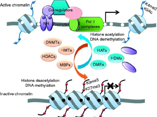
Figure 1.3: Regulation of chromatin by histone modifications
The two major epigenetic modifications are DNA methylation and histone modifications which regulate the active or inactive chromatin state. Hypermethylation of the promoter is associated with transcriptional silencing due to the loss of affinity for transcriptional factors such as the nuclear receptor (NR) and accessibility by the transcriptional machinery. The inactive chromatin has increased affinity for methylated DNA-binding proteins (MBPs), which further recruit histone
deacetylases (HDACs), DNA methyltransferases (DNMTs), histone methyltransferases (HMTs) and other corepressors. Methylated promoters are associated with unique repressive histone marks, which classically include trimethylation of histone 3 on lysine 9 (H3K9me3), and trimethylation of histone 3 on lysine 27 (H3K27me3). By contrast, unmethylated promoters are associated with gene transcription. They have increased affinity for histone acetylases (HATs), histone demethylases (HDMs), DNA demethylases (DMEs), and histone marks associated with active chromatin, including acetylation of histone H3 on lysine 9 (H3K9Ac) and trimethylation of histone H3 on lysine 4 (H3K4me3). The illustration is adapted from 7.
1.3.2 Transmission of epigenetic modifications
Epigenetic marks are transmitted through multiple cell cycles. While much is known about the epigenetic inheritance of DNA methylation, the exact mechanisms responsible for the stable transmission of histone modifications are less well understood. Replication-associated transmission, histone variants, and chromatin remodeling complexes are some of the factors that have been implicated.
Methylation of histone lysine residues has been shown to be transmitted through replication where the methyl-CpG binding protein recruits the HMT SETDB1 to chromatin during replication to methylate newly-deposited histones and thus coupling the transmission of H3K9 methylation with DNA methylation 89. Evidence has also
shown that the long-term silencing Polycomb complex remains bound to chromatin during replication, possibly binding more than one nucleosome and allowing nucleosomes to maintain contact with chromatin even as the replication fork passes through. Alternatively, the complex could interact directly with the replication machinery
90. Another study demonstrated that the Polycomb repressor complex 2 containing the
HMT EZH2 can bind H3K27me3 modifications at sites of ongoing replication, which would then allow transmission or copying of the parental H3K27me3 marks to the new histones deposited on the daughter strand 91. Interestingly the Polycomb complex
family has been recently found to play a role in the epigenetic regulation of pancreatic cell regeneration associated with diabetes 92.
Together, these results highlight the need for further investigation into the mechanisms by which diabetes-induced epigenetic changes might be stably transmitted through cell replications to establish a transcriptional or metabolic memory associated with diabetic complications.
1.3.3 Role for microRNAs in epigenetic regulation
One of the emerging areas of research involves the post-transcriptional regulation of gene expression through a recently discovered group of small RNAs, the microRNAs (miRNAs). MiRNAs are single-stranded, non-coding RNAs of 19-25 nucleotides that normally bind the 3’-untranslated region of target mRNAs, leading to either translational repression or RNA degradation 93.
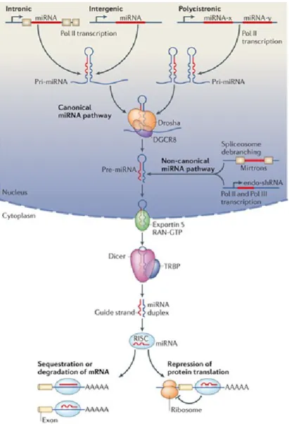
MiRNAs produced via the canonical pathway are transcribed in the nucleus by RNA polymerase II into primary miRNAs (pri-miRNAs) that are several hundred nucleotides in length. Subsequently, the microprocessor complex, which consists of Di George syndrome Critical Region gene (DGCR8) and the nuclease Drosha, cleaves the pri-miRNAs, with a stem-loop structure, into miRNA precursors (pre-miRNAs) of approximately 60 to 70 nucleotides. In the non-canonical miRNA pathway, miRNAs are transcribed directly as endogenous short hairpin RNAs (endo-shRNAs) or derived directly by splicing from introns (mirtrons) 94. Next, exportin-5 transports the
pre-miRNAs from the nucleus to the cytoplasm via a RAN-GTP-dependent process. The pre-miRNAs are further processed in the cytoplasm by RNase Dicer in association with Transactivation Response RNA Binding Protein (TRBP) to final miRNA duplexes of 19-25 nucleotides. Finally, the mature single-stranded miRNAs are generated and incorporated into the RNA-Induced Silencing Complex (RISC) and are ready to interact with specific mRNA targets 93. The miRNAs biogenesis pathways are depicted in
Figure 1.4.
MiRNAs provide a rapid but reversible system of gene regulation, which also allows the cell/tissue/organism to respond to environmental stimuli without changing the DNA sequence itself. MiRNAs have been identified as negative regulators in various pathways, targeting signaling molecules, transcription factors, and numerous other enzymes and proteins. There is also the potential for miRNAs to target chromatin-modifying enzymes which in turn could also affect transcription and expression of miRNAs 95. Therefore, miRNAs may themselves be epigenetically regulated.
Recent findings have demonstrated a critical role for miRNAs in various diseases. They have been found to play key roles in proliferation, differentiation, development, and in cancer, where they can act as oncogenes or tumor suppressors 93,96. MiRNAs
are also associated with the regulation of genes relevant to insulin secretion, cholesterol biosynthesis, fat metabolism, adipogenesis and angiogenesis, crucial pathways in the pathogenesis of diabetes and its vascular complications 97,98. The role
of miRNAs and their potential relationships to epigenetic mechanisms in diabetic complications are currently an area of great interest both as a means for understanding the molecular pathways leading to these complications and for discovering new potential therapeutic targets.
Figure 1.4: MiRNA biogenesis pathways
MiRNAs, produced via the canonical pathway are transcribed from genomic DNA in the nucleus as long primary miRNAs (pri-miRNA) by RNA polymerase II. The pri-miRNA transcript forms a stem-loop structure that is recognized and processed by Drosha in association with DGCR8 into a precursor miRNA (pre-miRNA). In the non-canonical miRNA pathway, miRNA are transcribed directly as endogenous short hairpin RNAs (endo-shRNAs) or derived directly by splicing from introns (mirtrons) 94. The pre-miRNA is then exported to the cytoplasm by exportin 5 and RAN-GTP-dependent process. In the cytoplasm, pre-miRNA is further processed by RNase activity of Dicer and Transactivation Response RNA-Binding Protein (TRBP) complex to form the mature double-stranded ~22 nucleotide miRNA. The mature miRNA is then incorporated into the RISC
complex which mediates small interfering RNA silencing by degrading the target mRNA or interfering with translation. The illustration is adapted from 97
1.3.4 MiRNAs as novel regulators of endothelial function
MiRNAs play a central role in endothelial function regulation. Elimination of most endothelial miRNAs by Dicer knockdown in ECs inhibits proliferation and tube formation in vitro 99. Moreover, Dicer knockout mice have impaired blood vessel
development 100. These findings suggest that miRNAs are essential for the
maintenance of vascular homeostasis.
The first large analysis of miRNA expression in ECs was carried out in HUVECs and identified 15 highly expressed miRNAs and some of their angiogenic mRNA targets (Flt-1, Nrp-2, Fgf-R, c-Met, and c-kit), according to prediction algorithms 101.
Additional studies also profiled the expression of miRNAs in ECs 102. The highly
expressed miRNAs that were common in at least two of the three studies included miR-15b, 16, 20, 21, 23a and 23b, 24, 29a and 29b, 31, 99a, 100, 103, 106, 125a and 125b, 126, 130a, 181a, 191, 221, 222, 320, let-7, let-7b and let-7c 99,101.Several miRNAs have also emerged as potential regulators of EC function under diabetic conditions. Wang et al. identified that high glucose levels induced miR-320 expression in myocardial microvascular ECs. Inhibition of miR-320 improved proliferation and migration, which has implications for diabetic-impaired angiogenesis 103. More recently, expression of
miR-503 was found to be increased in ECs exposed to high glucose levels in vitro. Consistently, miR-503 expression also increased in ECs enriched from ischemic limb muscles of STZ-induced diabetic mice 104. Moreover, miR-503 expression was
remarkably higher in the limb muscles and plasma from diabetic patients and inversely correlated with expression of cell division cycle 25A, a known target gene that controls cell cycle function. Local inhibition of miR-503 promoted vascular wound healing and blood-flow recovery in a diabetic mouse model of limb ischemia 104. Expression of
another miRNA, miR-29c, was increased in microvascular ECs exposed to hyperglycemic conditions and in the kidney from diabetic mice. Upregulation of miR-29c decreased Spry1 protein levels and promoted activation of Rho kinase, which suggests that miR-29c may play a role in kidney remodeling during diabetic nephropathy 105. Additionally, miR-221 expression increased in ECs in response to
advanced glycation end products or glucose, and its overexpression reduced c-kit expression 106.
MiR-101 is known to affect endothelial function and angiogenesis under normal glucose conditions 12 and to target the histone methyltransferase Enhancer of Zester
Homologue 2 (EZH2) for inhibition 12,107–109. However, its role in the context of diabetes
is still unexplored.
1.3.5 Enhancer of Zeste Homologue 2 and its role in angiogenesis
EZH2 is a well characterized epigenetic regulator and is a member of the Polycomb-Group (PcG) proteins. PcG proteins function as transcriptional repressors that silence specific sets of genes through chromatin modification 110. PcG proteins act
together in Polycomb Repressive Complexes (PRC). In this context, PRC2 is a multi-subunit complex which includes EZH2, Suppressor of Zeste 12 (SUZ12), and Embryonic Ectoderm Development (EED). A number of other proteins, such as retinoblastoma binding proteins 4 and 7 (RBBP4/7), enhance the enzymatic function of the complex 111.
EZH2 is the catalytically active component of PRC2, which holds the evolutionarily conserved SET domain that confers its HMT activity. Specifically EZH2 trimethylates the histone H3 at lysine 27 (H3K27me3) 112. The H3K27me3 mark subsequently
recruits PRC1, leading to the propagation of the repressed state through a variety of mechanisms, including chromatin compaction and recruitment of other chromatin-remodeling enzymes, such as DNA methyltransferases 113. PRC1 and PRC2 are
shown in Figure 1.5.
Expression profiling indicated that EZH2 promotes angiogenesis in ovarian carcinoma 114 and glioblastoma cells 12,109. Moreover, altered EZH2 expression was
described to affect angiogenesis in bladder cancer cells 115, in HUVECs 116 and in vivo,
in a mouse model of limb ischemia 117. It was also described that EZH2 expression is
regulated by miRNAs including miR-26 10, miR-214 11, and miR-101 12,107–109. On the
other hand, regulatory feedback loops between EZH2 and microRNAs have been recently identified in human tumorigenesis 95,118. In this context EZH2 has been recently
proposed to regulate miR-101 in non-ECs 115,119. The complexity of EZH2-mediated
gene silencing has further increased since the recent discovery of a novel isoform of EZH2 protein that function in mammalian cells 9.
Figure 1.5: The Polycomb repressor complexes
PRC2 is a multi-subunit complex which includes EZH2, which is the catalytically active component, Suppressor of Zeste 12 (SUZ12), and Embryonic Ectoderm Development (EED). A number of other proteins, such as retinoblastoma binding proteins 4 and 7 (RBBP4/7), enhance the enzymatic function of the complex. PRC2 is recruited to chromatin and allows trimethylation of histone H3 on lysine 27 (H3K27me3) by EZH2. PRC1 is then recruited to chromatin through the recognition of H3K27me3 mark by chromobox (CBX) proteins. RNF2/RING1 homologs are E3 ubiquitin ligases for H2A, which is monoubiquitylated at lysine 119. The RYBP repressor protein recognizes H2A monoubiquitylation, contributing to transcriptional repression. The grey circular shapes in the figure depict other PRC subunits and associated proteins. The figure is adapted from http://www.activemotif.com/stem-cell-identity.
1.3.6 Enhancer of Zeste Homologue 2 and its novel isoform: EZH2-β
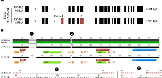
It has been recently shown that alternative splicing of the EZH2 locus yields two major transcriptional variants: EZH2α and EZH2β 9.
EZH2α, encoded by 20 exons, is the HMT classically associated with the function of the PRC2 complex. EZH2β, a novel isoform, skips exon 4 of EZH2α and utilizes an alternative 5′ splice donor on EZH2α exon 8/EZH2β exon 7, as presented in Figure 1.6 A. Compared with the EZH2α sequence, the shorter EZH2β shows conservation of several domains, namely the nuclear localization signals, SANT (DNA-binding domains) and the SET domain. Together, the conservation of these domains should confer this protein with the ability to localize to the nucleus and function as an HMT. Accordingly, this isoform localizes to the cell nucleus, complexes with EED and SUZ12, and binds to promoters where it increases H3K27me3 levels, all properties in common with EZH2α protein. At the protein sequence level, EZH2α and EZH2β differ by 44 amino acids, measuring 751 and 707 amino acids, respectively, as shown in Figure 1.6
B 9. A highly similar third splice variant encoding five fewer amino acids than EZH2α
has also been referred to as EZH2. Structural comparison of these closely related variants does not reveal any differences that would suggest differing function and thereby have been considered interchangeable in the literature.
Figure 1.6: Comparative analysis of EZH2 transcriptional variants
Alternative splicing of the EZH2 locus yields two major transcriptional variants: EZH2α and EZH2β. The sites of alternative splicing events are highlighted in red on the reference isoform
(A). At the protein structure level EZH2α and EZH2β show the conservation of functional
domains and binding sites of enzymatic co-factors, represented by different colors. Labels 1 and 2 indicate the locations of deletions in EZH2β compared with EZH2α. A comparison of the amino acid sequences highlighting the amino acid differences between the two isoforms is also presented (B). The illustration is adapted from 9.
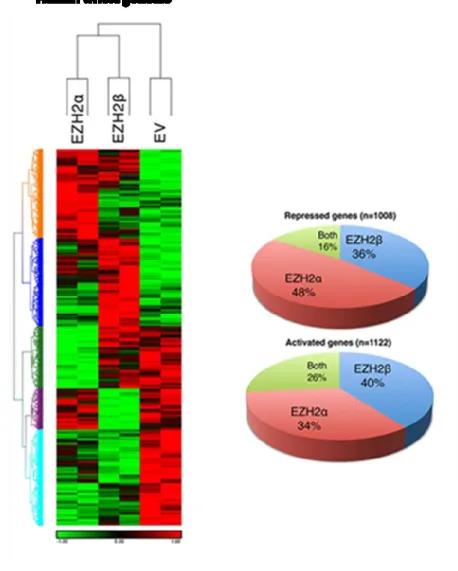
Expression profile experiments performed in pancreatic epithelial cell systems combined with adenoviral-mediated introduction of both EZH2 isoforms, using array analysis, showed that the two different isoforms can each not only regulate distinct targets but also share gene expression networks, making the regulation of transcription based on the deposition of the H3K27 histone mark more complex than previously anticipated 9.
Figure 1.7: Expression patterns of EZH2α and EZH2β
Arrays analysis shows that of the 28,869 well-annotated genes assessed, 366 unique targets (36% of total repressed) are uniquely repressed by EZH2β. EZH2α-generated expression profiles display a downregulation of 480 targets (48% of total repressed). Notably, 162 targets (16% of total repressed) are repressed by both EZH2α and EZH2β. Both isoforms also induce upregulation in the expression of a significant number of targets, which may reflect indirect effects mediated by the repression of upstream regulators. Of the genes assayed, 444 (40% of total activated) are activated by EZH2β, 382 (34%) by EZH2α and 296 (26%) by both isoforms. The overall array data indicate that the novel EZH2β isoform is responsible for the expression pattern of its own unique set of genes, in addition to a group of common targets with EZH2α. The illustration is adapted from 9.
1.4 Diagnostic and therapeutic potential of miRNAs in GDM
vascular complications
1.4.1 MiRNAs with diagnostic potential for vascular complications
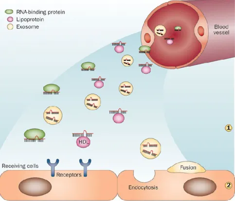
In addition to regulating gene expression inside the cells where they are produced, several miRNAs are found in blood and other body fluids in association with proteins, exosomes and/or lipoprotein complexes as illustrated in Figure 1.8 120,121. The function
of circulating miRNAs remains still to be elucidated. In vitro studies indicate that miRNAs transported by exosomes or HDL can be transferred in an active form to recipient cells thus representing a novel cell-to-cell communication mode 122.
Circulating miRNAs are very stable and resistant to treatment with ribonucleases, freezing/thawing cycles and other drastic experimental conditions 123,124. Consequently,
serum or plasma samples can be stored at −20 °C or −80 °C for up to several months without notable degradation of miRNAs, which suggests that these small RNA molecules are sufficiently robust to serve as biomarkers 125. Circulating miRNAs have
several other advantages as potential biomarkers, indeed they are found not only in blood but also in other easily accessible biologic fluids such as urine, saliva, amniotic fluid and breast milk 126. Moreover, they can be detected by quantitative real-time PCR
(qPCR), and most of them are evolutionarily conserved, which facilitates the translation of results obtained from in vivo animal studies to human healthcare. Although the identification of miRNAs as non-invasive biomarkers in pregnancy is still in the early stage, much progress has been made in the initial discovery phase. Some candidate miRNAs have been identified in the plasma of T2D patients, such as miR-126 127 and
miR-503 104, and they seem to be promising biomarkers for monitoring diabetic
vascular complications. GDM is associated with long-term macrovascular complications for both mother and child that can have a devastating effect on quality of life and life expectancy. The discovery of biomarkers that could be used to predict individuals at risk of serious complications such as cardiovascular disorders would enable clinicians to adapt therapeutic approaches and minimize the expected effects of the disease. However, reliable biomarkers for these long-term complications of GDM are still lacking.
Figure 1.8: Blood and other body fluids contain active miRNAs
MiRNAs can be released by cells and are found in a stable form in body fluids. The miRNAs present in the blood are associated with protein complexes such as Argonaute-2, with HDL particles, or are transported inside membrane-bound vesicles such as exosomes (1). Circulating miRNAs can be taken up in active form through different mechanisms, including receptor-mediated capture, endocytosis or fusion of exosomes with the plasma membrane of receiving cells (2). Transfer of miRNAs between distantly located cells constitutes a potentially new communication mode. The illustration is adapted from 128.
1.4.2 MiRNAs as therapeutic targets for angiogenesis
With the increasing characterization of miRNAs in numerous diseases, the field of miRNA therapeutics is emerging. MiRNA-based therapeutics involves modulation of disease-associated miRNAs by miRNA antagonists or mimics 129,130. Recent studies
have shown powerful therapeutic effects of miRNAs in several disease models. For example, a miR-122 inhibitor showed efficacy in vivo in suppressing Hepatitis C virus infection in primates 131.
MiRNA antagonists are typically chemically modified single-stranded oligonucleotides with complementary sequences to the miRNAs of interest, which competitively bind to the endogenous miRNAs. The commonly used chemical modifications include cholesterol-conjugated 2’-O-methyl modification (antagomiRs) and locked-nucleic acid modification (LNA) 131,132. MiRNA “sponges” contain
complementary binding sites for the miRNAs of interest and have the advantage of blocking a whole family of related miRNAs or achieving cell-type specific miRNA silencing 133. MiRNA mimics can be chemically modified double-stranded miRNAs or
miRNAs expressed by viral vectors, including retrovirus, lentivirus and adeno-associated virus.
MiRNA-based therapeutics is still in the early stages, however, inhibition of some miRNAs has been promising for the future treatment of cardiovascular diseases, such as miR-92a 134 and the miR-15 family 135. To date, there is only one antagomiR in
clinical trials; Miravirsen, an inhibitor of miR-122, against the Hepatitis C virus (HCV). Just recently, data from the phase 2a trial were released indicating that Miravirsen provided continuous and prolonged antiviral activity and was well tolerated in patients infected with HCV.
These promising clinical outcomes promote the research for additional miRNA therapeutic targets and new techniques for efficient in vivo delivery.
Aim of the study
Gestational diabetes mellitus (GDM) is a pregnancy-specific condition concerning approximately 7% of all pregnancies and is associated with long term adverse outcomes for the offspring. GDM leads to fetal hyperglycemia and endothelial dysfunction in the fetal circulation which might be responsible for the increased incidence of type 2 diabetes (T2D) and cardiovascular risk observed later in life in the offspring 2–4.
Molecular mechanisms involved in GDM-elicited outcomes are still unclear, however epigenetic regulation and microRNAs (miRNAs) are likely to play a role. Epigenetic mechanisms induce long-term gene expression changes in response to in utero environmental perturbations 32. Moreover, miRNAs control the function of
endothelial cells (ECs) under physiological and pathological conditions and can target the epigenetic machinery 99,101.
The aim of this study was to investigate the functional and expressional impact of GDM on human umbilical vein endothelial cells (HUVECs) focusing on the role of miR-101 and its target the Enhancer of Zeste Homologue-2 (EZH2). To this end, we isolated HUVECs from the umbilical cords of GDM and control healthy pregnancies. These cells represent an expedient in vitro model allowing for non-invasive analyses of the effect of the uterine environment on the fetal endothelium.
The first objective was to investigate the impact of GDM on the functional capabilities of HUVECs, with particular emphasis on apoptosis, migration, proliferation and tube formation. Secondly, we evaluated the expression of EZH2 (both EZH2 α and β isoforms) and miR101 to see whether they may affect endothelial function in diabetic conditions.
3.1 Study subjects and ethics
Umbilical cords were collected upon delivery from 24 full‐term healthy and 22 full‐term gestational diabetic pregnancies. The characteristics of the donors are presented in Table IV.
The study conforms to the principles outlined in the Declaration of Helsinki. Approval was granted by the ethics committee of the Faculty of Medicine, University of Sassari, and all mothers were fully informed and provided consent. Patients at 24‐28 weeks gestation with basal glycemia >95 mg/dL (i.e. following an overnight fast) and >155 mg/dL two hours after a 75 gram oral glucose load were diagnosed as having gestational diabetes mellitus (GDM).
Human umbilical vein endothelial cells (HUVECs) were isolated at the University of Sassari, Italy, and exported to the University of Bristol, UK, where all experiments were performed. Import, storage and use of HUVECs samples in Bristol for this study were approved by NRES Committee West Midlands (REC reference: 12/WM/0366; IRAS Project Code: 113869).
3.2 Isolation and culture of Human Umbilical Vein Endothelial
cells (HUVECs)
HUVECs were isolated from the umbilical cord vein of the newborn as described previously 136,137.
The two ends of the cord were cut with a sterile scalpel and a cannula was introduced at one end of the vein, which was the widest vessel having a thick lumen as shown in Figure 3.1. The vein was washed twice using a 30 ml syringe with 30 ml phosphate-buffered saline (PBS) so that the cord was clean and there was no blood or clot visible. HUVECs were flushed out after digestion for 10 mins at 37°C with 0.05% (w/v) type II collagenase from Clostridium histolyticum (Sigma-Aldrich, UK), prepared in medium M199 (Life Technologies, UK) supplemented with 100 U/ml of penicillin G sodium salt and 100 μg/ml streptomycin (Life Technologies, UK). The cellular suspensions were then collected and centrifuged at 1000×g for 10 minutes. The supernatant was carefully discarded and the pellet was resuspended in 5 ml of M199 medium supplemented with 10% (v/v) fetal calf serum (FCS), 10% (v/v) newborn-calf serum (NBCS) (Life Technologies, UK), 2mM glutamine and 1% penicillin/streptomycin.
The cells were then plated in 25 cm2 tissue culture flasks (Corning, US) pre‐treated
with 0.1 % gelatin (Sigma-Aldrich, UK) and cultured in 5% CO2 in a 37°C humidity
incubator. The medium was changed every two days. After one week culture, the adherent cells were harvested with 0.05% trypsin-EDTA (Life Technologies, UK) treatment and sub-cultured. From passage 2, HUVECs were cultured in endothelial cell basal medium EGM-2 (Lonza, UK), following the addition of a SingleQuots™ Kit (Lonza, UK) and additional 8% FBS (10% FBS in total).
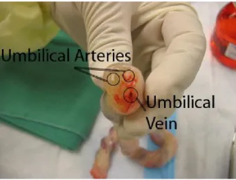
Figure 3.1 Human umbilical vein and arteries
The picture indicates the location of the umbilical artery and umbilical vein. The HUVECs are isolated from the umbilical vein. The image is adapted from http://www.slhd.nsw.gov.au
3.3 Immunocytochemistry
Isolated HUVECs were characterized by immunocytochemistry. HUVECs were seeded in round glass microscope slides on a 24 multiwell plate at a density of 4x10⁴
cells/well in EGM-2 10% FBS. The cells were then rinsed in PBS and fixed in 4% PFA for 20 minutes at room temperature. Cells were permeabilised with 0.1% Triton X-100 (Sigma Aldrich, UK) in PBS and then blocked in 0.5% goat serum (Vector Laboratories, UK) in PBS for one hour at room temperature. Primary antibodies for human CD-31/PECAM‑1 (R&D Systems, UK, 1:50), CD-144/VE-Cadherin (Santa Cruz Biotechnology, UK 1:50), Von Willebrand Factor (Dako, UK, 1:50) were added in 1% goat serum and incubated overnight at 4°C. The cells were washed in PBS three times