Received: August 4, 2017. Accepted: March 6, 2018. Pre-published: March 29, 2018.
©2018 Ferrata Storti Foundation
Material published in Haematologica is covered by copyright. All rights are reserved to the Ferrata Storti Foundation. Use of published material is allowed under the following terms and conditions:
https://creativecommons.org/licenses/by-nc/4.0/legalcode. Copies of published material are allowed for personal or inter-nal use. Sharing published material for non-commercial pur-poses is subject to the following conditions:
https://creativecommons.org/licenses/by-nc/4.0/legalcode, sect. 3. Reproducing and sharing published material for com-mercial purposes is not allowed without permission in writing from the publisher.
Correspondence:
[email protected] Ferrata Storti FoundationHaematologica
2018
Volume 103(6):949-958
doi:10.3324/haematol.2017.177980 Check the online version for the most updated information on this article, online supplements, and information on authorship & disclosures: www.haematologica.org/content/103/6/949D
iamond-Blackfan anemia (DBA) is a rare inherited bone marrow
failure disorder linked predominantly to ribosomal protein gene
mutations. Here the European DBA consortium reports novel
mutations identified in the RPL15 gene in 6 unrelated individuals
diag-nosed with DBA. Although point mutations have not been previously
reported for RPL15, we identified 4 individuals with truncating
muta-tions p.Tyr81* (in 3 of 4) and p.Gln29*, and 2 with missense variants
p.Leu10Pro and p.Lys153Thr. Notably, 75% (3 of 4) of truncating
muta-tion carriers manifested with severe hydrops fetalis and required
intrauterine transfusions. Even more remarkable is the observation that
the 3 carriers of p.Tyr81* mutation became treatment-independent
between four and 16 months of life and maintained normal blood counts
until their last follow up. Genetic reversion at the DNA level as a
poten-tial mechanism of remission was not observed in our patients. In vitro
studies revealed that cells carrying RPL15 mutations have pre-rRNA
pro-cessing defects, reduced 60S ribosomal subunit formation, and severe
proliferation defects. Red cell culture assays of RPL15-mutated primary
erythroblast cells also showed a severe reduction in cell proliferation,
delayed erythroid differentiation, elevated TP53 activity, and increased
apoptosis. This study identifies a novel subgroup of DBA with
muta-tions in the RPL15 gene with an unexpected high rate of hydrops fetalis
and spontaneous, long-lasting remission.
Recurring mutations in
RPL15 are linked to
hydrops fetalis and treatment independence in
Diamond-Blackfan anemia
Marcin W. Wlodarski,1,2 Lydie Da Costa,3,4,5,6 Marie-Françoise O'Donohue,7
Marc Gastou,3,4,8Narjesse Karboul,3,5 Nathalie Montel-Lehry,7Ina Hainmann,1
Dominika Danda,1,9Amina Szvetnik,1Victor Pastor,1,10Nahuel Paolini,11
Franca M. di Summa,11Hannah Tamary,12,13Abed Abu Quider,14
Anna Aspesi,15Riekelt H. Houtkooper,16 Thierry Leblanc,17 Charlotte M.
Niemeyer,1,2 Pierre-Emmanuel Gleizes7 and Alyson W. MacInnes16
1Department of Pediatrics and Adolescent Medicine, Division of Pediatric Hematology
and Oncology, Medical Center, Faculty of Medicine, University of Freiburg, Germany;
2German Cancer Consortium (DKTK), Freiburg, Germany and German Cancer Research
Center (DKFZ), Heidelberg, Germany; 3University Paris VII Denis Diderot, Faculté de
Médecine Xavier Bichat, Paris, France; 4Laboratory of Excellence for Red Cell, LABEX
GR-Ex, Paris, France; 5Inserm Unit 1149, CRI, Paris, France; 6Hematology Laboratory,
Robert Debré Hospital, Paris, France; 7LBME, Centre de Biologie Intégrative, Université
de Toulouse, CNRS, UPS, France; 8UMR1170, Gustave Roussy, Villejuif, France;
9Department of Tumor Pathology, Centre of Oncology, Maria Sklodowska-Curie Memorial
Institute, Poland; 10Faculty of Biology, University of Freiburg, Germany; 11Department of
Hematopoiesis, Sanquin and Landsteiner Laboratory, AMC/UvA, CX Amsterdam, the
Netherlands; 12Hematology Unit, Schneider Children's Medical Center of Israel, Petach
Tikva, Israel; 13Sackler School of Medicine, Tel Aviv University, Israel; 14Pediatric
Hematology/Oncology Department, Soroka Medical Center, Faculty of Medicine, Ben-Gurion University, Beer Sheva, Israel; 15Dipartimento di Scienze della Salute, Università
del Piemonte Orientale, Novara, Italy; 16Laboratory Genetic Metabolic Diseases,
Academic Medical Center, Amsterdam, the Netherlands and 17Pediatric Hematology
Service, Robert-Debré Hospital and EA-3518, Université Paris Diderot - Institut Universitaire d'Hématologie, Paris, France
ABSTRACT
Introduction
Diamond-Blackfan anemia (DBA) (OMIM# 105650) is an inherited bone mar-row failure disorder that typically manifests in children under the age of one year. While the central phenotype is pure red cell aplasia, developmental delay and a
number of physical malformations are also linked to DBA.1These include (but are not limited to) craniofacial malformations, growth retardation, abnormalities in the extremities, heart defects, as well as urogenital defects.2,3 While most patients (>90%) are diagnosed within the first year of life, a minority present with anemia at birth. Hydrops fetalis due to severe intrauterine anemia is con-sidered a very rare manifestation of DBA.4 To date, a total of 10 cases of DBA-associated hydrops have been reported in single cases.5-13 The clinical outcome of these patients was poor as compared to typical DBA (3 patients died perinatally, 4 patients were steroid unre-sponsive, 2 patients required steroid therapy, and 1 had unknown outcome).
Almost all of the mutations linked to DBA have been found in genes coding for ribosomal proteins (RPs).14 These RPs include: eS7 (RPS7), uS8 (RPS15A), eS10 (RPS10), eS17 (RPS17), eS19 (RPS19), eS24 (RPS24), eS26 (RPS26), eS27 (RPS27), eS28 (RPS28), uS14 (RPS29), uL18 (RPL5), uL5 (RPL11), eL15 (RPL15), eL18 (RPL18), uL24 (RPL26), eL27 (RPL27), eL31 (RPL31), uL29 (RPL35) and eL33 (RPL35A).15-28 The RPL15 gene has so far been reported in one patient who carried a large monoallelic microdeletion involving this gene.23 Non-RP genes linked to DBA, albeit very rarely involved, are TSR2 and GATA1.26,29 RP gene aberrations lead to haploinsufficien-cy, and in many cases result in reduction in the respective RP which impairs ribosome biogenesis, affecting both the processing pathways of pre-rRNA maturation and the assembly of the large or small ribosomal subunit.30,31 TP53 is a tumor suppressor protein and transcription fac-tor that stabilizes in response to cell stress, such as DNA damage or nucleolar stress induced by ribosome biogen-esis defects.32,33 Depending on the level of stress, stabi-lized TP53 will induce cell arrest, DNA repair, senes-cence and/or apoptosis. This pathway has been promi-nently implicated in the pathogenesis of DBA, with a number of studies suggesting that TP53 stabilization lies at the heart of the loss of erythroid progenitor cells in DBA bone marrow.34-36 In addition to activating apopto-sis, DBA-linked RP gene mutations can impair cellular differentiation, alter the landscape of mRNAs on ribo-somes, and induce autophagy.36-38
Therapy in DBA depends on the severity of anemia and on the response to oral glucocorticosteroids (GCS). GCS-non-responders receive chronic blood transfusions or can undergo hematopoietic stem cell transplantation (HSCT) which is often delayed because patients can achieve treat-ment independence.39 For unknown reasons, 20% of all DBA cases can become independent of steroid treatment and/or blood transfusions by the age of 25 years.39,40 To date, however, there are no predictive genetic markers that could improve decision making for timely HSCT. Given the significant risks that are associated with HSCT,41 especially if a matched sibling donor is not avail-able, such predictive markers would be very valuable.
Here we describe 6 DBA cases associated with muta-tions in ribosomal protein gene RPL15. These patients display an unexpectedly high rate of hydrops fetalis and treatment independence, pointing to a new genotype-phenotype correlation in DBA which could be important for risk stratification.
Methods
Patients
Diagnosis of DBA was made based on typical features includ-ing aregenerative anemia with erythroid hypoplasia.1 Written informed consent was obtained from patients and/or parents prior to inclusion in this study, which was performed in accor-dance with the ethical standards of the Declaration of Helsinki. The study was approved by the institutional ethics committee (University of Freiburg, IRB n. 205/99 and n. 351/17).
Cell culture
Lymphoblastoid cell lines (LCLs) were derived from Epstein-Barr virus (EBV)-immortalization of peripheral mononuclear cells isolated from whole blood using Ficoll (GE Life Sciences) and grown in RPMI (Gibco) containing 10% fetal calf serum (FCS), 1% L-glutamine, and 1% penicillin/streptomycin, as pre-viously described.42
DNA sequencing and bioinformatics
Targeted Sanger sequencing was performed as previously reported.43 Primer sequences are available upon request. Pathogenicity of mutations, evolutionary conservation across species, and the physicochemical difference between amino acids were evaluated using standard prediction tools as outlined in Online Supplementary Table S1.
Details of pre-rRNA processing analysis, Northern blots, polysome profiling, measurement of growth rate and de novo protein synthesis are available in the Online Supplementary Methods.
Erythroid red cell culture assays
Erythroid red cell culture assays were performed as previous-ly described.36,44Antibodies and stains for FACS analysis were PC7 or PE conjugated CD34 (Beckman coulter, Brea, CA, USA), APC conjugated CD36 (BD Biosciences San Jose, CA, USA), PE conjugated α4 integrin (Miltenyi, Paris, France), APC conjugat-ed Band 3 (kindly providconjugat-ed by Mohandas Narla’s lab, NYBC, New York, USA), PE/Cy7 conjugated IL-3R (Miltennyi, Paris, France), PE/Cy7 (PE)-coupled GPA (Life Technologies Carlsbad, CA, USA), and DAPI (Sigma). FACS analysis was conducted on a BD Biosciences Influx flow cytometer (BD Biosciences San Jose, CA, USA). Data were analyzed using Kaluza software (Beckman Coulter, Brea, CA, USA). Antibodies for western blotting were TP53 (Sigma #5816), phospho-TP53 (Ser15, Cell Signaling), p21 (Cell Signaling #2947), actin (Sigma #Ac-15), and eL15 (Abcam #ab130992). Primers for real-time PCR used are as follows: TaqMhupRPL15F:
CAGCCATCAGGTAAGCCAA-GA; TaqMhuRPL15R: CAGCGGACCCTCAGAAGAAA;
TaqMhupp21F: TTGCTGCCGCATGGG, TaqMhup21R: CCTTGTGGAGCCGGAGCT; TaqMhupactinF: CTGGAACG-GTGAAGGTGACA;TaqMhupactinR: AAGGGACTTCCTG-TAACAACGCA; TGTAGTGGATGGTGGTACAGTCAGA; TaqMB2MhF: GCGGCATCTTCAAACCTCC; TaqMB2MhR: TGACTTTGTCACAGCCCAAGATA. Real-time PCR was per-formed using the ABI 7900 Real Time PCR system and Taqman PCR mastermix (Life Technologies Carlsbad, CA, USA). Quantification of gene amplification was performed in dupli-cate using DDCt method. The expression level of each gene was normalized using the housekeeping genes: actin, and β2 microglobulin.
Results
Identification of novel RPL15 mutations in 6 patients
within EuroDBA registries
Approximately 30% of all registered DBA patients who have been tested for mutations in the most common DBA-linked genes (RPS19, RPL5, RPL11, RPS10, RPS26, RPS7, RPS17, RPS24, and RPL35a) still do not have an established genotype. Because a whole gene deletion of RPL15 has been reported before in one DBA patient but not in our cohorts,23 we used targeted Sanger sequencing of RPL15 to determine if mutations in this gene could be driving disease in patients without an established geno-type. The national patient registries from EuroDBA part-ners in Germany, France, Italy and Israel were included in this study. As of November 2017, these cohorts represent a total of 985 patients. A complete description of the his-tory and composition of the EuroDBA consortium has been recently published.45 Study outline and screening strategy are illustrated in Online Supplementary Figure S1.
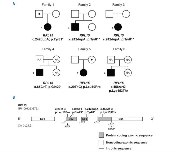
Four unrelated DBA patients from the German, and one each from the French and Israeli registries were identified carrying mutations in the RPL15 gene (NM_001253379.1), which encodes ribosomal protein eL15/RPL15 (Figure 1A and B). Out of the 6 RPL15-mutated patients, 3 unrelated patients (P1-3) carried the same novel indel mutation c.242dupA altering the tyrosine at position 81 to a stop codon: p.Tyr81* (Table 1 and Online Supplementary Table S1). Patient 4 (P4) carried another stop-gain mutation in RPL15 resulting in an even earlier protein truncation: c.85C>T; p.Gln29*. The Exome Aggregation Consortium (ExAC) reports that RPL15 is very intolerant to loss-of-function mutations with no reported cases present in over 60,000 individuals (pLI score =0.96), strongly suggesting the novel mutations p.Gln29* and p.Tyr81* found in our patients are highly deleterious (Online Supplementary Table S1). In addition, the analysis of family trios revealed that hotspot mutations arose de novo in two families (Table 1 and Figure 1A), while one patient (P1/DE071) inherited the mutation from her father who had been categorized as
Figure 1. Mutations in RPL15 are identified in patients with Diamond-Blackfan anemia (DBA).(A) Six unrelated pedigrees of individuals affected by DBA associated with RPL15 mutations. All families have one DBA-affected individual who is also a mutation carrier, as indicated with filled squares (male) or circles (female). Unaffected individuals are indicated by unfilled symbols. Unaffected mutation carriers are denoted by a dot symbol (). NA: unaffected family members who were not investigated for the presence of mutations. Families 1-4 harbor heterozygous stopgain mutations in RPL15; families 5-6 carry heterozygous missense RPL15 mutations. (B) Schematic representation of human RPL15 depicting localization of the mutations identified in families 1-6.
A
a DBA silent carrier due to very high erythrocyte adeno-sine deaminase (eADA) levels. The remaining two point mutations c.29T>C; p.Leu10Pro (P5) and c.458A>C; p.Lys153Thr (P6) affect highly conserved residues and, based on results from in silico prediction, are probably deleterious (Online Supplementary Table S1). Biomuta, DMDM, the Exac/GnomAD databases, and NCBI do not report any variants in the codon for Leu10. The GnomAD population database reports one variant in Lys153 (Lys153Arg; rs370700905) identified in 33 out of 232840 total alleles.
Genotype-phenotype association for truncating RPL15
mutations: severe hematologic phenotype and rapid
acquisition of treatment independence
All of the individuals with mutations in RPL15 present-ed with typical bone marrow erythroid hypoplasia,
ele-vated eADA, and most of them presented with increased fetal hemoglobin (HbF) levels (Table 1). Notably, hydrops fetalis (considered the most severe hematologic pheno-type of DBA) was associated only with truncating RPL15 mutations. The affected fetuses P2-4 required between four and nine intrauterine transfusions (Table 1). One indi-vidual (P2) with the p.Tyr81* hotspot mutation presented with various physical malformations, while the dysmor-phic features in other patients were less severe (Table 1). Unexpectedly, all 3 patients carrying p.Tyr81* substitution (P1-3) attained a rapid treatment independence both with and without steroid treatment (Figure 2A), while the fourth patient with the RPL15 mutation c.85C>T; p.Gln29* responded to steroids; however, the therapy was discontinued due to overt toxicity.
Based on published observations of genetic revertant mosaicism as a “repair mechanism” in other bone marrow
Table 1. Clinical characteristics of patients with RPL15 mutations.
Pat/ ID RPL15 gene; Hematology and therapies Gestational age; Age and status Family (sex) mutation malformations; other at last follow up history
1/DE071 c.242dupA; Onset: 3 months old; Hb 4.6g/dL 6.5 years, Father mutation
(F) p.Tyr81* Lab: MCV↑, eADA↑ (925U/Iec), HbF normal normal Hb, carrier, eADA↑
Evolution: spontaneous recovery after 1 transfusion 37 weeks, IUGR no therapies (1396U/Iec) but
at 6 months old. Relapse at 5.7 years (Hb 3.6g/dL), clinically silent
achieved remission after short course of steroids
2/DE189 c.242dupA; Onset: prenatal (4 intrauterine transfusions) 34+1 weeks, hydrops fetalis, ptosis, 5 years, Parents and
(M) p.Tyr81* Lab: MCV↑, eADA↑ (1526U/Iec), HbF (6.6%) flat nose, deep set ears, normal Hb, sister wild type
Evolution: 4 transfusions (birth-16 months), intermittent AV-block, duplex no therapies
achieved remission after short course left kidney, hypogonadism,
of steroids intersexual genitalia (46XY), microcephaly,
left cerebellar hypoplasia, developmental disorder with mental retardation
and cerebral palsy
3/DE115 c.242dupA; Onset: prenatal (6 intrauterine transfusions), 35+6 weeks, hydrops fetalis, 16 years, Parents
(F) p.Tyr81* Lab: MCV↑, eADA↑ (1284U/Iec), HbF↑ (10.6%) IUGR, PFO normal Hb, wild type
Evolution: 2 transfusions after birth, achieved no therapies
spontaneous remission at age of 4 months
4/DE202 c.85C>T; Onset: prenatal (9 intrauterine transfusions) 32+3 weeks, 18 years, Parents and
(M) p.Gln29* Lab: MCV↑, eADA ↑ (2628U/Iec), HbF↑ (2%) hydrops fetalis, regular sibling
Evolution: after birth irregular transfusions, hypogammaglobulinemia, transfusions healthy
steroid-responsive (4-9 years), discontinued plantar warts (carrier status
due to toxicity, transfusion-dependent from age during steroid therapy unknown),
of 9 years eADA/Hbf unknown
5/IL c.29T>C; Onset: 4 months old, Hb 5g/dL 27 weeks 2 years, Mother mutation
(F) p.Leu10Pro Lab: MCV↑, eADA and HbF unknown (placenta previa), steroids carrier: Hb, MCV
Evolution: transfusion dependent, recently started none and HbF normal,
steroids with good response eADA unknown
6/FR c.458A>C; Onset: 6 months old, Hb 7g/dL 41 weeks, low-set hair line; 22 years, Parents and
(M) p.Lys153Thr Lab: MCV normal, eADA unknown, HbF↑ (15%) growth retardation and regular sibling healthy
Evolution: initially steroid-responsive, mental retardation transfusions (carrier status
transfusion dependent from age of 5 years unknown)
All patients presented with DBA-typical erythroblastopenia in bone marrow. Pat: patient number; ID: patient identifier in respective national registry; RP: ribosomal protein; F: female, M: male; Hb: hemoglobin; Lab: supportive laboratory parameters; MCV: mean corpuscular volume; eADA: erythrocyte adenosine deaminase; Evolution: evolution of dis-ease and therapies; ↑ elevated for age; HbF: fetal hemoglobin; Remission: treatment independence; IUGR: intrauterine growth restriction; PFO: persistent foramen ovale.
failure syndromes,46 we speculated that the hematologic remission in our patients was attained via genetic rever-sion (e.g. uniparental disomy). Sequencing of DNA in P1-3 either at multiple time points or after the acquisition of treatment independence did not reveal any changes in mutation load (Figure 2B).
Mutations in RPL15 recapitulate specific pre-rRNA
processing defects found in RP depleted cells
Mutations in DBA-linked RP genes result in haploinsuf-ficiency of the encoded RP. Since the majority of RPs incorporate into pre-ribosomal particles in a progressive
manner concurrently with pre-rRNA maturation, a lack of one RP impairs pre-rRNA processing in an explicit way that is reproducible in most cell types.47 Online Supplementary Figure S2 illustrates the normal pre-rRNA processing pathway that begins with a single transcribed strand of 47S pre-rRNA that then undergoes a complex series of cleavages and trimmings to ultimately result in the mature 18S, 28S, and 5.8S strands of rRNA found in ribosomes. To investigate the functional consequences of the newly identified RP gene mutations reported here, EBV-immortalized lymphoblast cell lines (LCLs) were gen-erated and the pre-rRNA processing of these LCLs was
Figure 2. Longitudinal mutations in RPL15 are identified in individuals diagnosed with Diamond-Blackfan anemia (DBA). (A) Clinical evolution of P1-P3 carrying truncating hotspot mutation RPL15 p.Tyr81*. Patient 1 manifest-ed with DBA after birth and after one transfu-sion achieved spontaneous remistransfu-sion at the age of six months. A relapse occurred five years later and after a short course of steroids the patient attained treatment inde-pendence. Patients 2 and 3 had a similar clin-ical course with hydrops fetalis and prenatal intrauterine transfusions, and achieved treat-ment independence either spontaneously or after one course of steroids. (B) Sanger sequencing results of the recurring mutation in RPL15 from initial diagnoses as well as post remission. Arrows indicate the inserted A nucleotide. FUP: follow up; BM: bone marrow; PB: peripheral blood; LCL: lymphoblastoid cell lines.
A
compared to HeLa cells depleted of eL15.
As previously reported,23 depletion of eL15 in HeLa cells resulted in a decrease in 32S and 12S pre-rRNAs (Figure 3A and C). This was accompanied by the accumulation of the 36S and 36S-C precursors, which are inconspicuous in normal cells, and a concomitant drop in 32.5S pre-rRNA (Figure 3B). In addition, these cells displayed lower levels of 30S rRNA and higher levels of 41S and 18S-E pre-rRNAs (Figure 3A and C). This phenotype indicates that depletion of eL15 affects cleavage of the ITS1 at site 2, which promotes direct cleavage of early precursors at site E and formation of 18S-E and 36S pre-rRNAs. The 36S precursor is then trimmed by the 5’-3’ exoRNase XRN2 to produce the 36S-C and 32.5S pre-rRNAs.48Sucrose gradi-ent analyses confirmed the efficiency of the eL15 siRNAs in reducing the free 60S subunit peak and inducing the for-mation of half-mers, as expected from the deficiency of an RPL protein (Online Supplementary Figure S3). Consistent with eL15 partial loss-of-function, an increase of 36S and
36S-C precursors was observed in the patient LCLs carry-ing the eL15 p.Tyr81* variant (Figure 3B). In addition, both cell lines displayed a marked increase of the amount of 18S-E precursors translating into a higher 18S-E/21S ratio, similar to eL15-depleted cells (Figure 3A and C). This phe-notype is similar to that previously observed in LCLs har-boring a large heterozygous deletion in RPL15.23
RPL15 mutations impair 60S ribosomal subunit
formation, cell proliferation, and de novo protein
synthesis
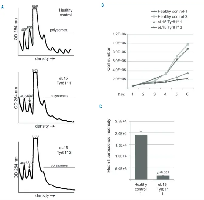
Defective processing of pre-rRNA in cells often results in cells that are unable to fully form ribosomal subunits. As such, pathogenic variants of RPs linked to DBA very often impair biogenesis of ribosomal subunits in LCLs.37 Polysome profiling was performed to determine if the observed pre-rRNA biogenesis defects resulted in impaired biogenesis of large ribosomal subunits. Figure 4A shows polysome profiles of LCL extracts derived from a
Figure 3. Mutations in RPL15 recapitulate specific pre-rRNA processing defects found in eL15 depleted cells. (A) Northern blot analysis of siRNA-treated HeLa cells or lymphoblastoid cell lines (LCLs) derived from individuals with Diamond-Blackfan anemia (DBA). Radiolabeled probes against ITS2 (top panel), ITS1 (middle panel), 18S or 28S (lower panel) rRNA sequences were used to blot 3µg total RNA isolated from cells. (B) Longer exposures of upper molecular weight pre-rRNA species observed in the northern blots from (A). Intensity profiles of the lanes is shown in the right-hand boxes. (C) Quantification of rRNA precursors in siRNA-treated HeLa cells (top) or LCLs (bottom) derived from individuals with DBA. The results of single experiments performed for each sample are displayed as multiple bars. Pre-rRNA ratios are normalized by dividing by the mean of the control samples.
A B
healthy individual or patients carrying the p.Tyr81* vari-ant in eL15. As expected, the LCLs from the healthy indi-vidual reveal an equivalent ratio of 40S to 60S peaks, while the LCLs derived from patients reveal a substantial reduction of 60S peaks compared to 40S peaks. These results suggest the pre-rRNA defects depicted in Figure 3 go on to impair biogenesis of large 60S ribosomal sub-units.
The impairment of ribosome biogenesis in cells can slow the rate of protein synthesis and cell proliferation. RP gene mutations linked to DBA are reported to slow the rate of cell proliferation and increase apoptosis.36 In agree-ment, we found that LCLs carrying the eL15 p.Tyr81*
vari-ant proliferated far more slowly than healthy control LCLs (Figure 4B). Moreover, measurement of de novo protein synthesis by Click-iT® labeling analysis revealed a severe reduction in the synthesis rate of LCLs carrying the eL15 variant (Figure 4C and Online Supplementary Figure S4).
Erythroid cell culture assays of primary RPL15
c.242dupA cells reveal severe proliferation defects,
differentiation delays, and TP53-related apoptosis
Erythroid cell culture assays show that hematopoietic progenitor cells can reveal reduced proliferation rates, delayed differentiation, and increased TP53-induced apoptosis in a manner that is largely dependent on which
Figure 4. RPL15 mutations impair cell proliferation, de novo protein synthesis, and 60S ribosomal subunit formation.(A) Representative (n=3) polysome profiles of lymphoblastoid cell line (LCL) extracts derived from healthy individuals or individuals with Diamond-Blackfan anemia (DBA) carrying the RPL15 c.242dupA muta-tion. The 40S small subunit, 60S subunit, 80S monosome, and polysomes are labeled. Arrows point to the reduced 60S peaks in cells with RPL15 mutations. (B) Growth curve of LCLs derived from individuals with DBA or healthy controls over six days. Standard Deviations for healthy control-1 cells on days 1-5 are 2.6e4, 1.5e4, 3.8e4, 6.9e4, 1.0e5; healthy control-2 cells are 8.8e3, 3.0e4, 2.9e4, 7.5e4, 8.5e4; RPL15 c.242dupA-1 are 2.1e4, 1.9e4, 1.6e4, 2.3e4, 2.2e4; and RPL15 c.242dupA-2 are 9.5e3, 1.7e4, 1.7e4, 2.2e4, 2.5e4. (C) Measurement of the amount of de novo protein synthesis in 30 minutes in LCLs derived from healthy indi-viduals or a DBA patient carrying the eL15 Tyr81* variant using Click-iT® analysis.
A B
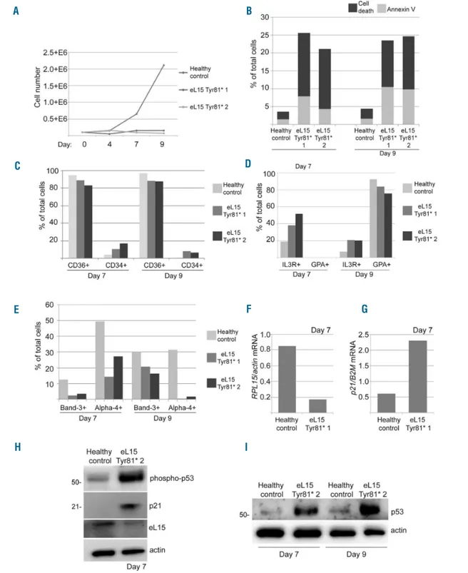
RP gene is haploinsufficient.36 We performed these assays in CD34+cells isolated from bone marrow mononuclear cells (BM-MNC) of 2 patients with truncating RPL15 mutations p.Tyr81*. Compared to healthy control,
patients BM-MNC showed a higher rate of cell death and apoptosis (Figures 5B and Online Supplementary Figure S5). FACS analysis revealed CD36 downregulation and CD34 upregulation on both day 7 and day 9 in the Tyr81*
Figure 5. Erythroid cell culture assays of primary RPL15 c.242dupA erythroid progenitor and precursor cells reveal severe erythroid proliferation defects, differen-tiation delays, and TP53-related apoptosis. (A) Proliferation curve of erythroid cells isolated from CD34+cells from peripheral blood of 2 individuals with Diamond-Blackfan anemia (DBA) and an RPL15 mutation or a healthy control over nine days in liquid culture medium. (B) FACS analysis results of the percent of dead cells or apoptotic cells staining positive for Annexin V on days 7, 10, and 13 after plating in red cell culture medium. (C) FACS analysis results on days 7 and 9 of the per-cent of cells staining positive for CD36 and CD34. (D) FACS analysis results on days 7 and 9 of cells staining positive for IL3R and GPA. (E) FACS analysis results on days 7 and 9 of cells staining positive for Alpha-4 and Band-3. (F) Real-time PCR results of the ratio between RPL15 mRNA and actin mRNA in cells. (G) Real-time PCR results of the ratio between p21 mRNA and actin mRNA in cells. (H) Western blot analysis of cells from a healthy control or an individual with DBA and a mutation in RPL15 using antibodies against phosphorylated TP53, p21, eL15, or actin on day 7. (I) Western blot analysis of cells from a healthy control or an RPL15 with DBA and a mutation in RPL15 using antibodies against TP53 or actin on days 7 and 9.
A B
C
E F G
H I
mutant cells as compared to healthy controls (Figures 5C and Online Supplementary Figure S5). In addition, Tyr81* mutant cells retain a higher expression of the IL3 receptor that is normally down-regulated in the erythroid lineage, and express less erythroid-specific glycophorin A (GPA), Band-3, or alpha-4 integrin compared to healthy control cells (Figure 5D and E and Online Supplementary Figure S5). Additionally, expression of RPL15 mRNA was lower (Figure 5F) and of p21 mRNA higher in Tyr81* mutant cells (Figure 5G). The observed p21 overexpression is like-ly TP53-mediated, as supported by western blot analike-lysis confirming an increase in TP53 phosphorylation and increased p21 protein expression (Figure 5H). Finally, TP53 protein stabilization was observed in Tyr81* mutant patient cells (Figure 5I).
Discussion
At present there are 19 RP genes associated with DBA, representing almost one-quarter of the 80 cytoplasmic RPs in human cells. Here, we describe a novel genetic subgroup of DBA due to mutations in the RPL15 gene and identify the truncating mutation c.242dupA; p.Tyr81* as a recurrent genetic cause in 3 unrelated patients. The experimental results show that LCLs carry-ing the p.Tyr81* variant reveal an array of molecular defects typically associated with a ribosomopathy phe-notype. These include impaired pre-rRNA processing, reduced 60S ribosomal subunit formation, a reduction in de novo protein synthesis, and impaired cell proliferation. Further, mutant LCLs phenocopied biological changes observed in HeLa cells depleted of eL15. These results, similar to previous reports that DBA-linked RP muta-tions impair pre-rRNA processing, indicate that the RPL15 mutations reported here are likely pathogenic and result in haploinsufficiency.
The altered ribosome biogenesis may explain the observed reduction in global protein synthesis, substanti-ating previous reports discussing altered translation of spe-cific mRNAs in DBA-mutant cells as a key component of disease pathogenesis.49,50In support of this pathogenesis, we also found that hematopoietic stem cells with the eL15 Tyr81* variant that were induced to differentiate into ery-throcytes revealed a substantial decrease in the number of erythroid colonies and delays in differentiation. These findings, in addition to increased apoptosis, TP53 stabi-lization, and p21 overexpression in RPL15-mutant hematopoiesis, suggest that eL15 plays a critical role in ribosome biogenesis and that the reduction of the protein by genetic haploinsufficiency drives severe stress in the nucleolus.
Hydrops fetalis arising from severe intrauterine anemia is an uncommon manifestation of DBA with 10 cases reported so far. The clinical outcome of these patients was poor and, unexpectedly, no spontaneous remission was observed. In contrast, 50% (3 of 6) of all patients with RPL15 mutations reported in our study showed prenatal manifestation with the necessity of intrauterine transfu-sions. Remarkably, the hydrops cases were observed only in the subgroup of patients carrying protein truncating mutations p.Tyr81* and p.Gln29*, with prevalence of hydrops reaching 75% (3 of 4).
A substantial percentage of individuals with DBA spon-taneously achieve treatment independence at some point; however, the reasons for this remain elusive and no pre-dictive biomarkers exist.39In line with this, another unex-pected finding was the rapid and sustained treatment independence achieved within four to 16 months after birth in all 3 unrelated DBA patients with the recurrent p.Tyr81* mutation. Although Patient 1 relapsed five years after achieving spontaneous remission, a short steroid course was very effective and resulted in treatment inde-pendence. These observations suggest that p.Tyr81* mutation carriers can achieve treatment independence with or without a previous course of steroids, and that a genotype itself may be an important biomarker for the expected clinical course. Our findings might help prospec-tive clinical stratification to determine which individuals are more likely to become treatment independent without the need for HSCT. The reasons why the specific muta-tion p.Tyr81* drives treatment independence remain unknown and cannot be explained by a potential genetic reversion, which was not observed in our patients. One tempting speculation is that the remaining wild-type allele compensates for the haploinsufficiency (e.g. by epigenetic mechanisms) in the adult hematopoiesis, but the compen-sation might not be present or sufficient in the fetus. These findings warrant further studies on the future avail-ability of more patient material.
In conclusion, our study establishes germline point mutations in the RPL15 gene as novel genetic etiology of DBA. Half of the individuals carrying these mutations manifest with hydrops fetalis, a phenotype that is very rarely observed in DBA patients. Finally, we establish that a recurrent DBA genotype, eL15 p.Tyr81*, is linked to treatment independence.
Acknowledgments
Very special thanks go to all the individuals who consented to participate in this study and their families. We thank Alexandra Fischer (Freiburg) for data management, Sandra Zolles, Sophia Hollander, Dirk Lebrecht, and Gunda Ruzaike (Freiburg) for technical assistance, Pritam Kumar Panda (Freiburg) for bioinfor-matic analysis, and Dr. Marije Bartels (UMC Utrecht) for criti-cal reading of the manuscript.
Funding
AM, ML, MW, LDC, NK, HT, PEG, and MFOD are sup-ported under the framework of E-Rare-2, ERA-Net for Research on Rare Diseases (ZonMW #113301205 and #40-44000-98-1008 in the Netherlands; #BMBF 01GM1301 and 01GM1609 in Germany; Chief Scientist Office, Israeli Ministry of Health # 3-12844 in Israel; #ANR-15-RAR3-0007-04 and #ANR-12-RARE-0007-02 in France). MW and CN are sup-ported by DKTK German Cancer Consortium, fot molecular diagnostics of pediatric malignancies. LDC was supported by the Laboratory of Excellence for Red Cells [(LABEX GR-Ex)-ANR Avenir-11-LABX-0005-02], the French National PHRC OFABD (DBA registry and molecular biology), and the “Fondation ARC pour la recherche contre le cancer”. LDC, MFOD and PEG are funded by the French National Research Agency [ANR-DBA-Multigenes-ANR-2015-AAP générique-CE12]. AA was supported by DBA Telethon grant GGP13177 and Fondazione Europea per la DBA and Gruppo di Sostegno DBA Italia.
References
1. Vlachos A, Ball S, Dahl N, et al. Diagnosing and treating Diamond Blackfan anaemia: results of an international clinical consen-sus conference. Br J Haematol. 2008; 142(6):859-876.
2. Ball SE, McGuckin CP, Jenkins G, Gordon-Smith EC. Diamond-Blackfan anaemia in the U.K.: analysis of 80 cases from a 20-year birth cohort. Br J Haematol. 1996;94(4):645-653.
3. Lipton JM, Atsidaftos E, Zyskind I, Vlachos A. Improving clinical care and elucidating the pathophysiology of Diamond Blackfan anemia: an update from the Diamond Blackfan Anemia Registry. Pediatr Blood Cancer. 2006;46(5):558-564.
4. Vlachos A, Klein GW, Lipton JM. The Diamond Blackfan Anemia Registry: tool for investigating the epidemiology and biology of Diamond-Blackfan anemia. J Pediatr Hematol Oncol. 2001;23(6):377-382.
5. Semmekrot BA, Haraldsson A, Weemaes CM, Smeets DF, Geven WB, Brunner HG. Absent thumb, immune disorder, and con-genital anemia presenting with hydrops fetalis. Am J Med Genet. 1992;42(5):736-740.
6. Van Hook JW, Gill P, Cyr D, Kapur RP. Diamond-Blackfan anemia as an unusual cause of nonimmune hydrops fetalis: a case report. J Reprod Med. 1995;40(12):850-854. 7. McLennan AC, Chitty LS, Rissik J, Maxwell DJ. Prenatal diagnosis of Blackfan-Diamond syndrome: case report and review of the literature. Prenat Diagn. 1996;16(4):349-353.
8. Rogers BB, Bloom SL, Buchanan GR. Autosomal dominantly inherited Diamond-Blackfan anemia resulting in nonimmune hydrops. Obstet Gynecol. 1997;89(5 Pt 2):805-807.
9. Scimeca PG, Weinblatt ME, Slepowitz G, Harper RG, Kochen JA. Diamond-Blackfan syndrome: an unusual cause of hydrops fetalis. Am J Pediatr Hematol Oncol. 1988 Fall;10(3):241-243.
10. Dunbar AE, 3rd, Moore SL, Hinson RM. Fetal Diamond-Blackfan anemia associated with hydrops fetalis. Am J Perinatol. 2003;20(7):391-394.
11. Saladi SM, Chattopadhyay T, Adiotomre PN. Nomimmune hydrops fetalis due to Diamond-Blackfan anemia. Indian Pediatr. 2004;41(2):187-188.
12. Valsky DV, Daum H, Yagel S. Reversal of mirror syndrome after prenatal treatment of Diamond-Blackfan anemia. Prenat Diagn. 2007;27(12):1161-1164.
13. Da Costa L, Chanoz-Poulard G, Simansour M, et al. First de novo mutation in RPS19 gene as the cause of hydrops fetalis in Diamond-Blackfan anemia. Am J Hematol. 2013;88(4):340-341.
14. Boria I, Garelli E, Gazda HT, et al. The ribo-somal basis of Diamond-Blackfan Anemia: mutation and database update. Hum Mutat. 2010;31(12):1269-1279.
15. Ban N, Beckmann R, Cate JH, et al. A new system for naming ribosomal proteins. Curr Opin Struct Biol. 2014;24:165-169. 16. Cmejla R, Cmejlova J, Handrkova H, Petrak
J, Pospisilova D. Ribosomal protein S17 gene (RPS17) is mutated in Diamond-Blackfan anemia. Hum Mutat. 2007; 28(12):1178-1182.
17. Doherty L, Sheen MR, Vlachos A, et al. Ribosomal protein genes RPS10 and RPS26 are commonly mutated in
Diamond-Blackfan anemia. Am J Hum Genet. 2010; 86(2):222-228.
18. Draptchinskaia N, Gustavsson P, Andersson B, et al. The gene encoding ribo-somal protein S19 is mutated in Diamond-Blackfan anaemia. Nat Genet. 1999;21(2):169-175.
19. Farrar JE, Nater M, Caywood E, et al. Abnormalities of the large ribosomal sub-unit protein, Rpl35a, in Diamond-Blackfan anemia. Blood. 2008;112(5):1582-1592. 20. Gazda HT, Grabowska A, Merida-Long LB,
et al. Ribosomal protein S24 gene is mutat-ed in Diamond-Blackfan anemia. Am J Hum Genet. 2006;79(6):1110-1118. 21. Gazda HT, Preti M, Sheen MR, et al.
Frameshift mutation in p53 regulator RPL26 is associated with multiple physical abnormalities and a specific pre-ribosomal RNA processing defect in diamond-black-fan anemia. Hum Mutat. 2012;33(7):1037-1044.
22. Gazda HT, Sheen MR, Vlachos A, et al. Ribosomal protein L5 and L11 mutations are associated with cleft palate and abnor-mal thumbs in Diamond-Blackfan anemia patients. Am J Hum Genet. 2008;83(6):769-780.
23. Landowski M, O'Donohue MF, Buros C, et al. Novel deletion of RPL15 identified by array-comparative genomic hybridization in Diamond-Blackfan anemia. Hum Genet. 2013;132(11):1265-1274.
24. Wang R, Yoshida K, Toki T, et al. Loss of function mutations in RPL27 and RPS27 identified by whole-exome sequencing in Diamond-Blackfan anaemia. Br J Haematol. 2015;168(6):854-864.
25. Mirabello L, Macari ER, Jessop L, et al. Whole-exome sequencing and functional studies identify RPS29 as a novel gene mutated in multicase Diamond-Blackfan anemia families. Blood. 2014;124(1):24-32. 26. Gripp KW, Curry C, Olney AH, et al.
Diamond-Blackfan anemia with mandibulofacial dystostosis is heteroge-neous, including the novel DBA genes TSR2 and RPS28. Am J Med Genet A. 2014;164A(9):2240-2249.
27. Ikeda F, Yoshida K, Toki T, et al. Exome sequencing identified RPS15A as a novel causative gene for Diamond-Blackfan ane-mia. Haematologica. 2017;102(3):e93-e96. 28. Mirabello L, Khincha PP, Ellis SR, et al.
Novel and known ribosomal causes of Diamond-Blackfan anaemia identified through comprehensive genomic character-isation. J Med Genet. 2017;54(6):417-425. 29. Sankaran VG, Ghazvinian R, Do R, et al.
Exome sequencing identifies GATA1 muta-tions resulting in Diamond-Blackfan ane-mia. J Clin Invest. 2012;122(7):2439-2443. 30. Choesmel V, Bacqueville D, Rouquette J, et
al. Impaired ribosome biogenesis in Diamond-Blackfan anemia. Blood. 2007;109(3):1275-1283.
31. Ferreira-Cerca S, Poll G, Gleizes PE, Tschochner H, Milkereit P. Roles of eukary-otic ribosomal proteins in maturation and transport of pre-18S rRNA and ribosome function. Mol Cell. 2005;20(2):263-275. 32. Golomb L, Volarevic S, Oren M. p53 and
ribosome biogenesis stress: the essentials. FEBS Lett. 2014;588(16):2571-2579. 33. Kastan MB, Onyekwere O, Sidransky D,
Vogelstein B, Craig RW. Participation of p53 protein in the cellular response to DNA damage. Cancer Res. 1991;51(23 Pt 1):6304-6311.
34. Dutt S, Narla A, Lin K, et al. Haploinsufficiency for ribosomal protein genes causes selective activation of p53 in
human erythroid progenitor cells. Blood. 2011;117(9):2567-2576.
35. Jaako P, Flygare J, Olsson K, et al. Mice with ribosomal protein S19 deficiency develop bone marrow failure and symptoms like patients with Diamond-Blackfan anemia. Blood. 2011;118(23):6087-6096.
36. Moniz H, Gastou M, Leblanc T, et al. Primary hematopoietic cells from DBA patients with mutations in RPL11 and RPS19 genes exhibit distinct erythroid phe-notype in vitro. Cell Death Dis. 2012; 3:e356.
37. Pereboom TC, Bondt A, Pallaki P, et al. Translation of branched-chain aminotrans-ferase-1 transcripts is impaired in cells hap-loinsufficient for ribosomal protein genes. Exp Hematol. 2014;42(5):394-403.e4. 38. Heijnen HF, van Wijk R, Pereboom TC, et
al. Ribosomal protein mutations induce autophagy through S6 kinase inhibition of the insulin pathway. PLoS Genet. 2014; 10(5):e1004371.
39. Vlachos A, Muir E. How I treat Diamond-Blackfan anemia. Blood. 2010;116(19): 3715-3723.
40. Narla A, Vlachos A, Nathan DG. Diamond Blackfan anemia treatment: past, present, and future. Semin Hematol. 2011; 48(2):117-123.
41. Peffault de Latour R, Peters C, Gibson B, et al. Recommendations on hematopoietic stem cell transplantation for inherited bone marrow failure syndromes. Bone Marrow Transplant. 2015;50(9):1168-1172. 42. Hui-Yuen J, McAllister S, Koganti S, Hill E,
Bhaduri-McIntosh S. Establishment of Epstein-Barr virus growth-transformed lymphoblastoid cell lines. J Vis Exp. 2011;(57).
43. Hirabayashi S, Flotho C, Moetter J, et al. Spliceosomal gene aberrations are rare, coexist with oncogenic mutations, and are unlikely to exert a driver effect in child-hood MDS and JMML. Blood. 2012; 119(11):e96-99.
44. Hu J, Liu J, Xue F, et al. Isolation and func-tional characterization of human erythrob-lasts at distinct stages: implications for understanding of normal and disordered erythropoiesis in vivo. Blood. 2013; 121(16):3246-3253.
45. Da Costa L, O'Donohue MF, van Dooijeweert B, et al. Molecular approaches to diagnose Diamond-Blackfan anemia: The EuroDBA experience. Eur J Med Genet. 2017 Oct 26. [Epub ahead of print] 46. Lo Ten Foe JR, Kwee ML, Rooimans MA, et
al. Somatic mosaicism in Fanconi anemia: molecular basis and clinical significance. Eur J Hum Genet. 1997;5(3):137-148. 47. Henras AK, Plisson-Chastang C,
O'Donohue MF, Chakraborty A, Gleizes PE. An overview of pre-ribosomal RNA processing in eukaryotes. Wiley Interdiscip Rev RNA. 2015;6(2):225-242.
48. Preti M, O'Donohue MF, Montel-Lehry N, Bortolin-Cavaille ML, Choesmel V, Gleizes PE. Gradual processing of the ITS1 from the nucleolus to the cytoplasm during syn-thesis of the human 18S rRNA. Nucleic Acids Res. 2013;41(8):4709-4723. 49. Ludwig LS, Gazda HT, Eng JC, et al.
Altered translation of GATA1 in Diamond-Blackfan anemia. Nat Med. 2014;20(7):748-753.
50. Horos R, Ijspeert H, Pospisilova D, et al. Ribosomal deficiencies in Diamond-Blackfan anemia impair translation of tran-scripts essential for differentiation of murine and human erythroblasts. Blood. 2012;119(1):262-272.