Università degli Studi del Piemonte Orientale
“Amedeo Avogadro”
Dipartimento di Scienze del Farmaco
Dottorato di Ricerca in Biotecnologie Farmaceutiche ed Alimentari
XXVIII ciclo a.a. 2012-2015
A JOURNEY THROUGH THE CHEMISTRY
OF DIAGNOSTIC IMAGING PROBES
Arianna Maria Giani
Supervisor: Prof. Giovanni Battista Giovenzana
Contents
Chapter 1
Introduction
1
Chapter 2
Outline of the thesis42
Chapter 3
cis-IPDTA: an Original Polyaminopolycarboxylic Chelating Agent from
Isophoronediamine. Synthesis and Thermodynamic Characterization of Metal Complexes
45
Chapter 4
Determination of Thermodynamic and Kinetic Parameters for the Sc(III)-AAZTA System
61
Chapter 5
Fluorescence Studies on 2-(Het)Arylperimidine Derivatives
74
Chapter 6
Unprecedented Formation of 2,5-Diaminoquinones from the Reaction of Vanillin with Secondary Amines in Aerobic Conditions
105
Chapter 7
Conclusions 113List of publications
115Acknowledgements
1171
CHAPTER 1
1.0 MOLECULAR IMAGING
“Imaging” refers to the generic process through which it is possible to observe an area of a living organism not visible from the outside and it is widely employed in clinical diagnostics in order to detect and locate physiological and pathological phenomena.
In "vivo" medical imaging has made wide improvement through advances in the engineering of imaging devices and developments in the chemistry of probes. Over the past years, the National Cancer Institute recognized Molecular Imaging as an extraordinary tool for studying diseases noninvasively and quantitatively at the molecular level.[1]
Molecular imaging is a scientific discipline and it is a result of combined advances in chemistry, biology, physics and engineering. It can be defined as the visualization, characterization, and measurement of biological processes at the molecular and cellular levels in humans and other living systems and molecular imaging agents are probes used to visualize, characterize, and measure biological processes. These two definitions were stated by the Society of Nuclear Medicine in 2007 as a way to highlight the interdisciplinary nature of this new field of research.[2]
Molecular imaging employs contrast agents or probes of different nature to gain more information. The advantages of using more specific molecular probes are evident for personalized medicine and for accelerating the time and the quality of the diagnosis. At the state of art, however, most of these molecules are non-targeted reagents.[3]
2
1.1 BASIC PRINCIPLES OF IMAGING MODALITIES: BASIC OF DIAGNOSTIC IMAGING TECHNIQUES
Imaging modalities can be simply divided into two categories:
Morphological and anatomical techniques including Computed Tomography (CT), Magnetic Resonance Imaging (MRI) and Ultrasound Sonography(US). They are characterized by high spatial resolution but they are not able to give any information about little changes at the tissue or cell levels. As a matter of fact, these techniques are able to give images of the body that allow to detect coarse changes in a tissue such as tumours in advanced stages.
Molecular Imaging techniques include Single Photon Emission Computed Tomography (SPECT), Positron Emission Tomography (PET), Optical Imaging (OI) and Photoacoustic Tomography(PAT). Thanks to the use of radiopharmaceuticals, luminophores and choromophores injected at nanomolar blood concentration they allow to detect subtle changes inside a tissue in a pathological situation (for example early stage tumours). Unfortunately nuclear techniques as SPECT and PET do not possess a great spatial resolution and Optical Imaging techniques suffer from a limited tissue penetration.
Combining morphological/anatomical and molecular imaging modalities, it is possible to obtain a better imaging of the pathophysiological changes in early disease phases with high structural resolution (for example, PET–CT and PET– MRI technology).
3
Table 1. Imaging techniques currently used in the context of biomedical research and/or medical diagnosis Technique Labels Signal
measured Strengths Weaknesses Cost Throughput
Sensitivity ([M] of label
detected)
Resolution CT None X-rays Fast, cross-sectional images Poor resolution of soft
tissues High Low 10
-6 50µm MRI Can use isotope-labelled molecular tracers magnetic fields Alterations in Harmless, high-resolution of soft tissues Cannot follow many labels High Low 10-9-10-6 50µm
US
Microbubbles, which can be combined with targeted
contrast agents Sound waves Quick, harmless Poor image contrast Low High 10
-8 50µm
SPECT Radiolabelled molecules γ-rays
Can distinguish between radionuclides, so more processes can be imaged at
once
Requires radioactivity High Low 10-14 1-2mm
PET Radiolabelled molecules
2 γ-rays from positron annihilation
Highly sensitive
Can detect only one radionuclide, implies
radioactivity
High Low 10-15 1-2mm
OI
Genetically engineered proteins and fluorescently
labelled probes
Light, particularly in
the infrared
Easy, non-damaging technique readily adapted to
study specific molecular events
Poor depth penetration Low High 10-12 50µm
PAT and create sound signals Probes that absorb light Sound waves Better depth resolution than light
Information processing and machines still being
optimized
4
1.1.1 Computed Tomography
Computed tomography (CT) was introduced in the clinical practice in 1972 and rapidly became a very important tool in the radiological diagnosis. For example CT of the brain became the procedure of choice for evaluation of brain tumours. It replaced invasive procedures such as pneumoencephalography and the progressive improvement of the image quality, reduction of costs, and reduction of scan times has resulted in significant expansion of CT applications.[4] CT is a medical imaging method that combines multiple X-ray projections taken from different angles to produce detailed cross-sectional images of areas inside the body. CT images allow to obtain very precise 3D views of certain parts of the body, such as soft tissues, blood vessels, lungs, brain, heart, abdomen, pelvis and bones. It is often used to locate bone and traumatic injuries, vascular condition/blood flow, abdominal aortic aneurysms, pulmonary embolism, cardiovascular disease and to measure the size and location of tumours.
5
1.1.2 Magnetic Resonance Imaging
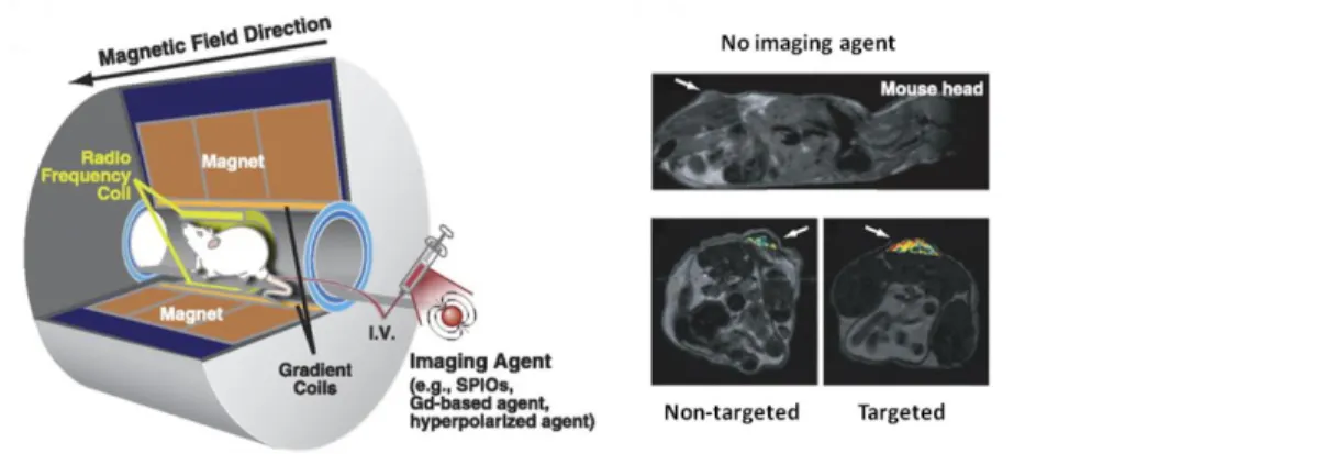
Magnetic resonance imaging (MRI) is a medical imaging technique commonly used to visualize structures and functions of the body providing detailed images in any plane using intravenous contrast agent.[6]
This technique is based on the principles of nuclear magnetic resonance. A powerful magnetic field aligns the nuclear magnetization of hydrogen atoms of the body water molecules. Radio frequency (RF) fields are used to systematically alter the alignment of this magnetization inducing the hydrogen nuclei to produce a rotating magnetic field detectable by the scanner.[7]
Figure 2. Schematic illustration of MRI: a mouse is placed inside a magnet. Unpaired nuclear spin within the
body either align parallel or anti-parallel with the direction of the magnetic field. MR signal is generated from a very small difference in number of parallel versus anti-parallel spins. MRI images with or without contrast agents are reported. [5]
1.1.3 Ultrasound Sonography
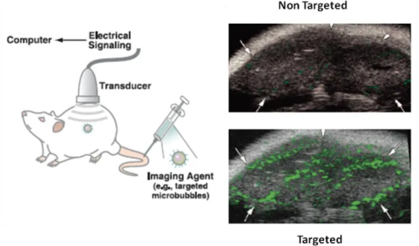
Ultrasound (US) imaging is based on the use of the sound at frequency higher than 20 KHz to obtain projections of the organism. Ultrasound contrast agents (UCAs) can be used to improve US-imaging and are based on materials characterized by different acoustic properties respect to other tissues. The most common approach is the use of intravenous injections of small air or gas bubbles (microbubbles) that
6
improve the Doppler signal from blood vessels. One of its most important application is the study of angiogenesis; it can be indeed used to image microcirculation.[8]
Figure 3. Schematic illustrating the general principles of molecular imaging using US. US images
demonstrating the significant advantage of using microbubbles targeted to vascular endothelial growth factor receptor type 2 (VEGFR2) compared with non-targeted microbubbles for visualizing tumour angiogenesis in mice. [5]
1.1.4 Single Photon Emission Computed Tomography
Single Photon Emission Computed Tomography (SPECT) is a medical imaging technique commonly used to obtain 3D biotopological data of the body. SPECT use -rays to collect images through a gamma-camera that provides a series of two-dimensional views of the distribution in the body of a radionuclide previously administered to the patient. The radionuclide may be introduced into the patient, normally through injection into the bloodstream or seldom per os.
7
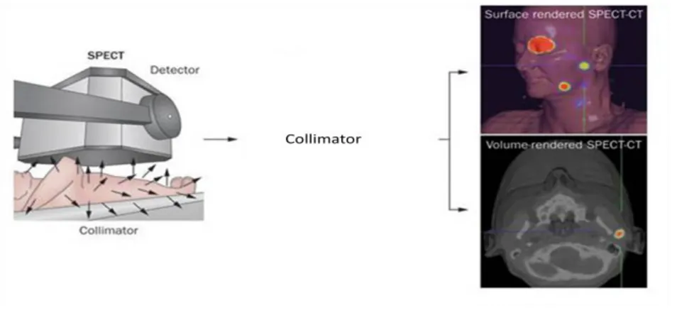
Figure 4. SPECT principles. The radiotracer previously administrated to the patient decay emitting photons.
Produced photons transfer energy to the gamma-camera detector. Because photons are emitted in all directions, a collimator is used to collect only photons that are travelling perpendicular to the detector surface. These detectors are made of a high density material, such as lead or steel, through which there are multiple parallel holes. To collect a tomographic image, the gamma camera rotates around the body, collecting a set of overlapping planar images that are elaborated through a 'back projection' technique.
1.1.5 Positron Emission Tomography
Positron Emission Tomography (PET) is one of the most recent techniques used in medical diagnostics and it is able to give functional imaging of the body. This technique exploits positrons issued following radioactive decay of the previously administered tracer. PET is very useful to locate functional changes inside an organ or an entire apparatus and it is essential for early diagnosis. In fact, physiological changes take place before structural alterations in a pathological condition.
8
Figure 5. PET scanners detect the simultaneous outcoming of two photons of a characteristic energy from opposed detectors (lower panels), a process is termed 'coincidence'. The photons have an energy of 511 keV, which is the energy released from an electron-positron annihilation reaction. A tomographic image is obtained by combining many 'lines of coincidence'.
1.1.6 Optical Imaging
Optical imaging uses light to visualize the body, and so it is sensitive to compounds and structures in tissues that are able to provide optical contrast. The acquired image depends on the light wavelength applied and the instrumental mode of detection. Generally, light tissue interaction is characterized by the processes of absorption, photon scattering and fluorescence emission. Optical contrast mechanisms include absorption, fluorescence, luminescence and scattering. Optical imaging combines different modalities with a great potential for biomedical imaging. These techniques are used in molecular imaging to visualize and quantify altered molecular mechanism in a non-invasive and ionising radiation-free manner, using relatively simple apparatus.[9-10]
9
Figure 6. General principles of molecular imaging using optical fluorescence [5]
1.1.7 Photoacoustic Tomography
First reported by Alexander Graham Bell in 1880 [11], the photoacoustic effect can be exploited for imaging purposes. It can be described as the production of sound waves resulting from the absorption of light. One of the main advantages of PAT is its depth of penetration (from 6 mm to 5 cm), which is superior to that of most optical techniques. In addition, the spatial resolution of PAT is not negatively influenced by increases in depth of penetration, as is typically the situation with other optical modalities and US.
10
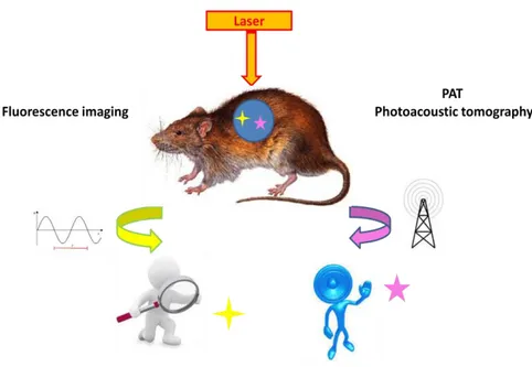
1.2 OPTICAL IMAGING
Optical Imaging is a light-based technology with a great potential to improve the in vivo and in vitro diagnosis and treatment of various diseases. It offers a number of important advantages over existing radiological imaging techniques using non-ionising radiation. Moreover, the use of dedicated “probes” offers the possibility to study selected biological pathways at the functional and molecular level in a living system or in a model tissue or cell.
Figure 8. Optical Imaging principles
The principles underlying selected techniques of molecular imaging are shown in Figure 8. The target is irradiated by a high intensity light source, usually a laser. The image can be obtained by detecting the luminescence of suitable fluorescent agents (fluorescence imaging), previously administered. In a different technique (PAT) the laser irradiation is modulated in the KHz range, and an acoustic signal is
11
detected, arising from the sound waves resulting by the local expression following the absorption of radiation by selected probes. In both imaging techniques, the molecular probe must be endowed with a strong ability to absorb the radiation of the source. Laser sources in the red and infrared region are preferred due to their significant penetration depth in tissues. In fluorescence imaging, the molecular probe must receive the absorbed energy in the form of a longer-wavelength radiation. In PAT, the molecular probe must undergo to a nonradiative decay, releasing thermal energy leading to adiabatic expansion of the local environment.
1.2.1 General requirement for fluorescent probes in Optical Imaging
A successful optical molecular probe for medical imaging should have several adjective properties such as brightness, bio- and photostability, and specific wavelengths and pharmacokinetics [12]
WAVELENGTH: fluorophores require excitation light to emit a signal. Excitation in the ultraviolet cannot be used because may cause direct tissue damage,[13] blue or green excitation light have poor tissue penetration and fluorophores which need excitation with about 600 nm (yellow light) are not applicable due to the presence of endogenous molecules responding at the same wavelength. In fact the tissue autofluorescence at these wavelengths could be too high to be distinguished from the exogenous fluorophore signal.[14] The best excitation wavelength of a fluorophore is between near-infrared and the deep red range due to a good tissue penetration and low autofluorescence.[15]
12
Figure 9. Electromagnetic spectrum
BRIGHTNESS: of course, the brighter the agent, better will be signal-to-noise ratio. To be able to increase the brightness, the size of the fluorophore must be larger. However, in this situation different problems ensues such as a difficult conjugation and higher toxicity.[12]
STABILITY: the in vivo stability is another crucial point. Once internalized into the lysosome, most of the fluorophores, such as fluorescein, BODIPY, and cyanine derivatives except rhodamines, lose fluorescence in several days. Rhodamine is an exception because it can keep fluorescence for longer than a week. [16-17] Furthermore, photodegradation is a very usual phenomenon for this type of molecules. Although the stability is essential some level of degradation is desirable so that the product could be easily conjugated, metabolized and excreted.
PHARMACOKINETICS: fluorophores are often conjugated with biomolecules such as proteins, sugars and antibodies. Most of small-molecules fluorophores may alter the pharmacokinetics of targeting portions to which they are conjugated. Pharmacokinetic and toxicological studies are vital to understand biological half-life, specific tissue accumulation and toxicity.
13
1.2.2 Classification
Fluorophores can be divided into three major classes:
SMALL-MOLECULE FLUOROPHORES: are synthetic fluorophores with various core structures including fluorescein, BODIPY, rhodamine, and cyanine derivatives. All of these are commercial available, with molecular ranges between 300 to 2000 Da and cover the emission spectrum from blue to NIR.[18]
GENETICALLY ENCODED FLUORESCENT PROTEINS: they are artificial proteins with emission wavelengths not found in nature, including infrared. [19] In particular three proteins can be cited including green fluorescent protein (GFP), yellow fluorescent protein (YFP), and red fluorescent protein (RFP). Some of them have unique features among which a large Stokes shift.[20]
NANOCRYSTALS: are synthetic materials characterised by a broad excitation range, a narrow emission peak, resistance to photodegradation, and extremely high brightness.[21] Numerous nanocrystals with unique optical properties have been reported. They are new products belonging to the field of nanotechnologies, however the pharmacokinetic and pharmacodynamic will be investigated together with many toxicological studies to report any shadows about their use.
1.2.3 Small-Molecule Fluorophores
Fluorescein is a synthetic organic compound soluble in water and alcohol. It is widely employed as a fluorescent tracer for many optical applications. Fluorescein is a fluorophore commonly used in microscopy, in forensics to
14
detect latent blood stains and it is a very important for Fundus Angiography.
Figure 13. Fluorescein structure
Fluorescein is used to perform retinal angiography to investigate ocular pathologies.[22]
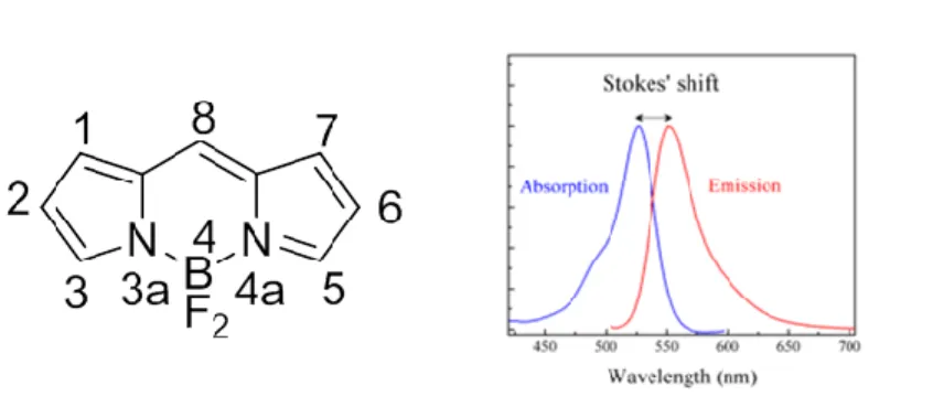
BODIPY dyes have been described for the first time in 1968 by Treibs and Kreuzer.[23] BODIPY dyes are notable for their uniquely Stokes shift, high fluorescence quantum yields and high solubility in many organic solvents. The combination of these qualities makes BODIPY fluorophore an important tool in a variety of imaging applications. With derivatives that span the entire visible spectrum BODIPY dyes are proving to be extremely versatile. Some of these compounds have very excellent spectral characteristics and they are largely better than fluorescein. [24] They are used for in vitro clinical diagnostics or biological, pharmacological and biochemistry research. Most of these dyes are used to generate fluorescent conjugates such as enzymes, fatty acids, phospholipids, lipopolysaccharides and receptor ligands. In addition, oligonucleotide conjugates BODIPY dyes have been reported to be useful for DNA sequencing,[25,26,27] nucleic acid hybridization [28] and other applications.
15
Figure 10. The structure and numbering of the BODIPY fluorophore,
4,4-difluoro-4-bora-3a,4a-diaza-s-indacene is reported. Stokes shift is the difference (in wavelength or frequency units) between positions of the band maxima of the absorption and emission spectra.
Cyanines represent one of the subclasses of the huge family of polymethine dyes.[29,30] Cyanines have been used as sensitizer for emulsion photography and in advanced photonic materials,[31,32] as well as photodynamic therapy agents or probes for near-infrared imaging.[33,34]These dyes embody a π-conjugated bridge composed of an odd number of sp2 carbon atoms linking electron-donating or -accepting groups. Photophysical properties are unequivocally related to the bridge length and to a minor extent to the terminal substituents.
Figure 11. General structure of cyanines
Rhodamine dyes are widely employed as fluorescent markers for labeling proteins, nucleic acids, lipids, carbohydrates, toxins, hormones and other biomolecules. They are used owing to their photostability, high extinction, high fluorescent quantum yields, and low degree of triplet formation. [35,36]
16
Figure 12. General structure of Rhodamine derivatives
Rhodamine derivatives are used for detecting nucleic acids,[37] for measuring membrane potential,[38] photodynamic therapy,[39] and apoptosis assays.[40]
17
1.3 MRI: CONCEPTS
Magnetic resonance imaging (MRI) is one of the most significant developments in medical imaging and it is based on the principles of nuclear magnetic resonance, discovered by Bloch and Purcell in 1946, for which they were awarded the 1952 Nobel Prize.[41,42] Since 1970s, MRI has evolved into an indispensable modality for routine clinical diagnosis and widely used tool for “in vivo” biomedical research. MRI was used at first for imaging the body and its role in biomedical research has expanded significantly in the past 20 years due to its ability to provide physiological and functional information. Although MRI is becoming mature, advances are still being made and promise to further expand its utility to many applications.[43] MRI is attractive in clinical medicine because it provides images with exquisite soft tissue contrast and it is non-invasive. The use of MRI eliminates the need for invasive diagnostic procedures, and it has been shown to provide physiological information earlier in clinical investigation. The technique also offers fast scan times, the capacity to produce excellent quality and high-resolution images. It has the greatest spatial resolution and clinical potential, and can image structures in the millimeter range without the use of ionising radiation such as that used in X-ray and CT scanning. [44] In 1973, Lauterbur applied the NMR principles to a combination of a strong constant magnetic field and weaker and variable magnetic field gradients. This allowed to identify the position of a particular nucleus, as the strength of the field is proportional to the radiofrequency. [45] Lauterbur did not make any hypothesis as to the potential application for this research, although a comment was made that there was a signal difference between cancerous tissue and normal tissue. Nevertheless, clear potential for the use in clinical imaging was realized, and as soon as few years after this idea was reported, prototype MRI machines were invented. In 1980, medical MRI scans started and since then MRI has become an essential tool for diagnoses in medicine. [45]
18
The simplest form of NMR imaging involves the application of a linear magnetic field gradient in addition to the main static field in order to “spatially encode” nuclei in the subject with different resonant frequencies. The free introduction decay signal (FID) following a radio frequency pulse is Fourier transformed to yield a one-dimensional projection of signal amplitude along a particular line through the subject. Briefly, the net macroscopic magnetization of proton spins, which is aligned parallel with the applied field along the z axis, is perturbed by application of one or more radio frequency pulses. The component of the magnetization along the z axis “relaxes” back to its equilibrium value with an exponential time constant, T1, the longitudinal (or spin-lattice) relaxation time. The
time dependence of the magnetization perpendicular to the z axis is characterized similarly by T2, the transverse (or spin-spin) relaxation time, which measures the
time for the decay of the transverse magnetization to its equilibrium value of zero. In image data acquisition, the pulses are rapidly repeated for each projection. Tissues with short T1, values generally yield greater image intensity than those with
longer values since the steady-state magnetization along the z axis is greater in the tissue with the fastest relaxation. On the other hand, short T2 values are always
associated with lower signal intensity since this diminishes the net transverse magnetization available for detection. Under conditions normally employed, the dominant effect of a paramagnetic agent in NMR imaging is to increase the signal intensity of the tissue containing the agent.
1.3.1 Spin-Lattice Relaxation (T1)
When a RF pulse is applied, M tips towards the xy plane while processing around the steady external magnetic field B0. When this perturbation is terminated, the
nuclear spin system will "relax" to the equilibrium by dissipating the excess energy to its surroundings via thermal exchange. This process is called spin-lattice
19
relaxation and the equilibrium condition is reached by a first order process characterized by a time constant, T1, the spin-lattice relaxation time.
1.3.2 Spin-Spin Relaxation (T2)
When the RF pulse is applied, the moments in the xy plane begin to lose their precessing phase coherence, due to the natural processes that cause nuclei to exchange energy with each other. As a result of this process, called spin-spin relaxation, the net Mxy magnetization decays to zero exponentially with time, characterized by the time constant T2. SI1 and SI2 represent the signal intensities of
two adjacent regions on an MR image. The magnitude of the detected signal depends upon the spin density (number of protons available), T1 and T2
characteristics of tissues, chemical shift, temperature, and flow phenomena. Among these parameters, the relaxation characteristics are most influential to the signal intensity. With conventional MR imaging techniques, however, the relaxation characteristics of the normal and pathological conditions are often very small, making the accurate diagnosis based on the contrast from the MR image difficult. Therefore, it is important to increase the contrast between the normal tissue and pathological conditions on an MR image to improve the diagnostic accuracy. Since the variation of relaxation characteristics has a profound effect on the tissue contrast, the selective enhancement of tissue contrast can be achieved by the administration of MR contrast agents, which change the local environment of protons and thereby change their relaxation characteristics. [46]
20 ] [ ) / 1 ( ) / 1 ( Ti Ti 0ri CA 1.3.3 Contrast agents
MRI contrast agents (MRI CAs) are chemical compounds able to alter the relaxation times of water protons in tissues where they are distributed. Contrast agents are today used in up 50% of all MRI scans in the clinic. These molecules contain paramagnetic metal ion that increase the relaxation of water protons. The relaxation rate (ri) represents the efficiency of a contrast agent and is defined by the
eq. 1 where (1/Ti) indicate the relaxation time of the solution of the contrast agent,
(1/Ti)0 that of water, [CA] the c0ntrast agent millimolar concentration.
Eq. 1
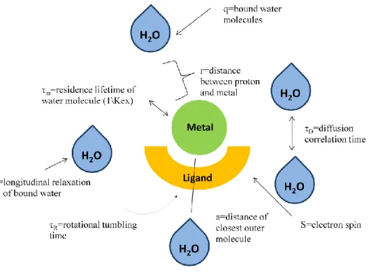
In Fig. 3 essential parameters influencing the relaxivity are reported. Contrast agents enhance both longitudinal (1/T1) and transverse (1/T2) relaxation rates.
Factors include the number of inner-sphere water molecules (q), the rotational tumbling time (τR), and the residence lifetime of inner-sphere water molecules (τM).
A larger inner-sphere hydration (i.e.: higher q values) leads to larger relaxivity values. Outer-sphere relaxivity also contributes to the overall relaxivity of a contrast agent. Values such as a (distance of closest approach of outer-sphere water molecules) and τD (diffusional correlation time of outer-sphere water molecules)
21
Figure 14. Variables contributing to contrast agent relaxivity
1.3.4 General features for MR Contrast Agents
In addition to nuclear relaxation properties, imaging contrast agents should be biocompatible, aside from standard pharmaceutical features such as water solubility and stability. The principal features requested to a good MRI contrast agent are:
RELAXIVITY: the efficiency with which the paramagnetic complex enhances the proton relaxation rates of water, referred to as relaxivity, must be sufficient to significantly increase the relaxation rates of the target tissue. The dose of the complex at which such alteration of tissue relaxation rates occurs must be non-toxic.
22
“IN VIVO” DISTRIBUTION: Ideally, to be of diagnostic value, the complex should be localized for a period of time in a target tissue. This is a basic requisite in any procedure of imaging based on complexes where detection of the agent is usually a simple function of its tissue concentration.
TOXICITY OF METAL COMPLEXES: the "in vivo" stability and the tissues clearance behaviour of intravenous metal complexes affect their acute and chronic toxicity. It is well known that the free lanthanide ions are relatively toxic at doses required for NMR relaxation rate changes thus, the dissociation of the complex must not occur. [49] It is clear that the integrity of the gadolinium(III) complex must be maintained in vivo in order to create a safe and efficacious MRI agent.
Toxic effects from a metal complex can derive from:
The free metal ion and the free ligand: In the latter two cases, metabolites may be more toxic than the starting compound. The decomplexation of the metal from the ligand generally leads to a higher toxicity due to the presence of the free metal in the body. [30] Dissociation of Gd(III) from an MRI contrast agent is highly undesirable. Free metal and unchelated ligands are generally more toxic than the complex itself. Free Gd(III) is known to bind strongly to serum proteins. In addition the released metal is irreversibly accumulates in the bones. The free ion manages to coordinate oxygen, nitrogen, or sulphur normally present in cell membranes macromolecules resulting in alterations of the dynamic equilibria necessary to life. Gd(III) can interfere and compete with Ca(II) binding sites whereas the toxicity of free ligands can be explained from the sequestration of common metal ions present in the body such as Ca(II).
23
L so l ML T
K [ ]
The complex: The toxicity of intact metal complexes can derive from a wide variety of effects. The injection of large quantities of the ionic complexes and appropriate counter ions induces a difference in osmolarity between intracellular and extracellular compartments. Water is drawn out of cells as a result of the osmotic gradient, causing cellular and circulatory damages. Other possible mechanisms of chelate toxicity include enzyme inhibition, nonspecific protein conformational effects, or alteration of membrane potentials. [50]
1.3.5 In vivo stability of metal complexes
The complex should be efficiently excreted from the body minutes or hours after administration; stability is required only for its lifetime. A wide range of coordinating molecules, proteins and metal ions can compete for either the paramagnetic metal ion or its multidentate ligand. An important thermodynamic point for trivalent metal ions in serum is their precipitation after the interaction with common anions like hydroxide, phosphate, or carbonate. Both phosphate and carbonate are significant with respect to in this parameter. Martell defined the solubilisation constant Ksol,(eq. 2) as the degree of conversion of the free ligand to
the metal chelate where TL is the total concentration of the ligand. Low values of Ksol show incapacity of a ligand to solubilize the ion; alternatively, it would predict
that the complex would be unstable with respect to metal ion precipitation. Very high values of Ksol would occur for a thermodynamically stable complex where no
precipitate would be present. [51]
24
1.3.6 Clinically approved Gadolinium, Manganese and Iron chelates
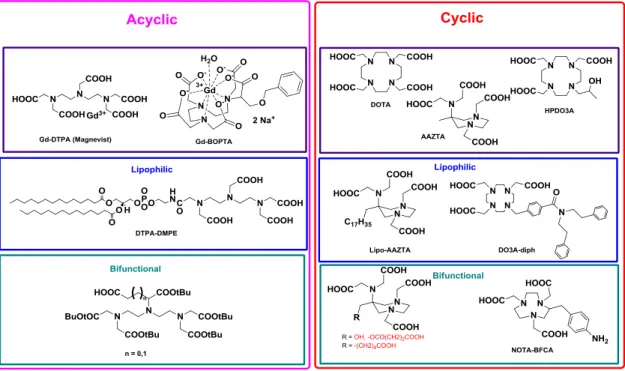
INTRAVENOUS AGENT: Acyclic and macrocyclic polyaminocarboxylic ligands form stable complexes with gadolinium leading to the clinically employed Magnevist®, Dotarem®, Omniscan® and Prohance® (Figure 15). Macrocyclic chelates are characterized by a superior thermodynamic and kinetic inertness with respect to the acyclic counterparts. In recent years, complexes of acyclic chelating agents were demonstrated to slowly dissociate and to be linked with pathologies such as Nephrogenic Systemic Fibrosis, observed in patient with kidney failure exposed to these CAs.[52]
Figure 15. Common commercially available probes
Manganese(II) can be used as an alternative paramagnetic metal ion complexed to a ligand too. Being an endogenous metal ion, its eventual release from corresponding chelates raises less concerns than with gadolinium. A paramagnetic complex based on this metal ion was previously formulated and commercialised for liver imaging: Teslascan® (Figure 16).
25
Another class of intravenous contrast agents includes nanoparticles formed by iron oxides that constitute superparamagnetic centers. Their large magnetic influence causes distortion of the local magnetic field followed by an important signal loss (T2). Currently, the most used contrast agents are
based on supermagnetic iron oxide (SPIO). After injection they are selectively absorbed by reticuloendothelial cells located in the liver, spleen and bone marrow.
ORAL AGENTS: oral agents are used for studying abdominal and pelvic structures. They are generally solutions of metal-chelate complexes (Magnevist®) or paramagnetic metal ions (ex.: ferric ammonium citrate).
1.3.7 Classification of MRI contrast agents
26
ACYCLIC CHELATING AGENTS: DTPA was one of the first chelating agents employed in clinical diagnostics with Gd (Gd-DTPA), also known as Magnevist®. It is defined as linear or acyclic octadentate ligand which coordinates Gd(III) permitting the coordination of only one molecule of water in the inner coordination sphere. Several octadentate ligands based to the DTPA scaffold were synthesized, substituting acetate moiety with different amides (DTPA-BMA) obtaining Gd-DTPA-bisamides complexes with relaxivity properties similar to DTPA. The linear structure entails low values of stability constant, while the high denticity is responsible of the relatively low values of relaxivity. Starting from the last observation, ligands characterized by lower denticity were recently investigated showing enhanced relaxivity properties.
CYCLIC CHELATING AGENTS: macrocyclic ligands as DOTA are preferable to the DTPA-like open-chain ones, as the latter show lower thermodynamic stability and kinetic inertness. During the last decades, the research in the field of cyclic chelating agents for lanthanides were widely explored: in this framework DO3A and its derivates as HP-DO3A were investigated leading to interesting results and clinically employed derivatives. Recently a new mesocyclic heptadentate ligand with efficient chelating properties was synthesized, i.e.: AAZTA. Its efficient size-match with small transition metal ions and lanthanides, the satisfactory thermodynamic and kinetic inertness award to this ligand interesting diagnostic application not exclusively in MRI, but also in PET, and SPECT techniques.
LIPOPHILIC CHELATING AGENTS: the introduction of a lipophilic alkyl chain on the skeleton of the common contrast agents supports the self-assembly into micelles or other lipid-based nanoassemblies that exhibit enhanced values of r1. In addition to this, lipophilic CAs can further form
27
increments in the relaxivity due to the reduced and lower mobility of the systems: example of lipophilic MRI-CAs are DTPA-DMPE and Gd-AAZTA-C17 with potential applications for in vivo imaging of tumours and
cardiovascular diseases.
BIFUNCTIONAL CHELATING AGENTS (BFCAs): they were introduced with the aim to add more selectivity to the contrast agents linking itself to a targeting vector. The most common BFCAs derive from cyclic and acyclic polyaminopolycarboxylic acids similar to DTPA, DOTA and AAZTA, on which a suitable versatile and reactive functional group is added, without altering the chelating properties of the coordination cage.
28
1.4 NUCLEAR MEDICINE IMAGING: SPECT AND PET
Nuclear medicine imaging is based on the principle of tracers and primarily gives images of function, including physiology, biochemistry or metabolism, by analyzing the dynamic behaviour of molecules in organs and tissues. Nowadays SPECT and PET are the dominant imaging techniques in nuclear medicine. Changes in function often anticipate changes in anatomy in various disease conditions. Accordingly, nuclear medicine imaging may be useful for early diagnosis of disease and for assessment of treatment effects in the early post-therapeutic stage. Single-photon emission computed tomography (SPECT) and positron emission tomography (PET) were the first molecular imaging modalities used clinically. [53]
PET requires the injected drug to be labelled with a positron-emitting radionuclide. When radionuclide decays, it expels a positron from its nucleus, which travels a short distance before being destroyed with an electron to release two γ rays (moving in the opposite direction), detected by the PET scanner.
SPECT requires the use of a contrast agent labelled with a γ-emitting radionuclide. These γ rays are recorded by the detectors of a dedicated gamma camera and after that the signal is processed, it can be converted into an image identifying the tracer localization. In both situations after an enough acquisition time, the data are elaborated by computer using complicated algorithms to produce images of the radiotracer’s location within the organism. PET and SPECT use radiopharmaceutical compounds to provides images. Radiopharmaceuticals are drugs containing a radionuclide, and are used routinely in nuclear medicine for the diagnosis or therapy of various diseases. [54] Intravenous injection is the favourite modality to administer radiopharmaceuticals. They are mostly small organic or inorganic compounds with a definite composition.
29
Radiopharmaceuticals are above all small organic or inorganic compounds but they can also be biomolecules such as monoclonal antibodies and antibody fragments labelled with a radionuclide.
Diagnostics and therapeutics are the two main classes of radiopharmaceuticals. Another kind of classification may be associated to the type of biodistribution: those whose distribution into the body is determined only by their chemical and physical properties; and those whose distribution is determined by their receptor binding or biological interactions. [54]
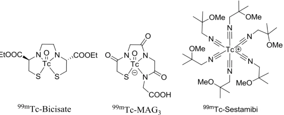
Radiopharmaceutical Trade name Primary uses
Indium-111 pentetate Indium-111 oxyquinoline Samarium-153 EDTMP Tc-99m Bicisate (ECD) Tc-99m Disofenin (DISIDA) Tc-99m Exametazine (HMPAO) Tc-99m Gluceptate Tc-99m Lidofenin (HIDA) Tc-99m Mertiatide Tc-99m Oxidronate (HDP) Tc-99m Pentetate (DTPA) Tc-99m Sestamibi Tc-99m Succimer (DMSA) Tc-99m Teboroxime Tc-99m Tetrofosmin Indium-111 DTPA® Indium-111 oxine® Quadramet® Neurolite® Hepatolite® Ceretec® Glucoscan® Technescan® HIDA Technescan® MAG3 Osteoscan® HDP Techneplex®, Technescan® Cardiolite®, Miraluma® DMSA® Cardiotec® Myoview® Imaging of CSF kinetics
Labeling leukocytes and platelets
Palliative treatment of bone pain
Cerebral perfusion imaging
Hepatobiliary imaging
Cerebral perfusion imaging
Renal imaging
Hepatobiliary imaging
Renal imaging
Bone imaging
Renal imaging and function studies
Myocardial perfusion imaging Breast tumor imaging Renal imaging
Myocardial perfusion imaging
Myocardial perfusion imaging
Table 2. List of selected examples of commercial “small molecule” radiopharmaceuticals, along with their medical applications.
30
Figure 18. Structures of selected radiopharmaceuticals based on small metal complexes.
Diagnostic radiopharmaceuticals are molecules labelled with positron-emitting isotopes for positron emission tomography (for PET) or gamma-emitting isotopes for single photon emission computed tomography (for SPECT). As a rule, diagnostic radiotracers do not have pharmacological effect and they are dosed in the range of 10-6 to 10-8 M.
The first 99Mo/99mTc generator has been developed in 1959 by the Brookhaven National Laboratory,[55] and 99mTcO42- has been used for diagnosis of thyroid
disease due to the similarity with iodide. Subsequently a lot of 99mTc complexes have been designed, synthesized and studied as fundamental radio-probes for nuclear medicine. [55,56]
1.4.1 Target-specific radiopharmaceuticals
Currently, one of the biggest issues in the molecular imaging research is the probe specificity. Development of targeted-specific radiopharmaceuticals will be the future in this field. In this strategy, radiotracers are conveyed to the diseased tissue where will interact with a specific receptor. Image quality may be increased by channelling a large amount of radiotracer in the desired tissue, then the toxicity may be reduced. The high specificity of receptor binding results in selective uptake and distribution of the radiolabelled receptor ligand at diseased tissues. A number
31
of biomolecules, including monoclonal antibodies, antibody fragments and small peptides, have been studied as ‘‘carriers’’ for radionuclides and a part of these have been approved by FDA for diagnosis or treatment of various diseases.
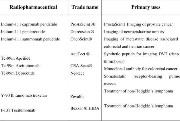
Radiopharmaceutical Trade name Primary uses
Indium-111 capromab pendetide Indium-111 pentetreotide
Indium-111 satumomab pendetide
Tc-99m Apcitide Tc-99m Arcitumomab Tc-99m Depreotide
Y-90 Ibitumomab tiuxetan
I-131 Tositumomab ProstaScint1® Octreoscan ® OncoScint® AcuTect ® CEA-Scan® Neotect Zevalin Bexxar ® HIDA
ProstaScint1 Imaging of prostate cancer Imaging of neuroendocrine tumors
Imaging of metastatic disease associated with colorectal and ovarian cancer
Synthetic peptide for imaging DVT (deep vein thrombosis)
Monoclonal antibody for colorectal cancer Somatostatin receptor-bearing pulmonary masses
Treatment of non-Hodgkin’s lymphoma
Treatment of non-Hodgkin’s lymphoma
Table 3. Selected target-specific diagnostic and therapeutic radiopharmaceuticals
.
32
1.4.2 Coordination chemistry and design of radiopharmaceuticals
The general strategies for the design of a radiopharmaceutical are:
Integrated approach: the substitution of a part of a known strong affinity receptor ligand with an "unnatural" metal chelate is the basic principle of integrated approach. In this way the conformation, size changes and receptor binding affinity remain unchanged. Unluckily this method often is complicated and the final receptor binding affinity is too low.[57]
Bifunctional approach: The bifunctional approach uses a high affinity receptor ligand as the targeting biomolecule such as monoclonal antibodies, small peptides, peptidomimetics, or nonpeptide receptor ligands. The typology and oxidation state of the radiometal are essential criteria to select macromolecules. This approach is the most used to design and development of target-specific radiopharmaceuticals.[54]
Hybrid approach: the radiometal is chelated by a tripeptide sequence (such as Gly-Gly-Gly, Cys-Gly-Gly, or Cys-Gly-Cys) containing an N4, N3S, or
N2S2 donor set.[54] the tripeptide sequence can be included in a bigger
peptide structure that can be linear or cyclic. Though this method has been used to obtain 99mTc-and188Re-labeled a-melanocyte-stimulating hormone peptide analogues the efficacy of this approach for the production of a big quantity of final radiopharmaceutical for the commercial market is yet to be proved.[58,59]
33
1.4.3 Radiometals
Traditionally non-metals isotopes as 18F, 15O, 13N, and 11C are used for PET investigation. They are produced by means of a cyclotron.
18
F-FDG is the radiolabeled analogue of glucose and today it is the most important tracer used for PET analysis. Because the supply of glucose is increased in many malignancies, the use of 18F-FDG is essential for detecting and monitoring the tumour presence or the therapy efficacy. It is an important tool for the evaluation of glucose metabolism in the heart and brain and it has been recently approved for Alzheimer's diagnosis.
Figure 20. Structure of Fludeoxyglucose (18F). Studies with 18F-FDG in a normal subjectand a subject with dementia due to Alzheimer’s disease.
Amyvid™ is another example of a molecular imaging agent containing 18
F and it is able to bind β-amyloid aggregates, and it is intended for use with PET imaging of the brain. It is a sterile, non-pyrogenic radioactive diagnostic agent for intravenous injection. It is useful to investigate the brain pathology and to estimate β-amyloid plaque density in adult patients with cognitive impairment who are being evaluated for Alzheimer’s disease and other causes of cognitive decline.
34
Figure 21. Amyvid™ (E)-4-(2-(6-(2-(2-(2[18
F]-fluoroethoxy)ethoxy)ethoxy)pyridine-3-yl)vinyl)-N-methylbenzamine.
Above mentioned isotopes are incorporated into small molecules but owing to their short half-lives and rapid clearance they are not suitable for long term analyses. [53] The time required to purify the radioisotopes, to incorporate it into a carrier agent, the packaging and transport of the resultant radiopharmaceutical across distant locations must be considered and the radiotracer half-life is a central issue. Radionuclides derived from Zr, Y, In, Ga, and Cu isotopes have a longer half-life and they have been investigated as radionuclide labels for biomolecules because they have the potential to combine a nearly optimal decay profile with the biological characteristics of the targeting molecule to become a useful radiopharmaceutical. [60]
Recently PET radioconjugates specific for tumour-associated receptors have been introduced in clinical routine use. They may extend PET applications for in vivo quantification of receptor expression and its possible changes during the course of cancer therapy. [61] In this regard the most prominent examples are 68Ga-labeled somatostatin analogues for detect and follow neuroendocrine tumours. [62,63,64] At the same time other radiometals have witnessed a growing interest for nuclear techniques as zirconium and scandium. In particular the 3.97 hours half-life of 44Sc and its high positron branching of 94.27% and promote the application of 44Sc as radiopharmaceutical for PET. [64] The ligands already used in gadolinium(III)-based magnetic resonance imaging (MRI) contrast agents may be used for the chelation of these radiometals and derivatives of DTPA and DOTA are the first choice. More recently it has been shown that DOTA derivatives are suitable ligands for scandium.[65-66]
35
1.4.4 Production of radionuclides
The radionuclides most commonly used are generated by a cyclotron or, less commonly, using a linear accelerator. The spectrum of radionuclides that may be produced is dependent of the accelerator energy. Nowadays accelerators with different energies are sold by several industries. 18F is a dominant example of radionuclide used in clinics and produced by accelerator. It is produced by proton bombardment of a stable oxygen-18 (18O) enriched water target, thanks to the nuclear reaction 18O(p,n)18F.
Radionuclide Half-life 11 C 20.385 min. 13 N 9.965 min. 15 O 2.037 min. 18 F 109.77 min. 61 Cu 3.33 hours 64 Cu 12.7 hours 86 Y 14.74 hours 124 I 4.176 days 110 In 1.2 hour 89 Zr 78.4 hours 66 Ga 9.5 hours
Table 4. Data on the half-life of radionuclides are reported
The “generator” is another tool able to provide radionuclides decaying into "daughter" radionuclides with much shorter half-lives. The "parent" radionuclide is chemically bound to an ion exchange column and the ‘daughter’ radionuclide (because of different chemical property) can be eluted and collected. In particular
36
the 68Ge/68Ge generator is remarkable for oncology: its long production useful life combined with the short half-life of 68Ga make it ideal for nuclear medicine practice. [66] Parent Radionuclide Half-life Daughter Radionuclide Half-life 62 Zn 9.26h 62Cu 9.73 min. 68 Ge 270.8days 68Ga 67.71min. 82 Sr 25.0days 82Rb 1.273 min.
Table 5. Data on the half-life of radionuclides (parent and daughter)
Figure 20. Schematic representation of post-processing technologies for commercial 68Ge/68Ga radionuclide generators is shown Figure. [67]
a) Cation exchange-based (Mainz): (1) generator elution through cation-exchange cartridge, (2) desorption of purified 68Ga using HCl/acetone or HCl/ethanol mixtures
37
b) Anion exchange-based (Hannover/Heidelberg): (3) generator elution into HCl reservoir, (4) second elution through anion-exchange catridge, (5) desorption of purified 68Gd using water c) Fractionation (Rotterdam): (6) identification of the eluate fraction and use without further purification
Kinetic and thermodynamic stability, redox properties, stereochemistry, coordination chemistry, the charge and lipophilicity are important criteria for the choice of the radiometal. In both production methods the security of final radiometals is essential. Chemico-physical analyses are routinely performed, such as impurity, pH, osmolarity, sterility and toxicity. [61,62,63]
38
REFERENCES
[1] B. Laxman, D. E. Hall, M. S. Bhojani, D. A. Hamstra, T. L. Chenevert, B. D. Ross and A. Rehemtulla; Proc. Natl. Acad. Sci. USA (2002), 99, 16551
[2] N. Gourtsoyiannis , I. McCall , M. Reiser , B. Silberman , A. Bischof Delaloye , I. Carrió , A. Cuocolo and W. Knapp; Eur Radiol. (2007) Aug;17(8):1926-30. [3] K. Hisataka, M. Ogawa, R. Alford, P. L. Choyke and Y. Urano; Chem. Rev. (2010), 110, 2620–2640
[4] A. Drevelegas and N. Papanikolaou; Imaging of Brain Tumors with Histological Correlations, Springer, (2011)
[5] M. L. James and S. S. Gambhir; Physiol. Rev. 92: 897–965, (2012)
[6] S. Aime, M. Botta, M. Fasano and E. Terreno; Chem. Soc. Rev.(1998), 27, 19 [7] M. Bottrill, L. Kwok and N. J.Long; Chem. Soc. Rev. (2006), 35, 557
[8] H-D. Liang and M. J. K. Blomley; Br. J. Radiol. (2003), 76, S140-S150 [9] E. M. C. Hillman and S. A. Burgess; Laser & Photon. Rev. (2009), 3, 159 [10] V. Ntziachristos; Annu. Rev. Biomed. Eng. (2006), 8, 1
[11] AG. Bell; Philos. Mag. J. Sci. XI 510–528, (1880).
[12] H. Kobayashi, M. Ogawa, R. Alford, P. L. Choyke and Y. Urano; Chem. Rev. (2010), 110, 2620–2640.
[13]Y. M. W. Janssen; Lab. In. Vest. (1993), 69, 261.
[14] T. Schaeffter; Prog. Drug Res. (2005), 62, 15.
[15]V. Ntziachristos, C. Bremer and R. Weissleder; Eur. Radiol. (2003), 13, 195. [16] Y.Hama, Y. Urano, Y. Koyama, M. Bernardo, P. L. Choyke and H. Kobayashi; Bioconj. Chem. (2006), 17, 1426.
[17] M. R. Longmire, M. Ogawa, Y. Hama, N. Kosaka, C. A. P. Regino, L. Choyke and H. Kobayashi; Bioconjugate Chem. (2008), 19, 1735.
[18] H. Kobayashi, M. R. Longmire and P. L. Choyke; Adv. Drug Deliv. Rev. (2013) Jul; 65(8): 1112–1119.
39
[20]R. Ando, H. Mizuno and A. Miyawaki; Science (2004), 306, 1370.
[21]C. Burda, X. Chen, R. Narayanan and M. A. El-Sayed; Chem. Rev. (2005), 105, 1025.
[22] A. Oishi and F.G. Holz; Am. J. Ophthalmol. (2015) Oct 23.
[23] A. Treibs and F. H. Kreuzer; Justus Liebigs Ann. Chem. (1968), 718, 208. [24] Handbook of Fluorescent Dyes and Probes, Wiley, (2015)
[25] L.M. Levine, M.L. Michener, M.V. Toth, B.C. Holwerda, Anal. Biochem. 247, 83-88 (1997)
[26] F. Perrin, J. Phys. et Radium (1926), 7(12), 390-401
[27] Weber, G.; in Hercules, D.M. Fluorescence and Phosphorescence Analysis. Principles and Applications, Interscience Publishers (J. Wiley & Sons), New York, pp. 217-240 (1966).
[28] A. Castro, Anal. Chem. (1997), 69(19), 3915-3920.
[29] J. Fabian, H. Nakazumi and M. Matsuoka;. Chem. Rev. (1992), 92, 1197−1226.
[30] A. Mishra, R. K. Behera, B. K. Mishra and G. B. Behera;. Chem. Rev. (2000), 100, 1973− 2011.
[31] J. M. Hales, J. Matichak, S. Barlow, S. Ohira, K. Yesudas, J.-L. Br das, J. W. Perry and S. R. Marder; Science (2010), 327, 1485−1488.
[32] Q. Bellier, N. S.Makarov, P.A. Bouit, S. Rigaut, K. Kamada, P. Feneyrou, G. Berginc, O. Maury, J. W. Perry and C. Andraud; Phys. Chem. Chem. Phys. (2012), 14, 15299−15307.
[33] X. Wang, J. Sun, W. Zhang, X. Ma, J. Lv and B. Tang; Chem. Sci. (2013), 4, 2551−2556.
[34] C. Hu, W. Sun, J. Cao, P. Gao, J. Wang, J. Fan, F. Song, S. Sun and X. Peng; Org. Lett. (2013), 15, 4022− 4025
[35] M. Beija, C.A.Mafonso and J.M.G. Martinho; Chem. Soc. Rev. (2009), 38, 2410-2433;
40
[37] O. Seitz and T. Grossmann; Eur. Pat Appl. EP 1860197 (2007). [38] D. Dickman; PCT Int. Appl. WO 2006054296 (2006).
[39] T. Woo, G. G. Miller and R. Madiyalakan; PCT Int. Appl. WO 2008011707 (2008)
[40] M. Singh and J. W. Gatson; US Pat. Appl. Pub. US 2007141581 (2007). [41] F. Bloch, W. W. Hansen and M. Packard; Phys. Rev. (1946), 69,127. [42] E. M. Purcell, H. C. Torrey and R. V. Pound; Phys. Rev. (1946), 69, 37. [43] X. Hu and D. G. Norris; Annu. Rev. Biomed. Eng. (2004), 6, 157–84. [44] M. Bottrill, L. Kwok and N. J. Long; Chem. Soc. Rev. (2006), 35, 557. [45] P. C. Lauterbur; Nature, (1973), 242, 190.
[46] R. B. Lauffer; Chem. Rev. 1907, 87, 901.
[47] E. L. Que and C. J. Chang; Chem. Soc. Rev. (2010), 39, 51. [48] I. Solomon; Phys. Rev., (1955), 99, 559.
[49] S. Aime , M. Botta, L. Frullano , S. Geninatti , G.B. Giovenzana, R. Pagliarin , G. Palmisano , FR. Sirtori and M. Sisti; J. Med. Chem. (2000);43(21):4017-24. [50] A. N. Oksendal and P-A Hals; J. Magn. Reson. Imaging, (1993), 3, 15. [51] A. E. Martell; Inorg. Chimica Acta, (1991).
[52] H.S. Thomsen, S.K. Morcos, T. Almén, M.F. Bellin, M. Bertolotto, G. Bongartz, O. Clement, P. Leander, G. Heinz-Peer, P. Reimer, F. Stacul, A. van der Molen and J. AW. Webb; Eur. Radiol. (2013) Feb;23(2):307-18.
[53] T. J. Wadas, E. H. Wong, G. R. Weisman, and C. J. Anderson; Chem. Rev. (2010), 110, 2858–2902
[54] S. Liu; Chem. Soc. Rev. (2004) 33 445-461.
[55] S. Banerjee, M. R. A. Pillai and N. Ramamoorthy; Semin. Nucl. Med. (2001), 31, 260.
[56] D. Jain; Semin. Nucl. Med. (1999), 29, 221.
[57] R. K. Hom and J. A. Katzenellenbogen; Nucl. Med. Biol. (1997), 24, 485. [58] M. F. Giblin, S. Jurisson and T. P. Quinn; Bioconjugate Chem. (1997), 8, 347.
41
[59] Y. B. Miao, D. Whitener, W. W. Feng, N. K. Owen, J. Q. Chen and T. P. Quinn; Bioconjugate Chem. (2003), 14, 1177.
[60] M. J. Welch, C. S. Redvanly; Handbook of Radiopharmaceuticals. John Wiley & Sons Inc.: Hoboken, NJ, (2003).
[61] G. Kramer-Marek and J. Capala; Curr. Pharm. Des. (2012), 18, 2657–2669. [62] P. W. Miller, N. J. Long, R. Vilar and A. D. Gee; Angew. Chem. Int. Ed. (2008), 47, 8998-9033.
[63] S. Liu; Chem. Soc. Rev. (2004), 33, 445–461.
[64] F. Roesch; Curr. Radiopharm. (2012) 5(3):187-201.
[65]J. Chen, Z. Cheng, T. J. Hoffman, S. S. Jurisson and T. P. Quinn; Cancer Res. (2000), 60, 5649.
[66]A Guide to Clinical PET in Oncology: Improving Clinical Management of Cancer Patients (IAEA Tecdoc Series No. 1605. Book)
42
CHAPTER 2
2.0 OUTLINE OF THE THESIS
The entire PhD period was devoted to the design, preparation and preliminary evaluation of novel potential probes for diagnostic imaging applications. Rather than selecting a single diagnostic technique among the large number of actually available ones, we decided to focus our research activity on the development of small-sized target compounds, holding specific molecular properties of potential interest for one or (better) more diagnostic techniques. In this contexts were developed the first two topics of this PhD thesis, both involving the study of chelating agents. Chelating agents are widely employed for the coordination of metal ions and when the metal ion plays a role in the diagnostic technique the chelator is essential to allow its safe introduction in the living organism, to mask its toxicity and optionally to enhance specific physico-chemical property and to target the complex towards selected organs and/or tissues. Acyclic chelating agents such as derivatives of the archetypal EDTA and the homolog DTPA are generally a first choice for the chelation of metal ions of diagnostic and therapeutic interest. Unsurpassed thermodynamic inertness of metal complexes of macrocyclic ligands led to the election of this family of chelating agents as the “gold standard” for in vivo applications. Nevertheless, their slow kinetic of complex formation is still an open issue in nuclear medicine, where short-lived isotopes are routinely used. The search for alternative chelating agents is very active because the properties of the currently employed compounds are far to be optimal. Much work has to be done in terms of stability, selectivity and other important features such as the easy conjugation to other molecules, (bio)degradability and not last an easy synthetic access leading to reduced costs.
43
The first topic of the present thesis involved the design and preparation of a novel chelating agent. The latter was built in a very simple process, taking advantage of a widely available commodity chemical. The structure of the hexadentate ligand includes two amines and four carboxyl groups, placed in a stereochemically controlled fashion on a substituted cyclohexane ring. The unusual arrangement of donor groups of this novel compounds was the subject of the potentiometric determination of its protonation and stability constants. The affinity data registered for different metal ions allowed us to delineate the behaviour of this original chelating agent.
The experience in the potentiometric determination of solution thermodynamic data gained during the period spent in the Department of Inorganic and Analytical Chemistry of the University of Debrecen was directly applied in the second section of the PhD activity. In this task, we explored the possibility to expand the use of a chelating agents previously developed in our research group (AAZTA) for a metal ion recently resurged to the interest of the nuclear medicine community, i.e.: Sc3+. The positron emitting isotope 44Sc is characterized by its relatively long half-life, allowing a wider time window for diagnostic imaging purposes; moreover, it can be obtained by a dedicated generator, allowing a more practical and cyclotron-independent in situ use. The coordination chemistry of Sc3+ is scarcely explored and we were prompted to evaluate the affinity of the chelating agent AAZTA towards this trivalent metal ion, using a combination of potentiometric and spectrophotometric techniques.
An additional topic of the present thesis involved the search for novel molecular scaffolds for optical imaging applications. The latter usually relies on established chromophores or luminophores, with the molecular diversity represented by large variations in the pattern of substituents.
We decided to explore different molecular bases for the development of optical imaging probes and turned our attention to two specific families of compounds: perimidines and aminoquinones.
44
Perimidines are trinuclear diazaheterocycles synthesised from 1,8-naphthalenediamines and easily functionalised on the pyrimidine moiety, allowing large structural modulations. During the PhD period a series of 2-substituted perimidines was synthesized in order to evaluate its photophysical properties. All the synthesized compounds showed significant fluorescence, and a detailed study of absorption, emission and quantum yield allowed to trace the general properties of perimidines and to learn on the influence of the substituents.
Aminoquinones, aminoquinomethides and derivatives were considered as potential scaffolds for optical imaging applications mainly due to their “push-pull” electronic features, leading to unusually high absorption wavelengths. An easy synthetic access, combined with an almost unlimited variation of the substituents and optional benzo- o hetareno- ring fusion, represent an interesting starting point for the design of chromophores reaching the near-infrared optical window of biological tissues. Exploring the chemistry of aminoquinomethides we met an unprecedented reaction sequence leading to the efficient preparation of highly coloured diaminoquinones, starting from simple compounds as vanillin. The reaction sequence was studied in detail, allowing to outline the likely mechanism and the structural versatility. Preliminary toxicological tests on diaminoquinones showed an almost complete absence of effects on different cellular lines and suggesting the general safety for these compounds.
45
CHAPTER 3
cis-IPDTA: an Original Polyaminopolycarboxylic Chelating Agent from Isophoronediamine. Synthesis and Thermodynamic Characterization of Metal Complexes
Arianna Maria Giani[a,b], Adrienn Vágner[a] Roberto Negri[b], Zsolt Baranyai[a], Giovanni Battista Giovenzana[b]*
[a] – Department of Inorganic and Analytical Chemistry, University of Debrecen, H-4010, Debrecen, Egyetem tér 1 (Hungary)
[b] – Dipartimento di Scienze del Farmaco, Università degli Studi del Piemonte Orientale “A. Avogadro”, Largo Donegani 2/3, I-28100 Novara (Italy)
Abstract
An original diaminotetracarboxylic acid chelating agent (IPDTA, cis-isophoronediamine-N,N,N’,N’-tetraacetic acid) was prepared starting from commercially available isophoronediamine. Its protonation constants and the stability of selected metal complexes were determined by potentiometric titrations. The coordination behavior of cis-IPDTA was compared with that of the linear congener BDTA and iminodiacetic derivatives.
Introduction
Polyaminopolycarboxylic acids represent the most important class of synthetic chelating agents. They are largely used in a wide range of applications, resulting in
46
a worldwide estimated consumption in the order of 105 tons/year.[1] Despite such impressive commercial interest, this huge market is dominated by just three compounds, NTA, EDTA and DTPA, depicted in Figure 1, with less than a dozen more ligands produced in industrially relevant amounts.[2]
N HOOC
HOOC
COOH HOOC N N COOH
COOH HOOC HOOC N N COOH HOOC N COOH COOH EDTA NTA DTPA
Figure 1: The polyaminopolycarboxylic acid ligands NTA, EDTA and DTPA.
The research in this area is still active as several issues in the design of complexing agents are far to be considered satisfactory, such as the specificity and selectivity towards specific metal ions[3][4] and (bio)degradability.[5] The large scale involved in the different applications of chelating agents lead to the inclusion of the production cost as a critical factor in an overall evaluation of new potential candidates.
In the search of original ligands with improved properties we recently designed and synthesized novel compounds with unusual molecular skeletons as AAZTA,[6] a mesocyclic triaminotetracarboxylic acid showing remarkable affinity for several metal ions of diagnostic and therapeutic interests,[7,8] and NORDATA, a bicyclic diaminotetracarboxylic derivative featuring a rigid molecular structure.[9]
Furthermore, heterocyclic s-triazine derivatives embodying two hydrazino-N,N-diacetic coordinating residues (TDITAs) were assembled resulting in a overall
47
heptadentate coordinating environment with a selective affinity for mid-series lanthanoids.[10,11]
Isophoronediamine (IPD) is a commodity chemical produced in large amounts as an intermediate for the preparation of isophorone diisocyanate, an important building block widely employed for the preparation of polyurethanes and in the modification of alkyd resins. IPD is used as such as hardener for epoxy resin and as intermediate in the preparation of the corresponding diisocyanate.[12] Despite its wide availability and low price, this diamine found scarce use in organic synthesis, a recent example being the preparation of a diurea-based catalyst used to perform a stereoselective variant of the Morita-Baylis-Hillmann reaction.[13]
Here we describe the synthesis and the complexation behavior of a new polyaminopolycarboxylic acid-based ligand, cis-IPDTA (cis-isophoronediaminetetraacetic acid), built by placing four coordinating carboxymethyl groups on the nitrogen atoms of the isophoronediamine skeleton.
Experimental section
2.1 - Material and methods
The chemicals used for the experiments were of the highest analytical grade. Isophoronediamine was obtained from Sigma-Aldrich; the resolution of the diastereomeric mixture to obtain diastereomerically pure racemic (±)-cis-isophoronediamine is reported in literature.[14] The metal chlorides solutions were prepared from the corresponding commercially available salts (Aldrich, 99.9%). The concentration of CaCl2, MnCl2, Pb(NO3)2, Cd(NO3)2, ZnCl2, CuCl2 and LnCl3
solutions were determined by complexometric titration with standardized Na2H2EDTA and Xylenol Orange (ZnCl2, Pb(NO3)2, CdCl2, LnCl3), murexide
(CuCl2), Patton & Reeder (CaCl2) and Eriochrome Black T (MnCl2) as indicators.
![Figure 7. Schematic representation of the basic concept of PAI. [5]](https://thumb-eu.123doks.com/thumbv2/123dokorg/4812993.49972/13.774.88.694.690.941/figure-schematic-representation-basic-concept-pai.webp)