DIPARTIMENTO DI SCIENZE DELLA VITA
DOTTORATO DI RICERCA IN SCIENZE DELLA VITA
XXXII CICLO
AUTOCHTHONOUS TUSCAN OLIVE LEAVES
(olea europaea var. Olivastra seggianesse) AS ANTIOXIDANT,
ANTIMICROBIAL AND IMMUNOMODULATORY SOURCE FOR
BIOMEDICINE AND TISSUE ENGINEERING
Settore Scientifico Disciplinare:
MED/11
Relatore: Prof. Rossella Di Stefano
Correlatore: Prof. Serena Danti
(Dept. of Surgical, Medical and Molecular Pathology (Dept. of Civil and Industrial Engineering) and Critical Care Medicine)Coordinatore: Prof. Massimo Valoti
Tesi di: Jose Gustavo De La Ossa Guerra
iii ABSTRACT
Background. Olea europaea is one of the most ancient trees of the Mediterranean region. Olive leaf extracts (OLE) have aroused interest in researchers from different scientific disciplines mainly due to the distinctive phenolic composition allegedly related to potent biological activities. Objectives. The aim of the present s study was to evaluate: A) the properties of OLE extracted from the Tuscan Olea europaea to protect endothelial cells against oxidative stress generated by reactive oxygen species (ROS) in 2D culture and innovative 3D scaffold (P(VDF-TrFE)); B) to investigate the antimicrobial effect of OLE versus Cold Atmospheric Plasma (CAP) technology or their combination against pathogens, i.e., Escherichia coli, Staphylococcus aureus and
Listeria innocua, grown to exponential (24h) or stationary (6h) phase; C) to characterize electrospun
OLE-loaded PHBHV based composite fibers for wound healing applications; D) to evaluate the ability of OLE incorporated in Polyhydroxyalkanoates (PHAs) fibers to modulate the release of cytokines from healthy Human Keratinocytes (HaCaT). Methods: OLE total polyphenols (TPs) were characterized by the Folin– Ciocalteu method. Endothelial cells were grown in conventional cultures (two-dimensional, 2D) and on three-dimensional scaffold fabricated via electrospinning. Cell viability and ROS production after H2O2 insults were
evaluated by WST-1, AlamarBlue and Probe CM-H2DCFDA assays. The E.coli, S.aureus and L.innocua
growth were assessed by CFU/mL and CAP methods; Real-time polymer chain reaction (PCR) was carried out to evaluate the immunomodulatory properties; Fourier Transform Infrared Spectroscopy (FT-IR) Analysis was performed to discriminate the chemical composition in both electrospun fibers. Moreover, Gel Permeation Chromatography (GPC) to allow biodegradation analysis, SEM microscopy to study fiber morphology and HPLC to carry out the release study, were performed. Results: OLE TP content was 23.29 mg of gallic acid equivalent (GAE)/g, and oleuropein was the principal compound. The dose-dependent viability curve highlighted the absence of significant cytotoxic effects at OLE concentrations below 250 µg GAE/ml TPs. OLE preconditioning at 100 µg GAE/ml was protective against ROS in both models. The combination of CAP and OLE resulted in substantial microbial inactivation against all strains at exponential phase showing a complete inactivation. OLE possess a significant anti-inflammatory activity, downregulating the expression of all proinflammatory cytokines, upregulating IL-8, IL- 1α and TNF-α in HaCaT model. Conclusion. OLE possess a significant antioxidant and anti-inflammatory activities; PHBHV+OLE retains OLE beneficial effects and represents a high-value 3D scaffold with great potential in tissue regeneration. CAP and OLE have synergistic antibacterial activity; therefore, CAP technology in combination with OLE can be utilized for effective decontamination when required for example in wound healing and biomedical devices.
Keywords: Olea europaea, Olive Leaves Extract (OLE), Endothelial cells, ROS, Scaffold,
Polyhydroxyalkanoates (PHA), PHBHV, Gene expression, Biodegradation, electrospinning, Cold Atmospheric Plasma (CAP), anti-inflammatory, wound healing, antioxidant, tissue regeneration.
iv RIASSUNTO
Background. L'Olea europaea è uno degli alberi più antichi della regione mediterranea. Gli estratti di foglie di olivo (Olive Leaf Extracts, OLE) hanno suscitato interesse nei ricercatori di diverse discipline scientifiche principalmente a causa della particolare composizione fenolica, presumibilmente correlata alla sua potente attività biologica. Obiettivi. Lo scopo di questo studio è stato valutare: a) le proprietà degli OLE di Olea
europaea Toscana per proteggere le cellule endoteliali dallo stress ossidativo generato dalle specie reattive
dell'ossigeno (ROS), sia in coltura 2D sia in innovativi scaffold 3D (P (VDF-TrFE)); b) studiare l'effetto antimicrobico di OLE rispetto alla tecnologia Cold Atmospheric Plasma (CAP), o la loro combinazione, contro agenti patogeni, ad esempio Escherichia coli, Staphylococcus aureus e Listeria innocua, cresciuti a livello esponenziale o in fase stazionaria; c) caratterizzare fibre composite elettrofilate a base di PHBHV caricate con OLE per applicazioni nella guarigione delle ferite; d) valutare la capacità di OLE incorporato nelle fibre di poliidrossialcanoati (PHA) di modulare il rilascio di citochine da cheratinociti umani sani (HaCaT). Metodi: I polifenoli totali (total polyphenols, TP) di OLE sono stati caratterizzati con il metodo di Folin-Ciocalteu. Le cellule endoteliali sono state coltivate in colture convenzionali (cioè bidimensionali, 2D) e su impalcature tridimensionali fabbricate tramite elettrofilatura. La vitalità cellulare e la misurazione dei ROS dopo lo stress indotto da H2O2 sono state eseguite tramite WST-1, alamar Blue e la sonda CM-H2DCFDA per ROS. La
crescita di E. coli, S. aureus e L. innocua è stata valutata mediante il test di unità (CFU) formanti colonie (espressa in CFU/ml) e CAP. È stata eseguita la reazione a catena della polimerasi in tempo reale (real-time PCR) per valutare le proprietà immunomodulatorie; analisi di spettroscopia infrarossa in trasformata di Fourier (FT-IR), per discriminare la composizione chimica in entrambe le fibre elettrofilate; cromatografia a permeazione su gel (GPC) per consentire l'analisi della biodegradazione; microscopia SEM per lo studio della morfologia delle fibre ed in fine, HPLC per eseguire lo studio di rilascio. Risultati: il contenuto in TP di OLE era 23,29 mg di acido gallico equivalente (GAE)/g e l'oleuropeina era il composto principale. La curva di vitalità dose-dipendente ha evidenziato l'assenza di effetti citotossici significativi a concentrazioni di OLE inferiori a 250 µg GAE/ml di TP. Il pretrattamento di 100 µg GAE/ml di TP di OLE ha avuto effetti protettivi sullo stress ossidativo indotto da H2O2 in entrambi i modelli (2D e 3D). La combinazione di CAP e OLE ha
determinato una sostanziale inattivazione microbica contro tutti i ceppi in fase esponenziale mostrando una completa inattivazione. OLE ha dimostrato una significativa attività antinfiammatoria, sottoregolando l'espressione di tutte le citochine proinfiammatorie e sovraregolando IL-8, IL-1α e TNF-α nel modello HaCaT. Conclusione: OLE possiede una significativa attività antiossidante e antinfiammatoria; la fibra a base di PHBHV + OLE preserva gli effetti benefici di OLE e rappresenta uno scaffold 3D di alto valore con un grande potenziale nella rigenerazione dei tessuti. CAP e OLE possiedono un'attività antibatterica sinergica; pertanto, la tecnologia CAP in combinazione con OLE può essere utilizzata per una decontaminazione efficace, ad esempio nella guarigione delle ferite ed in dispositivi biomedicali.
Parole chiavi: Olea europaea, estratto di foglie di olivo, cellule endoteliali, ROS, scaffold,
poliidrossialcanoati, PHBHV, espressione genica, biodegradazione, elettrofilatura, plasma atmosferico freddo, antinfiammatorio, cicatrizzante, antiossidante, rigenerazione dei tessuti.
v
To my dear
Mother, Father and Siblings
To my team of the Cardiovascular Research Laboratory and Oto-Lab Group
To the Real Amazing Spiderman, a.k.a. Gianfranco Serecchia
vi
ACKNOWLEDGEMENTS
The Doctorate Degree Program, PEGASO, Scholarship of the Tuscany Region, Italy, University of Siena is gratefully acknowledged for supporting these research activities.
My personal acknowledges are given to:
• The professors and the research groups at Cardiovascular Research Laboratory, Multifunctional Bio-Ecocompatible Materials Laboratories (University of Pisa) and Oto Lab (Azienda Ospedaliero-Universitaria Pisana), for their continuous support to my research activity.
• Dr. Chiara Piccini (University of Siena, Italy) and Dr. Claudio Cantini (CNR-IVALSA, Follonica, Italy) for providing the olive leaves for this study.
• Dr. Tommaso Neri, Valentina Scalise and Delfo D’Alessandro (University of Pisa, Italy) are thanked for their technical and scientific support.
• The 3R’s Center (University of Pisa, Italy) is acknowledged for supporting the development of new in vitro culture models.
• CISUP (Center of Instrument Sharing, University of Pisa) is acknowledged for scanning electron microscopy (SEM).
• Prof. Ipsita Roy & RoyLab, University of Sheffield, UK, for providing lab-synthesized polyhydroxyalkanoates.
• Dr. Caterina Cristallini, CNR-IPCF, Pisa, and Dr. Niccoletta Barbani, Dept. of Civil and Industrial Engineering, University of Pisa, for their support to biomaterials characterization. • Prof. Maria Digiacomo and Prof. Marco Macchia, Dept. of Pharmacy, University of Pisa, for
their support to olive leaf extract characterization.
• The European Union Horizon 2020 research program (BBI-H2020), PolyBioSkin project, grant number G.A. 745839.
vii
• Prof. Eirini Velliou, Dr. Hani El-Hari and Lisa Puth at the Bioprocess and Biochemical Engineering (BioProChem) group, Department of Chemical and Process Engineering, Faculty of Engineering and Physical Sciences, University of Surrey, Guildford, UK to permit development new capacities in Microbiology.
• The Dept. of Experimental Medicine, University of Campania, Naples, for all the collaboration in the execution of this project.
viii
List of Figures
Chapter 1.
Figure 1. Chemical structure of polyphenols……….…………...………....6
Figure 2. Predicted routes for absorption of dietary phenolics ………...….7
Figure 3. Structure of Oleuropein………...7
Figure 4. ROS resources during cardiovascular diseases………...9
Figure 5. Risk factor and Endothelial dysfunction………..…....10
Figure 6. Effect of oleuropein on interplay between oxidative stress, autophagy and inflammation..11
Figure 7. General schematic of wound healing………..14
Figure 8. The Wound Healing Phases………....15
Figure 9. Characteristics of OLE in the immune system studied…………...………....18
Figure 10. The TE Triad………....….23
Figure 11. Granules of PHA generate by the bacteria and its general structure ………25
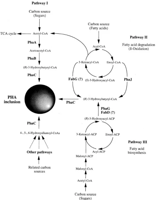
Figure 12. Metabolic Pathway of PHA Biosynthesis……….27
Figure 13. Chemical Structure of P(VDF–TrFE)………...30
Chapter 2. Figure 1. Electrospinning process and fiber morphology………...40
Figure 2. Dose- and time-dependent cell metabolic activity………..40
Figure 3. Antioxidant effect of OLE………..……….41
Figure 4. ROS production by HUVECs………..41
Figure 5. Metabolic activity of HUVECs on scaffolds and antioxidant activity of OLE on cell/scaffold constructs……….………...42
Figure 6. Results of ROS analysis performed on the 3D model……….43
Figure 7. SEM and fluorescence analyses of P(VDF-TrFE)/HUVEC construct……….………44
Chapter 3. Figure 1. Effects of OLE on Escherichia coli………..………51
Figure 2 Effect of OLE on Staphilococcus aureus………..………...52
Figure 3. Effect of OLE on Listeria innocua………..………....52
Figure 4. The effects of CAP (1 min exposure time) or OLE on E. coli………53
Figure 5. The effects of CAP (1 min exposure time) or OLE on S. aureus………...54
Figure 6. The effects of CAP (1 min exposure time) or OLE on L. innocua………...54
ix Chapter 4
Figure 1. Anulus graph showing the percentage of different polyphenols in OLE………64
Figure 2. SEM micrographs of (A) PHBHV, and (B) PHBHV/OLE electrospun fibers………64
Figure 3. FT-IR spectra of OLE, PHBHV and PHBHV/OLE…..………..………....65
Figure 4. Results of Chemical imaging analysis…………..………66
Figure 5. Graph showing molecular weight loss as obtained from GPC of PHBHV for 28 days and PHBHV/OLE fiber ………..………..……….67
Figure 6. SEM micrographs of PHBHV/OLE under degradation………...67
Figure 7. Cumulative release of polyphenols from PHBHV/OLE fiber...69
Figure 8. Cytocompatibility of PHBHV and PHBHV/OLE scaffolds………...69
Chapter 5 Figure 1. SEM micrographs of PHA electrospun fibers with and without OLE………77
Figure 2. . Bar graphs showing IL-1 expression by HaCaT cells at 6 and 24 h………...78
Figure 3. Bar graph showing IL-6 expression by HaCaT cells at 6 and 24 h
.
.………..79Figure 4. Bar graph showing IL-8 expression by HaCaT cells at 6 and 24 h. ……...………79
Figure 5. Bar graph showing TNF-α expression by HaCaT cells at 6 and 24 h……….80
x
List of Tables
Chapter 1
Table 1. Uses/applications of plant components in biomedical fields………4 Table 2. State-of-the.art of application of OLE in cardiovascular diseases and related…………..…13 Table 3. OLE and wound healing studies………16 Table 4. Plants containing antimicrobial compounds against Gram-positive and Gram-negative bacteria………..….19 Table 5. PHA applications in regenerative medicine……….28 Table 6. Studies conducted with (P(VDF–TrFE) in biomedical applications………31 Chapter 2
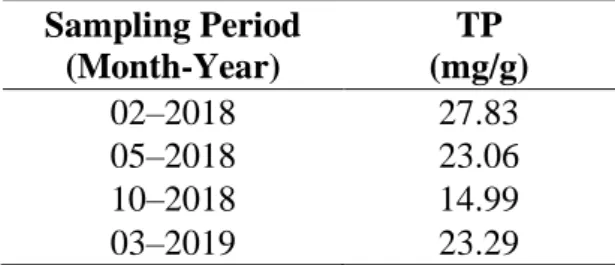
Table 1. Quantification of TP in olive leaf lyophilized extract in different periods……….…….…39 Chapter 3
Table 1. Concentrations of Polyphenols in OLE………51 Chapter 4
Table 1. Content of main phenols in OLE ………..…..63 Table 2. Weight loss of PHBHV/OLE fibers in different media………...………...67 Table 3. Cumulative release (µg) of the main OLE phenolic compounds in a 4 cm2 square of PHBHV/OLE fiber mesh………..………..68 Chapter 5
Table 1. Real-time PCR conditions for HaCaT cells………75 Table 2. Results of metabolic activity (AlamarBlue test)……….77
xi ACRONYMS
AA Acrylic Acid
ACN Acetonitrile
ADP Adenosin diphosphate
AMPK 5’ adenosine monophosphate-activated protein kinase
CAP Cold Atmospheric Plasma
CM-H2DCFDA 5-(and-6)-chloromethyl-2’,7’-dichloro-di-hydro-fluorescein diacetate, acetyl
COX Cyclooxygenase
CRP C Reactive Protein
CVD Cardiovascular Diseases
CRP C-Reactive Protein
DAPI 4’,6-Diamidino-2-phenylindole dihydrochloride
DBD Dielectric Barrier Discharge
DCM Dichloromethane
ECM Extracellular Matrix
EGM-2 Endothelial Cell Growth Medium
EVOO Extra Virgin Olive Oil
FBS Fetal Bovine Serum
GAE Gallic Acid Equivalent
H2O2 Hydrogen Peroxide
HaCaT Human keratinocytes
Hcy Homocysteine
HEPES 4-(2-hydroxyethyl)-1-piperazineethanesulfonic acid
HPLC High-Performance Liquid Chromatography
HT Hydroxytyrosol
HUVEC Human Umbilical Vein Endothelial Cells ICAM-1 Intercellular Adhesion Molecule 1
IL-1β Interleukin-6
iNOS Inducible form of nitric oxide synthase
M199 Medium 199
MD Mediterranean Diet
MeOH Methanol
MEK Methyl Ethyl Ketone
MMP-9 Metalloproteinases-9
mTOR Mammalian target of rapamycin
Na2CO3 Sodium Carbonate
NCD Non-communicable diseases
NF-kB Nuclear Factor Kappa-Light-Chain-Enhancer of Activated B Cells
NO Nitric Oxide
LC3 Autophagy-specifific marker
LDPE Low Density Poly-Ethylene
OLE Olive Leave Extract
oxLDL Oxidized low-density lipoprotein
PARP1 Poly (ADP-ribose) polymerase
PBMC Peripheral Blood Mononuclear Cells
PET Polyethylene Terephthalate
P(3HO –co-3HD) Poly(3-hydroxyoctanoate-co-3-hydroxydecanoate) PHBHV Poly(hydroxybutyrate-co-hydroxyvalerate)
xii
ROS Reactive Oxygen Species
RNS Reactive Nitrogen Species
SAR Structure Activity Relationship
SEM Scanning Electronic Microscopy
SIRT-1 NAD-dependent deacetylase sirtuin-1
SHR Spontaneously Hypertensive Rats
TE Tissue Engineering
TFEB Transcription factor EB
TP Total Polyphenol
UV Ultraviolet
WST-1 4-[3-(4-iodophenyl)-2-(4nitrophenyl)-2H-5-tetrazolium]-1,3-benzenedisulfonate ester
xiii INDEX Abstract………...………...iii Riassunto………...……….iv Dedication………....v Acknowledgements………....vi List of Figures………vii List of Tables………..ix Acronyms………....x CHAPTER 1 STATE-OF-THE-ART………...1 1. Introduction ……….…..………...……....1
1.1 Uses of Plants: Medicinal Approach ……….…...1
1.2 Olive Leaves Extract: Uses, Characteristics and Properties ……….………..….5
1.2.1 Endothelial Dysfunction and OLE….………...8
1.2.1.1 Endothelial dysfunction: HUVEC as model system..……....………...11
1.2.2 Wound Healing ………...……….……….……..14
1.2.3 Immunomodulation and OLE……….……….……...……...…..17
1.2.4 OLE as Antimicrobial Resource……….……….………19
1.2.4.1 Cold atmospheric Plasma…..……….……….20
1.3 Tissue Engineering…..………..………...………..….22
1.3.1 Polyhydroxyalkanoates………...25
1.3.2 Poly(vinylidene fluoride–trifluoroethylene) biopolymer in electrospun nanofiber…...29
1.4 Research Objectives…..………...……...32
CHAPTER 2 EXPERIMENTAL SECTION I……….….33
2. Autochthonous Tuscan OLE as antioxidant source for biomedicine…...…………..………34
xiv
2.1.1 Materials………...………...34
2.1.2 Sample Preparation ………...35
2.1.3. OLE Polyphenol Characterization………...……….35
2.1.4. HPLC Characterization…….…………..………..35
2.1.5. Endothelial Cell Isolation and Culture………...….………..36
2.1.6. Scaffold Fabrication and Characterization………...……..………...36
2.1.7. Investigation of OLE Effects………...……….36
2.1.7.1. 2D HUVEC Model………...………..36
2.1.7.2. 3D HUVEC Model………...………..37
2.1.8. DAPI Staining………….………..………....38
2.1.9. Statistical Analysis……...………...38
2.2 RESULTS………...………..39
2.2.1 Characterization of OLE from Tuscan Olea europaea…………..………39
2.2.2 Scaffold Characterization………..39
2.2.3 Dose- and Time-Dependent Effect of OLE on 2D Culture Model...40
2.2.4 Antioxidant activity of OLE………...………...40
2.2.5 ROS production………..…………...…....41
2.2.6 OLE effect in 3D culture model ………..………...……..42
2.2.7 3D model characterization……..………...44
2.3 DISCUSSION………...44
CHAPTER 3 EXPERIMENTAL SECTION II………48
3. Combined antimicrobial effect of cold atmospheric plasma (CAP) and bio-waste OLE against bacterial pathogens……….………49
3.1. MATERIALS AND METHODS………...….49
3.1.1 CAP Set-up……….…………...49
3.1.2 Inoculum of OLE ………..49
3.1.3 Effect of OLE and CAP/OLE on E. coli, S. aureus and L. innocua…..………...…….50
3.1.4 Statistical Analysis……….………...50
3.2 RESULTS AND DISCUSSION………....…..50
3.2.1 Polyphenol Characterization……….……….50
3.2.2 Effect of OLE on E. coli, S. aureus and L. innocua………...…..51
3.2.3 Effect of CAB on inactivation of E. coli, S. aureus and L. innocua .………53
3.3 DISCUSSION………...54
CHAPTER 4 EXPERIMENTAL SECTION III………..….58
xv
4. Polyhydroxyalkanoates: Electrospun fibers incorporating OLE as bio-based scaffolds for
wound healing……….………...59
4.1 MATERIALS AND METHODS………..………59
4.1.1 Fabrication of Electrospun PHBHV/OLE fiber meshes………...59
4.1.2 OLE Characterization by HPLC…….………60
4.1.3 Characterization of PHBHV/OLE electrospun fibers…………..…………....…...61
4.1.4 Degradation study of PHBHV/OLE electrospun fiber………...61
4.1.5 In vitro phenol release study ………..62
4.1.6 Cytocompatibility of PHBHV/OLE Fiber Scaffold …...………...62
4.2 RESULTS …………..………....63
4.2.1 OLE characterization………..64
4.2.2 Characterization of PHBHV/OLE fiber meshes……….64
4.2.3 Polyphenol release from PHBHV/OLE fiber meshes ………..……….68
4.2.4 Cytocompatibility of the scaffolds using HFFF2 cells ………..69
4.3 DISCUSION ……….……….……70
CHAPTER 5 EXPERIMENTAL SECTION IV……….…………...73
5. Immunomodulatory Effects of OLE incorporated in the scaffold………...74
5.1 MATERIALS AND METHODS……….……….…..74
5.1.1 Scaffolds fabrication……….…………...74
5.1.2 Epidermal cell culture and viability assay………..75
5.1.3 Evaluation of Immunomodulatory Properties……….………75
5.2 RESULTS……….……….….76
5.2.1 PHBHV and PHB/PHOHD Scanning Electronic Microscopy…...………...76
5.2.2 Cell metabolic activity …...………...……….……77
5.2.3 Cytokine Expression.……….……….78
5.2.4 Indirect antimicrobial activity……….80
5.3 DISCUSSION………...81
CHAPTER 6 CONCLUSIONS AND FURTHER PROSPECTIVES ………..86
xvi
CHAPTER 1
1
1. INTRODUCTION
1.1 Uses of Plant: Medicinal Approach
In many countries in the world, people still rely on traditional plant-based medicines for their primary healthcare. This is especially true for many rural communities in Africa, parts of Asia, and Central and South America, where plants and knowledge of their traditional use are accessible and affordable. In other countries, many of these traditional plant-based medicines are being integrated through regulations into mainstream health systems (Allkin, B., 2017). A variety of plant extracts have been used in traditional medicine over the centuries and some plants and herbs have been identified for provide evidence in the prevention or treatment in many diseases including cardiovascular diseases (CVD), diabetes, weight loss, inflammation, etc. (Poswal, F. et al., 2019), due to the presence of different polyphenols.
Polyphenols, both flavonoids and phenolic acids, are ubiquitous in plants and are mainly present as glycosides. Upon their chemical structure, polyphenols produce different colours from yellow-green to blue-red, especially in flowers. Therefore, they can act as attractants for honeybees or attraction for other pollinators. Moreover, polyphenols can implement structural functions, such as the defence against ultraviolet (UV) radiation or the protection of plants against microbial invasion and herbivores (Harborne and Williams 2000; Manach, C et al. 2004; Taiz and Zeiger, 2009). For human nutrition, polyphenols are well-known for their health properties. Dependent on their chemical structure, polyphenols are antioxidant (or pro-oxidant), anti-inflammatory, or anti-carcinogenic. However, their bioavailability is generally low (Sahin, S et al. 2018), due to the sugar moiety of the flavonoid glycoside can affect the bioavailability, even though only aglycones can be absorbed.
In Western-style diets, chlorogenic acid due to coffee consumption is the major source of polyphenols. Moreover, the quercetin and kaempferol are the most consumed flavonoids and are present in high concentrations in, for example, onion or Brassica vegetables.
Many plant species, such as Crataegus species, comprehends to be valuable in treating CVDs particularly angina heart failure and hyperlipidemia (Chan, Q. et al., 2002). The leaves, flowers and fruits of Crataegus species contain varying quantities of a number of biologically active substances such as oligomeric, procyanidins, flavonoids and catechins. The extract is suggested to have antioxidant properties and inhibits the formation of thromboxane (Bahorun, T. et al.,1994; Vibes, J. et al.,1994 ). In traditional Chinese medicine the fruit of the hawthorn (usually Crataegus) is ideally used for many indications including digestive disorders and for lowering cholesterol and blood pressure (Chang, W.T. et al., 2005). C. oxyacantha berry extract antagonized dietary induced increases in cholesterol triglycerides and phospholipid levels in D fractions and very low density
2
lipoprotein fractions in rats (Shanthi, S. et al., 2004). Thus, it could inhibit the progression of atherosclerosis (Rajendran, S. et al., 1996). Other species like Rubus spp. (Rosaceae) leaves have been used as antimicrobial, anticonvulsant, and muscle-relaxing agents. Morus alba (Moraceae) leaves have been applied in these examples clearly demonstrate that leaves can be at least equally interesting as fruits or other parts of plants. Importantly, they are also a much more accessible source of polyphenols than fruits. Abelmoschus esculentus also has a beneficial impact on the cardiovascular system. Sabitha et al. demonstrated that A. esculentus peel and seed powder reduced the blood glucose level and improved the lipid profile level in diabetic male Wistar rats (Sabitha, V et al., 2011). Tea (Camellia sinensis, Theaceae), the most important non-alcoholic beverage in the world, has been extensively studied for its putative disease preventive effects. Tea leaves are well known as an abundant source of polyphenols with strong antioxidant properties (Gramza, A. et al., 2005). Regular tea intake prevents cancer and vascular disorders and regulates the digestive system (Khan, N. and Mukhtar, H., 2013). Animal, as well as human studies, point to the cardioprotective effect of black tea by lowering cholesterol level. Also, the ability of black tea to decrease some inflammatory markers and mediators expressed by endothelium clearly points to the beneficial prosperities of this plant towards the vascular system (Dudzińska, D. et al., 2015). Also, Urtica dioica (Urticaceae) roots and leaves are a remedy for hypertension, diabetes, prostate hyperplasia, and cancer (Ziyyat, A. et al., 1997; Konrad, L. et al., 2000). Urtica dioica possesses anti-inflammatory, anti-hyperglycaemic, antimicrobial, antioxidant, anti-ulcer, and analgesic activity (Gülçin, I. et al., 2004). Urtica dioica leaf extract, when administered before glucose loading, has demonstrated strong ability to decrease glucose level in alloxan-induced diabetic rats (Bhouham, M. et al., 2003). Extract from U. dioica also appears to be an effective scavenger of free radicals, including superoxide anion radicals and hydrogen peroxide. Allium sativum (Amaryllidaceae), known since ancient times for its healing properties, may be a beneficial agent for the treatment of CVD (Agarwal, K.C. et al., 1996; Neil, H.A., 1996). Allium sativum can normalize plasma lipids, enhance fibrinolytic activity, inhibit platelet aggregation, and reduce blood pressure and blood glucose level. In experiments on platelet aggregation evoked by Adenosin diphosphate (ADP) , collagen, or arachidonic acid, Hiyasat et al. compared the anti-platelet activity of methanolic and aqueous extracts isolated from the leaves of A.
ursinum and sativum. Alcoholic extracts of both species and an aqueous extract of A. sativum most
efficiently inhibited ADP-induced platelet aggregation, while an aqueous extract of A. ursinum inhibited platelet aggregation, but the effect did not depend on the type of platelet agonist (Hiyasat, B. et al., 2009). Other plants like Ginkgo extract also demonstrates cardioprotective activity, which has been demonstrated in an experiment with HgCl2-induced oxidative damage in Wistar albino male and female rats. While HgCl2 has been shown to significantly increase thromboplastic activity and malondialdehyde levels or decrease glutathione levels in serum and tissue samples, this effect has
3
been effectively reversed by Ginkgo leaf extract (Tunali-Akbay, T. et al., 2006). Dicksonia sellowiana (Dicksoniaceae) is a common tree in Central and South America; its leaves are used in a folk medicine to treat scabies, pruritus, parasitic diseases, and asthma. Hydroalcoholic extract of D. sellowiana (HEDS) decreases hypertension and induces endothelium-dependent relaxation in spontaneously hypertensive rat (SHR) aortic rings. Hydroalcoholic extract of D. sellowiana induces aortic relaxation by activation of muscarinic receptors and stimulation of the NO pathway in SHR rat aortic endothelium. Others like sweet cherry (Prunus avium) and tart or sour cherry (Prunus cerasus) have global trading importance and are now growing widely around the world. Although the commercial cultivation of sweet cherries is more difficult and expensive than that of tart cherry, the first ones are prized for their excellent taste and nutritious nature, thanks to the high presence of sugars and the low level of acidity. Depending on pre- and post-harvest factors, sweet cherry also contains high levels of nutrients, including glucose, fructose, vitamin C, and bioactive compounds, which present various health benefits (Chockchaisawasdee, S. et al., 2016). Several clinical studies have shown that cherry fruit or juice consumption plays an important role in the struggle against inflammatory diseases (Kelley, D.S., 2018). Moreover, a recent study confirmed the protective effects of cherry extracts to reduce ROS accumulation in vitro and the main role of its polyphenols in inflammation reduction and endothelial dysfunction improvement (Beconcini, D. et al, 2019). One of the most common plant known for its properties in the human health is the olive tree, mainly the oil olive, fruit, and leaves (Romani, A. et al., 2019). Specifically, the use of the olive leaves (extract) has been explored due to its phenolic composition. Such bioactive ingredients are used in medicines, pharmaceuticals or cosmetics, to improve the human health as well as to develop functional foods (Abaza, L. et al., 2015). The main uses of plant components in the biomedical field are summarized in table 1.
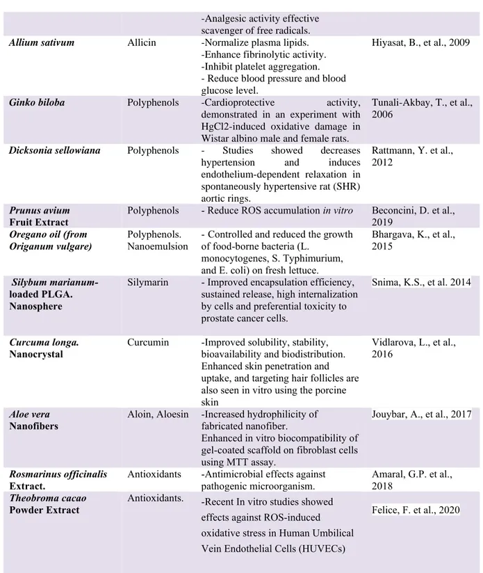
Plant species Bioactive component
Biomedical application/Uses References
Crataegus spp. Extract Oligomeric, procyanidins, flavonoids and catechins
-Angina heart failure and hyperlipidemia Chan, Q. et al., 2002 Rubus spp Leaves Extract Polyphenols -Antimicrobial -Anticonvulsant, -Muscle-relaxing agents. Rajendran, S., et al., 1996 Abelmoschus esculentus Peel and seed extract
Polyphenols - Peel and seed powder reduced the blood glucose level and improved the lipid profile level in diabetic male Wistar rats.
Sabitha, V., et al., 2011
Camellia sinensis (Tea) Leaves Extract
Antioxidants -Studied for its putative disease preventive effects.
- Studies for vascular disorders and regulates the digestive system
Gramza, A., et al., 2005
Khan, N., and Mukhtar, H., 2013 Urtica dioica Extract. Polyphenols -Anti-inflammatory. -Anti-hyperglycaemic, -Antimicrobial -Antioxidant -Anti-ulcer
Gülçin, I., et al., 2004
Bhouham, M., et al., 2003
4
Table 1. Uses/applications of plant components in biomedical field. -Analgesic activity effective
scavenger of free radicals.
Allium sativum Allicin -Normalize plasma lipids. -Enhance fibrinolytic activity. -Inhibit platelet aggregation. - Reduce blood pressure and blood glucose level.
Hiyasat, B., et al., 2009
Ginko biloba Polyphenols -Cardioprotective activity, demonstrated in an experiment with HgCl2-induced oxidative damage in Wistar albino male and female rats.
Tunali-Akbay, T., et al., 2006
Dicksonia sellowiana Polyphenols - Studies showed decreases hypertension and induces endothelium-dependent relaxation in spontaneously hypertensive rat (SHR) aortic rings.
Rattmann, Y. et al., 2012
Prunus avium
Fruit Extract Polyphenols - Reduce ROS accumulation in vitro Beconcini, D. et al., 2019
Oregano oil (from Origanum vulgare)
Polyphenols. Nanoemulsion
-Controlled and reduced the growth of food-borne bacteria (L.
monocytogenes, S. Typhimurium, and E. coli) on fresh lettuce.
Bhargava, K., et al., 2015
Silybum marianum-loaded PLGA. Nanosphere
Silymarin - Improved encapsulation efficiency, sustained release, high internalization by cells and preferential toxicity to prostate cancer cells.
Snima, K.S., et al. 2014
Curcuma longa.
Nanocrystal Curcumin -Improved solubility, stability, bioavailability and biodistribution. Enhanced skin penetration and uptake, and targeting hair follicles are also seen in vitro using the porcine skin
Vidlarova, L., et al., 2016
Aloe vera
Nanofibers Aloin, Aloesin -Increased hydrophilicity of fabricated nanofiber.
Enhanced in vitro biocompatibility of gel-coated scaffold on fibroblast cells using MTT assay.
Jouybar, A., et al., 2017
Rosmarinus officinalis Extract.
Antioxidants -Antimicrobial effects against pathogenic microorganism.
Amaral, G.P. et al., 2018
Theobroma cacao
Powder Extract Antioxidants. -Recent In vitro studies showed effects against ROS-induced oxidative stress in Human Umbilical Vein Endothelial Cells (HUVECs)
5
1.2 Olive Leaves Extract: Uses, Characteristics and Properties
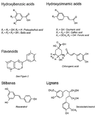
Olea europaea is one of the most ancient trees of the Mediterranean region. Olive leaf extracts (OLE) have been an interest in researchers from different scientific disciplines mainly due to the distinctive phenolic composition allegedly related to potent biological activities (Goulas V., et al., 2009). Olive leaves contain considerable bio-phenols as the other parts of the olive tree (Sahin, S., and Bilgin, M., 2017). OLE is used principally as a food supplement and an over-the-counter drug for a variety of beneficial effects, including anti-inflammatory and anti-atherosclerotic ones. Phenolic compounds in OLE are secondary metabolites of plant, which play important roles in disease resistance (Servili and Montedoro, 2002; Ryan, D., et al., 1999), protection against pests and species dissemination. The interest in these compounds is related with their antioxidant activity and promotion of health benefits (Ryan, D et al., 2002). The Mediterranean diet's healthy effects can in particular be attributed not only to the high relationship between unsaturated and saturated fatty acids in olive oil but also to the antioxidant property of its phenolic compounds. The main phenolic compounds, hydroxytyrosol (HT) and oleuropein, which give extra-virgin olive oil its bitter, pungent taste, have powerful antioxidant activity both in vivo and in vitro. (Manna et al., 1999; Servili et al., 2004; De la Torre-Carbot, et al., 2005). Besides, literature reports several compounds of the class of flavonoids that may occur in appreciable amounts (Savournin, et al., 2001) and become the major leaf constituents in certain phenological stages (Papoti & Tsimidou, 2009). Consequently, phenolic compounds have antioxidant activity, being attributed to the capacity of scavenging free radicals, donating hydrogen atoms, electrons, or chelate metal cations, characteristics studies in molecules as Quercetin and Catechin (Afanas, I.V., et al,1989). The number and positions of the hydroxyl groups, and the nature of substitutions on the aromatic rings, gives to phenolic compounds the capacity of inactivating free radicals, which is referred to as structure–activity relationships (SARs) (Minatel, I.V., et al, 2017). In Figure 1 is schematized the principal biomolecules in OLE.
6
Figure 1. Chemical structure of polyphenols. Source: © 2004 American Society for Clinical Nutrition
The OLE are characterized by the presence of polyphenols responsible for some health properties of olive oil, including anti-atherogenic, anti-inflammatory, anti-aging, anti-tumour, anti-viral, and immune modulatory activities (Sahin, S., et al, 2018; Susalit, E., et al., 2011; Boss, A., et al.,2016; Magrone, T., et al., 2018).
The polyphenols in OLE that are the most common in the human diet are not necessarily the most active within the body, either because they have a lower intrinsic activity or because they are poorly absorbed from the intestine, highly metabolized, or rapidly eliminated. In addition, the metabolites that are found in blood and target organs and that result from digestive or hepatic activity may differ from the native substances in terms of biological activity. Extensive knowledge of the bioavailability of polyphenols is thus essential if their health effects are to be understood. The aglycones can be absorbed from the small intestine. However, most polyphenols are present in food in the form of esters, glycosides, or polymers that cannot be absorbed in their native form. These substances must be hydrolyzed by intestinal enzymes or by the colonic microflora before they can be absorbed. When the flora is involved, the efficiency of absorption is often reduced because the flora also degrades the aglycones that it releases and produces various simple aromatic acids in the process. During absorption, polyphenols are conjugated in the small intestine and later in the liver. This process mainly includes methylation, sulfation, and glucuronidation (Figure 2).
7
Figure 2. Predicted routes for absorption of dietary phenolics. Source: Igor Otavio Minatel, Cristine Vanz Borges, Maria Izabela Ferreira, Hector Alonzo Gomez Gomez, Chung-Yen Oliver Chen and Giuseppina Pace Pereira Lima (March 8th, 2017). Phenolic Compounds: Functional Properties, Impact of Processing and Bioavailability, Phenolic Compounds - Biological Activity, Marcos Soto-Hernandez, Mariana Palma-Tenango and Maria del Rosario Garcia-Mateos, IntechOpen, DOI: 10.5772/66368. Available from: https://www.intechopen.com/books/phenolic-compounds-biological-activity/phenolic-compounds functional-properties-impact-of-processing-and-bioavailability
Oleuropein (Figure 3), related secoiridoids and other derivatives are the principal compounds of olive leaves, but other phenolic compounds are present in both olive (Olea europaea L.) fruit and leaves. These phenolic compounds include, among others, HT, tyrosol, rutin, verbascoside, luteolin-7-glucoside, and oleuropein.
Oleuropein, is the major and most abundant phenolic compound in olive leaves and fruits and is responsible for the characteristic bitterness of the olive fruit (Tayoub, G. et al., 2012). The concentration of oleuropein can reach up to 140 mg/g (14%) on a dry matter basis in young olives and 60-90 mg/g of dry matter in the leaves.
8 1.2.1 Endothelial Dysfunction and OLE
Oxidative stress is characterized by an imbalance between reactive oxygen species (ROS) and reactive nitrogen species (RNS) levels, and the enzymatic antioxidant protection system and plays a key role in the pathophysiological mechanisms leading to CVD. ROS refers to a group of small reactive molecules and are produced under both the normal life process and the various pathological conditions. ROS can function as a signaling molecule or a risk factor for the occurrence of diseases (Xu, T et al 2019). The levels of intracellular ROS are precisely regulated to limit it to a certain level. The central role in the pathogenesis belongs to the vascular endothelium is correlated to the oxidative stress. Vascular biology assumes a pivotal role in the initiation and perpetuation of cardiovascular tissue and organ damages. In various pathophysiological states, an imbalance due to a reduced nitric oxide (NO) production and an increased ROS production, so-called “oxidative stress”, may promote endothelial dysfunction and lead to cardiovascular complications, inflammation, increased expression of redox-sensitive proinflammatory genes and cell adhesion molecules (Kouka, P., et al., 2017; Woywodt, A. et al., 2002). All these are closely interrelated and establish a deadly combination that leads to endothelial dysfunction, vascular smooth muscle and cardiac dysfunction, hypertension, vascular disease, atherosclerosis, and CVD (Micucci, M., et al., 2014). Understanding the mechanism of ROS production, ROS-related signaling pathways, and their different roles played under different pathological conditions, is essential for increasing the chance of success during CVD treatment (Xu, T. et al., 2019). Figure 4 reports the pathways leading to the production of ROS. Due to high oxygen consumption by increased mitochondrial activity, the transfer of a single electron to molecular oxygen gives rise to a monovalent reduction of oxygen, which leads to the formation of superoxide ions. The enzymatic process can also promote superoxide production through NADPH oxidase enzymes or the xanthine/xanthine oxidase system. Moreover, in the presence of transition metal ions (e.g., Fe2+n3+, Cu+n2+), hydrogen peroxide (H2O2) produces the highly reactive hydroxyl radical (OH.) and hydroxyl ion (OH-), according to the Fenton reaction.
9
Figure 4: ROS resources during cardiovascular diseases. The NOX-derived ROS are the primary ROS resources. NOX1, NOX2, NOX4, and NOX5 are expressed in the endothelial cell. NOX1 and NOX2 are expressed in the VSMC. NOX2 and NOX4 are abundant in cardiomyocyte. The activity of NOX2 in the immune cells also contributes to the ROS production under pathological condition. NOX-derived ROS can uncouple the NO synthase and promote O2-generation. The xanthine dehydrogenase is transformed into xanthine oxidase by oxidation which uses oxygen as an electron acceptor and produces ROS. Ischemia disrupts the oxygen supply and promotes the electron accumulation of electron transport chain. Reperfusion recovers the oxygen and promotes O2-production. Monoamine oxidase (MAO) anchored on the mitochondrial outer membrane degrades the monoamines and produces H2O2. Source: Xu, T., et al., 2019.
Despite the fact that small quantities of intracellular ROS are constantly produced in the cells, the excessive generation of ROS, caused by pathological stimuli or by the failure of ROS clearance system, is the major cause of various vascular dysfunctions. Several evidences suggest that excessive ROS production contributes to the altered vascular functions including endothelial dysfunction, vascular smooth muscle cell overgrowth, and structural remodelling. Furthermore, oxidative stress could induce vascular inflammation and injury through activation of the transcription factors, upregulation of adhesion molecules, stimulation of chemokine production, and recruitment of inflammatory cells (Xu, T., et al., 2019; Förstermann, U., et al., 2008; Thomas, S.R., et al., 2008).
The leaves of the olive tree (Olea europaea L.) have been used since ancient times to combat issues related to high blood pressure, atherosclerosis, diabetes and also in endothelial dysfunction, hypertension and for other medicinal purposes (Romero, M., et al., 2016). Nowadays, endothelial dysfunction is a trademark underlying vascular disease caused by some risk factor like aging, diabetes, or arterial hypertension (Figure 5). In the aorta, NO is the leading factor accounting for endothelium-dependent relaxation (Vanhoutte, P.M., and Miller, V.M., 1985). Thus, the diminished acetylcholine-induced relaxation indicates an impaired agonist-induced NO bioactivity. In an animal model using hypertensive rats, OLE treatment can eliminate the altered responses to acetylcholine
10
observed in aortae, indicating a protective role in agonist-induced NO bioactivity (Romero, M., et al., 2016). Additionally, HT, the main metabolite of oleuropein (Del Boccio, P., et al., 2003), also reversed the reduced intracellular NO levels stimulated by acetylcholine in endothelial cells incubated
in vitro with high glucose and free fatty acids (Storniolo, C.E., et al., 2014). eNOS is a constitutive
enzyme, controlled at the transcriptional and post-transcriptional levels. The post-transcriptional eNOS regulation is dependent on the phosphorylation state, mainly on a serine residue (Ser-1177) on the reductase domain and on a threonine residue (Thr-495) within the calcium/calmodulin residue. Thus, the functional changes observed in endothelium-dependent relaxation should be attributed to an alteration in NO synthesis and/or its bioavailability. A key mechanism of endothelial dysfunction in hypertension involves the vascular production of ROS, particularly superoxide (O2˙−), which reacts rapidly with NO and inactivates it (Tschudi, M.R., et al., 1996; Kobayasi, R., et al., 2010). The enhancement of ROS production, in particular O2˙−, affects the endothelial function not only by reducing NO bioavailability, but also by promoting inflammation (Vila, E . and Salaices, M., 2005) via MAPK signaling pathways, that is also modulated by ROS (Griendling, K.K., et al., 2000). OLE administration, which reduces the ROS level, also reduces p38 activation and the inducible form of nitric oxide synthase (iNOS) enzyme found in the aorta from Spontaneously Hypertensive Rats (SHR) (Romero, M., et al., 2016)
Figure 5. Risk factors and endothelial dysfunction.
However, the mechanisms by which the biomolecules of OLE acts are unclear but possible path execution have been proposed for oleuropein and oleuropein metabolites absorption. As reported by Manna et al. (2003), oleuropein-glycoside may diffuse through the lipid bilayer of the epithelial cell membrane and be absorbed via a glucose transporter or, as additional mechanisms, via the paracellular or transcellular passive diffusion (Manna, C., et al., 2003).
Oleuropein presents two isoforms, glycosidic form and aglycone, that have recently attracted scientific attention by virtue of their health benefits not only in cardiovascular system, but also as antioxidants, anti-inflammatory, neuro-protective, and anti-cancer compounds. These
11
pharmacological activities are mainly due to their putative radical scavenging features, due to the ortho-diphenolic group. It is important to say that the mechanistic studies indicate that these compounds are also able to act at different sites, interfering with protein function and gene expression, or modifying cellular pathways relevant to the non-communicable diseases (NCDs) i.e. a group of long-lasting and slowly progressive chronic disorders pathological processes (Nediani, C., et al., 2019), suggesting that the actions of oleuropein in various disorders may result from shared molecular mechanisms. For example, dysregulated autophagy is a common feature of NCDs. This dysregulation seems to be due to increased oxidative stress, so, although these mechanisms are generally viewed as cell autonomous, recent evidence suggests an occurrence of an interplay between autophagy and oxidative stress that influences the inflammatory state of tissues, linked with NCD development (Pena-Oyarzun, D., et al., 2018) (Figure 6).
Figure 6. Effect of oleuropein on interplay between oxidative stress, autophaghy and inflammation in non-communicable diseases. AMPK, 5’ adenosine monophosphate-activated protein kinase; Beclin-1 autophagy-specifific marker; COX, Cyclooxygenase; CRP, C Reactive Protein; Hcy, homocysteine; ICAM-1, Intercellular Adhesion Molecule 1; IL-1β, interleukin-1β; IL-6, interleukin-6; iNOS, inducible form of nitric oxide synthase; LC3 autophagy-specifific marker; MMP-9, metalloproteinases-9; mTOR, mammalian target of rapamycin; NF-kB, Nuclear Factor Kappa-Light-Chain-Enhancer of Activated B Cells; oxLDL, oxidized low-density lipoprotein; p62 autophagy-specifific marker; PARP1, Poly (ADP-ribose) polymerase; ROS, Reactive Oxygen Species; SIRT-1, NAD-dependent deacetylase sirtuin-1; TFEB, Transcription factor EB; TNF-α, tumour necrosis factor-α; VCAM-1, Vascular Cell Adhesion Molecule 1. Source: Illustration Modified from Nediani et al., 2019.
1.2.1.1 Endothelial dysfunction: Human Umbilical Vein Endothelial Cells (HUVEC) as model system.
Endothelial cells (ECs) are important cell that line the blood vessels regulate both vascular tone and blood vessel permeability and are very sensitive to injury caused by oxidative stress (Dejana, E., et al., 2001). The results from the injury leads to compensatory responses that alter the normal
12
homeostatic properties of the ECs. Thus, the injury increases the adhesiveness of the endothelium to leukocytes and platelets, as well as its permeability. The injury also induces a procoagulant state and the release of vasoactive molecules, cytokines, and growth factors (Xu, T., et al., 2019).
The Human Umbilical Vein Endothelial Cells (HUVECs) are cells derived from the endothelium of veins from the umbilical cord, these cells offer a classic model system to study the different aspects associated to endothelial function and disease, such as normal, abnormal and tumor-associated angiogenesis, oxidative stress, hypoxia and inflammation related pathways in endothelial under normal and pathological conditions, cardiovascular-related complications associated with various diseases, mode of action and cardiovascular protection effects of various compounds, and so on (Park, et al., 2006). In previously works, the HUVECs have been selected and tested to demonstrate stimulation dependent angiogenesis and key endothelial cell signaling pathways (e.g., phosphorylation of VEGFR, Akt, MAPK, and expression of Tie2, eNOS, Axl and Etk/Bmx). The obtained evidence indicated that HUVECs are an important in vitro model useful in molecular medicine including pathophysiology of atherosclerosis and plaque formation, and mechanisms for the control of angiogenesis or neovascularization in response to hypoxia and inflammation in tumors, ischemic tissue, and in embryogenesis (De Paola and Burn 2005; Park et.al, 2006). A nutraceutical intervention with OLE using HUVEC as model could be a new perspective for the study of dysfunction of the endothelium and damages in the vascular system.
Other studies have been developed resulting in promising outcomes in cardiovascular diseases and problems related as is show in Table 2.
13
Aim of study Relevant outcomes References
Hyperlipidemia - OLE extract (1.200 mg/day for 28 days) decreased total cholesterol, LDL cholesterol, total cholesterol/HDL cholesterol ratio, oxidized LDL, and GGT in Granada, Spain 39 hypercholesterolemic subjects (aged 45.0 ± 8.8 years)
Fonolla, Diaz-Ropero, de la Fuente, & Quintela, 2010
Inflammation - In vitro study with Andalusian extract inhibited pro-inflammatory mediator NO in LPS stimulated RAW264.7 cells
- Double-blind randomized crossover using OLE polyphenols (6 mg hydroxytyrosol, 136 mg oleuropein) for 6 weeks reduced the interleukin−8 in 60 experimental participants.
Talhaoui, et al. 2016
Lockyer, et al. 2017
Antihypertensive - Double-blind randomized crossover. OLE polyphenols (6 mg hydroxytyrosol, 136 mg oleuropein) for 6 weeks reduced BP without the alternation in inflammation, glucose metabolism and vascular function biomarkers in 60 experimental participants. - In vivo study demostraded that Oleuropein reduced SBP in male SHR Sprague Dawley rats
- Study in human. OLE extract EFLA®943 (500−1,000 mg) tablets to hypertensive monozygotic twins (40n, age: 16–60), significant reduction in BP was observed after 8 weeks.
- In vivo study using OLE (30 mg/kg/day bw 5 weeks) to SHR rat reduced SBP by modulating the oxidative and pro-inflammatory status and improved vascular function. Lockyer, et al. 2017 Ghibu, et al. 2015 Perrinjaquet-Moccetti, et al. 2008 Romero, et al., 2019
Oxidative Stress - Animal model. OLE extract 50–100 g/kg bw for 8 weeks to the pigs significantly protected RBCs hemolysis from AAPH or H2O2 initiators in dose-dependent manner.
14
Table 2. State-of-the-art of application of OLE in cardiovascular diseases and related.
1.2.2 Wound Healing and OLE
Wound is still one of the main health concerns due to the complications from co-morbidities such as diabetes or infection (Sahana, T.G. and Rekha, P.D., 2018). Wound healing is a complex and dynamic process connecting a cascade of biological reactions initiated in response to an injury. The process mainly involves the interaction of immune and non-immune cells (i.e., endothelial cells, fibroblasts, keratinocytes) with soluble mediators (i.e., cytokines and growth factors), and extracellular matrix (ECM) components (Sahana, T.G., et al., 2018; Singer, A.J., 1999; Gurtner, G.C.,et al., 2008) (Figures 7 and 8). The rate of healing in acute wounds differs from that in chronic wounds and is also dependent on the immunological status of the patient (Demidova-Rice, T.N., et al., 2012). Skin ulcers represents a healthcare challenge for the medical sectors affecting several million people worldwide (WHO).
Obesity - Randomized, double-blind, placebo-controlled study, 77 healthy adult overweight/obese subjects (aged 56 ± 10 years and BMI 29.0 ± 2.7 kg/m2)
with total cholesterol levels of 5.0– 8.0 mmol/L (5.9 ± 0.7 mmol/L) were randomly assigned to receive 500 mg of OLE (n = 39) or placebo (n = 38) for 8 weeks. OLE supplementation did not significantly affect blood lipid levels after 4 weeks or after 8 weeks compared to placebo (all p > 0.05).
Stevens, Y., et al., 2020
Cardioprotective. hepatic, and metabolic signs of a
high-carbohydrate
- Animal model. Results strongly suggest that an OLE containing polyphenols such as oleuropein and hydroxytyrosol reverses the chronic inflammation and oxidative stress that induces the cardiovascular, hepatic, and metabolic symptoms in this rat model of diet-induced obesity and diabetes without changing blood pressure.
15
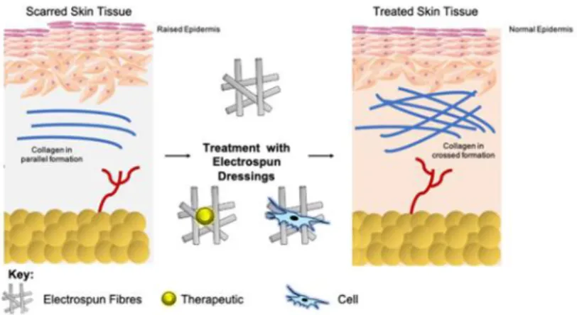
Figure 7. General schematic of wound healing scar. Source: Mulholland EJ (2020) Electrospun Biomaterials in the Treatment and Prevention of Scars in Skin Wound Healing. Front. Bioeng. Biotechnol. 8:481. doi: 10.3389/fbioe.2020.0048
Moreover, some processes skin regeneration involving the expression of defensin in the epidermal cells of tissue-engineered skin can be activated, which may play an important role in the effectiveness ulcer replacements (Rezaie, F., et al., 2019; Baltazar, T., et al., 2020). However, several factors affecting the specific tissue microenvironment, sustained by the perseverance of inflammation, still make ulcer healing an unmet clinical need, which deserves additional research (Sen, C.K., et al., 2009). On the other hand, other critical process in skin regeneration is the re-epithelialization of wounded skin due to requires the rapid and coordinated migration of keratinocytes (KCs) into the wound bed. Almost immediately after wounding, cells present at or attracted to the wound site begin to secrete a complex milieu of growth factors. Growth factors are molecules that stimulate cellular growth, proliferation, differentiation, and/or migration. Growth factors can be small molecules such as hormones or macromolecules such as proteins and can be secreted as fully functional molecules or as molecules that require further posttranslational processing to be activated. These growth factors exert mitogenic and motogenic effects on KCs, inducing the rapid proliferation and migration of KCs at the wound edge (Seeger, M.A., and Paller, A.M., 2014). In fact, these growth factors in KCs have a role in the motility of the cells. The activities of these growth factors are regulated both temporally and spatially in the wound site to establish the well-choreographed process of wound healing. On wounding, KCs along the wound edge begin a dramatic rearrangement of their cytoplasmic and membrane structures. These changes include the disassembly of most hemidesmosomes (e.g., cell-ECM contacts) and many desmosomes (e.g., cell–cell contacts), retraction of cytoplasmic keratin intermediate filaments from the periphery of the cell, and rearrangements of the actin cytoskeleton that facilitate the formation and retraction of lamellipodia and focal adhesions (Seeger, M.A and Paller, A.M., 2014).
16
Figure 8. The Wound Healing Phases. Source:https://www.skincol.com/en/info/mechanic-of-action.html
Leaves and fruits of Olea europaea L. have been used externally as an emollient for skin ulcers and for healing of inflammatory wounds. Moreover, in animal study the aqueous extract of O. europaea leaves displayed wound healing activity (Koca, U., et al., 2011). It is believed that OLE acts as a free radical scavenger by a synergy of its many polyphenols. It is thus important a means for local delivery of OLE inside ulcers, which at the same time, induce connective tissue regeneration. In Table 3 is summarized some relevant studies of the utilization of OLE in wound healing.
Plant origin Outcomes References
Razi Herbal
Medicine Institute, Lorestan, Iran.
- Results suggested that Oleuropein accelerates skin wound healing in aged male Balb/c mice. These findings can be useful for clinical application of Oleuropein in expediting wound healing after surgery.
Mehraein, F., et al., 2013
Olea europaea var. europaea were collected from
Bahcelievler region, Turkey.
- In vivo model resulted in animals treated with the aqueous extract demonstrated increased contraction (87.1%) on excision and a significant increase in wound tensile strength (34.8%) on incision models as compared to other groups
Koca, U., et al., 2011
Olea europaea from Turkey In vitro study. NIH-3T3 cells were treated with crude OLE and H2O2 as controls in wound scratch assay. In stress condition results indicated that lower concentrations (between 1-50 µM) of H2O2 promoted cell migration.
Erdogan, et al., 2018
Olea europaea leaves from Iran.
The results showed that control group significantly showed higher wound area, total bacterial count, and higher expressions of IL-1β and TNF-α (P<0.05) and lower expressions for IL-10 and TGF-β (P<0.05). The treatment with OLE could significantly decrease wound area, total bacterial count, expressions of
17
Table 3. OLE and wound healing studies.
1.2.3 Immunomodulation and OLE
The complexity of immune reactions occurs via the production of both pro- and anti-inflammatory cytokines and antibacterial molecules with different time scale, which ultimately ensure an efficient tissue response to foreign agents. The biological mediators of innate immunity and inflammation are cytokines, biological molecules that act as soluble mediators of natural immunity and the immune response. They are described as multi-functional molecules, together with chemokines and adhesion molecules, which play important biological activities (Mantovani, A., et al., 2004).
In recent years numerous studies have attributed to the polyphenols a broad range of biological activities including anti-inflammatory and immune-modulatory actions (Santangelo, C., et al., 2007; Yahfouti, N. et al., 2018). Inflammation is known to be a significant reason connected to various human issues including cancer, diabetes type II, arthritis, neurodegenerative diseases, and cardiovascular issues i.e. a multifactorial response (Bengmark, S., 2004). Polyphenols have demonstrated positive effects in vitro and in vivo studies and beneficial role as therapeutic tools in multiple acute and chronic disorders (Gonzalez, R., et al., 2011). Numerous epidemiological and experimental researches have evidenced the anti-inflammatory and immune modulation activities of dietary polyphenols, including OLE, showing the ability of these natural compounds to modify the expression of several pro-inflammatory genes, like multiple cytokines, lipoxygenase, iNOS, cyclooxygenase (COX), in addition to their antioxidant characteristics (Santangelo, C., et al., 2007). The first evidences about the anti-inflammatory and immunomodulatory effects of natural compounds was in 2006 in the clinical trial called PREDIMED. The research showed the anti-inflammatory effect of the Mediterranean Diet supplemented with extra virgin olive oil (EVOO) for a period of three months in comparison with a low fat diet. The authors found a significant decrease of serum C-Reactive Protein (CRP), interleukine (IL) IL-6, adhesion molecules (ICAM-1 and VCAM-1), and chemokines, in a group of 722 participants (Estruch, R., 2010). Also, in another
1β and TNF-α, and increase the expression of IL-10 and TGF-β (P<0.05).
Olea europaea from Turkey. - OLE incorporated in dressing showed in a Spraque Dawley rats diabetic group; wounds closure time of 24.80 days ± 1.48 in OLE wound dressing and 28.00 ± 2.31 days in classical wound dressing.
Samancioğlu, S. et al., 2015
18
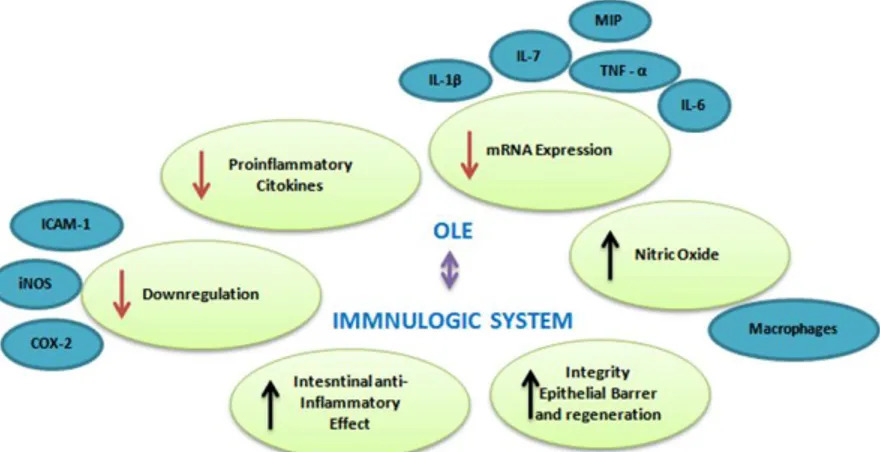
human trial, 18 healthy volunteers, who had consumed on a single occasion OLEs (51 mg oleuropein and 10 mg HT), were evaluated for vascular function via digital volume pulse (DVP) and cytokine production from endotoxin-stimulated peripheral blood mononuclear cells (PBMCs) (Lockyer, S., et al., 2015). DVP stiffness index and IL-8 production were significantly reduced in comparison to matched untreated controls. Oleuropein and HT urinary metabolites peaked after 8-24 h from administration. Considering the evidences based in MD together with administration of OLE, data suggest that oleuropein and HT exert various protective effects in the host, such as enhanced NO generation by murine macrophages (Manna, C., et al., 2004), induction of the anti-inflammatory pathway in human whole blood cultures (Magrone, T., et al., 2017), prevention of oxidative myocardial injury due to ischemia and reperfusion (Vinson, J.A., et al., 1995; Miao, J., et al., 2019), decrease in blood pressure, and platelet aggregation, inhibition of 5- and 12-lipooxygenases and increased free radical scavenging (Miles, E., et al., 2005), as well as to reduce chronic inflammation and oxidative stress responsible for cardiac and hepatic damage, also preventing the outcome of metabolic syndrome, as reported in an animal model (Pirozzi, C., et al., 2016). Significant advances have been elucidated with respect to the anti-inflammatory mechanisms exerted by OLEs. Macrophages represent one of the principal factors in the inflammatory response, because can produce ROS, but also pro-inflammatory cytokines and chemokines, including IL-1, IL-6, TNF-a, and IFN-γ (Nediani, C., et al., 2019) (Figure 9).
Figure 9. Characteristics of OLE in the immune system studied. MIP (Macrophage inflammatory protein-2), ICAM (intercellular adhesion molecule 1), iNOS (inducible nitric oxide synthase), TNF (tumor necrosis factor), IL (Interleukine).
Therefore, besides the release of several inflammatory cytokines or mediators, damaged tissues also release monocyte chemoattractant proteins (MCP-1), COX, iNOS, metalloproteinases (MMP), and adhesion molecules. In addition, nuclear factor Kappa β (NF-kβ) occupies a key upstream position in a complex signal transduction pathway, controlling the production of countless pro-inflammatory mediators (Hassen, I. et al., 2015). Notwithstanding, Toll-like receptor (TLR) and TNF receptor activation leads to inflammatory gene expression and production of COX-2, IL-1 and IL-8 (Cohen,
19
P., 2014). In this context, in vitro oleuropein-mediated down-regulation of TLR activation by lipopolysaccharide (LPS) has been documented as a result of iNOS, COX-2, extracellular signal-regulated kinases (ERK)1/2, c-JunN- Terminal kinase (JNK) and NF-k light poly-peptide gene enhancer in B-cell inhibitor (IKB) phosphorylation decrease, respectively (Matsuguchi, T., et al., 2003; Ryu, S.J. et al., 2015).
1.2.4 OLE as Antimicrobial Resource
There is an increased societal interest for sustainable industrial routes, with special emphasis in the food and biomedical sector to manufacture safe yet sustainable packaging for edible products and surgical devices equivalently (Guillard, V., et al., 2018; Isbary, G., et al., 2013). Consequently, there is a growing interest towards minimal processing technologies able to replace conventional decontamination benchmarks. To this end, natural antimicrobial compounds, including vegetable bioactive molecules, offer an emerging strategy to control microbial contamination by virtue of their specific bioactive molecules. Indeed, some molecules and plant-derivatives demonstrate good antimicrobial activity: against pathogenic bacteria present in (i) either packaging or processing steps in the food industry (Guillard, V., et al., 2018) or in (ii) biomedical devices, such as surgical tools and supporting parts, the latter being very important in healthcare since the inaccurate sterilization is responsible for at least 1.5%–7.2% of post-operational complications (Isbary, G., et al., 2019; Ben-Othman, S. et al., 2020). Antibacterial properties of OLE have been widely explored exhibiting antimicrobial activity against a wide range of Gram-positive and Gram-negative bacteria, as reported in Table 4.
20
Table 4. OLE containing antimicrobial compounds against Gram-positive and Gram-negative bacteria
According to some studies, OLE exerts antimicrobial effects due to its high phenolic compound content (Poudyal, H., et al., 2010; Lee, O.H. and Lee B.Y., 2010). Some studies with OLE reported the growth inhibition in some bacteria species but these findings are contradictory and may depend on the concentration and/or the bacteria used. For example, Sudjana et al., demonstrated that OLE does not show broad-spectrum activity having appreciable activity only against C. jejuni, H. pylori
Specie Olive variety and origin TP Extraction Method
Concentration Reference
Klebsiella pneumoniae
Olea europaea (Turkey, west Anatolian) Aqueous 30 µl OLE; 15% (w/v) Tranter, H.S., et al., 1993 Staphylococcus aureus Olea europaea (Portugal)
Aqueous 5 mg/mL Pereira, A.P., et al., 2007
Tranter, H.S., et al., 1993
Bacillus cereus Olea europaea (Portugal) Aqueous 5 mg/mL Pereira, A.P., et al., 2007
Bacillus subtilis Olea europaea (Portugal) Ethanol 27.2 mg/g Pereira, A.P., et al., 2007
Pseudomonas aeruginosa
Olea europaea (Portugal) Aqueous 5 mg/mL Pereira, A.P., et al., 2007
Campylobacter jejuni Olea europaea (Australia)
No described No described Friedman, M., 2015 Sudjana, A.N., et al., 2009
Helicobacter pylori Olea europaea (Australia)
No described No described Sudjana, A.N., et al., 2009
Escherichia coli Olea europaea (Several countries)
Water, ethanol. Variable Pereira, A.P., et al., 2007
Albertos, I., et al., 2017
Salmonella enterica Olea europaea var. Sylvestris (Algeria)
Methanol/water 198.7 mg GA/g (1 g)
Friedman, M., 2015 Albertos, I., et al., 2017 Djanane, D., et al., 2019 Listeria monocytogenes Commercial Extract (USA)
Water/ethanol 62.5 mg/ml Albertos, I., et al., 2017
Bourke, P., et al., 2017