Università degli Studi di Ferrara
DOTTORATO DI RICERCA IN
FARMACOLOGIA E ONCOLOGIA MOLECOLARE
CICLO XXII
COORDINATORE Prof. Pier Andrea Borea
LOCAL SUPPLEMENTATION OF
BRAIN-DERIVED NEUROTROPHIC FACTOR FOR
THE TREATMENT OF NEURONAL DAMAGE
Settore Scientifico Disciplinare BIO/14
Dottorando
Tutore
Dott. Tao Su
Prof. Michele Simonato
CONTENTS
ABSTRACT………...……….……… 3
CHAPTER I: BACKGROUND………. 7
Preface 9
1 Neurotrophins 11
1.1 Gene and protein structure 11 1.2 Neurotrophin receptors: identification and structure 15 1.3 Signaling pathway 17
2 BDNF 20
2.1 Distribution and cellular localization 20 2.2 Expression regulation 22 2.3 BDNF secretion 23 3 The role of BDNF in the pathogenesis of some neurological diseases 25
3.1 BDNF in neurodegenerative diseases 25 3.2 BDNF and epilepsy 27 3.3 BDNF and other neurological disorders 28 4 BDNF as a potential therapeutic agent for neurological diseases 30
5 Strategies of NTFs therapy 32 5.1 Direct infusion 32 5.2 BDNF mimetics 33 5.3 Local delivery 35
5.4 Gene therapy 36
5.5 Stem cell-based gene therapy 37 5.6 Mesoangioblast: an alternative cell source for NTF delivery 39
6 REFERENCES 40
CHAPTER II: THE FINDINGS……… 57
1. Overview of the research program 591.1 Rationale 59
1.2 Research Basis 59 1.3 Unsolved issues 60
1.4 Aim 61
1.5 Scheme 61
2. PART I - The neuroprotective effects of MABs-delivered BDNF 63
2.1 Introduction 63
2.2 Methods 64
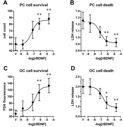
2.3 Results 68
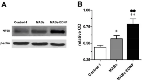
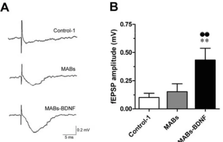
2.3.1 Recombinant BDNF exhibits concentration-dependent protective effects on cell survival 68 2.3.2 Soluble mediators from MABs engineered to produce BDNF highly improve cell survival 70 2.3.3 The MABs-BDNF-produced mediators sustain neuronal structural integrity in adult
organotypic slices 72 2.3.4 Functional benefits afforded by the MABs-BDNF conditioned media in the cultured
Contents
2.4 Discussion 75
2.4.1 Protective Effects of rBDNF on Neurons 75 2.4.2 Protective effects of MABs and MABs-BDNF produced factors 76 2.4.3 The possible mechanism of BDNF protective effects 77 2.4.4 Neuroprotective strategies with NTFs 79 3. PART II - Effects of MABs-delivered BDNF on neurogenesis 80
3.1 Introduction 80
3.2 Methods 81
3.3 Results 84
3.3.1 Neurogenesis in hippocampal slice cultures 84 3.3.2 Modulation of neurogenesis by NTFs and MABs-BDNF 85
3.4 Discussion 87
3.4.1 Neurogenesis in OHSC 87 3.4.2 Neurogenesis modulated by NTFs 88 3.4.2 Potential application 91
4. PART III - Transplantation of MABs-BDNF 93
4.1 Introduction 93
4.2 Methods 94
4.3 Results 95
4.4 Discussion 98
4.4.1 Epilepsy and NTFs 98 4.4.2 Ineffectiveness of MABs-BDNF systemic infusion in the TLE model 99 4.4.3 Alternative MABs-based NTF delivery strategies 100
5. Conclusion 103
6. Future outlook: opportunities and challenges 104
7. References 107
ACKNOWLEDGEMENT……… 130
APPENDIX………...………131
Appendix I………. Page 1 Appendix II……… Page 7 Appendix III……….. Page 17
Local supplementation of brain-derived neurotrophic factor for
the treatment of neuronal damage
Tao SU
Tutor: Prof. Michele Simonato
Department of Clinical and Experimental Medicine, Section of Pharmacology, Neuroscience Center, University of Ferrara, Ferrara 44100, Italy; National Institute of Neuroscience, Italy
Abstract
T
he importance of nerve growth factors, especially brain-derived neurotrophic
factor (BDNF) in the regulation of neuronal survival and plastic changes in
morphology and function has been increasingly studied during the recent years. It
has been proposed that the pathogenesis of some neurological diseases may be due
to an alteration in neurotrophic factor and/or Trk receptor levels. The use of
neurotrophic factors as therapeutic agents is a promising approach aimed at
restoring and maintaining neuronal function in the central nervous system (CNS).
This study is undertaken to develop a novel stem cell-based gene therapy to deliver
neurotrophic factors to vulnerable regions of the CNS. Stem cell-based gene
therapy is a potential delivery option by which cells are engineered to produce
neurotrophic factors in vitro and then transplanted to the target area where
neurotrophic factors are secreted to exert protective and/or restorative effects on the
host tissue. A recently isolated mesodermal stem cell, mesoangioblast (MAB), has a
high adhesin-dependent migratory capacity and may selectively cross the
blood-brain barrier and home in the lesioned areas. Therefore, MABs provide an
ideal cellular source for BDNF delivery. In this study, we generated a genetically
modified mesoangioblast producing BDNF (MABs-BDNF). These engineered
MABs maintained transgene expression and secretion of bioactive BDNF in time.
We investigated the protective effects of MABs-BDNF in vitro using primary
cultures and organotypic cultures of hippocampal slices. The viability of the
cultured slices was assessed in several ways: fluorescein diacetate (FDA)
hydrolysis assay, lactate dehydrogenase (LDH) release assay,
immunohistochemistry for MAP2, immunoblot for neurofilament 68, and field
potential recordings. Direct exposure of recombinant BDNF to primary cultured
neurons and adult slices resulted in a concentration-dependant protective effect.
The conditioned medium from MABs-BDNF highly promoted cell survival, while
the conditioned medium from control cells (MABs) or an equivalent amount of
rBDNF showed beneficial effects on cell survival to a lesser extent. The protective
effects of MABs-BDNF were attenuated by adding either with the TrkB receptor
blocker K252a or the BDNF scavenger TrkB-IgG.. This indicates that the
conditioned medium from MABs-BDNF can foster the adult slice culture through
secreting the engineered BDNF and unknown pro-survival factors produced
intrinsically by MABs. The MABs-BDNF conditioned medium was optimal for
retention of morphologic characteristics and viability in organotypic cultures from
adult hippocampal slices. Moreover, MABs-BDNF were found to promote
4 Abstract
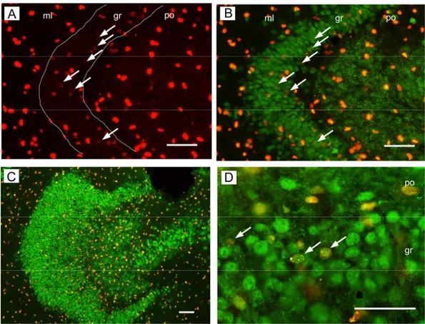
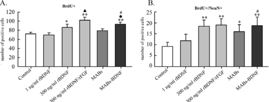
neurogenesis and glia proliferation. Treatment with the MABs-BDNF conditioned
medium was found to increase the number of BrdU-labeled and BrdU/NeuN double
labeled cells in the dentate gyrus of cultured slices. These in vitro findings
demonstrate the beneficial effects of MABs-BDNF on neurons and provide a
rationale for transplanting MABs-BDNF in the damaged brain as a therapeutic
approach. Thus, we tested the transplantation of MABs-BDNF in an animal model
of neuronal loss, the hippocampal sclerosis induced by status epilepticus. So far, we
have not detected a deposition of MABs-BDNF in the epileptic brain after their
systemic administration. Future experiments will aim at optimizing the
transplanting conditions, changing the delivering routes, and assessing their
therapeutic value in other neurological diseases associated with cell death. In terms
of their prominent beneficial by-stander effects on neurons, MABs-BDNF hold
substantial promise as therapeutic agents in the treatment of neurological diseases.
Keywords: BDNF, mesoangioblasts, gene therapy, delivery, brain damage
Rilascio localizzato di brain-derived neurotrophic factor per il
trattamento del danno neuronale
Tao SU;
Tutore: Prof. Michele Simonato
Dipartimento di Medicina clinica e Sperimentale, Sezione di Farmacologia, Centro
di Neuroscienze, Università di Ferrara, Ferrara 44100, Italia; Istituto Nazionale di
Neuroscienze, Italia.
Abstract
L’importanza dei fattori neurotrofici, in particolare del brain-derived neurotrophic
factor (BDNF), nel controllo della sopravvivenza neuronale e nelle modificazioni
plastiche morfo-funzionali del sistema nervoso è diventata sempre più rilevante
negli ultimi anni. Si è infatti ipotizzato che un’alterazione di questo fattore
neurotrofico e/o dei livelli del suo recettore Trk possano rappresentare tappe
patogenetiche importanti nello sviluppo di alcune patologie neurologiche. Da ciò
deriva che l’utilizzo dei fattori neurotrofici come agenti terapeutici sia un approccio
farmacologico promettente per il recupero ed il mantenimento delle funzioni
neurali del sistema nervoso centrale (SNC). Scopo dello studio intrapreso è stato
quello di cercare di sviluppare una nuova terapia genica basata sulle cellule
staminali per ottenere il rilascio di fattori neurotrofici in aree vulnerabili del SNC.
La terapia genica basata sull’impiego di cellule staminali è una delle possibili
tecniche di bio-delivery per mezzo della quale le cellule, ingegnerizzate per
produrre fattori neurotrofici in vitro, vengono trapiantate nell’area bersaglio dove
rilasciano i fattori neurotrofici con effetti protettivi e/o rigenerativi sul tessuto
ospite. Il mesoangioblasto (MAB) è una cellula staminale, recentemente isolata,
che possiede un’alta capacità migratoria adesina-dipendente, può attraversare
selettivamente la barriera emato-encefalica e raggiungere l’area neurale lesionata. I
MAB, perciò, offrono una sorgente cellulare ideale per il rilascio di BDNF. Nella
nostra ricerca sono stati generati mesoangioblasti geneticamente modificati per la
produzione di BDNF (MABs-BDNF). I mesoangioblasti in questione mantengono
nel tempo l’espressione del transgene e le proprietà secretorie di BDNF
biologicamente attivo. Successivamente abbiamo studiato i loro effetti
neuroprotettivi in vitro, su colture primarie e su colture organotipiche ippocampali.
Si è valutata la vitalità delle fettine tissutali ippocampali mantenute in coltura
mediante diverse metodiche: test di idrolisi della Fluoresceina DiAcetato (FDA),
saggio di rilascio della LattatoDeidrogenasi (LDH), immunoistochimica per MAP2
(Microtubule Associated Protein 2, proteina associata ai microtubuli di tipo 2),
immunoblot per il neurofilamento 68, registrazione dei potenziali di campo. Si è
visto che l’esposizione diretta delle colture primarie neuronali e delle colture
d’ippocampo adulto al BDNF ricombinante (rBDNF) produce effetti protettivi
dose-dipendente. Il medium condizionato dai MABs-BDNF promuove in maniera
rilevante la sopravvivenza cellulare, mentre sono assai minori gli effetti benefici
sulla sopravvivenza cellulare indotti dal medium condizionato ottenuto dalle cellule
di controllo (MABs) o da un equivalente quantità di rBDNF. Gli effetti protettivi
6 Abstract in Italian
dei MABs-BDNF vengono attenuati dall’aggiunta sia di K252a, bloccante
recettoriale di TrkB, sia da TrkB-IgG, sottrattore anticorpale di BDNF. Questo sta
ad indicare che il medium condizionato dai MABs-BDNF può proteggere le colture
organotipiche di adulto non solo mediante la liberazione di BDNF ma anche tramite
il rilascio di non ancora noti fattori neuroprotettivi prodotti intrinsecamente dai
MABs. Il medium condizionato MABs-BDNF è risultato ottimale per il
mantenimento delle caratteristiche morfologiche e di vitalità delle colture
organotipiche ippocampali di adulto. I MABs-BDNF, inoltre, sono risultati
promuovere la neuronogenesi e la proliferazione gliale. Il medium condizionato dai
MABs-BDNF ha incrementato, infatti, il numero di cellule marcate con la
BromoDeossiUridina (BrdU) e delle cellule doppiamente marcate, BrdU/NeuN
(BromoDeossiUridina/Neuronal Nuclear protein, proteina nucleare neuronale) nel
giro dentato delle fettine in coltura. Queste scoperte ottenute in vitro dimostrano gli
effetti positivi dei MABs-BDNF sui neuroni e forniscono un razionale per l’utilizzo
terapeutico del trapianto di MABs-BDNF nelle aree cerebrali danneggiate. In modo
preliminare abbiamo testato il trapianto di MABs-BDNF in un modello animale di
perdita neuronale, la sclerosi ippocampale indotta dallo stato epilettico, non avendo
purtroppo riscontrato una localizzazione di MABs-BDNF nel cervello epilettico, in
seguito alla loro somministrazione sistemica. Successivi esperimenti saranno
necessari per ottimizzare le condizioni del trapianto, modificando le vie di delivery
e valutando il loro valore terapeutico in altre patologie neurologiche associate alla
morte cellulare. I MABs-BDNF, per il loro preminente effetto benefico, seppur
indiretto, a tipo “spettatore innocente” sui neuroni, risultano promettenti agenti
terapeutici per il trattamento delle malattie neurologiche.
Chapter I
BACKGROUND
Preface1 Neurotrophins
1.1 Gene and protein structure
1.2 Neurotrophin receptors: identification and structure
1.3 Signaling pathway 2 BDNF
2.1 Distribution and cellular localization 2.2 Expression regulation
2.3 BDNF secretion
3 The role of BDNF in the pathogenesis of some neurological diseases
3.1 BDNF in neurodegenerative diseases
3.2 BDNF and epilepsy
3.3 BDNF and other neurological disorders
4 BDNF as a potential therapeutic agent for neurological diseases
5 Strategies of NTFs therapy 5.1 Direct infusion 5.2 BDNF mimetics 5.3 Local delivery 5.4 Gene therapy
5.5 Stem cell-based gene therapy 5.6 Mesoangioblast: an alternative cell source for NTF delivery
Chapter I: BACKGROUND 9
Preface
Back in the early 1950s, a group of embryologists discovered that a series of
signalling molecules secreted by the target tissues in the developing nervous system,
which were called neurotrophic factors (NTFs). NTFs were defined as
target-derived, antiapoptotic molecules that maintain embryonic or adult neuronal
cells. The word ‘trophic’ is derived from Greek ‘trophé’ meaning nourishment or
taking up of nutrients. Various families of neurotrophic factors exist, e.g., the
neurotrophin (NT) family, the fibroblast growth factor (FGF) family, the epidermal
growth factor (EGF) family, the insulin-like growth factor (IGF) family, the
vascular-endothilial growth factor (VEGF) faimily, the glial cell-derived
neurotrophic factor (GDNF) family, and the ciliary neurotrophic factor (CNTF).
Currently, six neurotrophins have been isolated: NGF, BDNF, NT-3, NT-4 (also
known as NT-5), NT-6, and NT-7. The NT-6 and NT-7 genes have been identified
only in fish and probably do not have mammalian or avian orthologues
(Gotz et al.,
1994; Nilsson et al., 1998)
. The FGF family includes 22 known members in human,
which initially were found to influence the growth of fibroblasts and later their
ability in many other cell types were demonstrated
(Burgess and Maciag, 1989)
.
For example, basic FGF (bFGF, also called FGF-2, EDGF, or HBGF-2) can
influence the differentiation and migration of neurons and glia
(Walicke et al.,
1986).
All these NTFs signal via factor specific multicomponent receptor
complexes. The NGF-superfamily of receptors includes p75 and the receptor
protein tyrosine kinases (Trk): TrkA, TrkB and TrkC. The mammalian FGF
receptor family has 4 members, FGFR1, FGFR2, FGFR3, and FGFR4. The GDNF
family of receptors includes a receptor complex of Ret and growth factor receptor
α1-4. The neurokine superfamily ligands act via the receptors gp130 and leukemia
inhibitory factor receptor-β (LIFR-β).
Since their discovery, research in the field of NTFs has provided a vast amount
of data not only on their biochemical properties but also on how they function
during development and how they maintain neuronal activity in the adult nervous
system. It has been evidenced that NTFs regulate almost all aspects of neuronal
development and function, including precursor proliferation and commitment, cell
survival, axon and dendrite growth, membrane trafficking, synapse formation and
function, neurotransmitter release, synaptic plasticity, as well as glia differentiation
and interaction with neurons. It is well accepted that NTFs are critical to the
development and maintenance of the mammalian CNS. Alterations in NTF levels
have profound effects on a wide variety of phenomena, including myelination,
regeneration, pain, aggression, depression and substance abuse. The influence of
neurotrophic factors spans from developmental neurobiology to neurodegenerative
and psychiatric disorders. Recently, NTFs have received considerable interest as
possible means for the treatment of neurodegenerative diseases, psychiatric
disorders and CNS trauma. There are many successful paradigms with respect to
pre-clinical and clinical applications of NTFs, especially neurotrophin family
member (e.g. NGF, BDNF) to prevent, slow the progression of, or even reverse the
effects of a number of neurodegenerative diseases and other types of insults in both
the central and peripheral nervous system, since the idea that degenerative diseases
of the nervous system may result from insufficient supply of these neurotrophins
has generated great interest in these NTFs as potential therapeutic agents.
Besides the neurotrophin family, many other NTFs show significant
therapeutic potential in a variety of diseases states, from neurological diseases,
Parkinson’s disease (PD), Alzheimer’s disease (AD), amyotrophic lateral sclerosis
(ALS), stroke, schizophrenia, and depression to diabetic neuropathies, neuropathic
pain, macular degeneration, and obesity. These factors include members of the
fibroblast growth factor (FGF) family, GDNF family, CNTF family, and
insulin-like growth factors I and II (IGF-I and -II). Here, we will focus primarily on
the neurotrophin family and their cognate receptors.
In this chapter, we will summarize the knowledge acquired on the gene and
protein structure of neurotrophins and their molecular and cellular biology during
the last decade. Because of the importance of BDNF on neuronal survival and
regeneration, we will review particularly the distribution and alteration of BDNF
and its therapeutic potential in the neurological diseases. In addition, the various
therapeutic strategies against neurodegenerative diseases are introduced. Among
these strategies, a novel cell source (mesoangioblast) for cell based therapy
attracted our interest.
Chapter I: BACKGROUND 11
1 Neurotrophins
The neurotrophins are a family of structurally related secretory proteins that
are widely expressed in neurons and their target cells
(Alderson et al., 1990)
. These
secreted proteins play a crucial role in the control of neuronal numbers and of
dendritic growth. To date five members have been identified. They are called nerve
growth factor (NGF), brain-derived neurotrophic factor (BDNF), and neurotrophin
(NT) 3, 4/5, and 6. The first neurotrophin identified was the NGF
(Levi-Montalcini,
1966)
. It was found, however, that only a few CNS neurons were responsive to
NGF. The second neurotrophin, BDNF, initially purified from the pig brain as a
trophic factor for dorsal root ganglion cells, helped to establish the concept that the
diffusible growth factors could regulate the fate and the shape of most vertebrate
neurons
(Hofer and Barde, 1988)
. Molecular cloning of BDNF revealed that its
amino acid sequence had a strong homology to that of NGF. By using the
contiguous regions between NGF and BDNF to design oligonucleotides for
polymerase chain reaction cloning, a third related protein called neurotrophin 3
(NT-3) was cloned
(Maisonpierre et al., 1991; Hallbook et al., 1993; Dawson et al.,
1995)
. This is located on human chromosome 12 (12p13)
(Maisonpierre et al.,
1991)
. Finally, the last member of the neurotrophin family (NT-4) was identified
and cloned in Xenopus
(Hallbook et al., 1991)
. The equivalent human cDNA is
different from Xenopus NT-4 and is thought to be a separate gene and called
NT-5
(Berkemeier et al., 1991; Berkemeier et al., 1992)
. Subsequently it was
realized that they are homologueous genes and this neurotrophin is often termed
NT-4/5. NT-4/5 is present on human chromosome 19 (19q13.3)
(Berkemeier et al.,
1992; Ip et al., 1992)
. NT-6 has only been found in fish
(Gotz et al., 1994)
.
To better exploit the NTFs therapeutic potential, we need to understand the
details of the molecular biology of the neurotrophin system and integrate them with
the physiology of neurotrophins. In the following sections, we review their gene
and protein structure, biosynthesis and secretion, receptors, signaling pathways, and
regulation of neuronal activity. The data on the molecular mechanisms of gene
regulation from other fields of biomedical science, together with the physiological
data on neurotrophic factor action and known intracellular signal transduction
pathways help us to get insight into the function and modulation of these NTFs,
thereby contributing to the exploitation of potential therapies for neurological
disorders.
1.1 Gene and protein structure
The recent explosion in genomics research has given us dozens of amino acid
sequences of NTFs from dozens of species. In addition, the crystal structures of
many NTFs are now available after a decade of intense research. Comparing the
amino acid sequences and structural features of the NTFs can give insight into the
evolutionary history of these families and can help reveal functionally important
regions of the molecules.
Sequence alignments
All genes encoding neurotrophins have a basically similar structure. A single
discontinuous exon contains all the information for encoding the prepropeptide.
The coding exon is preceded by several noncoding exons that are subject to
alternative splicing and give rise to alternative 5’- untranslated regions (UTRs) of
mRNA. Multiple promoters have been demonstrated for all neurotrophins.
The BDNF gene (bdnf), mapped to chromosome 11p in humans, has four
upstream exons (exons I-IV) that are associated with distinct promoters, and one 3’
exon (exon V) that encodes the mature BDNF protein
(Metsis et al., 1993;
Timmusk et al., 1993)
. Recently, some novel exons had been identified in the
upstream noncoding exons
(Aid et al., 2007)
. According to the new nomenclature,
BDNF transcripts contain 9 exons (I-IXA) in the upstream unstranslated region,
while the common coding region previously called exon V is now named as exon
IX. The usage of different promoters gives rise to mRNAs that contain only one of
the upstream noncoding exons spliced to the coding exon. Upstream exons of the
BDNF gene do not contain a splicing acceptor site thus preventing the splicing of
the more upstream exons to those located proximally to the coding exon. As many
as 11 different BDNF transcripts can be generated in both humans and rodents by
alternative splicing. They differ only in their upstreanm untranslated region but
share a common exon IX
(Aid et al., 2007)
. All these BDNF mRNAs are expressed
in the brain at very different levels and generating a spatial diversity of expression.
Moreover, different promoters in the BDNF gene have specific developmental
patterns of expression and differential activation capacities. For instance, a 5-kb
region of promoter Ⅲ is involved in BDNF gene induction after sciatic nerve
transaction
(Timmusk et al., 1995)
.
Following the detailed description of BDNF gene structure, many attempts
have been made to localize structural elements of the gene governing mRNA
regulation in vivo or in vitro. For instance, the first intron of the BDNF gene
contains a neuron-restrictive silencer element (NRSE)-type regulatory sequence
(Timmusk et al., 1999)
. Alterations in the NRSE affected expression levels of
promoters II and I that are located nearby. In the brain, mutation of the NRSE
modulated the responsiveness of BDNF promoters I and II to activation of the
glutamatergic system
(Timmusk et al., 1999)
. Short regions flanking promoters I, II,
and III in the BDNF gene have been localized as putative calcium responsive
regions
(Bishop et al., 1997)
. Two different regulatory sequences involved in the
activation of transcription were localized in the proximal promoter region. One is a
novel calcium response element required for calcium-dependent BDNF expression
in both embryonic and postnatal cortical neurons. The second element matches the
consensus sequence of a cAMP response element (CRE) and is required for
transactivation of the promoter in postnatal but not in embryonic neurons
(Tabuchi
et al., 2002)
. Differential cell-specific activation of different BDNF promoters via
activation of the glutamatergic system most likely reflects involvement of specific
kinase pathways in the cells
(Marmigere et al., 2001)
.
The human NGF gene is located on the short arm of chromosome 1 (1p22)
(Francke et al., 1983)
and codes for a polypeptide of 307 amino acids. It contains
three upstream exons and the promoter activity has been linked to the first and the
third exon. The NGF gene has the most complex splicing pattern. The majorities of
transcripts are transcribed from the distal promoter and contain exon I, while the
Chapter I: BACKGROUND 13
other noncoding exons are combined by alternative splicing and give rise to a
variety of mRNAs with different 5’-UTRs. In addition to ngf and bdnf, two other
neurotrophin genes, neurotrophin-3 (nt3) and neurotrophin-4/5 (nt4/5), have been
identified in mammals
(Sekimoto et al., 1998)
. The NT-3 gene contains two
upstream exons flanked by promoters.
Molecular evolution
The evolution of the NTFs has been studied using phylogenetic trees that
organize the relationships between their amino acid sequences
(Kullander et al.,
1997)
. The works have divided neurotrophin residues into two categories:
conserved and variable, based on early sequence alignments
(Thoenen, 1991)
. With
the exception of NT4/5, neurotrophin sequences are highly conserved. For instance,
BDNF shares about 50% amino acid identity with NGF, NT-3. Based on sequence
comparisons and on the isolation of neurotrophin genes in various vertebrates, it is
thought that ngf / nt3 and bdnf / nt4/5 evolved from separate duplication events
(Hallbook, 1999)
. The most primitive neurotrophin genes have been isolated from
jawless fishes, a river lamprey and the Atlantic hagfish. In bony fishes, more
neurotrophin and receptor genes have been isolated than in mammals
(Hallbook,
1999)
.The neurotrophin receptors of the trk family seem to have coevolved with the
neurotrophin genes
(Hallbook, 1999)
. So far, no neurotrophin-like sequences have
been detected in invertebrates
(Bargmann, 1998)
. No ortholog of the NT-4 gene has
been found in teleost fishes, and no ortholog of the NT-6 or NT-7 genes has been
found in tetrapods (although the names are similar, the human NT-6 genes are
closer to NGF).
Proteins structure
All the neurotrophins are synthesized as precursor proteins a little over 200
amino acids long (approximately 30 kDa in size), containing a signal sequence for
secretion. Following proteolytic cleavage, a mature C-terminal active peptide
slightly shorter than 120 amino acids long (approximately 13 kDa) is released
containing six cysteine residues at identically spaced positions in all mammalian
neurotrophins
(Chao and Bothwell, 2002)
. The mature part is very well conserved
and approximately 50% of the amino acids are common to all neurotrophins. The
neurotrophins are secreted as noncovalent-linked dimers, containing a signal
peptide following the initiation codon and a pro-region containing an N-linked
glycosylation site. They are all basic proteins with isoelectric points above 9.0, a
somewhat unusual property for secreted proteins, which may serve the purpose of
limiting their range of action
(Chao and Bothwell, 2002)
.
Three-dimensional structures of most of the human neurotrophins have been
determined. These structures can be aligned to reveal that the regions of similarity
are much larger than was suggested from sequence alignments. Most of the residues
that participate in secondary structure are strongly conserved both within and
between sub-families, but there are numerous exceptions.
The core structure consists of two pairs of intertwined two-strand beta sheets,
joined by three disulfide bonds. There are also three shorter beta strands leading to
beta turns and loops (
Fig. 1
). The four core beta strands are virtually
superimposable across all the structures. The characteristic formation of a double
loop formed by two disulphide bonds, penetrated by a third disulphide bond, is
named cysteine ‘knot’
(McDonald and Hendrickson, 1993)
. The neurotrophins and
GDNF family members are members of a large superfamily of growth factors that
contain a cysteine knot motif
(Holland et al., 1994; Saarma and Sariola, 1999)
. This
reveals strong conservation, especially within secondary-structure elements. This
family includes transforming growth factor (TGF)- β , human chorionic
gonadotropin, platelet-derived growth factor, vascular endothelial growth factor,
and many others
(McDonald and Hendrickson, 1993)
. The cysteine knot consists of
three cysteine disulphide bonds. The crystal structures of the neurotrophins show
the classic cysteine knot growth factor structure with head-to-head sub-units
forming a noncovalently linked dimer. These residues implicated in binding that are
conserved across families are thought to represent a common interface to the Trk
receptors, while the unique ones may represent elements of specificity
(Robinson et
al., 1995)
.
The only significant differences
between neurotrophins are found in the
loops and turns between beta strands
(McDonald and Hendrickson, 1993)
,
especially loop 3. Loop 3 is the most
different among the four neurotrophin
structures. NT-3 has a single loose
helical turn in this region. NT-4 and
NT-6 sub-families have a large
insertion in this portion, including a
small eight-residue beta turn.
Interestingly, two cysteines that make
up the knot are missing from the
human NT-6 molecules
(Lai et al.,
1998)
. The N- and C-termini are
highly variable in both sequence and
structure among the neurotrophins, and
the temperature factors of these loosely
structured residues are comparatively
high. The N-terminus assumes a
helical structure upon binding the Trk
receptor. The role of the C-terminal
residues is not known, although they
may participate in p75 binding in NT-3
(Carraway and Mitra, 1987)
and NGF
(Bax et al., 1997)
.
Fig. 1. Schematic of the neurotrophin
molecule. Dashed blue lines represent the three disulfide bonds of the cysteine knot. The N terminus is disordered in the unbound structures and is shown by a dashed line. (Butte
et al., Cell. Mol. Life Sci. 58 (2001) 1003–1013)
By comparing the residues involved in the interface between TrkA-d5 and
NGF with the corresponding sequence alignments for the neurotrophins and the Trk
receptors, they can be seen to form two groups of conserved and non-conserved
residues
(Wiesmann et al., 1999)
. One set of residues making up a large patch and
centered on Arg 103 form a common set across all the neurotrophins and receptors.
As these residues are highly homologous and consist mainly of hydrophobic and
aromatic side chains, they probably mediate the bulk of common binding affinity
between neurotrophin and receptor. The other portion of the TrkA interface centers
Chapter I: BACKGROUND 15
on the N-terminal residues of the neurotrophin. These residues show little
conservation, either on the neurotrophin or the receptor side
(Hallbook et al., 1991)
.
The N-terminus, which was disordered in all the unbound neurotrophin structures,
forms a one-and-a-half-turn helical arrangement in the complex between TrkA and
NGF
(Wiesmann et al., 1999)
. NGF binding buries two helical hydrophobic
residues and creates a salt bridge across the interface. BNDF, NT-3, and NT-4 do
not share the same pattern of residue types at the N-terminus, and TrkB and TrkC
also differ in their corresponding interacting residues
(Robinson et al., 1995)
.
Together, these results suggest that the N-terminal residues help determine receptor
binding specificity and that each neurotrophin probably uses a different specific
interface with its cognate receptor in this region
(Kullander et al., 1997)
. Other
specificity-determining residues appear to lie scattered within the common binding
site.
The neurotrophins are initially synthesized as precursors or pro-neurotrophins,
which are cleaved to produce the mature proteins. Pro-neurotrophins are cleaved
intracellularly by FURIN or pro-convertases at a highly conserved dibasic
amino-acid cleavage site to release carboxy-terminal mature proteins
(Mouri et al.,
2007)
. The mature proteins exist exclusively as dimers, though a variety of
arrangements are seen, including heterodimers and homodimers, covalent and
non-covalent association of the promoters, and different spatial configurations
(head-to-toe, head-to-head, and skew). They are normally expressed at very low
levels during development. The amino-terminal half of the pro-neurotrophin is
believed to be important for the proper folding and intracellular sorting of
neurotrophins
(Mouri et al., 2007; Nomoto et al., 2007)
. It is worthy to note that the
discovery that pro-neurotrophins are biologically active has revolutionized the field
of neurotrophin research, necessitating the re-evaluation of much published data
(Nomoto et al., 2007)
. Biological activity has also been attributed to two other
peptides produced as a result of proNGF cleavage, LIP1 and LIP2
(Dicou et al.,
1997)
, which have been shown to protect against excitotoxin-induced cell death
(Dicou, 2006)
.
1.2 Neurotrophin receptors: identification and structure
Neurotrophins bind to two different classes of transmembrane receptor
proteins: the Trk (tropomyosin receptor kinase) family of RTKs (receptor tyrosine
kinases) and p75
NTR(p75 neurotrophin receptor). This dual system allows the
transduction of very different signals following ligand binding, which can be as
contrasting as signaling cell death through p75
NTRor cell survival through the Trk
receptors. These two classes of receptors also directly interact, allowing fine tuning
and cross talk
(Chao and Hempstead, 1995)
. Different neurotrophins show binding
specificity for particular receptors: TrkA preferentially binds NGF; TrkB
preferentially binds BDNF and NT4, while TrkC displays preference for NT3.
These interactions have generally been considered to be of high affinity. However,
in reality, the binding of NGF to TrkA, and of BDNF to TrkB is of low affinity
(Kaplan and Miller, 1997)
, but it can be regulated by receptor dimerization,
structural modifications or association with the p75 receptor
(Heumann, 1994)
. The
p75
NTRbinds all mature neurotrophins with approximately equal low affinity and
has, in recent years, been demonstrated to bind the proneurotrophins with high
affinity
(Massa et al., 2006)
. The ability of Trk and p75 receptors to present
different binding sites and affinities to particular neurotrophins determines both
their responsiveness and specificity. The ratio of receptors is important in dictating
the number of surviving cells, and interactions between p75 and Trk receptors
provide greater discrimination between different neurotrophins
(Chao and
Hempstead, 1995)
. Upon ligand binding, Trk receptors dimerize and become
catalytically active, resulting in receptor autophosphorylation
(Jing et al., 1992)
and
subsequent activation of a number of signalling cascades, including the Ras/
Raf/MAPK (mitogen-activated protein kinase)
(Thomas et al., 1992)
, PI
3K
(phosphoinositide 3-kinase)
(Soltoff et al., 1992)
, and phospholipase C-γ 1
pathways
(Widmer et al., 1993)
. Independent of Trk, the binding of neurotrophins
to the p75
NTRreceptor results in activation of NF-κb, a transcription factor
(Carter
et al., 1996)
, and c-Jun N-terminal kinase
(Harrington et al., 2002)
.
P75
NTRreceptor
The receptors for NGF were first identified on chick sensory ganglia and
dorsal root ganglia (DRG) using receptor binding techniques
(Sutter et al., 1979)
.
For many years this was believed to be a low-affinity receptor specific for NGF. It
was eventually established that P75
NTRbinds to all of the neurotrophins with a very
similar affinity
(Rodriguez-Tebar et al., 1991)
. The gene is present on the human
chromosome 17 (17q21–22)
(Huebner et al., 1986)
. P75
NTRis a transmembrane
glycoprotein receptor of ∼75 kD. There are four cysteine repeats (CR1-CR4) in the
extracellular domain. It contains a 28 amino acid signal peptide, a single
transmembrane domain and a 155 amino acid cytoplasmic domain. The
cytoplasmic domain of this receptor contains a “death” domain, known to be
involved in apoptosis
(Liepinsh et al., 1997)
. Signaling of P75 occurs through
cytoplasmic interactors. P75
NTRis a distant member of the tumor necrosis factor
receptor family
(Chao, 1994; von Bartheld et al., 1995)
. The defining motifs of this
receptor family are cysteine repeats in the extracellular domain, which form the
ligand-binding domain. With the exception of the neurotrophins, all other known
ligands of this receptor family are trimeric proteins that lead to the trimerization of
the receptor following ligand binding. Neurotrophin binding to p75
NTRhas been
shown to affect cell survival
(Barrett and Bartlett, 1994)
and axonal outgrowth
(Walsh et al., 1999; Bentley and Lee, 2000)
. Signaling by this receptor is discussed
at length below.
Trk receptors
In 1986 a human oncogene was isolated from colon carcinoma and was called
trk (tyrosine receptor kinase)
(Martin-Zanca et al., 1986)
. The protooncogene TrkA
was first identified as an NGF receptor
(Kaplan et al., 1991; Klein et al., 1991a)
,
followed by TrkB and TrkC through screening of cDNA libraries
(Barbacid, 1994)
.
TrkB protein was shown to be a receptor for BDNF
(Klein et al., 1991b)
and
NT-4/5
(Klein et al., 1992)
and TrkC was found to be a receptor for NT-3
(Lamballe
et al., 1991)
. These specificities are not absolute, and NT3 is also a ligand for TrkA
and TrkB. Members of other neurotrophic factor families have also been shown to
Chapter I: BACKGROUND 17
activate Trk. These include GDNF, CNTF and other neuropoietic cytokines
(Neet
and Campenot, 2001)
. These Trk activate many of the same intracellular signaling
pathways regulated by the receptors for mitogens. The protein Trk domains are
highly homologueous ( ∼ 80% amino acid identity), whereas the extracellular
domains are more divergent (∼30%). The TrkA gene is located on chromosome 1
(1q21 – 22) near ngf; TrkB is on chromsome 9 (9p22.1) and TrkC is on
chromosome 15 (15q25). The Trk receptors are transmembrane glycoproteins of ∼
140 kD, comprising about 800 amino acids with half of the residues at the amino
terminus forming the extracellular portion of the receptor. Examination of sequence
motifs in the extracellular region of the Trk receptors showed that there are five
distinct domains. They are tyrosine kinases with an extracellular ligand-binding
domain containing multiple repeats of leucine-rich motifs (LRR1-3), two cysteine
clusters (C1, C2), two immunoglobulin-like domains (Ig1, Ig2), and a single
transmembrane domain
(Schneider and Schweiger, 1991)
. The most important site
at which Trk receptors interact with neurotrophins has been localized to the most
proximal immunoglobulin (Ig) domain of each receptor
(Urfer et al., 1995; Urfer et
al., 1998)
. The three-dimensional structures of each of these Ig domains has been
solved
(Ultsch et al., 1999),
and the structure of NGF bound to the TrkA membrane
proximal Ig domain has also been determined
(Wiesmann and de Vos, 1999)
. This
exciting structural information has provided detailed information about interactions
that regulate the strength and specificity of binding between neurotrophins and Trk
receptors.
1.3 Signaling pathway
Binding of neurotrophins to the Trk receptors causes signaling events that
promote neuron survival, whereas activation of the p75
NTRpathway triggers
apoptosis and cell death
(Kaplan and Miller, 2000)
. Through Trk receptors,
neurotrophins activate Ras, phosphatidyl inositol-3 (PI3)-kinase, phospholipase
C-g1 and signalling pathways controlled through these proteins, such as the MAP
kinases. Activation of p75
NTRresults in activation of the nuclear factor-κB (NF-κ
B) and Jun kinase as well as other signalling pathways. Neurotrophins can bind
both Trk and p75
NTRand activate the different singalling pathways simultaneously,
and the signaling pathways through Trk receptors and p75
NTRmay interact with
each other
(Gargano et al., 1997)
.
Signaling through the P75
NTRreceptor
The mechanisms of transduction mediating the biological effects of p75
NTRin
neurons are poorly understood. On the one hand, p75
NTRcan modulate cellular
responses to neurotrophins, by interacting with Trk. Modulation of Trk interaction
with neurotrophins has been considered as the main p75
NTRmechanisms of action
since the discovery of Trk receptors
(Barbacid, 1994; Chao, 1994; Chao and
Hempstead, 1995)
. On the other hand, ligand engagement of p75
NTRhas been
shown to promote survival of some cells and apoptosis of others
(Barrett and
Bartlett, 1994)
and affects axonal outgrowth
(Yamashita et al., 1999; Bentley and
Lee, 2000)
through interacting with the intracellular binding proteins. To date,
several proteins had been identified that directly interact with p75
NTR, such as
RhoA, a member of the Ras superfamily of GTP-binding proteins
(Yamashita et al.,
1999)
, tumor necrosis factor receptor-associated factor-6 (TRAF6)
(Khursigara et
al., 1999)
, and a zinc finger protein named NT receptor interacting factor (NRIF)
(Casademunt et al., 1999).
RhoA has been shown to control the organisation of the
actin cytoskeleton in many cell types
(Ridley, 2006)
. Like other members of the
Ras superfamily, RhoA cycles between active, GTP-bound and inactive,
GDP-bound states. These proteins are presumptively the mediators of p75
NTR-induced changes in NF-κB and c-Jun kinase activities, and activation of the
sphingomyelin cycle, which result in the biological effects of p75
NTRactivation
(Chao, 1994)
. An important pathway promoting cell survival of many cell
populations involves activation of NF-κB
(Barrett and Bartlett, 1994)
. As is the
case with TNF receptor 1 signaling, it appears that activation of NF-κB prevents
cell death
(Liu et al., 1996; Hamanoue et al., 1999)
. Beyond the activation of
NF-κ
B, there are also clear indications that p75
NTRactivation by neurotrophins causes
programmed cell death. Cell death signaling involves caspase activation, as well as
Bax/Bad, Bcl-2, and Bcl-xL
(Coulson et al., 1999; Soilu-Hanninen et al., 1999)
, but
the details of the pathway linking p75
NTRwith cell death execution are far from
clear. Inhibition of Jun kinase (JNK) activity blocks apoptosis through p75
NTR,
suggesting that JNK plays a significant role in p75-mediated apoptosis
(Yoon et al.,
1998; Bhakar et al., 2003)
. It is assumed that the different use of cytoplasmic
interactors for signal transduction causes the considerably various consequences of
p75
NTRactivation associated with the cellular context
(Barker, 1998)
.
Signaling through the Trk receptors
Binding of the neurotrophins activates the Trk receptors by a two-step process:
ligand-induced receptor dimerization, and autophosphorylation of tyrosine residues
in its autoregulatory loop
(Schlessinger and Ullrich, 1992)
. The activated receptors
become able to interact and phosphorilate several intracellular targets
(Segal and
Greenberg, 1996)
. Among the proteins that can be activated by autophosphorilated
Trk receptors are phospholipase C-1g
(Obermeier et al., 1994; Stephens et al.,
1994)
; the adapter proteins Shc, rAPS and SH2-B
(Obermeier et al., 1994; Stephens
et al., 1994; Qian et al., 1998)
; phosphatidylinositol-3’ kinase (PI
3K)
(Stephens et
al., 1994)
; Fyn, a protein tyrosine kinase involved in regulation of cell adhesion and
synaptic plasticity
(Iwasaki et al., 1998)
; the brain immunoglobulin-like molecule
with tyrosine-based activation motifs (BIT), also known as SHPS-1 and SIRPa
(Ohnishi et al., 1999)
; and fibroblast growth factor receptor substrate 2 (FRS2)
(Easton et al., 1999)
. In turn, these activated proteins lead to activation of the
Ras/mitogen-activated protein kinase (MAPK) signaling pathway and
phosphorylation of extracellular signal-regulated kinases (ERKs)
(Kaplan and
Stephens, 1994)
, to an increase in intracellular calcium concentration and
subsequent activation of calcium/ calmodulin-dependent kinases and casein kinase
2
(Finkbeiner et al., 1997; Blanquet, 1998)
, CREB phosphorylation
(Finkbeiner et
al., 1997)
, and further activation of phosphatidylinositol-3 ‘ kinase
(Baxter et al.,
1995)
. Three main signaling cascades are activated by the Trk receptors and their
substrates. First, the activation of the Ras/Raf/MEK/MAPK pathway results from
the formation of a variety of complexes of adapter molecules. Phosphorylated Shc
Chapter I: BACKGROUND 19
leads to the activation of the Ras/Raf/MEK/MAPK pathway
(Kaplan and Miller,
2000)
. Phospho-Shc binds to the Grb2–SOS complex, which activates Ras, and
MAP kinase is activated through Raf and MEK. Similarly, phosphorylated FRS2
recruits a complex of the tyrosine phosphatase SHP-2/Grb2/SOS and activates
Ras/Raf/MEK/MAPK
(Kouhara et al., 1997; Hadari et al., 1998)
. Also, SNT seems
to activate this pathway, which mediates neuritogenesis
(Stephens et al., 1994)
.
Other docking molecules seem to form complexes with Grb2/SOS, such as rAPS
and SH2-B
(Qian et al., 1998)
. They were identified in developing neurons and
may be involved in neuronal differentiation. This complexity of activation allows a
sustained activation of the MAPK pathway in response to neurotrophins, ultimately
leading to activation of gene expression, neuronal survival and neurite outgrowth
(Kaplan and Stephens, 1994)
.
Fig. 2. Multicomponent receptor systems of neurotrophic factors. The neurotrophins family of
NGF, BDNF, NT-3 and NT-4 bind to specific Trk receptors. Cross-talk is also apparent in these interactions as more than one neurotrophin may bind to the same Trk receptor, and more than one Trk may bind to the same neurotrophin with less efficiency (e.g. NT3 activate TrkA and TrkB) in some cellular contexts. All members of the neurotrophin family interact with p75NTR. Each proneurotrophin also binds p75NTR, but not the Trk receptors. Each Trk receptor activates
several signal transduction pathways. Neurotrophin binding to the Trk receptors mediates survival, cell migration and myelination through several signalling pathways. The Trk receptor extracellular domain consists of two cysteine rich domains (C1,C2), three leucine-rich motifs (LRR 1-3), and two IgG-like (Ig 1-2) domains; the intracellular domain is a protein tyrosine kinase. The extracellular portion of p75NTR contains four cysteine-rich repeats, and the intracellular part contains one or two death domains. Additional details are provided in the text.
2 BDNF
As reviewed above, neurotrophins are critical molecules that support the
development, differentiation, maintenance and plasticity of brain function
throughout life. Among these molecules, BDNF is the most abundantly expressed
and widely distributed in the central nervous system (CNS), and has survival
promoting actions on a variety of CNS neurons including hippocampal and cortical
neurons
(Ghosh et al., 1994; Lindholm et al., 1996; Lowenstein and Arsenault,
1996)
, cholinergic neurons
(Alderson et al., 1990; Nonomura and Hatanaka, 1992)
,
and nigral dopaminergic neurons
(Hyman et al., 1991; Ostergaard et al., 1996)
.
Moreover, BDNF regulates almost all aspects of neuronal development and
function including precursor proliferation and commitment
(Davies, 1994)
, axon
and dendrite growth
(Davies, 2000)
, membrane trafficking, synapse formation and
function (Li et al., 1998), as well as glial differentiation and interactions with
neurons
(Lykissas et al., 2007)
. Recent research suggests that alterations in the
levels of BDNF or its main receptor TrkB can lead to neuronal death and contribute
to the pathogenesis of many CNS disorders such as Parkinson disease, Alzheimer
disease, Huntington disease, amyotrophic lateral sclerosis, and also aging
(Dawbarn
and Allen, 2003)
. There is widespread interest in the use of BDNF to treat these
CNS disorders. To better exploit its potential application in the therapy of CNS
disorders, here we describe in detail BDNF distribution, regulation, secretion and
function.
2.1 Distribution and cellular localization
BDNF mRNA and protein levels have been detected in the hippocampus,
amygdala, thalamus, projection areas of the olfactory system, inner and outer
pyramidal layers of the neocortex, claustrum, septum, cerebellum and the superior
colliculus
(Murer et al., 2001)
, indicating that BDNF has a more widespread
distribution than NGF. In the rat
(Nishio et al., 1994; Yan et al., 1997)
and human
(Quartu et al., 1999)
hippocampus, BDNF mRNA and protein levels have been
visualized in both the pyramidal and granule cell layers. BDNF-immunoreactivity
has also been observed in the hilar region of the dentate gyrus as well as the
pyramidal and apical dendritic processes of the CA3, CA2, CA1 and subiculum
regions
(Goutan et al., 1998)
. There are only few brain areas like the striatum that
completely lack BDNF mRNA
(Yan et al., 1997)
. BDNF expression is low in
developing regions of the CNS and increases as these regions mature
(Maisonpierre
et al., 1990)
. TrkB, receptor for BDNF, is also found in neuronal perikarya in
neocortex and striatum and in reactive astrocytes
(Soontornniyomkij et al., 1998)
.
The presence of both receptor and factor in the neocortical perikarya suggests an
autocrine function in those neurons while the presence of factor in neurites of
striatum suggests that the factor is not synthesized locally in striatum but is in the
process of being taken up by retrograde transport
(Canals et al., 2001)
. It is
important to keep in mind that BDNF-IR can result either from targeting of the
newly synthesized NT in BDNF producing cells or from BDNF uptake and
Chapter I: BACKGROUND 21
redistribution of endocytosed BDNF, which was originally released from
neighboring cells.
In the cortex, BDNF-immunostained cell bodies with a pyramidal morphology
were present in all regions, including the primary visual cortex and other occipital
areas, the motor and somatosensory cortex, the insular cortex, and cortex of the
temporal pole
(Murer et al., 1999)
. Non-pyramidal neurons were rarely stained.
Pyramidal BDNF-immunoreactive neurons were preferentially located in layers V
and III. The distribution is more abundant in the insular and temporal cortices than
primary motor and sensory cortices. Scattered pyramidal neurons show intense
labeling in the cells of deeper pyramidal layers. BDNF-immunoreactive neurons
were also noticeable in the white matter immediately adjacent to the cortex (see
(Conner et al., 1997)
and
(Yan et al., 1997)
, for a detailed description in the rat
brain).
In the hippocampal formation of the adult human brain, a minority of neurons
displayed a strong cell body BDNF-immunoreactivity
(Goutan et al., 1998)
. The
density of perikaryal staining seemed higher in the granule cell layer of the dentate
gyrus than in the Ammon’s horn
(Nishio et al., 1994)
. Within the latter, the density
of immunoreactive pyramidal cell bodies was lowest in CA1. Intensely labeled
axon-like fibers were present in the polymorph layer and hilar region of the dentate
gyrus, and in the pyramidal layer of fields CA3 and CA4. In fields CA3 and CA4,
the intensity of neuropile staining revealed the location of non-labeled pyramidal
neuronal cell bodies. The disposition of BDNF-immunoreactive axon-like fibers in
the CA3 and CA4 fields strongly suggested that they were in close contact with the
proximal dendrites of pyramidal neurons
(Dawson et al., 2001)
. In conjunction, the
anatomical distribution of BDNF in the hippocampal formation supports that a
large part of hippocampal BDNF is contained in the mossy fiber system, that is, in
the axons of dentate gyrus granule cells which make synaptic contacts on the
proximal dendrites of pyramidal neurons in the Ammon’s horn after traversing the
polymorph layer and hilar region of the dentate gyrus
(Wetmore et al., 1994)
.
Moderately intense neuropile staining was also found in the inner molecular layer
of the dentate gyrus. The two outer thirds of the molecular layer were almost
devoid of labeling, however, suggesting scarce or no BDNF in afferent fibers
arriving from the entorhinal cortex (the perforant path). In the molecular layer,
labeled neuronal profiles were rarely found
(Conner et al., 1997)
.
With respect to the subcellular localization of endogenous BDNF,
(Fawcett et
al., 1997)
showed BDNF-IR in the vesicular fraction of isolated synaptosomes from
rat cortex homogenates, and Western blot analysis revealed colocalization with the
synaptic marker synaptotagmin, which is associated with small synaptic vesicles as
well as with dense core vesicles (DCV)
(Berg et al., 2000)
. A postsynaptic
localization of BDNF at glutamatergic synapses is directly evident in the
postsynaptic density of rat cortical synapses
(Aoki et al., 2000)
. Interestingly, Berg
et al. (2000) reported about the ultrastructural localization of BDNF in DCV, which
had been biochemically isolated by density gradient centrifugation from the axons
of the optic nerve, from tissue of the superior colliculus and from the lateral
geniculate nuclei of the thalamus, respectively. Taken together, the ultrastructural
localization of endogenous BDNF in central neurons is not yet completely resolved,
which might in part be due to the low abundance of BDNF protein in central
neurons under basal conditions. The available data are consistent with a vesicular
storage in both axons and presynaptic structures as well as in dendrites and
postsynaptic structures
(Altar and DiStefano, 1998)
.
Earlier efforts showed that BDNF is retrogradely transported by peripheral and
CNS neurons
(Altar and DiStefano, 1998; Mufson et al., 1999)
. Locally applied
BDNF can be taken up by axons and retrogradely transported to neuronal cell
bodies in the adult mammalian CNS
(Mufson et al., 1999)
. Both p75
NTRand TrkB
seem to be able to mediate retrograde transport of BDNF
(Curtis et al., 1995; Pease
et al., 2000; Hibbert et al., 2006; Iwabe et al., 2007)
. The distribution of exogenous
BDNF protein following localized brain injections, is compatible with BDNF
anterograde transport from cell bodies to nerve endings. There are many studies
that demonstrated the existence of anterograde axonal transport of BDNF
(Spalding
et al., 2002; Butowt and von Bartheld, 2005)
. Evidence regarding a functional role
of anterogradely transported BDNF in the CNS, however, is scarce. There are
studies showing a functional role of BDNF in the adult mossy fiber and CA3
pyramidal neuron synapse, suggesting that anterogradely transported BDNF can be
involved in normal synaptic function
(Scharfman, 1997)
and epileptogenesis
(Binder et al., 1999b; Scharfman et al., 1999)
. A report by Fawcett et al. (1998)
provided evidence supporting a trophic action of anterogradely transported BDNF
in the developing rodent CNS
(Fawcett et al., 1998)
. Another means of delivering
BDNF would be via dendritic targeting of BDNF protein or mRNA. Such locally
synthesized BDNF could regulate synaptic strength in a site-specific manner.
Indeed, dendritic accumulation of BDNF mRNA and protein was found selectively
in discrete dendritic lamnias during epileptogenesis
(Tongiorgi et al., 2004)
.
2.2 Expression regulation
The expression of the neurotrophins in the mammalian brain is regulated
during development and by neuronal electrical activity. BDNF expression levels are
low at birth but increase dramatically during the first weeks of postnatal
development
(Dieni and Rees, 2002)
. BDNF expression shows distinct
modifications during fetal and early postnatal development. Expression of the
BDNF gene in the adult CNS can be regulated by a series of events, either
physiological signals or insults, including an increased expression following
hypoxia-ischemia and hypoglycemic coma
(Lindvall et al., 1992; Merlio et al.,
1993; Korhonen et al., 1998; Hughes et al., 1999)
, an increased expression in
interneurons located close to axotomized cortical projection neurons
(Wang et al.,
1998)
, and a reduced expression after stress
(Smith et al., 1995; Altar, 1999)
. Some
spontaneous changes in bioelectrical activity, or changes in activity induced by
pharmacological manipulations can modify BDNF mRNA expression in vitro
(Rutherford et al., 1997; Rutherford et al., 1998; Gorba et al., 1999)
. The regulation
of BDNF expression occurs in an activity dependent manner. Activity-induced
changes in BDNF expression have a very short latency, suggesting that BDNF can
function as an immediate-early gene. BDNF expression can also be regulated by
neurotransmitters and hormones
(Cosi et al., 1993; Vaidya et al., 2001).
Other
synaptic mediators and hormones involved in the regulation of BDNF expression in
the CNS are acetylcholine
(Lapchak et al., 1993; French et al., 1999)
, serotonin
Chapter I: BACKGROUND 23