Connexin expression in CNS:
Pathological significance and
Lithium-mediated neuroprotection
Doctorate thesis
Valentina Cicirata
UNIVERSITY OF CATANIA
UNIVERSITY BORDEAUX II
International Ph.D program in Neuropharmacology
XXV cycle
Connexin expression in CNS:
Pathological significance and
Lithium-mediated neuroprotection
Doctorate thesis
Valentina Cicirata
UNIVERSITY OF CATANIA
UNIVERSITY BORDEAUX II
International Ph.D program in Neuropharmacology
XXV cycle
Coordinator: Prof. Filippo Drago
Tutor: Prof. Filippo Drago
Table of contents
Aknowledgement……….5 Abstract……….6 List of abbreviations………8 Introduction………..9 1.Gap junction………..9 1.1.Connexins……….111.1.1 Gap Junnctions function………15
1.1.2.Functionl control of gap junction conductance………..16
1.2. Pannexins………..19
1.2.1. Pannexins in CNS……….22
1.3. Anatomical distribution of Gap Junction in the mammalian brain……….23
Neurons……….23
Astrocytes………25
Oligodendrocytes……….26
Schwann cells……….26
1.4. Gap Junction in Neuropathological conditions……….27
1.4.1. Brain Ischemia………..28
1.4.2. Epilepsy………..28
1.4.3. Demyelinating syndromes………..29
1.4.4. Neurodegenerative diseases………....30
Chapter I………34
Dynamic expression of Cx47 in mouse brain development and in the cuprizone model of myelin plasticity
Chapter II………77
Expression of connexin57 in mouse development and in harmaline-tremor model
Chapter III………..113
Expression pattern of Connexins and Pannexins in primary human astroglial cell cultures exposed to Glutamate or Lipopolysaccharide
Chapter IV………139
Effects of lithium treatment in the expression of Cx43, Cx30 and Cx26 by using both
in vitro and in vivo models of Alzheimer‘s pathological conditions
3. General Discussion and Conclusion...153 References list………156
AKNOWLEDGEMENT
ABSTRACT
The communication between cells in physiological and pathological conditions is mediated by clusters of intercellular channels called Gap Junctions which are composed by transmembrane proteins family called connexin (Cx). The expression and function of connexins in several pathological conditions is a topic of great interest and with unknown aspects to be investigated further. This is the theme of my thesis, which investigates the expression of connexins in different models of pathology of the CNS and also the possibility to modulate the response by neuroprotective agents like Lithium Chloride.
Three different studies were performed:
i) Dynamic Expression of Cx47 in Mouse Brain Development and in the Cuprizone Model of Myelin Plasticity: the study shows the dynamic expression of connexin47 (Cx47) in oligodendrocytes and myelin of mice, either in myelinogenesis occurring in early development or in an ex- perimental model of new-myelinogenesis of adult mice.
ii) Expression of Connexin57 in mouse development and harmaline-tremor model: the up-regulation of the Cx57 transcripts reported in this model suggested a possible involvement of Cx57 in the electrotonic coupling of the cerebellar system.
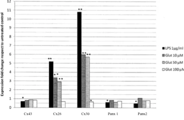
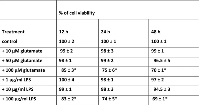
iii) Expression oattern of connexins and pannexins in primary human astroglial cell cultures exposed to glutamate or lipolysaccharide: the effects of the administration of LPS (1,10, 100 µg/ml) or glutamate (10, 50, 100 µM) for different time intervals (12, 24 and 48 h) were tested in human astrocytes. Four parameters were compared: cytological modification, cell viability, level of ROS and GSH, the level of mRNA of connexins (Cxs): Cx26, Cx30, Cx43 and pannexins (Panx): Panx 1 and Panx2. Main findings were:1) the stressors (LPS and glutamate) increased the mRNA level of Cx26 and Cx30, while Cx43, Panx1 and Panx2 were not significantly modified; 2) the level of Cx26 and Cx30 increased strongly at lower doses of stressors while increasing the doses of the stressors, the increment of Cx26 and Cx30 progressively lowered. Conversely, the level of the ROS increased with the doses of the stressors; 3) an inversed relation occurred between level of Cxs (Cx26 and Cx30) and grade of injury of the cells (as proved by astrocytosis and by MTT test).
Since there will be a close correlation between neurodegenerative disease and expression of connexin, the last step of the work concerns the possibility to control
the expression of connexins by the administration of a neuroprotective agent such as lithium.
LIST OF ABBREVIATIONS CNS: central nervous system
Cx: connexin GJ: gap junction
GJC: gap junction communication HCs: hemichannels
Panx: pannexin
GJIC: gap junctional intercellular communication Cx43: connexin 43
Cx26: connexin 26 Cx30: connexin 30 Panx1: pannexin 1 Panx2:pannexin 2
qRT-PCR: Quantitative real-time Polimerase Chain Reaction Vm: membrane voltage
Vj: transjunctional voltage CaM: calmodulin
MAPK: mitogen-activated protein kinase CBX: carbenoxolone
PD: Parkinson‘s disease
INTRODUCTION
1. Gap Junctions
Cells in tissues share ions, second messengers, and small metabolites through clusters of intercellular channels called Gap Junctions. The intercellular communication coordinated cellular activity and is a key condition for the existence of pluricellular organisms. Gap junctions are clusters of intercellular channels that allow direct diffusion of ions and small molecules between adjacent cells. Without this type of direct transmission, the exchange of information would not be possible. Indeed GJs regulate cellular synchronization, cells growth and metabolic coordination in tissues. Gap Junctions are intercellular channels composed by transmembrane proteins family called connexin (Cx) and recently, a new important class of GJ proteins has been recognized in vertebrates, the pannexins (Panxs), which are considered homologous to the innexins of the invertebrates. Phylogenetic analysis revealed that Panxs are highly conserved in
Nematoda, Mollusca, Arthropoda and also in mammals 1 . The high maintenance of
the Panxs in classes which are so distant in terms of phylogenesis suggests the importance of their functions 2 . They are present in all Metazoan kingdom. The first proteins identified have been the connexins, found only in Chordates. Indeed, in invertebrates are present the innexins, similar in the structure and membrane topology to connexins but not in the amino acidic sequence. In recent years, by sequencing of mammalian genomes, pannexins have been identified as genes homologues to innexins [3,4]. Three Panxs and more than 20 Cxs were cloned in mammals. Each cell type express a specific set of these proteins. The set of Cxs and Panxs expressed in single cells is functionally important because their combination in GJ channels is critical for permeability to specific signaling molecules or ions 5 . Each cell type expresses a given set of GJ proteins, but their expression pattern is not stable in time. In fact, the pattern expressed in resting condition usually changed after injury both in vitro 6 or in vivo models 7 , suggesting altered permeability to signaling molecules in suffering conditions. These proteins are characterized by a similar characteristic structure,
comprising four alpha helix transmembrane domains (TM1-TM4), N- and C-terminal intracellular regions, two extracellular loops (E1-E2) and one cytoplasmatic loop (I1) [8, 9]. This structure is essential for the formation of a hemichannel, indeed six Cxs, Panxs or Inxs oligomerize to form a hexameric pore complex, respectively called connexon, pannexon or innexon. Two opposing hemichannels, each arising out from a cell, give rise to a gap junction, most commonly assembled as GJs plaque, characterized by a reduced space between the cells (about 2-4 nm) and composed by clusters of few or hundreds of gap junction channels. The association of the two hemichannels is mediated by H-bonds occurring between the extracellular loops of the proteins (Figure 1).
Figure.1 Schematic representation of gap junctions and their components. Two cells
are interconnected by a plaque of gap junction channel, every formed by the opposition of two hemichannels. (On the right) Structure of a protein forming GJs characterized by: four transmembrane domains (M1-M4); N- and C-terminal intracellular regions; two extracellular loops (E1-E2) and one intracellular loop.
1.1 Connexins
Connexins are the molecular constituents of Gjs, which are clusters of intercellular channels that allow the direct intercellular exchanges of ions and small molecules (e.g., IP3, ATP, glutamate, and energy metabolites). The connexins proteins are encoded by a family of genes divided in five subfamilies α, β, γ, δ and ε. The five groups show differences in the structure and sequence due to a different phylogenetic origin.
Up to now, the family of connexins genes comprises 21 members in human and 20 in mouse genome, of these 19 are orthologous. The nomenclature of connexins includes the indication of the species of origin, ―h‖ for human and ―m‖ for mouse (the species in which they have been first studied), the family name ―Cx‖ and the number corresponding to the predicted molecular mass deduced on the basis of cDNA sequence (range of 23-62 kDa) 12, 13 . There is also another nomenclature, based on the use of the abbreviation ‗‗Gj‘‘, for gap junction, the group they belong (A, B, C, D, E for α, β, γ, δ and ε respectively) and by a number in according to the order of discovery. For example, mCx43, the first discovered connexin, belonging to the α-group, is also called Gja1.
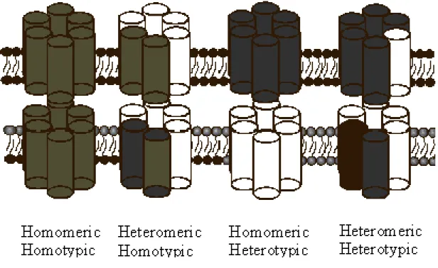
Figure 2. Gap junction channels formed from two identical hemichannels are called
homotypic, while those with differing hemichannels are heterotypic. In turn,
hemichannels of uniform connexin composition are called homomeric, while those with differing connexins are heteromeric. Channel composition is thought to influence the function of gap junction channels.
Various tissues and cell type can express more than one type of connexins. Connexins can form a large variety of channels; consequently, the connexons can be homomeric, if composed by the same type of connexins, or heteromeric in the case of two or more type of Cxs. Furthermore, a gap junction can be homotypic, if constituted of two identical hemichannels made from one type of connexin, or heterotypic, i.e. formed by two different hemichannels, each of which is made of a different type of connexin. Not all the connexins are able to form a heterotypic GJ. The different composition dictates different physiological properties as gating, single-channel conductance and the permeability to biological molecules [14-16] (Fig.2). The oligomerization of the connexins into a hemichannel (HC) occurs in the endoplasmic reticulum (ER), where the neoformed connexons pass in the Golgi apparatus and then in trans-Golgi network. They interact with chaperone proteins and inside to vesicles are transported along microtubules and actin filaments to the membrane cell, through which they freely
diffuse and are integrated in the outer edges of existing plaque. The connexon arrived at the membrane cell, is aided by E- and N-cadherins, to bind with another connexon arising from adjacent cells and to form the gap junction channel [16, 17-19]. Gap junction plaques are highly dynamic regions, in which new connexons are added at the periphery and old connexons are removed from the centre of the plaques. The removal occurs with invagination of a vesicle containing a portion of membrane with all or a part of GJ. This structure, named ―annular junction‖, is released in cytosol and its components are degraded through the lysosomal and proteosomal pathway [20-22]. Gap junctions biosynthesis and assembly are tightly regulated, indeed these structure have a half-life of only few hours. This rapid process is probably fundamental for a quick adaptation of the cells to mutated physiological or environmental conditions (Figure 3).
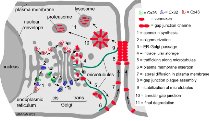
Figure 3. Schematic representation of the steps that lead to synthesis, assembly, and
degradation of gap junction membrane channels based on the current literature. Gap junction biosynthesis and degradation involves (1) synthesis of connexin polypeptides at endoplasmic reticulum membranes, (2) oligomerization into homo- and heteromeric gap junction connexons (hemi-channels), (3) passage through the Golgi stacks, (4) intracellular storage within trans Golgi membranes, (5) trafficking along microtubules, (6) insertion of connexons into the plasma membrane, (7) lateral diffusion of connexons in the plasma membrane, (8) aggregation of individual gap junction channels into plaques, (9) stabilization of peripheral microtubule plus-ends by binding to Cx43-based gap junctions, (10) internalization of the channel plaque leading to cytoplasmic annular junctions, and (11) complete degradation via lysosomal and proteasomal pathways. (Image from Segretain and Falk, 2004)
1.1.1Gap junctions functions
Gap junctions are implicated in a large variety of functions as embryonic development, morphogenesis, cell differentiation, cell proliferation and migration, electrical and mechanical synchronization (cardiac, muscle and brain cells), transmission of trophic or death molecules. All these functions have been discovered using targeted mutated connexins or through the over expression of some connexins isoforms. The importance of these proteins and, hence, of gap junctional communication is evident by the large number of human genetic diseases associated with connexins mutations or with pathogenic single nucleotide polymorphisms. Among this for example the X-linked Charcot-Marie-Tooth syndrome, a peripheral neuropathy with atrophy of distal muscles and low number of myelinating fibers, has been linked to mutations in Cx32 gene, suggesting its participation in myelination of peripheral nerves. Mutations in Cx43 can cause oculodentodigital dysplasia characterized by craniofacial, neurologic, limb and ocular abnormalities. Still mutations of Cx46 and Cx50 result in cataracts [23-27]. Connexins proteins are expressed in all tissue except in differentiated skeletal muscle, erythrocytes and sperm cells. This almost ubiquitary presence is a further confirmation of their importance for the correct functioning of the organisms. Recent studies show that connexons are also active in single plasma membranes and that they might be essential in intercellular signalling beyond their incorporation into gap junctions, so they may act as hemichannels (HCs) in different physiological and pathological process. In contrast to GJs, HCs show low open probability and low permeability to small molecules under resting conditions. HCs have been implicated in autocrine/paracrine signalling to provide a pathway for release of ATP, glutamate, NAD+ and prostaglandins. The role of hemichannels seems to be very important, because they act in the cells of various organs in response to extracellular signaling, injury, ischemic preconditioning and mechanical stimulation [28 - 30].
1.1.2 Functional control of Gap Junction conductance
A gap junction forms a hydrophilic channel pore of about 100-150 Å in length and 12.5 Å in width. It allows the passage of small molecules under 1kDa like ions, water, nucleotides, small peptides, metabolites. In this way, GJs provide to ionic and metabolic coupling among the cells. For some substances, this coupling is bidirectional and is driven by an electrochemical gradient, for others GJs there is a high degree of selectivity. Indeed the channels composed by different connexins show different permeability; for example some channels are specific for the cations or the anions, while others are able to discriminate between similar molecules as cAMP and cGMP. The junctional conductance, i.e. the passage of molecules or ions through GJ channel, is subjected to regulation by a number of physiological factors, such as voltage, intracellular pH and calcium, second messengers, or phosphorylation. Conductance of a single homo-connexins channel ranges from ~10 picoSiemens (pS) to ~300 pS [31-34]. The most important factor for the permeability is the structure of channel pore. Indeed the pore width, the electrical field and the electrical charge on the pore surface affect the permeability of ions and molecules passing through the channel. The permeation pathway of a gap junction channel consists of an intracellular channel entrance, a pore funnel and an extracellular cavity. The pore funnel surface is formed by the six N-terminal regions of connexins. Because the funnel forms a constriction site at the cytoplasmic entrance of the pore, the size and electrical character of the side-chains in this region should have a strong effect on both the molecular cut-off size and the charge selectivity of the channel. Indeed the substitutions or deletions of residues in N-terminal regions can affect single channel conductance, molecular permeability and charge selectivity [34].
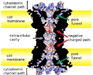
Figure 4 Pore structure of the Cx26 GJ channel. The permeation pathway of Cx26 GJ
consists of an intracellular entrance (the pore funnel formed by N-terminal regions), a negative charged path and an extracellular cavity. (Image adapted from Maeda et al., Cell. Mol. Life Sci, 2011)
The gap junctional communication is regulated at multiple levels. The regulation can be at short or long-term; the more rapid mechanism involves changing in the conductance or in the probability of opening of a single channel while in the slow mechanism occur an alteration of number of channels for changes in synthesis and degradation. Gap junction channels have different gating mechanisms are voltage sensitive. Indeed, they are under the influence of two types of electrical field, the membrane voltage (Vm), i.e. the voltage difference between intra and extracellular space, and the transjunctional voltage (Vj), that is established when the membrane voltages are not equal in the two coupled cells. Some connexins are sensitive to both Vj and Vm, others only to Vj. As regard the dependence by Vm, different connexins show different sensitivity; for example Cx43, Cx30 and Cx26 channels close with depolarization, whereas Cx45 upon hyperpolarization. The dependence of junctional conductance by Vj regards all GJs analyzed; so this mechanism is specific for GJs channels and is characterized by two forms of gating, fast or low, depending by the time reaction of opening and closing. Different studies revealed that N-terminal domain, forming the pore funnel, determine the magnitude and polarity of Vj. A cytoplasmic movement of the N-terminal portion,
where the voltage sensor is believed to reside, has been suggested to initiate voltage-dependent gating (Figure 4). Therefore, the substitution of the residues on N-terminal, changes the conductance or sensitivity. Indeed there are hemichannels that are close at positive voltage, as Cx26, Cx30, while others at negative voltage, Cx32, Cx43, Cx45 [36 - 40].
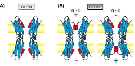
Figure 5 Plug gating model for transjunctional voltage-dependent gating of the Cx26
gap junction channel. (A): When there is no difference in membrane voltages between two neighbouring cells N-term region (in red) form the pore funnel and attach to TM1 by hydrophobic interactions. (B): When there is a difference in membrane voltages between two cells, the positive electric field causes the releasing of N-term from TM1. Once released, term region will assemble on the top of the pore with the others N-term region. (Image adapted from Maeda et al., Cell. Mol. Life Sci, 2011).
Others factors modulating gap junction conductivity are the Ca2+ and pH. An increase in intracellular calcium concentration reduces GJ communication. Indeed it is a signal to
and cell types, in a range from 500-600 nM to M concentration of Ca2+. There are also experimental evidences that the gating of GJs, Ca2+-dependent, could be mediated by calmodulin (CaM), an intermediate messenger protein that transduces calcium signals by binding calcium ions and then modifying its interactions with various target proteins, among which there are connexins [41, 42]. Also an increase in intracellular pH decreases junctional conductance of gap junction channels and some connexins are markedly more sensitive to acidification than others, although it‘s not still clear whether H+ acts directly on GJ channel [43- 45]. An extensive way with which connexins are regulated regards several post-translational modifications. The C-terminal region of Cxs contains serine, tyrosine and threonine, residues that may be phosphorylated by different protein kinases. At least eight kinases, including protein kinase A, protein kinase C, mitogen-activated protein kinase (MAPK) and so many phosphatases control the phosphorylation/dephosphorylation of various connexins. Except for the not phosphorylable Cx26, these modifications in Cxs are associated with changes in gating, conductance, permeability, but this depends on specific connexin isoforms or cell type. However, other modifications have been identified such as hydroxylation, methylation, and acetylation. Other effects regard the assembly into GJ plaque, their biosynthesis or their proteolytic degradation, regulating in this way their half-life [46-49]. In addition, ubiquitination would seem an important mechanism which connexins are regulated; the most studied Cx43 for example, can be ubiquitinated at plasma membrane or at ER level. Recently it has been implicated also a stimolation for Cx43 [50-52]. There are chemical agents able to block the GJ communication, they belong to multiple class and structures. Some are lipophilic such as heptanol, octanol and halothane, glycyrrhetinic acid derivatives such as carbenoxolone (CBX), quinine derivatives and some others different for structure and action mechanism. These molecules have been used to study the functions of GJs in vitro and in vivo, but although many of these are used to treat common diseases, as quinine for malaria, they give a plethora of side effects, due sometimes by blocking other channels. The development of selective blockers will allow better understanding of GJs communication [53].
1.2. Pannexins
Pannexins belong to a new family of proteins, also able to form GJs. They have more similar homology with innexins than with connexins, so that initially it was thought
they belonged to the innexins family. Many later studies showed that Panxs are a distinct family of proteins; they do not share the strongly conserved residues of Inxs even if as these last possess two cysteine residues in extracellular loops, and in contrast to connexins have a larger intracellular loops (~68 aa vs. ~30 aa).In the genomes of human and rodent species, are coded three proteins, Panx1, Panx2 and Panx3. Their calculated molecular mass is respectively 48, 73 and 45 kDa. In human genome Panx1 and Panx3 gene are located on chromosome 11 and Panx2 on chromosome 22, while in rat genome the first two are located in chromosome 8 and Panx2 on chromosome 7 [ 54, 55]. Panx1 and Panx2 transcripts are found in many rodents tissue, as brain and spinal cord, eye, thyroid, prostate, kidney; in humans are found also in hearth, gonads and skeletal muscle (in which there is no presence in rodents). Panx1 protein is expressed in numerous tissues in both human and rodents. Instead Panx2 is present in many tissues in rodents, but in humans is expressed only in the central nervous system (CNS). The expression of Panx3 seems confined to skin and cartilage cells, even if real-time PCR screening indicate that Panx3 mRNA is present also in kidney, spleen and brain [ 56, 57]. Pannexins have been found in large accumulation in ER and Golgi apparatus, suggesting that post-translational modifications and assembly in pannexons occur in the same way as demonstrated for connexins. The first studies on this protein were made by Bruzzone et al. using an expression system in Xenopus oocytes, in which they discovered that the channel formed by pannexins, if activated by voltage, had given rise to passage of molecules. But they concluded that Panx1 alone was able to form homomeric channels, while Panx2 could form only heteromeric channels with Panx1 [58]. To date, increasing evidences show that also Panx2 alone is capable to form a functional homomeric channel. Indeed the heteromeric channels Panx1/Panx2 are unstable and disaggregate in few hours, this could explain because the heteromeric is present in systems as Xenopus oocytes or in HEK 293, but in mammalian cells and brain tissue the two proteins exist separately [59]. Another controversy regards the ability of Panxs to form or not gap junction channels, but recent studies revealed evidence that they may form GJs in vitro cells, but to date it hasn‘t been demonstrated in vertebrate. As connexons, after activation pannexons open into large non-selective pores permeable to ions and small molecules up to 1 kDa and under a wide range of membrane depolarization. Panxs hemichannels differ from connexins HCs because they exhibit larger currents, faster kinetics of pore opening and a conductance of over 500 pS [58]. Different stimuli are able to open pannexons, among these mechanical stress, positive membrane potential, extracellular ATP, elevation in intracellular calcium, ischemic insult and inflammation, while acidification of cytoplasm cause the closing of
the channels. The presence of mechanisms distinct from those of connexins, makes possible the co-expression of the two proteins types in the same cells [60]. The pannexons may play a role in generation of oscillatory and synchronization activity in brain. They can be involved in the propagation of calcium waves, to which normally have been implicated the GJs connexins-formed [61]. Panxs would use a mechanism based on release of adenosintriphosphate (ATP). This has been demonstrated in blood endothelium, where mechanical stress or ischemia open pannexons in endothelial cells and cause the extracellular release of ATP. The released ATP acts on purinergic receptors P2Y, whose activation has effect on phospholipase C, increasing the IP3 and causing the release of calcium from intercellular stores; this at the end cause a consequent release of other ATP that can propagate using classical way of GJs or via pannexons. In this way, the propagation is ensured also without direct cell contact. This action probably is essential for the cells in normal tissue because Panxs channels, contrary to Cxs channels gated by cations as Ca2+ or Mg2+, are able to open when external concentration of calcium is at physiological level. So pannexons may propagate calcium waves and to have a role in vasodilatation, inflammatory response and ischemic death of neurons [62- 64]. Other evidences of calcium waves propagation are in erythrocytes, where there is expression only of Panx1 and not of Cx43 (the main Cx involved in calcium waves). In these the release of ATP occurs in response to ischemia and mechanical stress and after exhibit the same pathway observed in endothelium [65]. The Pannexins are the only ones, between the three families, to be extensively glycosylated. There is the presence of consensus sequence on the second extracellular loop and this modification seems important for intracellular trafficking and insertion in the cell membrane. Moreover, the glycosylation could prevent docking with other pannexons in adjacent cells [11, 66]. By analysis of amino acids sequence there are one phosphorylation site for Panx1 and multiple site for Panx2 and, as in connexins, these allow the activation/deactivation of the channels [67]. Another possible regulator of Panxs is the β-subunit of voltage-dependent potassium channel (Kvβ3), a protein belonging to the family of regulatory beta-subunits of the voltage-dependent potassium channels. The co-expression of Panx1 and Kvb3 has been showed in principal neurons of the hippocampus and in Purkinje cells of the cerebellum. The mechanism would involve the binding of Kvβ3 to the carboxy-terminus of Panx1 and maybe in response to changes of the intracellular redox potential, it would control the inactivation mechanisms of Panx1 hemichannels 68 .
1.2.1. Pannexins in CNS
Pannexin 1 and Panx2 transcripts are detected in many regions in rodents CNS, retina, cortex, hippocampus, cerebellum, olfactory bulb, spinal cord. Panx1 is expressed primarily in neurons and maybe in oligodendrocytes. Panx2 seems to be brain-specific, but the two proteins are inversely regulated during the development of the rodents brain. Panx1 show high levels of expression in the embryonic and young postnatal brain and decline considerably in the adult, whereas Panx2 mRNA expression is low in the prenatal brain but increase substantially during subsequent postnatal development with peaking at postnatal day 15 [69, 70]. Pannexins transcripts are particularly abundant in the adult cortex, in hippocampal and neocortical pyramidal cells but also in GABAergic interneurons, (to note the presence in both excitatory and inhibitory neurons, while Cx36 is present only into inhibitory interneurons), reticular thalamus, the inferior olive, magnocellular hypothalamic neurons, midbrain and brain stem motoneurons, Purkinje and Golgi cells in the cerebellum [6]. Panx1 protein is widely expressed in mammalian tissues; as regard brain is found in several regions including cortex, striatum, olfactory bulb, hippocampus, thalamus, inferior olive, inferior colliculus, amygdala, spinal cord, retina, and cerebellum. At the cellular level, Panx1 has been localized in different neuronal types, including olfactory bulb mitral cells, Purkinje cells, dopaminergic, cholinergic and glutamatergic neurons. In the cerebral cortex and hippocampus is localized in the postsynaptic cell membranes.
Panx1 is also detected in cultured astrocytes, immature oligodendrocytes and neurons, using immunofluorescence and Western blot analysis, but it has not been found in the cell surface of astrocytes [6, 69- 73]. Panx2 protein is expressed exclusively in the brain including the olfactory bulb, hippocampus, amygdala, superior colliculus, substantia nigra, cerebellum, hypothalamus and spinal cord. Its presence it has been confirmed in the majority of pyramidal cells and in GABAergic interneurons but as regard the expression in glial cells there are still contrasting opinions; under resting conditions, hippocampal astrocytes do not express Panx2 even if it appears in hippocampal astrocytes several hours after ischemia/reperfusion [6, 71-74]. The presence in brain becomes important because pannexins are present in all regions in which lack connexins but are alike coupled both metabolically and electrically. For example, the abundance of Panxs in Purkinje cells or in hippocampal pyramidal cells, that do not express Cx36 or Cx45, suggests that these are responsible for electric coupling and generation of high-frequency oscillations. Experiments in mice KO for Cx36 show a reduction in gamma frequency, while ultrafast oscillations are not modified. These data are consistent with
the absence of Cx36, present only in hippocampal interneurons, while other channels may determine fast oscillations occurring in pyramidal cells [75-77]. However, the impact of Panxs channels on neuronal network synchronization remains to demonstrate. It remains also unknown whether pannexins form functional GJCs and/or hemichannels. There are many controversial works regard the formation of GJs; in some systems as Xenopus oocytes or C6 glioma cells an over-expression of Panx1, but not Panx2, mediate intercellular coupling, while in other systems and other groups report no communication. In contrast, functional hemichannels formed by both Panx1 and 2 are demonstrated in different cellular system and in hippocampal neurons [70, 71, 78 .
1.3. Anatomical distribution of Gap Junctions in the mammalian brain
Electrical synapses are prevalent during the early phase of neurogenesis. Studies both in vitro and in vivo showed that gap junctions coupled all progenitor cells, neuroblasts and proliferating cells, in order to coordinate the neuronal communication. After about two weeks of postnatal development this communication is replaced by chemical synapses, but in mature neurons the expression of connexins is maintained and has a role in the coordination of neuronal activity and in mediation of synchrony and network oscillations [79- 81]. The expression of different connexins depends by developmental stage, cell type and brain region. With electrophysiology, transgenic animals, cell imaging and freeze-fracture replica immunolabeling (FRIL) it has been possible examine the presence of these proteins in CNS vertebrates. Several different connexins were detected in different nervous cells type.
Neurons
Electrotonic coupling between mammalian neurons has been shown in many areas of the CNS and has been implicated in neuronal synchronization [82-85]. During development, there is a high degree of intercellular coupling between neurons. Studies in the rat indicate that neuroblasts are coupled with approximately 30–60 others in columns within the ventricular zone of the developing cerebral cortex [86-89]. The zone is comprised of mitotically active epithelial cells lining the ventricles. These cells have been shown to express Cx43 and Cx26 [90], and the coupling appears to involve both neural precursors and radial glia [91]. Thus gap junctional intercellular communication (GJIC) may establish cortical domains in the developing neocortex that underlie the
adult pattern of functional archi- tecture [88,89,92]. Although Cx43 has been reported in neurons of the cortical plate, the expression of Cx32 in mature neurons [93] is controversial [94, 95]. The expression pattern of Cx26 [96] and Cx36 [97,98] in the developing brain is more consistent with the transient gap junctional coupling observed in the neocortex. In addition, Cx47 is also observed in the CNS [99], but its expression seems to be different from that of Cx36 in neurons of developing CNS, but there is some co-localization with Cx36 in cerebellum [99]. Cx37 is observed in motor neurons and Cx40 is expressed in developing neurons of spinal cord [100]. In the adult CNS, neurons are coupled via gap junctions mainly composed of Cx36 [95] and Cx45 [101] in the cortex and hippocampus. These neuronal gap junctions play an important role in forming electrical synapses [102,103]. Cx45 is expressed widely in the developing brain and in the adult brain localized in cerebral cortex, hippocampus and thalamus [101]. Cx45 is also observed in olfactory nerves [104]. In the retina, Cx26 is expressed in horizontal cells [105] and Cx36 in AII amacrine cells [105], although Cx26 is not shown in the neurons in the adult cortex and hippocampus [106]. Recently, it has been reported that hemichannels of horizontal cells in the retina are mainly composed of Cx26 and regulate the activity of the Ca2 + channels and subsequent glutamate release [105]. The availability of various antibodies has made it possible to detect connexins which compose gap junctions [95,107] as well as hemichannels [108, 109], allowing characterization of the functional state, distribution and colocalization of connexins. In order to understand the role of specific connexins in the CNS, a knockout strategy has been employed by several investigators. Initial examination suggested some subtle neural changes may be apparent in the Cx43 knockout mice at birth [110]. However, the neural phenotype of the Cx43 knockout mice is confounded by cardiac malformation and neonatal death [111]. In these mice, the abnormal migration of neural crest was observed [112,113]. More recently, we have reported a disturbance in the migra- tion of neurons in the neocortex of the Cx43 null mice [114]. Cx32 knockout mice are viable and fertile, and display demylenation in the PNS [115,116]. Moreover, neuronal hy- perexcitability and myelin defects in the neocortex were observed [117], and the accumulation of oligodendrocyte progenitor cells and amplified apoptosis has been reported in the CNS of Cx32 knockout mice [118]. Cx36 knockout mice are viable and display no obvious anatomical abnor- malities [119], however detailed neurodevelopmental and anatomical studies have not been reported. Studies using Cx36 knockout mice in vitro suggest that the neuronal gap junctions are critical in the formation of gamma frequency oscillations in the hippocampus [103] and in generating synchronous activity in the cortex [102]. These mice show selective impairment of
hippocampal gamma oscillations [120]. Knockout of the other neuronal candidate, Cx26, is lethal at E9 – 10[121], and thus additional strategies must be followed to investigate its function in neuronal development [122]. Cx45 knockout mice die at E8-9 due to abnormal vascular development. Therefore, it is impossible to study the gap junctional function using adult Cx45 knockout mice. The expression of Cx45 has also been reported in the embryonic cortex and hippocampus [123], but its role in the development of the CNS remains to be determined. In vitro studies have suggested a role for gap junctional coupling in neuronal differentiation. When NT2 human embryonal carcinoma cells differentiate into neurons in response to retinoic acid (RA), the expression of Cx43, and the level of gap junctional coupling, progressively disappear [124]. Blocking of gap junctions disrupts RA-induced neuro- nal differentiation of both human NT2 [125] and mouse P19 cells [126]. Moreover, the differentiation of NT2/D1 progen- itor cells are reduced by blocking of gap junctions and hemichannels, suggesting hemichannels also play a role in the neuronal differentiation [127]. It therefore appears that the temporal pattern of connexin expression and gap junctional coupling during neuronal differentiation is critical.
Astrocytes
The main cell type in the brain coupled by gap junctions is the astrocyte. Astrocytes have traditionally been viewed to have a role in the metabolic and trophic support of neurons [128]. Intimate interactions have been shown to be involved in the role of radial glia in directing migration of neurons in the cortex [129]. Indeed, gap junctional coupling between radial glia and neural precursors may be critical for this process [30]. Gap junctions in astrocytes are primarily composed of Cx43 [130,131], in addition to other connexins, including Cx30 [132,133], Cx47 [134], Cx40, Cx45, Cx46 and Cx26 [134]. Moreover, gap junctions provide a substrate for formation of a functional astrocytic syncytium [130,131,136,137], implicated in the spatial buffering capacity of astrocytes, particularly in dealing with extracellular K+ arising from neuronal activity [138,139]. The propagation of intercellular Ca2 + waves is an important feature of astro- cytes in response to activation [140]. Studies from several laboratories have shown that gap junctions are involved in mediating intercellular Ca2 + signaling throughout the glial syncytium [141-143]. Astrocytic hemichannels also play a role in the release of adenosine triphosphate (ATP) associ- ated with Ca2 + signaling
[144,145]. Moreover, the inhibition of glycolytic and oxidative metabolism resulted in an increase of astrocytic hemichannels [146], suggesting in- volvement of hemichannels in this pathological condition. Astrocytes cultured from Cx43 knockout mice exhibit reduced gap junctional coupling and Ca2 + wave propagation [147, 148]. Meanwhile, Cx43 knockout astrocytes express other connexin subtypes (Cx30, 40, 45, 26, 46) [135], suggesting that connexins other than Cx43 could not compensate for the reduction of GJIC in astrocytes. Even astrocytes derived from heterozygote Cx43 knockout mice showed a significant reduction in gap junctional coupling [147, 149]. These hetero- zygote mice have been used for in vivo studies (see below). Recently, mice lacking Cx43 specifically in astrocytes have been generated by using the cre-recombinase system [150]. These mice can survive to adulthood, unlike Cx43 knockout mice, and exhibit amplified motor activity with increased hippocampal spreading depression, providing a valuable model for in vivo studies. Although Cx30 has been shown only in astrocytic gap junctions in the CNS, no major abnormality was observed in the brains of Cx30-deficient mice, although they exhibit severe hearing loss [151].
Microglia
Microglia are important cells in the CNS, participating in the reactive gliosis. Although resting microglia do not show phagocytosis, activated microglia behave like phagocytes [152]. Under normal conditions, microglia show little expres- sion of Cx43, however, this increases following stimulation by inflammatory cytokines [6] or Ca2 + ionophores [153], allowing for enhancement of gap junctional coupling. Cx36 has been also found to form gap junctions in microglia [155], suggesting the direct communication between microglia. Moreover, brain macrophages/microglia decrease Cx43 expression and gap junctional coupling in co-cultured astro- cytes [155, 156]. It is important not only to understand the phagocytic function of microglia but also to understand the role of microglial gap junctions in the inflammatory response of the CNS.
Oligodendrocytes
Several reports have indicated that oligodendrocyte gap junctions are composed of Cx32 [157-162], Cx36 [154], Cx29 [163] and Cx47 [164]. We have shown that
expression of Cx32 coincides with maturation of oligodendrocytes temporally and spatially [159,165]. The function of gap junctions in oligodendrocytes is thought to be primarily metabolic to allow ions and nutrients to pass from the somata to all the layers of the myelin [166,167]. The importance of this channel has recently been realized by the reported mutations of Cx32 associated with X-linked Charcot – Marie – Tooth disease, a peripheral demyelinating disorder [168 – 172]. It was initially thought that human and rodent Schwann cells are susceptible to pathology, leading to peripheral nerve demyelination, while oligodendrocytes appeared not to be affected. Cx32 knockout mice showed a reduced myelin volume and an enhanced excitability in the CNS [117], and a progressive peripheral neuropathy has been observed after 3 months of age [115,116]. Cx29 has also been specifically observed in oligodendrocytes and Schwann cells [163]. The expression of Cx29 exists mainly at the paranodes and juxtaparanodes [163], whereas Cx32 is not observed in the paranodes [176], indicating a difference in the distribution of these two connexins. In the pathogenesis of demyelin- ation associated with the Cx32 deficiency, the compensatory role of Cx29 may have to be explored. Recently, Cx47 was observed to be expressed mainly in oligodendrocytes [164]. Cx47 null mice exhibit degeneration of nerve fibers, particularly in the optic tract [164]. Moreover, Cx47 and Cx32 double knockout mice exhibited a more severe demyelination in the CNS [164,177], indicating that both connexins play a critical role in myelination.
Schwann cell
The Schwann cells of peripheral nervous system. These cells proliferate after nerve development or after injury to promote the myelination; maybe GJs play a role in this process, distributing and synchronizing important signals to proliferation. Cx32 and Cx29, identified in Schwann cells, show the same distribution pattern at paranodal regions and in Schmidt-Lanterman incisures (small canals that interrupt the myelin sheath present in nerve cells; these incisions allow the nourishment of the cells isolated from the myelin). Cx32 is required for normal functions in peripheral nerve, as demonstrated with Cx32 knocked out (KO) animals or in Charcot-Marie Tooth disease. The role of Cx29 is not clear for controversial effects described in KO animal models [178-181].
1.4. Gap Junction in neuropathological condition 1.4.1. Brain ischemia
Cerebrovascular diseases rank are the third leading cause of death in the USA and fourth in Canada, most commonly manifesting as ischemic brain stroke [182,183]. In the context of experimental brain ischemia models, the neuro- protective role of astrocytic gap junctions is still controversial. Blocking astrocytic gap junctions enhances neuronal vulnerability to glutamate cytotoxicity in culture [184]. Moreover, blocking gap junctions in a hippocampal slice culture enhanced neuronal damage under experimental ischemia using oxygen and glucose depletion [185]. In vivo, heterozygote Cx43 knockout mice showed a significantly increased stroke volume compared to wild-type mice following middle cerebral artery occlusion (MCAO) and exhibited enhanced apoptosis in the penumbra [186]. These results suggest that astrocytic gap junctions play a neuroprotective role in oxidative and metabolic stress. On the other hand, neuronal death caused by oxygen and glucose depletion was decreased when Cx43 was blocked by specific antisense oligodeoxynucleotide in the hippocampal slice culture [187]. Similarly, the stroke volume following MCAO in the rat model was reduced by blocking gap junctions using octanol [188]. Therefore, it has been suggested that the spreading depression caused by ischemic insult goes through astrocytic gap junctions which remain open during the ischemic condition, resulting in the expansion of the stroke volume [189]. However, there are problems in interpreting the results obtained with gap junction blockers such as octanol because of the lack of specificity, particularly when administered systemically. In addition, there is no selectivity with regard to astrocytic gap junctions since all gap junctions in the tissue are affected. The use of more specific approaches to target gap junctions is desirable, for example using Cx43 antisense and interfering RNA. More recent use of targeted deletion of connexins, specifically in astrocytes, is providing evidence that astrocytic gap junctions play a neuroprotective role in ischemic insults through reduction of apoptosis and inflammation [190]. The role of astrocytic hemichannels in ischemic insult is still unknown. Recently, it has been reported that astrocytic hemichannels, which are closed under normal conditions, remained open under experimental ischemia induced by glucose and oxygen depletion. It is possible that open hemichannels allow for the release of glutamate causing loss of membrane potential [191], or hemichannels may contribute to the distribution of anti-apoptotic factors in the lesion area [192]. Cx32 knockout mice have been reported to exhibit an enhanced vulnerability of hippocampal neurons against brief global brain ischemia compared to wild-type mice [193],
suggesting that gap junctions of hippocampal interneurons play a neuroprotective role in ischemic insults. Moreover, expression of astrocytic gap junctions can be affected by macrophages and microglia express Cx43 following activation. In this context, the regulation of connexin expression can be mediated by various types of cells other than neurons and astrocytes following ischemic stress.
1.4.2. Epilepsy
There are two major clinical symptoms of epilepsy: the partial seizure where excessive electrical discharge is re- stricted to a given area in the brain, and the general seizure involving the entire brain [194]. The pathogenesis of seiz- ures may be associated with abnormal stimulation occurring in a certain region of the brain causing depolarization of the membrane, expanding to the surrounding cells. Gliosis at the lesion is usually observed in epileptic brain tissue. However, the participation of Cx43 which composes mainly astrocytic gap junctions is controversial (reviewed in Ref. [195]).In models of experimental epilepsy, strong recurrent excitatory activity, such as that produced by GABA receptor blocking, K+ pump blocking, and repetitive stimulation is used as the epileptic trigger [196]. Electrical coupling through neuronal gap junctions is reported to play an important role in the expansion of the epileptic wave [197]. Blocking of astrocytic gap junctions decreases Ca2 + oscillations in co-cultured neurons [197]. An increase of Cx32 expression was observed in the hippocampus in a model of bicuculline-induced epileptiform activity [198], although a decrease in the level of Cx36 mRNA has been reported in the hippocampus of the kainate-treated rat [199]. Meanwhile, neuronal gap junctions are required for the appearance of very fast oscillations associated with seizure activity [200]. Cx43 mRNA levels were increased in the temporal cortex of epilepsy patients [201]. Therefore, the role of gap junctions in epilepsy is still controversial. In the future, the gap junctional coupling between various types of cells in vivo should be evaluated under epileptic stimulation.
1.4.3. Demyelinating Syndromes
Alterations in connexins present in the myelynating glial cells (forming intercellular junctions in oligodendrocytes and autaptic, within themselves, in Schwann cells) all promote demyelination diseases. Interestingly, connexins present in the astrocytes, the major macroglial cell type in the nervous ystem and not traditionally associated with the
myelination process, also contribute to some myelin pathologies. Oligodendrocytes and Schwann cells express three different connexins: Cx47, Cx32 and Cx29, although only the first two are believed to form gap junction channels 202 . Whereas, Cx47 forms extensive gap junctions with astrocytes in soma and outer myelinated fibers, Cx32 is the most abundant within the layers of myelin itself (‗‗reflexive‘‘ or ‗‗autologous‘‘ gap junctions), between loops of the myelin sheath in individual oligodendrocytes and Schwann cells 203 , although it can also form gap junctions with other astrocytic connexins. These more direct pathways between the myelin layers allow a much shorter route for metabolite exchange. The precise mechanism by which deficient gap junction communication alters myelin formation and maintenance, and why some axonal fibers are more affected than others are questions that remain to be answered. Myelin gap junctions seem to be in an unique position not only to regulate metabolite trafficking to and from the myelin sheath but also to guarantee myelin structure and proper compaction by regulating ionic and water fluxes. Further studies on the interdependence between connex- ins, aquaporins, ion channels, the gliovascular interface, and cell–cell junctions and on how astrocytic proteins interact, in turn, with the myelinating cells, oligodendrocytes and Schwann cells, will lead us into a new exciting time to understand the role of glial cells in myelination 204 .
1.4.4. Neurodegenerative Disease
One of the major neurodegenerative diseases is Alzheimer‘s disease (AD). Clinically, cerebral atrophy is observed mainly in the frontal cortex of AD patients and pathologically, neurofibrillary degeneration and senile plaques are shown in the lesions [205]. The senile plaque is a round deposit composed of amyloid protein surrounded by astrocytic processes. An increase in the expression of Cx43 was observed at the site of these amyloid plaques. Pathological evaluations have revealed the importance of astrocytic participation in the lesion using presenilin mutant knockin mice [206,207]and apoE null mice 208 – 210]. However, there is no report clarifying the relation between gap junctions and AD. AD is a progressive disease and the lesion exhibits a successive expansion, suggesting that the glial network may play a critical role in the pathogenesis of AD. Parkinson‘s disease (PD) is a common neurodegenerative disease. Clinical symptoms are progressive tremor, muscle rigidity and gait disturbance [205]. In the brain of PD patients, the loss of dopaminergic neurons is observed in the substantia nigra-striatum [205]. The 1-methyl-4-phenyl- 1,2,3,6-tetrahydropyridine (MPTP)-model
of PD exhibited an increase of Cx43 expression in the striatum, although the coupling of astrocytes was not increased [211]. The alteration of gap junctions in the brains of PD patients has not yet been reported. Therefore, the pathological role of gap junctions in PD is still ambiguous. Although most of the mechanisms of tremors and dyskinesias which are commonly observed in PD patients are still obscure, the inferior olive has been focused on as the pathological generator of tremors [212, 213]. Neurons of the inferior olive are electrically coupled through gap junctions which plays a role in creating oscillatory activity [140]. Some studies have reported that the GJIC of inferior olive neurons is responsible for tremors [213,214], however, no difference of the severity of tremor induced by harmaline was observed between Cx36 knock- out mice and wild-type mice [215]. More experiments will be required to clarify the mechanism of tremors. In neurodegenerative diseases, it is also important to evaluate the inflammatory response because both AD and PD exhibit inflammation in the lesion [216]. Indeed, anti- inflammatory drugs may reduce the incidence of AD and delay the progression of the disease [217, 218]. As mentioned previously, activated microglia express gap junctions composed of Cx43 [6]. Moreover, activated macrophages decreased the expression of astrocytic gap junctions [7]. Therefore, further investigation of the possible gap junctional neuroprotective role of glial cells and inflammatory cells in neurodegenerative diseases is warranted.
2. Lithium and neuroprotection
Literatures data show the protective effects of lithium at the neuronal level. These include lithium modulation of autophagy, growth factors, excitotoxicity, and a variety of mechanisms underlying cell death, neurogenesis, and neuronal differentiation. All these effects represent the result of a multifaceted pharmacology, which is becoming more and more complex. Nonetheless, when trying to dissect the various mechanisms of action of lithium, two primary targets emerge: glycogen synthase kinase 3beta and
phosphatidylinositol phosphatase. The numerous lithium effects on biochemical
systems are placed downstream of these two main mechanisms. At several steps, these mechanisms interconnect to each other, thus making it difficult to keep distinct the biochemical cascades promoted by lithium. In this way, it is not surprising that, despite being described as different phenomena at the behavioral level, molecular mechanisms underlying the effects of lithium on mood, motor activity, and sensitization overlap with those responsible for neuroprotection and neurorestoration. It is likely that the ancestral
role of this ion as a modulator of cell survival, cell growth, movement, and mood is the consequence of a few molecular mechanisms operating in different neuronal networks, where a variety of cascade events take place. A variety of experimental models show neuroprotective effects of Li +, ranging froM acute neurological insults (ischemic, 219 ; epileptic, 220 ; traumatic, 221 ., and brain damage) to neurodegeneration [Huntington‘s disease, 222 ; Parkinson‘s disease (PD), 223 ; Prion diseases, 224 ; ALS, 225 ; cerebellar ataxia, 226 ; inclusion- body myositis, 227 ; dementia, 228 ;
229 ; 230 ]. Interestingly, the neuronal basis of neuroprotection might share the same mechanisms as the psychotropic effects of the drug. In fact, there is converging evidence that mood disorders and neuronal damage are often associated while both conditions seem to benefit from the modulation of the same intracellular pathways. Altogether these pre- clinical studies provide a strong background for the trans- lation of the neuroprotective effects of Li+ into modern neurological, and psychiatric practice. In switching from intracellular mechanisms to diseases, the powerful effects of Li+ administration on the expression of specific growth factors seem to be a key effect for the therapeutic role of Li+ both in neuronal degeneration and psychopathology. However, Li + induced neurotrophin changes in the brain are not always conclusive, and the mechanism of action remains partly unknown, calling for further studies aimed at elucidating this critical feature of Li + . A few experimental conditions in which Li+ provides neuroprotection involve glutamateneurotoxicity. Infact, Li+ pretreatmentisable to protect different cell types against excitotoxicity caused by glutamate, a-amino-3-hydroxy-5-methyl-4-iso- xazolepropionic acid, and kainate ( 231 ; 232 ; 225 ). Consistent with excitotoxicity, the protective effects of Li+ are likely to extend beyond VEGF or BDNF expression. In fact, Li + dose-dependently decreases the levels of p53 and Bax, and causes a time-dependent and dose-dependent increase in Bcl-2 233 . The p53 protein is a nuclear protein that binds to specific DNA sequences and functions as a transcription activator: it promotes the expression of Bax proapoptotic gene, but suppresses the expression of Bcl-2, an antiapoptotic gene 234 . Lithium- induced changes in the levels of Bax and Bcl-2 are likely to be because of inhibition of p53 expression. Lithium may also reduce directly the expression of Bax/Bcl-2; in fact, Li+ activates other transcription factors, such as cyclic AMP response element binding proteins ( 235 ; 236 ) and activating protein-1 ( 235 ; 237 ). All these Li + -induced changes in transcription factors depend on a direct inhibition of GSK-3b or PI3K/Akt/ GSK-3b. Finally, the powerful autophagy-inducing
the clearance of altered proteins and mitochondria, which provide a powerful defense for several subsets of neurons. Given the multiple steps affected by Li+ in the biology of the neuron and the presence of different targets contributing to its nonconventional pharmacology, it remains hypothetical to firmly propose a single mechanism of action for Li +-induced neuroprotection. Even focusing on excitotoxicity does not help very much as there are several potential mechanisms contributing to Li + -induced protection against glutamate overactivity. Moreover, it is very likely that protective effects of Li+ extend far beyond excitotoxicity. In fact, as reported at the beginning of the review, Li+ prevents detrimental processes within neurons, but also promotes neuronal sprouting and produces a neurogenetic effect. The ancestral role of this ion in mediating the basic functions of the cell seems to be bound to innumerable metabolic pathways, which call for extensive investigation to cover the complex pharmacology of such a small ion 238 . Among a plethora of intracellular transduction mechanisms affected by Li, two main effects emerge: the inhibition of GSK-3b and PI phosphatase. Several biochemical pathways located downstream to these enzymes are modulated by Li . A number of these pathways are interconnected, making it difficult to distinguish which effect derives from the sole modulation of a specific biochemical event. Consequently, when trying to translate such a complex biochemical pharmacology into clinical outcomes, it is puzzling to decipher which intracellular pathway plays a major role. For the same reason it is often arbitrary to distinguish between the mechanisms responsible for desired therapeutic effects and detrimental side effects. Lithium pharmacology remains under debate; nonetheless a growing body of evidence indicates novel biochem- ical targets of Li+ action. These include Li+ modulation of autophagy, growth factors, excitotoxicity, apoptotic cell death, neurogenesis, and neuronal differentiation. Remarkably, all these effects may well apply to the novel experimental therapeutics promoted by Li, and the classic effect of the ion as a mood stabilizer. In fact, the molecular mechanisms underlying the effects of Li on mood overlap with those responsible for neuroprotection and neurorestoration. It is likely that the ancestral role of this ion as a modulator of cell biology extends to a variety of neuronal networks producing several behavioral effects. In this way the novel pharmacology of Li is a sort of template that recapitulates the novel insights into the biology of neuropsychiatric diseases.
GLIA 58:1594–1609 (2010)
Dynamic expression of Cx47 in mouse brain development and in the cuprizone model of myelin plasticity
Rosalba Parenti1*, Federico Cicirata1, Agata Zappalà1, Angela Catania1, Francesco La Delia1, Valentina Cicirata1, Oliver Tress2, Klaus Willecke2.
1 Department of Physiological Science, University of Catania, V.le A. Doria, 6 95125 Catania, Italy
2 Institut fuer Genetik Rheinische Friedrichs-Wilhelm-Universitaet Bonn, Germany
Running title: Cx47 in development and plasticity process. Number of words: 9135 ; figures: 9
*Correspondence to: Dr. Rosalba Parenti, Department of Physiological Science, University of Catania, V.le A. Doria, 6 95125 Catania, Italy.
E-Mail: [email protected]
ABSTRACT
The study shows the dynamic expression of connexin47 (Cx47) in oligodendrocytes and myelin of mice, either in myelinogenesis occurring in early development or in an experimental model of new-myelinogenesis of adult mice. Cx47 first appeared in the embryonic mouse brain at E10.5; successively the expression increased, principally in regions populated by developing oligodendrocytes. The expression declined postnatally towards adulthood and was restricted to a few specific areas, such as the corpus callosum, the striatum, the cerebellum and the spinal cord. Since the expression of Cx47 in developing oligodendrocytes preceded those of Cx32 and Cx29, a role of Cx47 in myelinogenesis was postulated.
This hypothesis was tested in a model of re-myelination, which principally involved the corpus callosum, occurring in adult mice by treatment with cuprizone. Cx47 was up-regulated during demyelination and recovered during the remyelination phase. During demyelination, Cx47 was first over-expressed in the corpus callosum and later, when the myelin virtually disappeared in the injured areas, Cx47 was expressed in astrocytes located inside and closely around the demyelinated areas. The remyelination of injured areas occurred after stopping the administration of cuprizone and continued to complete recovery. In this period the expression of Cx47 shifted from astrocytes to new-formed myelin. So, Cx47 exhibits in this model a transient and de novo expression in astrocytes with a topographic segregation in the injured areas, only when oligodendrocytes and the myelin were most severely affected.
Taken as a whole the evidence would suggest that Cx47 play a key role in myelination.
INTRODUCTION
Intercellular communication through gap junctions (GJ) allows metabolic and electrical coupling of cellular networks. GJs are extensively distributed in the nervous system throughout life (Bruzzone et al., 1996). GJ channels are formed by oligomerized proteins called connexins (Cxs). Many Cxs have been identified in the central nervous system (Dermietzel, 1998). Evidence would suggest that each cell type usually expresses a specific set of Cxs which influences the permeability of channels formed, according to the metabolic or functional necessity (Bruzzone et al., 1994; Veenstra, 1996; Bevans et al., 1998; Cottrell and Burt, 2005; Johnstone et al., 2009).
Much experimental evidence has shown that Cxs are expressed during the whole life time, showing an evident expression during development. This is likely due to the role played by GJs in the establishment of regulatory compartments, and the formation of morphogenic gradients. Dynamic changes of Cxs expression may result in divergent cellular differentiation patterns by influencing the spread of intercellular signaling molecules, (Nadarajah et al., 1998; Rozental et al., 1998, 2000; Bittman and LoTurco, 1999; Prime et al., 2000; Mercier and Hatton, 2001; Parenti & Cicirata, 2004).
Oligodendrocytes, which myelinate nervous axons in the CNS, are extensively coupled via GJs (Dermietzel et al., 1997; Nagy et al., 2003; Nagy and Rash, 2003; Kamasawa et al., 2005). Oligodendrocytes are not coupled together; rather, together with astrocytes, they form a "glial syncytium." (Ahn et al., 2008; Orthmann-Murphy JL et al., 2008; Scherer et al., 1995). GJs may connect different parts of single cells surrounding nervous axons or different cells; so they provide essential pathways for intra- and intercellular ionic homeostasis (Nagy and Rash, 2003). Connexins expressed in oligodendrocytes are principally Cx32, Cx29 and Cx47 (Nagy et al., 2004). Cx32 is mostly expressed in perikarya of oligodendrocytes (Nagy et al., 2003; Kleopa et al., 2004). Cx29 is mostly expressed on sheets of mature myelin (Nagy et al., 2003; Kleopa et al., 2004). Cx47 is co-localized with Cx32 in oligodendrocyte somata (Menichella et al., 2003; Nagy et al., 2004; Li X et al., 2004; Kamasawa et al., 2005; Kleopa et al., 2004).
During development, Cx32 and Cx29 are temporally regulated. In fact, both Cx32 and Cx29 expression levels increase during the first few postnatal week concomitant with
the process of myelination in the brain and are highest in the adult brain (Dermietzel et al, 1989; Scherer et al., 1995; Nadarajah, 1997; Melanson-Drapeau et al., 2003; Nagy et al., 2003).
The expression of Cx47 was studied by Menichella (et al., 2003), who reported the temporal profile of Cx47 mRNA in extracts of brain regions during postnatal development. No information is yet available regarding Cx47 expression in prenatal life.
This study was planned to analyze in detail the expression of Cx47 protein from early stage oligodendrocyte development to adult life. Firstly we generated, purified and tested polyclonal antibodies against Cx47 in chicken. Our material showed that Cx47 was expressed in early embryonic life and that this expression was topographically and temporally organized. When compared to Cx32 and Cx29, Cx47 showed a temporal pioneering expression, suggesting a key role of Cx47 in the differentiation of oligodendrocyte progenitors and in myelinogenesis.
We tested this hypothesis in a model of experimental remyelination occurring in adult mice. Consequently, the expression of Cx47 was studied in oligodendrocytes following administration of cuprizone, a toxic copper chelator, which induces a transient demyelination process, followed by remyelination leading to complete normal recovery. In this model olidendrocytes degenerated during demyelination and re-generated during recovery. The expression pattern of Cx47 dynamically changed in the demyelination - remyelination process and thereafter progressively reduced to resting levels in coincidence with full morphological restoration. When the injury of both oligodendrocytes and myelin was more severe (resulting in substantial absence of both them in inured areas), Cx47 was de novo and transiently expressed by astrocytes principally located inside demyelinated areas. The shifted expression of Cx47 from oligodendrocytes/myelin to astrocytes and thereafter from astrocytes to oligodendrocytes/myelin, inside demyelination areas, would suggest that Cx47 plays a role in myelin remodelling.