UNIVERSITA' DEGLI STUDI DI CATANIA
DOTTORATO “BASIC AND APPLIED BIOMEDICAL SCIENCES”Lucia Salvatorelli
CYCLIN D1 AND WILMS TUMOR TRANSCRIPTION FACTOR-1 (WT1): POTENTIAL DIAGNOSTIC AND THERAPEUTIC MARKERS IN
SMALL ROUND BLUE CELL TUMORS OF CHILDHOOD
Tesi di Dottorato
Relatore Chiar.mo Prof. Gaetano Magro
Summary
1. Introduction ... 3
WT1 protein ... 3
WT1 antibodies: nuclear versus cytoplasmic immunoreactivity ... 4
WT1 protein expression in human fetal tissues ... 4
WT1 protein expression in neoplastic tissues ... 6
Cyclin D1 ... 15
Cyclin D1 in human fetal tissues ... 15
Cyclin D1 in neoplastic tissues ... 16
2. Aim of the study ... 16
3. Materials and methods ... 17
Embryonal/fetal tissues ... 17
Neoplastic tissues ... 18
Cell culture and reagents ... 21
Human Ewing sarcoma mouse xenograft ... 21
Immunohistochemistry ... 22
4. Results ... 24
WT1 protein expression in human embryonic/fetal tissues ... 24
Cyclin D1 expression in human embryonic/fetal tissues ... 29
WT1 protein expression in small round blue cell tumors ... 32
Cyclin D1 expression in small round blue cell tumours ... 34
WT1 and Cyclin D1 expression in Human Ewing sarcoma mouse
xenograft ... 37
5. Discussion ... 38
WT1 protein ... 38
Cyclin D1 ... 41
6. Conclusions ... 48
WT1 protein ... 48
Ciclin D1 ... 48
7. References ... 50
3
1. Introduction
WT1 protein
Wilms’ tumor 1 (WT1) gene, firstly cloned in 1990 in the childhood kidney cancer Wilms’ tumor [1,2], is a gene located on chromosome 11p13, which encodes zinc-fingers protein characterized by multiple alternative isoforms, with important regulatory functions in cell growth and development [3]. WT1 complexity in different tissues likely reflects its numerous functions (regulation of transcription; RNA metabolism and translation; nucleo- cytoplasmic shuttling properties; association with actively translating polysomes) [4] which seem to be dependent on the context in which WT1 operates, as well as on its interactions with several co-factors [5]. In this intriguing scenario, the different and apparently opposite roles of WT1 in proliferation/apoptosis and tumor suppressor/oncogene activity [3,5-13] may be explained by the various WT1 protein facets, as shown by its extremely variable, tissue-specific expression profile at nuclear, cytoplasmic or concurrently nucleo-cytoplasmic level throughout lifetime [14]. Comparative immunohistochemical studies on several immunomarkers in fetal and neoplastic tissues are useful in providing a unique insight into the link between development and cancer. This basic research can pose the basis for a translational approach, which can transform the discovery of immunomarkers,
including WT1 protein, into innovative diagnostic tools and therapeutic treatments of malignant pediatric tumors [15-17].
WT1 antibodies: nuclear versus cytoplasmic immunoreactivity
For a long time, it was believed that immunohistochemical expression of WT1 protein was exclusively limited to the nucleus and that the cytoplasmic localization was an artefact, likely due to cross-reactivity of the antibodies used. Nuclear staining has been obtained especially with antibodies directed against the C-terminal portion of WT1 protein (clone WT 1C19) [18,19]. However with the advent of new available antibodies against the N-terminal portion of WT1 protein (clone WT 6F-H2), more recent studies have shown that cytoplasmic WT1 staining can be truly obtained in the nucleus or cytoplasm, or concurrently in both nucleus and cytoplasm [11,12,17,20-24]. This variable immunoreactivity may be explained assuming that WT1 acts as regulator of either transcriptional or translational processes, shuttling between the nucleus and the cytoplasm [3,4].
WT1 protein expression in human fetal tissues
WT1 is necessary for normal embryogenesis as shown by embryonic lethality, loss of kidneys, inhibited gonad development and defects in various mesothelium-derived structures in WT1 null embryos [25-29]. In addition,
5
genitourinary malformations have also been observed in hemizygosity for WT1 in humans, suggesting that a commensurate WT1 gene dosage is necessary for normal development [30]. Tissue expression of WT1 during embryonic development has been examined in humans, rat, and mouse [18,19,26,27,31-36]. In the past, WT1 protein expression appeared restricted to nuclei of some fetal tissues including kidneys, gonads and related ducts, spleen, bone marrow, lungs, heart and arteries, intestine, smooth muscle of ureter and bladder wall, skeletal muscle, choroid plexus of brain and spinal cord [18,19,31-33,37-39]. However, some differences in the immunolocalization of the protein have been reported, especially for skeletal and smooth muscle, heart, and uterus [18,19,35]. These immunohistochemical results have been obtained by using antibodies directed against the C-terminal portion the molecule (clone WT C19). WT1 was mainly expressed in the nuclei of the urogenital tissues and mesothelial cells. In fact, both metanephric and mesonephric glomeruli, as well as developing sex cords, showed a strong and diffuse nuclear immunoreactivity. Apart from epithelial cells, a nuclear staining was also obtained in cells located in the peri- Müllerian and peri-Wolffian mesenchyme and in the stroma of developing gonads. Notably, we observed that a significant number of mesenchymal submesothelial cells also expressed WT1 at the nuclear level, as did the overlying mesothelial cells. The close contact of the submesothelial cells with the overlying mesothelial cells would indicate a migration of the latter into the
underlying mesenchyme, as previously proposed by other studies using antibodies against cytokeratins and extracellular matrix components [11,40- 42]. The most striking finding was the immunodetection of WT1 in the cytoplasm of both developing skeletal and cardiac muscle cells in human fetuses (gestational age of 7–24 weeks). The early detection of WT1 in the cytoplasm of cells composing the above tissues would suggest that this transcription factor may undergo nuclear-cytoplasmic shuttling, acting as complex regulator of transcriptional/translational patterns during ontogenesis of both skeletal and cardiac muscle cells. Apart from the skeletal/cardiac muscle cells, WT1 is expressed in the cytoplasm of endothelial cells of developing blood vessels. Interestingly, WT1 cytoplasmic expression has been documented in reparative neoangiogensis and in most benign and malignant vascular tumors [43,44]. These findings are also in line with the proposed WT1 involvement in tumor vascularization where it may participate in the regulation of endothelial cell proliferation and migration [45].
WT1 protein expression in neoplastic tissues
With the advent of new available antibodies against the N-terminal portion of WT1 protein (clone WT 6F-H2), some authors obtained WT1 expression within the cytoplasm of normal and neoplastic tissues. In the last two decades WT1 nuclear expression has also been detected in other tumors, such as
7
mesothelial and ovarian tumors, Sertoli cell tumor, desmoplastic small round cell tumor [14,46-49]. Since new recently generated antibodies against the N- terminal portion (clone 6F–H2) of WT1 are commercially available, an increasing number of benign and malignant tumors which exhibit exclusively WT1 cytoplasmic expression has been documented [15,21-23,43,50-61]. Among these tumors, cytoplasmic expression of WT1 was also reported in a few series of rhabdomyosarcomas [20,54,62] or in single case reports [21,55,63]. Our preliminary immunohistochemical results confirmed a diffuse cytoplasmic expression of WT1 in some cases of pediatric rhabdomyosarcoma [58]. Although it has been postulated an oncogenic role of WT1 in rhabdomyosarcoma [20], the explanation of its cytoplasmic expression in this malignant tumor is still largely unknown.
In vitro studies, Western blot and molecular analyses have confirmed the specificity of the cytoplasmic immunoreactivity [20,51,52]. In addition a potential role of WT1 in developing neural tissue seems to be demonstrated by the finding that WT1-null mice fail to form retinal ganglia [28] and olfactory epithelia [64]. WT1 expression has also been reported in various neuroepithelial tumors [14,38,53,65,66],such as gliomas [53,67] and peripheral nerve sheath tumors (neurofibromas and schwannomas) [53]. Apart from neoplastic tissues, WT1 involvement has also been proposed in neurodegenerative disorders, such as Alzheimer’s disease [39] and Huntington’s disease [68].
WT1 in Wilms’ tumor
It is a malignant pediatric embryonal neoplasm that occurs as a result of a disturbance of cellular differentiation of the metanephric blastema [69-72]. Accordingly, this neoplasm replicates, at least partially, the morphology of the developing metanephros. Histologically, the majority of Wilms’ tumors usually exhibits triphasic histological components, consisting of blastematous, epithelial, and stromal components [72]. Some cases of Wilms tumors are composed almost exclusively of the blastematous component and, thus, may result diagnostically challenging, especially when dealing with small biopsies (differential diagnosis with other small round blue cell tumors). The immunohistochemical profile depends on the different tumor components examined. Blastematous component shows diffuse expression of vimentin and WT1, while a variable staining is found with CD56, CD57, cytokeratins, EMA, desmin and PAX2 [14,18,19,73-76]. The epithelial elements stain for cytokeratin, EMA, CD56 and variably for PAX2 and WT1 [18,19,73,74]. The cells of the stromal component are usually vimentin positive, while the heterologous skeletal muscle component is reactive for desmin, myogenin and MyoD1. Among the above mentioned markers, WT1 is certainly the most sensitive and specific marker for the diagnosis of Wilms tumor, being detected in more than 90% of cases [14,18,19]. WT1 protein is expressed mainly at nuclear level, using antibodies directed both to C-terminal or N- terminal portions of the WT1 protein [14,18,19], with an immunoreactivity
9
which can be detected in blastematous, and less frequently, in epithelial and stromal cells. This immunoreactivity mirrors the developmental expression of WT1 protein during human nephrogenesis. A concurrent or exclusively (more often in blastematous and stromal cells) cytoplasmic WT1 expression can be found, especially by using WT1 N-terminus antibodies [14].
WT1 in desmoplastic small round cell tumor
This highly aggressive tumor, originally described as multiple intra- abdominal tumor masses, is now recognized to arise from many other sites, including pleura, kidney, ovary, scrotum, meninges, bone, scalp, paranasal sinuses, pancreas, and parotid gland [77]. The most common sites of origin are the mesentery, omentum, surface of the liver, and pelvic peritoneum [77]. Histologically, it is composed of variably-sized nests, trabeculae, or lobules of malignant small cells, usually separated by a prominent fibro-sclerotic stroma. Neoplastic cells are round in shape, with scant cytoplasm, indistinct cell borders and hyperchromatic round to oval, or slightly angulated, nuclei that have finely granular chromatin and small nucleoli. Central necrosis and calcification may be seen within the nests. Mitoses are usually observed. In some cases, unusual morphological features, such as rhabdoid or signet ring appearance, as well as glandular or pseudorosettes formation have been described [77]. Immunohistochemically, desmoplastic small round cell tumor is characterized by a polyphenotypic profile, with co-expression of vimentin,
desmin, epithelial markers (cytokeratin; epithelial membrane antigen), and WT1 [77]. Although other markers, such as CD99, smooth muscle actin, neuron-specific enolase, CD57 and synaptophysin are variably found, the co- expression of epithelial markers, desmin and WT1 have a major diagnostic value. With regard to WT1, it should be emphasized that most cases (>90%) of desmoplastic small round cell tumor show strong nuclear staining with antibodies directed against the C-terminal portion of WT1 protein (clone WT1 C19) [78,79]. This unexpected WT1 positivity was originally considered as an evidence of the possible origin of the tumor from mesothelial cells, which are WT1-positive during fetal development [11,80]. However, the lack of staining for other mesothelial markers, such as cytokeratins 5/6 and calretinin, argues against this hypothesis. It is widely accepted that the aberrant nuclear expression of WT1 protein in this tumor is due to a recurrent chromosomal translocation t(11;22)(p13;q12), which can be found in about 90% of cases [77]. Two genes, EWSR1 gene and WT-1 gene, are fusion partners, resulting in EWSR1-WT1 fusion transcript [77]. Some authors state that only nuclear staining obtained with the WT1 C-terminus antibodies (clone WT C19) is of diagnostic utility for desmoplastic small round cell tumor, because they are predictive of the EWS-WT1 translocation with high sensitivity and specificity [20,79]. Infact nuclear staining is found with the former antibodies in >95% of cases, in contrast to a minority of cases which results to be positive also with WT1 N-terminus antibodies [14,20,81]. It has been suggested that the
11
unusual nuclear WT1 immunoreactivity with N-terminus antibodies is likely due to novel fusion transcripts [81]. Interestingly, apart from nuclear staining, a weak to moderate cytoplasmic WT1 positivity has been reported in some cases of desmoplastic small round cell tumor, especially by using N-terminus antibodies [14,21,82].
WT1 in malignant rhabdoid tumor
Malignant rhabdoid tumor is a highly aggressive neoplasm that usually occurs in the kidney of children, but less frequently may arise from other sites, including central nervous system, somatic soft tissues, abdomen, pelvis, retroperitoneum, liver, heart, and gastrointestinal tract [83]. Histologically, it is composed of round/epithelioid to polygonal cells, variably arranged in solid or trabecular growth patterns. Characteristically, neoplastic cells have abundant, deeply eosinophilic cytoplasm with a paranuclear eosinophilic, PAS-positive inclusion and large, round, vescicular nuclei with finely dispersed chromatin containing a prominent eosinophilic nucleolus. Mitoses and necrosis are commonly seen. A minor tumor component is represented by smaller, round, undifferentiated cells with scant cytoplasmic rim. In some cases, this unusual cellular component may be prominent, posing serious differential diagnostic problems with small round cell tumors, especially Ewing’s sarcoma/peripheral primitive neuroectodermal tumors (EWS/pPNET) [84-86]. Immunohistochemically, malignant rhabdoid tumor,
like desmoplastic small round cell tumor, exhibits a poliphenotypic profile, with variable co-expression of different markers, including vimentin, cytokeratin, epithelial membrane antigen, and CD99 [83]. However, the most useful diagnostic marker is the complete absence of nuclear immunoreactivity for INI1 protein [84,86-88]. Additional markers, such as muscle specific actin, alpha-smooth muscle actin, S100 protein, synapthophisin, CD56 and also WT1 can be occasionally expressed [18,19,83]. As far as WT1 protein expression is concerned, immunostaining, exclusively nuclear or nucleo- cytoplasmic, can be detected using antibodies directed against the C-terminal portion of WT1 protein (clone WT C19) also in some cases of malignant rhabdoid tumor [18,19].
WT1 in rhabdomyosarcoma
Rhabdomyosarcoma (RMS) is a malignant tumor composed of cells which show variable morphological, immunohistochemical and ultrastructural evidence of skeletal muscle differentiation. Based on morphological, immunohistochemical and molecular features, at least four major subtypes can be recognized: (i) embryonal;(ii) alveolar; (iii) spindle cell/sclerosing; (iv) pleomorphic. While pleomorphic rhabdomyosarcoma is typically a tumor of adults, the other subtypes occur predominantly in children and adolescents. Histologically, the typical pattern of embryonal rhabdomyosarcoma consists of alternating areas of loose, myxoid, paucicellular stroma and densely
13
cellular areas which contain a proliferation of small, undifferentiated, hyperchromatic round to ovoid to stellate or, less frequently, spindle-shaped cells, variably admixed with a minority of rhabdomyoblasts. Alveolar rhabdomyosarcoma is a distinct subtype of rhabdomyosarcoma usually associated with aggressive behaviour. The most common sites of occurrence are the deep soft tissues of the extremities and axial musculature. Histologically, it is characterized by cellular nests separated by fibro-vascular septa. Neoplastic cells, with scant cytoplasm and large hyperchromatic nuclei, are mainly discohesive in the center of the nests, while they are attached to the fibro-vascular septa at the periphery of the nests. This histological growth pattern is designated with the term “alveolar” because it is vaguely reminiscent of lung alveoli. Some cases, which lack the alveolar growth pattern, are classified as “solid variant. Immunohistochemically, virtually all cases of rhabdomyosarcoma, whatever the subtype, are positive, albeit with a variable extension, to desmin, myogenin and MyoD1, currently considered the most reliable markers of this tumor [89]. Although it is true that immunohistochemistry staining pattern is not reliable in subtyping rhabdomyosarcoma, there is evidence that alveolar rhabdomyosarcoma usually exhibits a more diffuse staining for both desmin and myogenin compared with embryonal rhabdomyosarcoma. Among these markers, myogenin and MyoD1 are considered highly specific markers of skeletal muscle differentiation, as desmin can be expressed by several myofibroblastic/leiomyomatous lesions [89]. Interestingly, both embryonal
and alveolar rhabdomyosarcoma may also variably express CD99, cytokeratin, S100 protein, alpha-smooth muscle actin, and neuroendocrine markers, such as chromogranin A and synaptophysin [89]. In addition some cases of rhabdomyosarcoma have been reported to be WT1-positive. In this regard it should be emphasized that nuclear WT1 staining can be focally demonstrated only in a few cases, by using WT1 C-terminus antibodies [78]. Conversely, with the advent of new available WT1 N-terminus antibodies (clone WT6F- H2), several studies have reported a diffuse and strong cytoplasmic expression in embryonal, sclerosing/spindle cell and alveolar subtypes of rhabdomyosarcoma [17,20,21,55,62].
WT1 in neuroblastic tumors
Neuroblastic tumors occur in children and adolescents, especially from the adrenal gland, retroperitoneum or, more rarely, in the posterior mediastinum. They represent a heterogeneous group of lesions characterized by a wide morphological spectrum which reflects a different degree of differentiation from immature neuroblastic cells to mature ganglionic cells [90]. According to the degree of maturation, the following histological categories are defined:
(i) neuroblastoma (Schwannian stroma-poor tumors), including undifferentiated, poorly differentiated and differentiating neuroblastomas; (ii) ganglioneuroblastoma (Schwannian stroma-rich tumors), including intermixed and nodular ganglioneuroblastomas; (iii) ganglioneuroma
15
(Schwannian stroma predominant tumors), including maturing and mature ganglioneuromas [90]. Neuroblastoma is traditionally considered as a WT1- negative tumor, with only a few studies reporting focal and weak nuclear WT1 staining in neuroblastoma [20,62,78]. A more recent study showed WT1 expression preferentially in ganglioneuroblastoma and ganglioneuroma compared with neuroblastoma [82].
Cyclin D1
The protein Cyclin D1 (CD1), encoded by the CCND1 gene, belongs to the highly conserved cyclin family, whose members function as regulators of CDKs (cyclin-dependent kinase) through-out the cell cycle [91-95]. CD1 serves as a key sensor and integrator of extracellular signals of cells to promote progression through the G1–S phase of the cell cycle, playing several biological roles in promoting cellular proliferation/differentiation, apoptosis/survival, migration, metabolism, and neuronal regeneration [96].
Cyclin D1 in human fetal tissues
In humans, only a few data are available about the developmental expression profile of CD1, excepting for its overexpression in proliferative cardiomyocytes during normal heart development [97-99].
Cyclin D1 in neoplastic tissues
CD1 over-expression has been found to occur early during tumorigenesis, suggesting it may serve as a drive oncogene through its cell-cycle regulating function [96]. In this regard, CD1 expression has been reported in a wide variety of human tumors, including those of parathyroid, breast, esophagus, bladder, lung, prostate, colon, as well as in lymphomas and melanomas [96,100-109]. In vitro studies have shown that CD1 is overexpressed in Ewing’s sarcoma/PNET, while cyclins D2, D3 and E1 do similarly in rhabdomyosarcoma [110]. However, the immunohistochemical diagnostic utility of this marker has not been tested in these sarcomas.
2. Aim of the study
We focused on the immunohistochemical expression profile of WT1 protein and CD1 in:
• human fetal tissues in order to provide suggestions about their role in the development of tissues and organs; in this regard, we report and discuss the results obtained by immunohistochemical studies performed in a large collections of human fetuses, providing illustrations of the dynamic expression of WT1 in the different tissues, as well as its cellular distribution (nuclear versus cytoplasmic);
• a large series of pediatric small round blue cell tumors, including peripheral neuroblastic tumors (neuroblastomas, ganglioneuroblastoma,
17
ganglioneuromas), Ewing’s sarcoma/pPNET, embryonal and alveolar rhabdomyosarcomas, lymphoblastic lymphoma and Wilms’ tumor, to assess the potential utility of these markers in the differential diagnosis of these tumors. By comparison of the immunohistochemical results between fetal and neoplastic tissues, it is possible to establish a potential oncofetal expression of WT1 and Cylin D1
• Ewing Sarcoma derived by culture of human Ewing Sarcoma cell lines, inoculated in a group of mice, in order to establish if the immunohistochemical expression of WT1 and Cyclin D1 is comparable to that observed in human neoplastic tissues.
3. Materials and methods
Embryonal/fetal tissuesTissue samples were selected from paraffin embedded blocks available from the files of the Section of Anatomic Pathology, G.F. Ingrassia Department of Medical, Surgical, and Advanced Techonology, University of Catania. Tissues were collected from 3 human embryos of 6 weeks gestational age (wGA) and 30 fetuses ranging from 8 to39 wGA, obtained from legal interruptions or autoptic specimens. Fetal developmental age was based on size, including crown-heel, crown-rump and heel-toe measurements [111]. The above mentioned tissues have been previously used for other published immunohistochemical studies with the approval of the appropriate ethical
boards and are in accordance with the Declaration of Helsinki 1995 (revised in Edinburgh 2000) [11,12,17,40-42,112-115]. All tissue samples were fixed in 10% neutral buffered formalin for 12 h and embedded in paraffin. Sections from paraffin-embedded tissues were cut, stained with hematoxylin and eosin and checked histologically to exclude pathological changes.
Neoplastic tissues
A total of 27 cases of rhabdomyosarcomas (15 cases of embryonal type and 12 of alveolar type), 20 cases of EWS/pPNET, 31 cases of neuroblastic tumors, 10 cases of lymphoblastic lymphoma, 10 cases of Wilms tumor and13 cases of BCOR ITD–positive sarcomas, including 7 cases of undifferentiated round cell/Ewing-like sarcomas and 6 cases of PMMTI in children and adolescents were selected from the pathology files of the Section of Anatomic Pathology, G.F. Ingrassia Department of Medical, Surgical, and Advanced Techonologies, University of Catania and from the pathology files of the Italian reference center for pediatric soft tissue sarcomas at the University of Padova. Clinical data were obtained from the original pathology reports. Hematoxylin and eosin (H&E)-stained slides and a variable number of paraffin blocks or unstained sections were available. All cases of rhabdomyosarcomas, whatever the subtype (embryonal or alveolar), were positive for vimentin, desmin and myogenin, while they were negative for CD99, NB84, TdT, alpha-smooth muscleactin, S-100 protein, and pan-
19
cytokeratins. As expected, alveolar rhabdomyosarcoma stained more diffusely positive for myogenin than did embryonal rhabdomyosarcoma. Pre-operative core biopsies were also available in two cases of conventional type embryonal rhabdomyosarcoma and in one case of alveolar rhabdomyosarcoma. Based on morphological and immunohistochemical studies, tumors were classified as follows: (i) 12 cases of embryonal rhabdomyosarcoma (9 cases of conventional type; 3 case of botryoid variant); (ii) 15 cases of alveolar rhabdomyosarcomas (12 cases of conventional type; 3 cases of solid variant). Patients with embryonal rhabdomyosarcoma were 10 males and 2 females, with an age ranging from 1 to 14 years (median age 7.4 years). Tumors occurred in soft tissues of palate (n. 3 cases), testis (n. 2 cases), abdominal wall (n. 2cases), bladder (n. 1 case), orbit (n. 1 case), groin (n. 1 case), oropharynx (n. 1 case) and pelvic region (n. 1 case). Patients with alveolar rhabdomyosarcoma were 8 males and 7 female, with an age ranging from 1 to 15 years (median age 9.7 years). Tumors occurred in soft tissues of extremities (n. 6 cases), abdominal wall (n. 4 cases), testis(n. 2 cases), orbit (n. 1 case), groin (n. 1 case), and pelvic region (n. 1case). Patients with EWS/pPNET were 16 males and 4 females, with an age ranging from 4 to 17 years (median age 9.5 years). Tumors raised from soft tissues of extremities (n. 8 cases), chest wall (n. 6 cases), paravertebral (n. 3 cases), intra-abdominal (n. 1 case) and pelvic (n.1 case) regions, and retro-peritoneum (n. 1 case). For EWS/pPNET needle core biopsies were also available in three cases. All
cases of EWS/pPNET, morphologically classified as conventional/classic type, were positive with vimentin and CD99, while they were negative for desmin, myogenin, NB84, TdT, alpha-smooth muscle actin, and pan-cytokeratins. FLI-1 was positive in all cases (FLI-10/FLI-10) of EWS/pPNET in which this antibody was tested (10/20). Molecular data were also available for some tumous, confirming the pathologic diagnosis of EWS/pPNET. All cases were tested for WT1 and CD1 antibodies. Patients with neuroblastic tumor were 26 males and 18 females. The youngest patient was 1-month old while the oldest was 151.5 months old. Tumor sites were adrenal glands, retroperitoneum, superior and posterior mediastinum. The histological diagnosis of the different tumor types was based on well established morphological criteria [116-120]. In addition the prognostic categorization according to the International Neuroblastoma Pathology Classification (INPC) was also applied for each case [121-122]. The following histological categories were defined: (i) 15 Schwannian stroma poor tumors (1 undifferentiated neuroblastoma; 12 poorly differentiated neuroblastomas; 2 differentiating neuroblastomas); (ii) 11 Schwannian stroma-rich tumors (3 intermixed ganglioneuroblastomas; 8 nodular ganglioneuroblastomas); (iii) 5 Schwannian stroma predominant tumors (1 maturing ganglioneuroma; 4 mature ganglioneuromas). Among neuroblastomas and ganglioneuroblastomas, 11 cases and 3 cases, respectively, were classified as “tumors with unfavorable histology” based on the prognostic categorization according to the International Neuroblastoma
21
Pathology Classification (INPC) [121,122]. The patient with lymphoblastic lymphoma were 7 males and 3 females with a age ranging from 10 to 17 years. In all cases the tumors occurs in mediastinal region. The patient with Wilms tumour were 5 males and 5 females with a age ranging from 2 to 8 years. Histologically, 8 cases showed three major components: epithelial, stromal and blastematous, while the others two cases were constituted only by epithelial and stromal component, respectively. All the above mentioned cases were immunohistochemically tested for WT1 and CD1 antibodies.
Cell culture and reagents
The human Ewing Sarcoma cell lines, A673, derived from a 15 year old female patient were obtained from Sigma Aldrich. A673 cell lines were cultured in DMEM ((Sigma-Aldrich, Italy)), supplemented with 10% fetal bovine serum ((Sigma-Aldrich, Italy)), 2 mM L-glutamine, 100 U/ml penicillin and 100 mg/ml streptomycin (Euroclone, UK). Cells were maintained in a humidified environment at 37°C and 5% CO2/95% air atmosphere and cultured in 75 cm2 culture flasks. The medium was replaced twice a week and cells were split at about 80%–90% of confluence.
Human Ewing sarcoma mouse xenograft
Five weeks old female Athymic Nude-Foxn1nu mice (n =5) were purchased from Harlan (Harlan Laboratories, San Pietro al Natisone, Italy) and acclimated for a
week prior to experimentation. Each mouse was inoculated subcutaneously in the right leg with 2x10⁶ A673 suspended in PBS a final volume of 0.2 ml. Xenograft tumors were measured and mice were weighed twice a week, for 2 weeks. Tumor volume was determined with both caliper by using the following formula: L X W2/2 =mm3 where L and W are the longest and shortest perpendicular measurements in millimeters and Vevo 2100 Ultra High Frequency ultrasound. The animals were euthanized at day 15 and the tumor resected. After injection of the cells, the animals will be monitored at different timepoints through the use of in vivo imaging techniques, including: Vevo ultrasound, Pet/CT after having been previously anesthetized using gaseous anesthesia. The innovative ultra-high frequency ultrasound imaging system (Vevo ultrasound) will allow us to monitor the anatomy and physiology of the tumor in a non-invasive method. The animals will be kept for 2 weeks and then sacrificed with CO2 for histological characterization.
Immunohistochemistry
Immunohistochemical analyses were performed using the standard streptavidin–biotin-labeling technique using the LSAB kit (Dako, Glostrup, Denmark) with appropriate positive and negative controls. Sections derived from paraffin embedded specimens were deparaffinized in xylene for 15 min, rehydrated, and treated with 3% H2O2 for 10 min to block endogenous peroxidase activity, followed by extensive rinsing in double-distilled water
23
and further rinsing for 15 min in 0.01 M phosphate-buffered saline (PBS), pH 7.4. Deparaffinized sections were incubated with anti-WT1 antibody (clone WT 6F-H2) (Dako, Glostrup, Denmark). Incubation with primary CD1 (polyclonal anti-CD1;dilution 1:100, Dako, Glostrup, Denmark) was performed overnight at 4◦C followed by incubation with the linking antibody (biotiny lated anti-mouse immunoglobulins, Dako) and with the peroxidase- conjugated streptavidin (Dako) for 20 min at room temperature. Microwave pretreatment was crucial to enhance the staining in all samples examined. Accordingly, all sections were pretreated with citrate buffer (pH 6.0) and exposed to radiation in a microwave oven. To reduce the commonly seen non- specific immunoreactivity due to endogenous biotin, sections were pretreated with 10 mg/mL of ovalbumin in PBS followed by 0.2% biotin in PBS, each for 15 min at room temperature. Bound antibody was revealed by incubation with 3,3’-diaminobenzidine (Sigma–Aldrich, St. Louis, MO, USA) in 0.01% H2O2 for 5 min at room temperature. Sections were then counterstained with hematoxylin, dehydrated, and mounted. Negative controls involving the omission of the primary antibody were included. With regard to WT1 immunostaining, the percentage of positively stained cells was assessed by semi-quantitative optical analysis according to a four-tiered system (<1% positive cells, negative staining; 1–10% positive cells, focal staining; 11–50% positive cells, heterogeneous staining; >50%, diffuse staining). Staining intensity was graded into weak, moderate, or strong intensity. With regard to
CD1immunostaining, the percentage of positively stained cells was assessed by semi-quantitative optical analysis according to a four-tiered system (<1% positive cells: negative staining; 1–10% positive cells: focal staining; 11–50% positive cells: heterogeneous staining;>50% positive cells: diffuse staining). Staining intensity was graded into weak, moderate, or strong intensity.
4. Results
WT1 protein expression in human embryonic/fetal tissues
From gestational weeks 6 to 24, WT1 expression was found in several tissues at both nuclear and cytoplasmic level. Nuclear expression was mainly detected in epithelial tissues of the urogenital tract. A strong and diffuse WT1 nuclear staining was observed in metanephric and mesonephric podocytes, in the parietal layer of the Bowman’s capsule and in developing sex cords. Notably, a similar WT1 nuclear expression was also found in the mesothelial cells of all celomic-derived membranes, such as the pleura, the peritoneum and the serosal surfaces covering the abdominal and pelvic visceral organs (stomach, small and large intestine; pancreas, uterus and ovaries; bladder). With regard to mesenchymal tissues, nuclear immunoreactivity for WT1 was detected in metanephric blastema, gonadic stroma and mesenchymal cells surrounding Müllerian and Wolffian ducts. It was noteworthy that also
25
showed a strong and diffuse nuclear staining similar to that seen in the overlying mesothelial cells.
Interestingly a moderate to strong staining intensity for WT1 was also diffusely observed in the cytoplasm of endothelial cells of blood vessels in all developing tissues. A further unexpected finding was a strong and diffuse WT1 cytoplasmic expression in the developing skeletal and cardiac muscle cells throughout the gestational period that was examined. In this regard, in human embryos of 6 wGA, occipital, cervical, thoracic, lumbar and sacral myotomes were easily recognizable. Embryonic myoblasts were represented by spindle-shaped cells longitudinally oriented, with scant, slightly eosinophilic cytoplasm and elongated nuclei (Fig.1A). These cells were strongly stained with both desmin and myogenin which confirmed their skeletal muscle differentiation. From the 8 wGA, primary (early) myotubes, cells more elongated and larger in size than myoblasts, containing centrally located multiple nuclei and vacuolated cytoplasm, could be seen. They formed cellular aggregates destined to become skeletal muscle fibers. From 8 to 11 wGA, developing muscles were variably, most commonly, composed of primary myotubes with interspersed secondary (mature) myotubes represented by larger spindle-shaped cells with an increased number of closely packed nuclei that are centrally located, and with more abundant, deeply eosinophilic cytoplasm. Intracytoplasmic cross striations could be better identified from 11 wGA. With the increasing of gestational age, developing skeletal muscles were
predominantly composed of secondary myotubes which gave rise to muscle fibers. These latter were easily recognizable in that their nuclei were peripherally located and their eosinophilic cytoplasm showed numerous cytoplasmic cross striations. Interestingly the number of secondary myotubes progressively declined from the 21 wGA, and they were replaced by muscle fibers. Notably embryonic myoblasts exhibited strong cytoplasmic staining for WT1 as early as 6 wGA (Fig.1B). At this age, a similar diffuse cytoplasmic immunoreactivity for WT1 was also seen in the tissues surrounding myotomes, including the progenitor cells of the developing dermis. Interestingly, a diffuse and strong cytoplasmic staining for WT1 was maintained during all the other phases of myogenesis in both primary and secondary myotubes of the developing muscles of the trunk, head and neck, and extremities (Fig.2A and B). From 20 wGA, WT1 cytoplasmic expression decreased in the skeletal fibers which resulted to be composed of a mixture of strongly WT1-positive fibers alternating with fibers with a heterogeneous expression (Fig. 2C). In the latter fibers, cytoplasmic immunostaining ranged from focally strong to weak, or absent within the same fiber. This variable intracellular expression of WT1 resulted in a mosaic staining pattern which was better appreciated in cross sections. WT1 nuclear immunostaining was not observed in any skeletal muscle tissue during all the stages of myogenesis.
27 C B A
Fig. 1 Human embryo of 6 wGA.
(A) Embryonic myotubes, among vertebral bodies, are stained with hematoxylin and eosin. (B) WT1 protein is strongly expressed in the cytoplasm of myotubes.
Fig. 2
(A) Human fetus of 10 wGA. Panoramic view showing a strong and diffuse WT1 cytoplasmic expression in developing muscles of the pelvic region.
(B) Human fetus of 10 wGA. Higher magnification showing developing muscles composed predominantly of secondary WT1-positive myotubes.
(C) Human fetus of 21 wGA. A mixture of strongly WT1 stained-muscle fibers alternating with muscle fibers with a heterogeneous intracellular expression, ranging from focally strong to weak or absent staining.
Another interesting and unexpected finding was the cytoplasmic strong and diffuse expression of WT1 in the neuroblasts of human peripheral sympathetic nervous system (PSNS) and gastro-enteric nervous system (GENS). WT1 also high-lighted neuropil among the sympathetic neuroblasts
B A
that was not easily identified by light microscopy alone. Schwann cells of the interconnecting nerve fibers showed a weaker cytoplasmic staining. The cytoplasm of endothelial cells of blood vessels was strongly stained with WT1, and served as an internal control. WT1 nuclear expression was not found in the sympathetic neuroblasts at any stage of development investigated. From 9 wGA, immature ganglion cells could be identified within the developing sympathetic ganglia and myoenteric nervous plexuses. These cells, larger than neuroblasts, exhibited abundant eosinophilic cytoplasm and eccentrically located nuclei with a single prominent nucleolus. Immature ganglion cells, in contrast to neuroblasts, showed weak and focal to absent WT1 cytoplasmic staining. Schwann cells of interconnecting nerve fibers
were also stained with WT1.
From 8 wGA, differentiating chromaffin cells were identifiableas single cells, closely intermingling with sympathetic neuroblasts, within the developing extra- and intra-adrenal sympathetic ganglia. In addition they formed paraganglia, namely small cell clusters closely adjacent to ganglia and adrenal medulla. The latter was better appreciated from 15 wGA and consisted of closely packed clusters of differentiating chromaffin cells in close proximity to the central veins of the dep portion of the adrenal gland. Extra- and intra-adrenal differentiating chromaffin cells were not stained with WT1.
29
Cyclin D1 expression in human embryonic/fetal tissues
A focal nuclear CD1 expression was found in most epithelial tissues of gastro-intestinal and respiratory systems. In addition, only rarely mesenchymal cells were stained with CD1. However, the most striking result was CD1 expression in human developing peripheral sympathetic nervous system.
Neuroblastic cell lineage
During the early phases of development studied (from the 7 to 24 wGA), clusters of sympathetic neuroblasts (round to oval-shaped cells with scanty cytoplasm and hyperchromatic nuclei with dense chromatin without, or with only a few small nucleoli) interconnected by nerve fibers, were found from the paravertebral regions to the adrenal glands (Fig. 3A). These clusters were arranged in the primitive paravertebral, pre- and para-aortic, peri- and intra- adrenal (from the cortex to the central veins of the deep portion of the gland) sympathetic ganglia. From the 18th to the 28th wGA, these above mentioned cell clusters progressively decreased in number, until disappearing from neonatal tissues. Despite the gestational age, the sympathetic neuroblasts exhibited a strong and diffuse (>90% of cells) positivity for CD1 (Fig. 3B). As expected, immunoreactivity was restricted to nuclei, while no cytoplasmic staining was observed. Only a few Schwann cells of the interconnecting nerve fibers were stained with CD1. Notably nuclear CD1 immunostaining was also detected in most of the developing adrenocortical cells.
Ganglionic cell lineage
From the 10 wGA, cells with early morphological features of ganglion cell differentiation could be better identified as single cells or in the form of small nests, within the developing extra- and intra-adrenal sympathetic ganglia. These cells, larger than neuroblasts, exhibited relatively abundant eosinophilic cytoplasm and eccentrically located nuclei with fine chromatin containing a single or two small nucleoli. CD1 nuclear staining showed a variable staining intensity: the strongest immunoreactivity was seen in neuroblasts, while the weaker was detected in neuroblasts exhibiting the early features of ganglion cell differentiation. From the 18 wGA, the fully differentiating ganglion cells were recognizable for their progressive cytoplasmic enlargement and vesicular nucleus containing one or more prominent nucleoli. No immunoreactivity, or only focal staining (2–5% of cells) for CD1 was obtained in these cells. Similar results were found in mature ganglion cells of neonatal and adult sympathetic ganglia and adrenal glands. Schwann cells of nerve fibers associated with ganglion cells lacked any CD1 immunoreactivity.
Chromaffin cell lineage
From the 8 wGA, differentiating chromaffin cells, in the form of cells larger than neuroblasts with larger nuclei showing a finely chromatin granular pattern and pale cytoplasm were especially identified as single cells within the
31
clusters of intra-adrenal sympathetic neuroblasts, or as small clusters outside from sympathetic neuroblasts, closely intermingling with adrenal cortical cells. In addition these chromaffin cells formed extra-adrenal paraganglia, namely small-sized, round-shaped cell clusters closely adjacent to ganglia. From the 15 wGA, differentiating chromaffin cells were better appreciated as multiple clusters in close proximity to the central veins of the deep portion of the adrenal glands. They progressively increased in number and size from the 15 to 28h wGA to develop the adrenal medulla. Despite the gestational age of the human fetuses, the differentiating chromaffin cells, like the sympathetic neuroblasts, were strongly stained with chromogranin A. Extra- and intra- adrenal differentiating chromaffin cells were not stained with CD1.
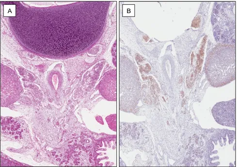
Fig. 3 Human fetus of 10 weeks of gestational age
(A) Hematoxylin & eosin staining and (B) immunoperoxidase staining with anti-CD1 antibody. The clusters of sympathetic neuroblasts migrating from the paravertebral region to the adrenal glands are highlighted by CD1.
B A
WT1 protein expression in small round blue cell tumors
As expected, Wilms’ tumor expressed WT1 protein mainly at nuclear level, with an immunoreactivity predominanly detected in blastematous, and less frequently, in epithelial and stromal cells. With the using antibodies directed against N-terminal portions of the WT1 protein [14,18,19], a concurrent or exclusively (more often in blastematous and stromal cells) cytoplasmic WT1 expression was found. However the most intriguing finding was the WT1 expression in rhabdomyosarcomas. Indeed all cases (27/27) of rhabdomyosarcoma were stained with WT1. Of note, all neoplastic cells, regardless of size, morphology, and cytoplasmic amount, exhibited strong WT1 cytoplasmic immunoreactivity (Fig. 4B and D). The immunostaining extension and intensity of WT1 among the different histologic sub-types of rhabdomyosarcoma was as follows: (i) diffuse (70–100% of neoplastic cells) and strong in all cases (15/15) of alveolar (Fig. 4A and B); (ii) diffuse (60– 100% of neoplastic cells) and strong in most cases (9/12) of embryonal rhabdomyosarcoma (Fig. 4C and D); (ii) heterogeneous and strong in two cases of conventional-type embryonal rhabdomyosarcoma (30 to 40% of neoplastic stained cells), and in one case of botryoid-type rhabdomyosarcoma (30% of neoplastic stained cells). Similarly, diffuse and strong WT1 cytoplasmic immunostaining was also observed in pre-operative core biopsies available from one case of alveolar rhabdomyosarcoma and two cases of
33
conventional type embryonal rhabdomyosarcoma. In all cases of rhabdomyosarcoma, WT1 was expressed in the cytoplasm of endothelial cells of blood vessels, and it served as internal positive control. WT1 nuclear staining was not observed in any case of rhabdomyosarcoma.
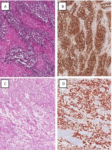
Fig. 4 Rhabdomyosarcomas
(A) Alveolar rhabdomyosarcomas, conventional type. Hematoxylin and eosin staining showing interconnecting tumor nests separated by fibrous stroma (original magnification×150). (B) Neoplastic cells show diffuse and strong cytoplasmic immunoreactivity for WT1.
(C) Embryonal rhabdomyosarcomas, conventional type. Hematoxylin and eosin staining showing small round cell tumor with alternating hypercellular and hypocellular myxoid areas (original magnification ×80). (D) Neoplastic cells show diffuse and strong cytoplasmic immunoreactivity for WT1.
Neither nuclear nor cytoplasmic expression of WT1 protein was found in the others examined small round blue cell tumors, including Ewing’s sarcoma, BCOR ITD–positive sarcomas, including 7 cases of undifferentiated round cell/Ewing-like sarcomas and 6 cases of PMMTI, neuroblastoma and lymphoblastic lymphoma. However, a cytoplasmic WT1 staining was obtained in the neoplastic ganglion cells present both in ganglioneuroblastoma and ganglioneuroma. Thus, the expression of WT1 in neuroblastic tumors does not reflect its developmental expression, but it seems to follow a reversal of its expression profile.
Cyclin D1 expression in small round blue cell tumours
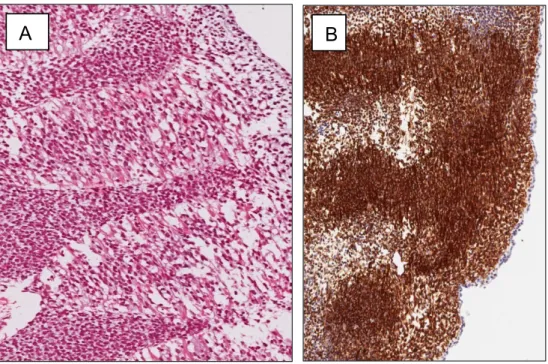
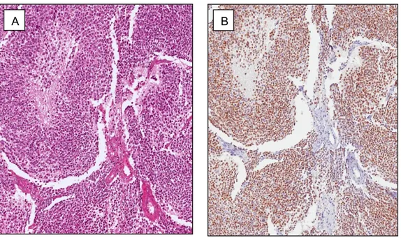
All 20 cases (100%) of EWS/pPNET exhibited strong and diffuse nuclear immunoreactivity for CD1 (Fig. 5). Notably the percentage of immunoreactivity varied in the different cases, ranging from 60% to 95% of neoplastic cells. Immunostaining, as expected, was exclusively confined to the nuclei of tumor cells, with no immunoreactivity in their cytoplasm. Interestingly a similar, diffuse CD1 immunostaining was also observed in pre- operative small biopsies available from three cases. In all cases of EWS/pPNET tested, CD1 was detected in the nuclei of endothelial cells of intra- and extra-tumoral blood vessels, and served as internal control. Interestingly, in all but one case of BCOR ITD–positive sarcomas a strong and diffuse (>70% of neoplastic cells) cyclin D1 immunoreactivity was obtained.
35
Fig. 5 EWS/pPNET
(A) Tumor area with solid growth pattern stained with haematoxylin and eosin. (B) Strong and diffuse nuclear staining for CD1 is seen.
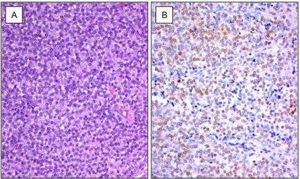
In the neuroblastic tumors examined, we observed a diffuse CD1 nuclear staining (>80% of these cells) restricted to neuroblastic component, while it was absent or only focally (<5% of neoplastic cells) found in tumor cells that showed clear-cut morphological features of ganglion cell differentiation (so-called “immature ganglion cells”) and in mature neoplastic ganglion cells. In details, CD1 immunoreactivity was restricted to neuroblastic cells which did not exhibit any morphological evidence of ganglion cell differentiation. These cells were the exclusive or predominant component, respectively, of undifferentiated or poorly differentiated neuroblastomas (Fig. 6), while they represented a minority component in differentiating neuroblastomas and in both intermixed and nodular ganglioneuroblastomas. On the contrary CD1 immunostaining was lacked, or only focally (<5% of neoplastic cells) retained, in immature ganglion cells found in differentiating neuroblastomas, and variably represented, in the form
B A
A B
of well-delineated microscopic foci or well-defined macroscopic or microscopic nodular areas, or as a minor microscopic component of scattered collections but without formation of distinct nests, respectively, in intermixed, nodular ganglioneuroblastoma or maturing ganglioneuroma. In mature ganglioneuroma no immunoreactivity for CD1 was observed in ganglion cells. Lymphoblastic lymphomas and the mesenchymal/stromal component of Wilms’ tumors exhibited only a focal immunoreactivity for CD1 (2-5% of neoplastic cells). In contrast CD1 expression was not detected in any case of rhabdomyosarcoma, regardless embryonal (12/12) or alveolar (15/15) subtypes. In all cases of rhabdomyosarcoma tested, CD1 was detected in the nuclei of endothelial cells of intra- and extra-tumoral blood vessels, and served as internal control.
Fig. 6 Poorly differentiated neuroblastoma.
(A) Hematoxylin & eosin staining showing a small round blue cell tumor composed almost exclusively of undifferentiated neuroblasts with formation of neuropil aggregates.
37
WT1 and Cyclin D1 expression in Human Ewing sarcoma mouse xenograft
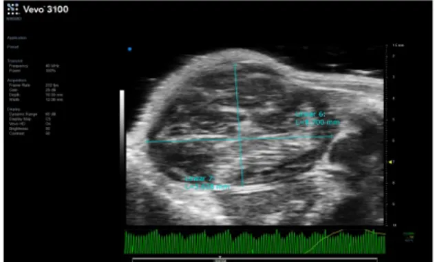
During the 15 days the animals are monitorized with the use of in vivo imaging techniques (Vevo ultrasound Pet / CT) (Fig. 7,8) to allow to valuate the increase in the size of the tumor masses. The dimensions of tumoral mass were between cm 2,91 and 0,55 (Tab.1).
All 5 cases of EWS/pPNET exhibited strong and diffuse nuclear immunoreactivity for CD1. Similarly to human tumor tissues, no immunoreactivity for WT1 was observed in Human Ewing sarcoma mouse xenograft.
Fig. 7 Vevo ultrasound in vivo imaging techniques
The image shows the tumor mass size.
Fig.8 PET /TC in vivo imaging techniques
Tumor Mass (cm-calliper)
Mice 0 day 3 days 4 days 5 days 6 days 7 days 10 days 15 days
Cage 44_ewing 1 0 0,2 0,1 0,1 0,1 0-0,1 0,1 e 0,2 2,91*2,91 2 0 0 0 0,3 0,5*0,2 0,6*0,3 0,85*0,862 1,036*0,843 Cage 45_ewing 3 0 0,1 0 0 0 0 0,85*0,497 0,7*1,039 4 0 0,3 0,2 0,2 0,3 0,1 e 0,3 0,408*0,521 0,556*0,693 5 0 0 0,3 0,3 0,3 0,3 1,051*0,582 0,629*0,965
Tab. 1 Tumor volums
5. Discussion
WT1 proteinThe subcellular localization of the WT1 protein has been a matter of debate over the last two decades. The cytoplasmic immunoreactivity obtained by using some antibodies directed against the N-terminal portion of WT1 was originally questioned and judged to be due to cross-reactivities of these reagents or to correspond to non-specific staining caused by formalin-fixation as previously documented for other transcription factors such as c-myc gene product [18,123,124]. Carpentieri et al. [20] postulated that the WT1 cytoplasmic immunostaining detected in human rhabdomyosarcomas could be explained by the presence of an inactive form of the protein, which is activated by phosphorylation and translocated into the nucleus. The different
39
results, i.e. nuclear versus cytoplasmic WT1 localization reported in the literature are likely due to the different specificities of the antibodies used by the various authors. It should be emphasized that WT1 nuclear expression has been mainly observed by using antibodies directed against the C-terminal portion of the molecule (WT C-19 polyclonal antibody), while an exclusive cytoplasmic expression or coincident cytoplasmic and nuclear expression has been noticed with more recently generated available antibodies against the N- terminal portion (clone 6F-H2). Currently WT1 cytoplasmic localization is widely accepted as being a true localization of the molecule [14,51,52,125]. This is in line with WT1 involvement, not only in transcriptional regulation in the nucleus, but also in RNA metabolism and translational regulation in the cytoplasm as well as to nucleo-cytoplasmic shuttling properties of WT1 and its association with actively translating polysomes [4].
Diagnosis of pediatric small round blue cell tumors can be difficult because of overlapping histologic and/or immunohistochemical features. In addition, the increasing use of small biopsies in daily practice, for planning presurgery neoadjuvant therapy (chemotherapy and/or radiotherapy), makes the diagnosis of these tumors more challenging [15,16,85,89,126,127]. In this regard, there is the need to render a precise diagnosis, specifying not only tumor histotype but, if possible, also its subtype. The advent of immuno- histochemistry has revolutionized the diagnostic approach to small round blue cell tumours. It is largely known that rhabdomyosarcoma was originally
considered as a WT1-negative tumor, as no immunoreactivity was obtained in the past by using antibodies directed against the C-terminal portion the molecule (clone WT C19). On the contrary, with the advent of new available antibodies against the N-terminal portion of WT1 protein (clone WT 6F-H2), there is increasing evidence of WT1 cytoplasmic expression not only in several tumors [15,22,23,44,45,59], including rhabdomyosarcoma [20,21,55,58,62], but also in human fetal tissues [11,12,80]. Moreover it was postulated that WT1 is a gene playing a role in the pathogenesis of human rhabdomyosarcoma [20].
We first showed that WT1 is abundantly expressed in the cytoplasm of human myoblasts from the 6 wGA and down-regulated from the 21 wGA and in adult benign tissues, including adult normal and benign tumors with skeletal muscle differentiation (rhabdomyomas). These findings suggest that this transcription factor may act as complex regulator of transcriptional/translational patterns during the early phases of human skeletal myogenesis. With regard to neoplastic tissues, we confirm a strong and diffuse WT1 cytoplasmic expression in both embryonal and alveolar rhab- domyosarcomas. Based on our results, we first show that WT1 is expressed in both developing and malignant skeletal muscle tissues, suggesting that it can be considered an oncofetalprotein useful as immunomarker in confirming the diagnosis of both embryonal and alveolar rhabdomyosarcoma. Although this finding is helpful in confirming diagnosis of rhabdomyosarcoma, especially in
41
tumors with ambiguous immunoprofile, WT1 should not be used alone in daily practice. This is due to the evidence that rhabdomyosarcoma mimics, such as EWS/pPNET and desmoplatic small round cell tumor, may express, even if less frequently and usually with focal extension, WT1 at the cytoplasmic level. With regard to desmoplastic small round cell tumor, a cytoplasmic WT1staining can be obtained by using WT1 (N-terminus-clone 6F-H2) antibodies [14,21], while a similar cytoplasmic immunoreactivity has been reported in up 43% or 63% of cases of EWS/pPNET by using antibodies against both C-terminus or N-terminus of WT1 protein, respectively [14]. This strongly suggests that a small round blue cell tumor which expresses a diffuse and strong cytoplasmic WT1 staining is likely to be a rhabdomyosarcoma, but this finding should be interpreted in the context of other antibodies, such CD99, FLI-1, desmin, myogenin, MyoD1, cytokeratins, and INI1 protein.
Cyclin D1
Among the pediatric small round blue cell tumors arising from soft tissues, distinguishing EWS/pPNET from alveolar rhabdomyosarcoma may be challenging [128]. The former tumor may occasionally exhibit a pseudo- alveolar growth pattern, while the latter may show an exclusively/predominantly EWS/pPNET-like solid growth pattern [129]. In addition EWS/pPNET may be occasionally desmin-positive [130-133],
whereas in some cases of rhabdomyosarcoma CD99 may be expressed with a membrane or paranuclear dot-like immunostaining pattern [134,135]. Although concurrent expression of desmin and myogenin strongly supports the diagnosis of rhabdomyosarcoma, the immunohistochemical approach to EWS/pPNET is mainly based on the combination of CD99 and FLI-1, usually positive in the majority of EWS/pPNETs, along with negative results for desmin and myogenin [128,129]. The diagnosis of EWS/pPNET is usually confirmed, especially for morphologically and/or immunohistochemically ambiguous cases, by the identification of the recurrent translocation t(11;22) (EWS–FLI1) or one of the variant translocations, such as t(21;22) (ESW–
ERG) and t(7;22) (EWS–ETV1) [129]. Unfortunately, a molecular diagnostic
approach is usually available at only a limited number of large centers and, thus, the most widely applied ancillary technique today is immunohistochemistry to confirm the diagnosis of EWS/pPNET and to exclude other similar-appearing round blue cell tumors [128]. Accordingly, there is an increasing need to identify positive diagnostic immunomarkers of EWS/pPNET that would be easily accessible to practicing pathologists. Previous in vitro studies demonstrated consistent upregulation of CD1 in EWS/pPNET but not in rhabdomyosaroma cell lines [110]. Notably CD1 overexpression in EWS/pPNET was only partially validated by one immunohistochemical study which revealed that only 42% of cases of this sarcomas were CD1-positive [136]. These conflicting results prompted us to
43
pediatric/adolescent soft tissue EWS/pPNET and rhabdomyosarcomas, respectively, to determine if it is exploitable as useful immunomarker in their differential diagnosis. We first showed that CD1 is a highly sensitive, albeit not specific, marker of EWS/pPNET, being diffusely and strongly expressed in all cases examined. Interestingly, CD1 nuclear immunoreactivity was obtained not only in surgically excised, but also in small biopsy specimens, suggesting that it can be successfully used as an immunomarker of EWS/pPNET in daily practice. Another interesting result is the overexpression of cyclin D1 in BCOR ITD–positive sarcomas, suggesting that Ewing-like undifferentiated round cell sarcoma and PMMTI are likely in the spectrum of a single morphobiological entity [137]. Apart from EWS, our results suggest that cyclin D1 is helpful in identifying Ewing-like sarcomas with EWS/CIC-DUX4 fusion or BCOR ITD, prompting pathologists to perform genetic and/or molecular studies as confirmatory tests.
In addition, the expression of cyclin D1 in Human Ewing sarcoma mouse xenograft could suggest the implication of this molecule in the development of the tumor, so it would be interesting to evaluate if the CD1-silencing could induce a reduction of the tumor masses.
However, the expression of CD1 in a variety of malignant tumors [96,104-109,138] necessitates caution in applying CD1 as a single marker for diagnosis of EWS/pPNET. Therefore we suggest that CD1 should be included and evaluated in the context of a wide immunohistochemical panel, together
with desmin, myogenin, MyoD1, CD99, FLI-1, TdT, LCA, NB84, INI-1, andS-100 protein, when dealing with a small round blue cell tumor of soft tissues in pediatric/adolescent patients. If the tumor examined is stained exclusively/predominantly with CD99 and FLI-1, an additional strong and diffuse nuclear CD1 immunoreactivity is helpful in confirming the diagnosis of EWS/pPNET. On the contrary, this latter immunohistochemical finding argues against the diagnosis of rhabdomyosarcoma, regardeless embryonal or alveolar subtypes, and may successfully used in the rare cases in which the interpretation of myogenic markers is ambiguous for technical artifacts.
Several morphological, immunohistochemical, and in vitro studies indicate that childhood peripheral neuroblastic tumors recapitulate the developmental stages of normal PSNS [113,116,118,128,139-143]. This has prompted for the search for specific cell differentiation markers suitable for diagnostic purposes and for a better understanding of the biology of peripheral neuroblastic tumors [40,112-114,141,142,144-147]. The focus on developing human PSNS, which arises from a common neural crest-derived precursor cell [148,149], is of special interest because it represents an in vivo model of ganglion cell differentiation, which can be exploitable for better understanding the molecular mechanisms involved in the maturation of some neuroblastomas into ganglioneuroblastomas with excellent prognosis (intermixed ganglioneuroblastoma; nodular ganglioneuroblastoma with favorable histology) [90]. Previous immunohistochemical findings have
45
ganglion cells is regulated by high levels of expression of several molecules, including Bcl-2 protein, HNK-1/carbohydrate epitope, neurofilament proteins, endothelin-B receptor (ET-B), cathepsin D, growth associated protein-43 (GAP-43), in association with a concurrent lack of tyrosine hydroxylase, chromogranin A, insulin growth factor-II (IGF-II), and Wilms Tumor-1 (WT1) [13,115,146,150,151]. D-type cyclins play a crucial role in cell cycle progression through the activation of their cyclin-dependent kinase partners CDk4 and CDk6.
We first investigated the developmentally regulated expression and distribution of CD1 in human PSNS and compared the results with those obtained in peripheral childhood neuroblastic tumors. We showed that CD1 is transiently expressed in the nuclei of neuroblasts during human PSNS development. Unlike other neuroblast-associated markers [115,146,150,151], CD1expression is lacked, as documented by a progressively loss of nuclear staining, in developing ganglion and chromaffin cells with increasing gestational age. The absence of CD1 immunoreactivity is also maintained in the ganglion cells of sympathetic ganglia and in adrenal medulla in neonates and adults. Accordingly, we suggest that CD1, when evaluated in the appropriate morphological context, is a reliable marker of sympathetic neuroblasts of human PSNS, which can be used routinely in formalin-fixed tissues. CD1 expression in human sympathetic neuroblasts is not at all surprising, as previous studies have documented its involvement in neuronal differentiation processes [152-154]. As previously suggested for
neuroblastoma [138,155], it is likely to postulate that CD1 expression in human sympathetic neuroblasts acts to maintain their undifferentiated phenotype, preventing their ganglion or chromaffin cell differentiation. In peripheral childhood neuroblastic tumors, we confirmed immunohistochemical data previously reported by Molenaar et al. [138]. However, that study did not specify in details the results obtained for each single hystotype, stating only that “CD1 overexpression was found in
neuroblasts, in contrast to a low expression in all cell types in ganglioneuromas” [138]. Our study shows that CD1 immunoreactivity was
restricted to neuroblastic cells, which do not exhibit any morphological evidence of ganglion cell differentiation, whereas it was virtually lacked in immature and mature ganglion cells. Accordingly, the higher expression of CD1 is obtained in undifferentiated and poorly differentiated neuroblastomas, and it was also maintained in the more undifferentiated component (neuroblastic component) of ganglioneuroblastomas. This is consistent with the current concept that ganglioneuroblastoma, especially the nodular subtype, represents an attempt of ganglioneuromatous maturation, not fully developed, by an original neuroblastoma [90]. Our findings suggest that CD1 is a highly sensitive, albeit not specific, immunomarker of malignant neuroblasts, which typically are the constitutive cells of neuroblastomas, the most undifferentiated tumors in the spectrum of peripheral childhood neuroblastic tumors. We suggest that CD1 can be used in daily practice, along with NB84
47
undifferentiated/poorly differentiated neuroblastoma, especially in small incisional biopsies. CD1 can also be useful in decorating the microscopic foci of neuroblastic collections, which are present, albeit with a variable extension and configuration, in both intermixed and nodular ganglioneuroblastomas. Apart from diagnostic purposes, our results seem to confirm the previously suggested hypothesis that CD1 expression in human neuroblastoma is crucial to maintain the undifferentiated phenotype of neuroblasts, preventing their ganglion cell differentiation [138]. In this regard, previous studies have documented not only high expression of CD1 at both mRNA and protein level in human neuroblastomas, but also of CDK4 and CDK6, the kinase partners of CD1, indicating a role for G1 entry checkpoint dysregulation in the etiology of neuroblastoma [138,155,157,158]. The pathogenetic role of CD1 in neuroblastoma has also been confirmed by in vitro studies which, using RNA interference against CD1 and CDK4 and CDK6, showed significant reduction of cell proliferation, a G1-specific cell cycle arrest, and extensive neuronal differentiation [138]. The comparative evaluation of the immunohistochemical findings in fetal and neoplastic tissues indicates that CD1 expression in peripheral childhood neuroblastic tumors mirrors its normal developmental regulation in PSNS, as already reported for Bcl-2 protein, c-ErbB2, insulin-like growth factor 2, beta-2-microglobulin, and cathepsin D [113,140,141,145,146]. This strongly supports the view that peripheral childhood neuroblastic tumors recapitulate morphologically and immunophenotypically the different developmental stages of the PSNS