A
A
l
l
m
m
a
a
M
M
a
a
t
t
e
e
r
r
S
S
t
t
u
u
d
d
i
i
o
o
r
r
u
u
m
m
–
–
U
U
n
n
i
i
v
v
e
e
r
r
s
s
i
i
t
t
à
à
d
d
i
i
B
B
o
o
l
l
o
o
g
g
n
n
a
a
DOTTORATO DI RICERCA IN APPLICAZIONI
BIOTECNOLOGICHE IN NEUROMORFOFISIOLOGIA
Ciclo XXI
Settore scientifico disciplinare di afferenza VET/01
TITOLO DELLA TESI
Mechanosensitivity in the myenteric plexus of the
guinea pig ileum
Presentata da
Gemma Mazzuoli
Coordinatore Dottorato Relatore
Index
INTRODUCTION ... 2
Enteric nervous system ... 4
Morphological classification of the enteric neurons ... 5
Electrophysiological classification of the enteric neurons ... 6
Functional and neurochemical classification of the enteric neuron ... 8
The IPANs theory ... 14
Mechanosensitivity of IPANs ... 15
Immunoreactivity of IPANs ... 16
From the IPANs theory to the multifunctional enteric neurons theory ... 18
Extrinsic afferent neurons ... 23
Extrinsic sensory neurons theory ... 28
Mechanosensitivity during evolution ... 29
Mechanosensitivity in the gut: ion channels ... 35
Aim of the study ... 42
MATERIAL AND METHODS ... 44
Tissue preparations ... 44
Multisite optical recording technique (MSORT) with voltage sensitive dye ... 46
Staining method ... 47
Optical recording method ... 49
Duration of the acquisitions ... 50
Technique validation ... 50
Advantage and disadvantages of MSORT ... 52
Behaviour of ganglia in freely contracting tissues ... 53
Different techniques for mechanical stimulation of ganglia and neurons ... 54
Von Frey hair technique ... 54
Intraganglionic injections technique ... 56
Immunohistochemistry ... 61
Data analysis and statistic ... 63
RESULTS ... 66
Behaviour of ganglia in freely contracting tissues ... 66
Von Frey hair technique ... 68
Intraganglionic injection technique ... 71
Experiments to characterize the stimulus modality ... 77
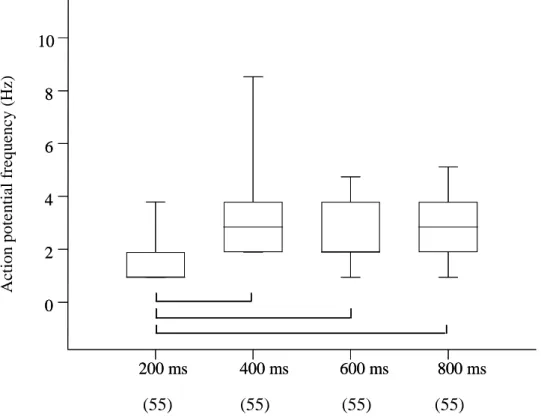
Relation between stimulus duration and discharge pattern ... 77
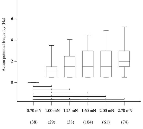
Relation between injection pressure and discharge pattern ... 78
Relation between injected volume and discharge pattern ... 80
Relation between rate of volume injection and discharge pattern ... 82
Response to electrical stimulations of interganglionic fiber tracts ... 83
Pharmacology of mechanosensitivity ... 88
Effect of nifedipine... 88
Effect of hexamethonium ... 90
Effect of ω-conotoxin GVIA ... 92
Effect of capsaicin ... 94
Neurochemical coding of mechanosensory neurons ... 96
Morphology of mechanosensory neurons ... 99
DISCUSSION ... 100
TABLES ... 110
ACKNOWLEDGEMENTS ... 114
Abbreviations
5-HT 5-hydroxytryptamine
AHP After-hyperpolarization
AMCA Aminomethylcoumarin-acetat
ATP Adenosine triphosphate
Calb Calbindin
CGRP Calcitonin gene-related peptide
ChAT Choline acetyltransferase
CNS Central nervous system
DRG Dorsal root ganglion
EC Enterochromaffin
ENK Enkephalin
ENS Enteric nervous system
EPSP Excitatory postsynaptic potential
GI Gastrointestinal
HT High-threshold
IBD Inflammatory Bowel Disease
IBS Irritable Bowel Syndrome
ICC Interstitial cell of Cajal
IGLE Intraganglionic laminar ending
IPAN Intrinsic primary afferent neuron
LT Low-threshold
LTSC Low threshold small conductance
MEC/DEG Mechanosensoryabnormal/degenerin
MP Myenteric plexus
MSC Mechanosensitive channels
MSORT Multi-Site Optical Recording
Technique
NeuN Neuronal nuclei antibody
NFP Neurofilament protein
NO Nitric oxide
NOS Nitric oxide syntethase
NSC Non-selective cation
SMP Submucosal plexus
SOM Somatostatin
SP Substance P
TRP Transient receptor potential
TTX Tetrodotoxin
Mechanosensitivity in the
myenteric plexus of the
Introduction
Jackie Wood, professor of physiology, cell biology and internal medicine at Ohio State University said: “What Mother Nature had done, rather than packing all of those neurons in the big brain in the skull and sending long lines to the gut, is distribute the microcomputer, the little brain, right along the systems that require control”. The idea that the gut can function by its own dates back to 1899 when Bayliss and Starling made their observations in dogs and rabbits on local reflexes in the isolated or extrinsically denervated intestine (Bayliss and Starling 1899; Bayliss and Starling 1901). During the First World War the German scientist Trendelenburg showed that a self-contained, self-regulating nervous system that is embedded within the wall of the gut could function on its own without input from the brain or the spinal cord (Trendelenburg 1917). In 1921 Langley published the great book “The Autonomic Nervous System” setting the basis for the first classification of the autonomic nervous system in the parasympathetic and sympathetic divisions. He also included a third division coning the term “Enteric Nervous System”, considering the fact that this system differs from the parasympathetic and sympathetic in its anatomical and functional independence from the brain and spinal cord. For reasons that still mystify researchers today, all the data collected until this point went into hibernation for nearly half a century but are still the basis for today research. Around 1965 some neurobiologists began to realise the clinical relevance of the autonomy
(or little) brain in the gut”. Nowadays it is well known that the enteric nervous system (ENS) regulates all the reflex pathways that control blood flow, motility, water and electrolyte transport and acid secretion, autonomously from the central nervous system (CNS). Despite that the CNS can influence and modulate the gut activity the ability of the gut to function in isolation is one of the most intriguing phenomenons in neurogastroenterology. This requires coding of sensory stimuli by specialized cells in the gut wall, which function as sensory neurons. These neurons are capable to detect chemical and/or mechanical stimulations and to orchestrate the appropriate responses via the neuronal network constituted by the two ganglionated plexus embedded in the gut wall. A lot of studies were carried out in the last years regarding the pathways used by enteric neurons to detect chemical environmental changes and to respond to them. On the other hand, despite all the studies on mechanosensitivity carried on non-neuronal mechanism (e.g. smooth muscle, interstitial cell of Cajal) in the gut, there is a lack of knowledge regarding mechanosensory neuronal pathways. Enteric neurons seemed to be prominent candidates to relay mechanosensitivity. Surprisingly, the identity of mechanosensitive neurons in the ENS as well as the appropriate stimulus modality is unknown.
Enteric nervous system
The ENS is composed by over 100 millions nerve cells embedded in the gut wall.
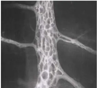
The nerve cell bodies are grouped in small aggregates, the enteric ganglia, which are connected by bundles of nerve cell processes to form two major ganglionated plexuses in the tubular digestive tract: the myenteric plexus (MP), also called the Auerbach plexus, that mainly regulates motility, and the submucosal plexus (SMP), which is often referred to as the Meissner plexus, that mainly controls secretion. The MP lies between the outer longitudinal and the inner circular muscle layers of the intestine and forms a continuous network around the circumference and along the gastrointestinal (GI) tract, from the upper oesophagus to the internal anal sphincter. The myenteric ganglia vary in size, shape and orientation between animal species and from one part of the intestine to another (Furness 2006). In the ileum of the guinea pig, ganglia range in size from 5 to over 200 nerve cell bodies (Furness 2006). The SMP is significant only in the small and large intestines. In general, the interconnecting strands of the SMP are finer and the ganglia are smaller than those of the MP (Furness 2006).
Ultrastructure analysis show that enteric ganglia are remarkably compact, consisting of the cell bodies of neurons, enteric glia, and nerve cell processes (Furness 2006). Particularly an electron microscope study described the ganglia of the MP of the guinea pig ileum like very compact structures, completely surrounded by a basal lamina and isolated from the connective tissue and blood vessels
elements, constituting a dense neuropil, with an intervening gap of 20 nm between the adjacent membranes (Gabella 1972).
Enteric neurons can be divided into different functional classes according to morphology of their cell body, projections to target, neurochemistry and pharmacological properties.
Morphological classification of the enteric neurons
The first and most enduring classification of enteric neurons by their shape was made by Dogiel more than 100 years ago: he provided a comprehensive description of neuron morphologies in the MP and SMP of the intestine from human, guinea pig, rabbit, rat, dog and cat (Dogiel 1895; Dogiel 1896; Dogiel 1899). He described three types of neurons now generally referred to as Dogiel types I, II and III.
The type I are flattened cells, slightly elongated, with stellate or angular outlines, they have 4 to 20 lamellar dendrites and long process which is likely the axon (Dogiel 1899). These neurons do not appear to belong to a single functional class: some are inhibitory motor neurons to the muscle, some are excitatory motor neurons and some are interneurons.
The Dogiel type II neurons have large oval or round cell bodies. Dogiel described them as having 3-10 dendrites and one axon (Dogiel 1899), however is now recognized that the principal processes are all axons. Between 80 and 90% of Dogiel type II neurons in guinea pig MP are immunoreactive for the calcium binding protein Calbindin (Calb) (Furness et al. 1988; Iyer et al. 1988; Song et al. 1991; Costa et
al. 1996) and nearly all are immunoreactive for choline acetyltransferase (ChAT) (Steele et al. 1991).
Type III cells were described as having 2 to 10 dendrites that became thinner and branched distant from the cell body; these dendrites appear to be relatively short, smooth but sometimes they have varicosities. The axon of these cells begins from a protrusion of the cell body or from a dendrite (Dogiel 1899). These cells are today called neurons with filamentous dendrites. Other morphological types of cells were also identified by Dogiel (1899) and in later studies (e.g. Stach 1989), but there is less of a consensus about the general use of this extended classification. Dogiel also suggested a correlation between the morphology and the functions of type I and type II neurons studying the dendrites and the axonal projection, he hypothesized that type I neurons fulfil motor whereas type II neurons have sensory functions (Dogiel 1895; Dogiel 1896; Dogiel 1899).
Electrophysiological classification of the enteric neurons
The first intracellular recordings from enteric neurons that provided the basis for classification were reported in 1970s (Nishi and North 1973; Hirst et al. 1974; Wood and Mayer 1978).
The two major electrophysiological classes of myenteric neurons are:
• AH neurons characterized by phasic spike discharge, spike component partially carried by calcium influx, slow afterspike hyperpolarization (AHP) following a single action potential discharge. They receive little fast synaptic input but can
in other AH neurons and S neurons. These cells have a distinctive Dogiel II morphology with large smooth cell bodies and multipolar processes. However, a few myenteric AH neurons are filamentous, uniaxonal neurons and shows fast excitatory postsynaptic potential (fast EPSPs). Many of them are Calb positive (Hirst et al. 1974).
• S neurons which exhibit tonic spike discharge. These neurons lack slow AHP, show sodium-driven action potentials and receive abundant fast EPSPs. These neurons are slowly adapting neurons considered to function as excitatory and inhibitory motoneurons or interneurons. They show a typical Dogiel type I morphology and are usually uniaxonal (Hirst et al. 1974).
While S neurons have abundant synaptic input only relatively few AH neurons generate fast EPSPs, and these are of small amplitude compared with those in S neurons (Kunze and Furness 1999; Blackshaw et al. 2007).
Functional and neurochemical classification of the enteric
neuron
Since more than hundred years it has been known that the ENS contains motor neurons, interneurons, and sensory neurons organised into functional circuits (Bayliss and Starling 1899; Langley and Magnus 1905; Trendelenburg 1917).
•
Motor neurons
: there are excitatory and inhibitory motor neurons innervating the longitudinal, the circular smooth muscle and the muscularis mucosae throughout the digestive tract. These neurons are uniaxonal. The primary transmitters of the excitatory motor neurons are acetylcholine and tachykinins. The inhibitory neurons have multiple transmitters, including nitric oxide (NO), vasoactive intestinal peptide (VIP) and adenosine triphosphate (ATP) (Furness 2006).The majority of neurons innervating the circular muscle have their cell bodies in the MP. In the guinea pig they are all in myenteric ganglia (Wilson et al. 1987), in other species, including rat (Ekblad et al. 1987; Ekblad et al. 1988), dog (Sanders and Smith 1986, Furness et al. 1990a), pig (Timmermans et al. 2001), and probably human, a component of circular muscle innervation comes from submucosal ganglia. A consistent finding of retrograde labelling studies has shown that the cell bodies of inhibitory motor neurons, supplying the circular muscle layer, are located oral to the circular muscle that
Brookes et al. 1997; Pfannkuche et al. 1998; Yuan and Brookes 1999) and project aborally for 0.5-25 mm in the myenteric plexus before entering the muscle. In contrast, the cell bodies of excitatory motor neurons, also supplying the circular muscle layer, mostly lie aboral to the circular muscle that they innervate, at distances up to about 8 mm (Brookes et al. 1991). However, a few cholinergic motor neurons project for short distances (up to 1 mm) aborally to the circular muscle (Brookes et al. 1991). The longitudinal muscle motor neurons are about 25% of nerve cells in the myenteric ganglia of the guinea pig small intestine and their axons do not project more than 3 mm from their nerve cell bodies (Brookes et al. 1992). The cell bodies of the neurons supplying the longitudinal muscle layer are in the MP of small animals. In the pig the majority of them are in the MP, but some longitudinal muscle motor neurons have cell bodies in the outer SMP (Timmermans et al. 2001).
•
Interneurons
: these neurons have been identified in all gut regions, and vary between regions more than the other types of neurons. In the guinea pig small intestine there classes of descending interneurons and one class of ascending interneurons have been identified (Costa et al. 1996; Furness 2006).The ascending interneurons are all located in the myenteric plexus and have medium/large-sized cells bodies with Dogiel type I morphology with lamellar dendrites and a single orally
projecting axon. They project up to 14 mm orally from their nerve cell body. They have a unique chemical coding containing immunoreactivity for ChAT, Calretinin, Substance P (SP), Neurofilament protein (NFP), and Enkephalin (ENK) (Brookes et al. 1997). Their synaptic transmission is predominantly cholinergic, through nicotinic receptors (Furness 2006). Ascending interneurons account for 5% of all myenteric neurons (Costa et al. 1996). In the guinea pig colon there are three classes of ascending interneurons all ChAT immunoreactive, which is consistent with ascending reflexes in the colon utilizing cholinergic neuroneuronal transmission (Lomax and Furness 2000).
The descending interneurons in the guinea pig small intestine include those immunoreactive for ChAT plus Somatostatin (SOM), ChAT plus Nitric oxide synthase (NOS), VIP and others substances and those immunoreactive for ChAT plus 5-hydroxytryptamine (5-HT) (Furness 2006). Their synaptic transmission is also cholinergic, as indicated by their immunoreactivity for ChAT, but transmission in local reflexes is not purely cholinergic (Furness 2006). The ChAT /5-HT neurons do not make connections with the inhibitory muscle motor neurons; they are involved in descending excitatory reflexes (Young and Furness 1995).
The ChAT /SOM neurons are present in the myenteric plexus; they have a characteristic soma-dendritic morphology, with smooth, medium-sized cell bodies with numerous filamentous
dendrites and a single axon (Portbury et al. 1995; Song et al. 1997).
The ChAT /NOS interneurons are involved in descending inhibitory reflexes (Yuan et al. 1995).
•
Intestinofugal neurons
: these neurons have the cell bodies embedded in the gut wall and send their processes to the prevertebral ganglia, where they form synapses with post-ganglionic sympathetic neurons (Kuntz 1938; Szurszewski and Miller 1994). In the small intestine of the guinea pig, the intestinofugal neurons are small Dogiel type I cells with short lamellar dendrites or sometimes short filamentous dendrites, but all have a single axon (Tassicker et al. 1999). They are immunoreactive for VIP/ChAT and calcitonin gene-related peptide (CGRP) but not for NOS, unlike those in the large intestine (Anderson et al. 1995; Mann et al. 1995). The sympathetic neurons that are innervated by intestinofugal neurons inhibit motility as well as secretion (Furness 2006).•
Sensory neurons
: these cells are the first neurons in a reflex pathway which encode information about the nature and intensity of the stimulus. Using the immediate early gene marker Fos, neurons in the SMP of the guinea pig small intestine that produced Fos in response to either cholera toxin or to mechanical stimulation by puffs of nitrogen onto the mucosal villi were identified (Kirchgessner et al. 1992). When thenicotinic blocker hexamethonium was present (to reduce fast synaptic activation of neurons) one particular class of cells was labelled. These cells were immunoreactive for Calb and/or SP (or a related tachykinin) (Kirchgessner et al. 1992). Sensory neurons make up approximately 13% of all neurons in the SMP of the guinea pig small intestine (Song et al. 1992) and have been dye filled during intracellular recordings (Bornstein et al. 1989). The SMP neurons with Calb and/or tachykinin immunoreactivity have AH-like properties with multipolar morphology (Bornstein et al. 1989; Evans et al. 1994). These cells have extensive projections within the submucous ganglia, often appearing to contact other nerve cell bodies (Bornstein et al. 1989; Evans et al. 1994). They can be retrogradely labelled by tracers applied to both the mucosa (Kirchgessner et al. 1992; Song et al. 1992) and to the MP (Kirchgessner et al. 1992; Song et al. 1998) and thus project to both targets. Sensory neurons are immunoreactive for ChAT (Furness et al. 1984) and are likely to make cholinergic and non-cholinergic synapses onto other classes of SMP and MP neurons. These sensory neurons do not appear to receive fast nicotinic synaptic inputs (Bornstein et al. 1984; Evans et al. 1994). The SMP neurons with immunoreactivity for Calb and/or tachykinins have Dogiel type II morphology.
The identification of myenteric Dogiel type II neurons as sensory neurons was achieved using electrophysiological recordings. A large proportion of myenteric Dogiel type II
neurons in the guinea pig small intestine respond to mucosally applied chemicals, such as solutions with low or high pH or short-chain fatty acids (Kunze et al. 1995; Bertrand et al. 1997). It is not currently clear whether Dogiel type II cells are directly activated by mucosal chemicals or whether they are activated indirectly, perhaps via enteroendocrine cells as demonstrated for SMP primary afferent neurons. However, the observation that responses to mucosal acid were not blocked by low [Ca2+], high [Mg2+] Krebs solution, which blocked synaptically evoked responses, suggests that the axons of these neurons may be capable of directly transducing some chemical stimuli (Kunze et al. 1995). It is not known whether or not the SMP sensory neurons also respond to chemical stimulation.
The IPANs theory
The term intrinsic primary afferent neurons (IPANs) has been proposed and used for enteric neurons that encode for sensory stimuli and represent the first neurons in the intrinsic reflex circuits that influence motility, secretion and blood flow (Furness et al. 1998; Pan and Gershon 2000; Clerc et al. 2002). The morphological, electrophysiological and neurochemical characteristics of IPANs have been most thoroughly studied in the guinea pig ileum (Brookes and Costa 2002; Furness et al. 2004). In this region all IPANs identified so far appear to have Dogiel type II morphology, e.g. ovoid cell soma with a pseudo-uniaxonal or multiaxonal appearance with terminals in the myenteric and submucosal ganglia, as well as projections to the mucosa. Electrophysiologically, they are AH/type 2 nerve cells, displaying a long lasting after spike hyperpolarisation (Iyer et al. 1988; Hendriks et al. 1990; Kunze and Furness 1999). In the guinea pig small intestine, IPANs respond to electrical stimulation of interganglionic fiber tract with slow EPSPs. Fast EPSPs to these neurons are rarely recorded and when present they are of low amplitude (Kunze and Furness 1999). These features are distinct from the other major class of enteric neurons, which have unipolar morphology (Dogiel type I), receive fast EPSPs, and are considered to function as interneurons and motor neurons. Electropyhsiologically, they belong to S/type 1 neurons.
Mechanosensitivity of IPANs
In few studies it has been shown that in the guinea pig small intestine, myenteric AH neurons, unlike S neurons, respond to chemical stimulants applied to the mucosa (Smith 1994; Smith 1996; Kunze et al. 1995; Bertrand et al. 1997; Bertrand and Bornstein 2002). In addition, AH neurons in the guinea pig small intestine respond to both, stretch and contraction, with an ongoing action potential discharge (Kunze et al. 1998; Kunze and Furness 1999). This response depends on an increase in smooth muscle tension (tone) and is dependent on the opening of gadolinium-sensitive mechanosensitive channels. Action potentials were also abolished by drugs (e.g. isoprenaline or nicardipine) that abolish muscle tension despite maintained stretch (Kunze et al. 1998; Kunze and Furness 1999). Muscle tone contributes to the mechanosensitivity of enteric neurons in the guinea pig ileum. In this tissue the neural response to sustained stretch depends on opening of stretch activated channels in the muscle followed by muscle contraction and mechanical communication from the contracting muscle mostly to myenteric AH neurons and very rarely to myenteric S neurons (Kunze et al. 1999; Kunze et al. 2000).
The action potentials were generated in the processes of Dogiel type II neurons, not in the nerve cell bodies. Thus in the guinea pig ileum these AH neurons are suggested to function as intrinsic mechanosensory and/or chemosensory neurons that may be responsible for initiating ascending excitatory and descending inhibitory peristaltic reflexes (Kunze et al. 1998, Kunze et al. 1999; Kunze and Furness 1999). Other myenteric AH neurons are also
activated during the stretch by synaptic transmission from neurons that respond directly to distortion (Kunze et al. 2000). Nevertheless, some of the S neurons also seem to react directly to the stretch, thus some uniaxonal neurons could also function as intrinsic sensory neurons (Kunze et al. 1998; Kunze and Furness 1999). Moreover, enteric neurons seem to show different responses depending on which mechanosensitive region of the neuron is activated. Thus, it seems that when the deformation is on the interganglionic nerve processes that occurs when the intestine is distended or the muscle contracts, this leads to action potential discharge; while deformation of the neuron soma inhibits spike discharge in myenteric AH neurons in the guinea pig ileum; this compression of the soma by pressure causes increased opening of potassium channels and thus has an inhibitory effect which may be protective (Kunze et al. 2000). However, in other mammalian sensory neurons, mechanical distortion increases excitability of both the neurites and the cell bodies (Cunningham et al. 1995; Cunningham et al. 1997; Kraske et al. 1998; Raybould et al. 1999).
Immunoreactivity of IPANs
IPANs are cholinergic (Furness et al. 1984; Steele et al. 1991), containing the peripheral form of choline acetyltransferase (Chiocchetti et al. 2003), and express tachykinins (Kirchgessner et al. 1992; Song et al. 1991), neuromedin U (Furness et al. 1989), the neurokinin receptor 3 (Johnson et al. 1998; Jenkinson et al. 1999; Furness 2000) and the P2X2 receptor (Castelucci et al. 2002).
A large proportion of IPANs is immunoreactive for the calcium-binding protein Calb (Furness et al. 1988; Iyer et al. 1988; Quinson et al. 2001). Thus, Calb is a marker for multipolar sensory neurons in the guinea pig small intestine (Furness et al. 1990b). Another study also supports the evidence that Calb immunoreactive neurons appear to be sensory neurons (Costa et al. 1996). Later, it has been shown that NeuN, a neuronal nuclei antibody, seems to be a selective marker for enteric IPANs in the guinea pig small intestine (Costa et al. 2001; Costa et al. 2002; Brody et al. 2002; Poole et al. 2002). In the MP of the guinea pig ileum quantitative data indicate that NeuN immunoreactive neurons are about 38% of all myenteric neurons (Costa et al. 2001, Costa et al. 2002). Furthermore all Calb immunoreactive neurons express NeuN (Costa et al. 2001, Costa et al. 2002). In the guinea pig ileum, NeuN labels the nuclei of almost all the enteric neurons, but the cytoplasmic expression of NeuN appears to be restricted to Dogiel type II neurons (Chiocchetti et al. 2003; Van Nassauw et al. 2005). While all Calb immunoreactive neurons express NeuN, not all NeuN immunoreactive neurons are positive for Calb. There are 2 NeuN subpopulation, one which is also immunoreactive for Calb (NeuN+/Calb+) (67±2%) and one which is not (NeuN+/Calb-).
From the IPANs theory to the multifunctional
enteric neurons theory
The use of the term IPANs and in particular the concept behind it was recently challenged (Spencer and Smith 2004; Wood 2004; Smith et al. 2007; Blackshaw et al. 2007; Wood 2008). The functional distinction of AH cells as the only enteric nerve population responding to mechanical stimuli was challenged for the first time by a study from Spencer and Smith (2004). In this study, performed in the guinea pig distal colon, they show that in circumferentially stretched preparations AH neurons appear to be electrically silent, while S interneurons respond with an ongoing action potential discharge. It is also noteworthy that action potential discharge in mechanosensory S neurons does not cease when the muscle is paralyzed. Further studies performed by this group allow them to conclude that in the guinea pig AH neurons appear to be necessary for initialing peristaltic waves, where muscle tone is required, while S neurons generate a more rapidly occurring motor pattern (called “ongoing peristaltic reflex activity”), where muscle tone is not required (Smith et al. 2007). Thus, they propose the existence of two different intrinsic sensory neurons populations and suggest that unipolar S neurons may be mechanosensory and at the same time function as interneurons (Smith et al. 2007).
These findings show that mechanosensitivity in the ENS is not a unique property of a single neuron type and that mechanosensory
stretch, tension, stress, and/or strain sensitive. In other words they may be multifunctional and, as such, they would have specialized regions for coding sensory information whereas other regions are responsible for synaptic transmission within the ENS (Grundy and Schemann 2005). Moreover, different populations of mechanosensitive neurons may be reflection of region-specific control of muscle activity. Thus phasic muscle activity is predominant in the ileum whereas tonic muscle activity prevails in the distal colon (Blackshaw et al. 2007). The rapid accommodating AH neuron is well suited to respond to phasic activity as spike discharge is phasic and self-limited by the slow AHP. The slowly accommodating S neuron, however, is able to respond to sustained changes in muscle tone as it is capable to generate tonic spike discharge. AH neurons are present in the guinea pig distal colon but their mechanosensitivity may have been missed as Spencer and Smith recorded neuronal activity during sustained stretch rather than the immediate responses to stretching the tissue (Blackshaw et al. 2007). Mucosally projecting AH neurons appear to respond to tension (tone) of smooth muscle, whereas S interneurons respond to changes in smooth muscle length or gut diameter (Smith et al. 2005). Functionally, these sensory modalities appear analogous to those in skeletal muscle where Golgi tendon organs and muscle spindles within the same muscle bundle give complementary information about changes in muscle force and length, respectively (Smith et al. 2005). A region with predominant tonic muscle activity is the gastric fundus/corpus region; no AH neurons are present in this region (Schemann and Wood 1989). However, distension evoked,
neurally mediated muscle responses can be evoked in flat sheet gastric corpus preparation suggesting mechanosensitive S neurons may also be present in the gastric MP (Schemann et al. 2008). AH neurons have been identified in the gastric antrum, a region that primarily exhibits phasic activity (Blackshaw et al. 2007). Moreover, if we consider different regions of the gut in species other than guinea pig it is difficult to ascribe function based solely upon the morphology and electrophysiological behavior of neurons. For example in the MP of the human colon there is a relative paucity of AH neurons although distension evoked peristaltic reflex activity can be readily evoked (Brookes et al. 1987; Bjornsson et al. 1998). Another example is the MP of the pig ileum, where only 17% of Dogiel type II neurons exhibit slow AHP and the majority of myenteric plexus neurons with multiple long processes receive fast EPSPs (Cornelissen et al. 2001). Thus, species and region-specific differences in morphology, electrophysiology, neurochemistry and neuropharmacology mean that is not possible to ascribe all sensory function to a single type of neuron (Schemann and Neunlist 2004; Grundy and Schemann 2005; Blackshaw et al. 2007).
The studies that will be presented in this thesis are based on the concept that mechanosensitive neurons in the ENS do not belong to a particular class with unique properties. We propose the existence of multifunctional mechanosensitive enteric neurons. It is important to note that this concept has ancient roots. The first idea of spontaneous pacemaker units in the gut that are multifunctional and provide simultaneous tonic input to both neurons and muscle fibers is dated
1970 (Wood 1970). After 30 years another study pointed out the idea that IPANs are multifunctional,serving both a primary afferent and an interneuronal function secretomotor purpose (Pan and Gershon 2000). In order to validate the concept of multifunctional neurons, we aimed to identify and characterize mechanosensory neurons in the MP of the guinea pig ileum. We studied the response to these neurons to the application of physiological mechanical stimuli that mimick contractile activity rather than using sustained stretch which hardly occurs in the ileum. In the review by Smith et al. (2007), it is noted that as enteric neurons are embedded between the two muscle layers, it is extremely difficult record from intrinsic neurons while dynamically stretching the tissue. This is the reason that such experiments are carried out using sustained stretch. We think that, especially in the ileum, where phasic muscle activity is predominant, a brief mechanical stimulation seems a more physiological stimulus. Our hypothesis is that stress and strain are important stimulus modalities, therefore, mechanosensory neurons respond to deformation. It is also likely that mechanosensory neurons receive fast synaptic input suggesting that their activity can be highly modulated by other neurons and hence that there is a low stimulus fidelity which allows adjusting for rapid gain changes in sensory networks. This should be an important feature, as mechanosensitive neurons must rapidly adapt to changes in contractile activity. Local adjustment of the excitability level in a sensory network which constantly modulates the responsiveness to a particular stimulus is thus a “smart strategy” to integrate many sensory inputs for a sensible adjustment of spatially
and temporarily coordinated gut behaviour. Therefore, the gain in the sensory networks may be important in determining gut behaviour. The gain is adjusted by excitatory or inhibitory synaptic inputs from other enteric neurones, input from the parasympathetic and sympathetic innervation, crosstalk with extrinsic afferents, release of mediators from enteroendocrine cells, immune cells and is probably also influenced by glia, interstitial cells of Cajal (ICC) and muscle cells. Integration of such multiplexed information is a remarkable capability of the ENS (Blackshaw et al. 2007). The network concept is supported by the findings that the ENS contains many multitasking neurones which implies that many enteric neurones are multitargeted and multifunctional. For example, the mechanosensory myenteric S neurone in the guinea pig distal colon functions at the same time as an interneuron receiving synaptic inputs from other enteric neurones (Spencer and Smith 2004). The same is true for mechanosensitive myenteric AH neurones that make interneuronal connections and are important to gate excitability spread in sensory and interneuronal networks. In addition, in the myenteric plexus of the stomach, numerous multitargeted neurones with projections to the muscle layers, mucosa and other myenteric ganglia have been identified (Schemann et al. 1995).
Extrinsic afferent neurons
A relatively rich afferent innervation conveys sensory information from the GI tract to the CNS where gut reflex function is coordinated and integrated with behavioural responses. Afferent innervation also mediates sensations from the gut. Three anatomical divisions of extrinsic sensory neurons can be distinguished. Vagal afferents that have their cell bodies in the nodose ganglia and project centrally to the nucleus tractus solitarius, innervate the oesophagus, the stomach and, with decreasing density, the intestine. Thoraco-lumbar spinal afferents with cell bodies in the dorsal root ganglia (DRG) innervate the entire length of the GI tract. The third division consist of spinal afferents with cell bodies in the sacral (or in some species, lumbar-sacral) DRG, which innervate the distal bowel via the pelvic nerves. Central projections of these afferent neurons enter the brain stem and spinal cord and make synaptic connections with second order neurons that distribute visceral information throughout the central nervous system. There are both anatomical and functional differences between different populations of sensory afferents supplying the GI tract. Functionally, three distinct and characteristic patterns of terminal distribution can be identified within the gut wall. One population of afferent fibres has responsive endings in the serosal layer and in the mesenteric connections often in association with mesenteric blood vessels. Another population has been traced into the muscularis externa and forms endings either in the muscle layers (Berthoud and Powley 1992; Fox et al. 2000; Wang and Powley 2000) or in the MP
mucosal lamina propria, where they are positioned to detect material absorbed across the mucosal epithelium or released from epithelial and sub epithelial cells including enterochromaffine and immunocompetent cells (Berthoud et al. 1995; Berthoud and Patterson 1996; Williams et al. 1997; Ward et al. 2003).
The three different populations of afferent endings have different sensory modalities responding to both mechanical and chemical stimulation generated within and outside the bowel wall (Lynn and Blackshaw 1999; Grundy 2002; Blackshaw and Gebhart 2002). Nerve terminals in the serosa and mesentery are activated by distortion of the mesenteric attachments; this means that they do not signal distension or contraction of the bowel wall unless it is strong enough to evoke mesenteric or serosal distortion. Afferents within the muscle layers of the gut wall also respond to distension and contraction, but have lower thresholds for activation and reach maximal responses within physiological levels of distension. Vagal and pelvic afferents show maintained responses to distension whereas splanchnic afferents seems to be more rapidly adapting (Berthound et al. 2004). Mucosal afferents in all three pathways do not respond to distension or contraction but are exquisitely sensitive to mechanical deformation of the mucosa, such as might occur with particulate material within the lumen (Berthound et al. 2004).
Terminals in the longitudinal and circular muscle layers have been described as intramuscular arrays, consisting of several long (up to a few mm) and straight axons running parallel to the respective layer and connected by oblique or right-angled short connecting branches
(Berthound et al. 2004). These intramuscular arrays have been suggested to be in-series tension receptor endings, possibly responding to both passive stretch and active contraction of the muscle (Powley and Phillips 2002), although direct evidence for this is currently lacking. Vagal afferent terminals in the MP throughout the GI tract have been described as intraganglionic laminar endings (IGLEs) (Rodrigo et al. 1975; Neuhuber 1987). These endings are in intimate contact with the connective tissue capsule and enteric glial cells in the myenteric ganglia and have long been hypothesized to detect mechanical shear forces between the orthogonal muscle layers (Neuhuber 1987). Evidence for such a mechanosensory function of IGLEs has been elaborated by mapping the receptive field of vagal afferent endings in the oesophagus and stomach and showing morphologically that individual “hot spots” of mechanosensitivity correspond to single IGLEs with the von Frey hair technique (Zagorodnyuk and Brookes 2000; Zagorodnyuk et al. 2001). Recording from nerve bundles running between the pelvic ganglia and the colon and rectum has revealed low-threshold slowly adapting mechanoreceptors, similar to those in the oesophagus and stomach, which could be activated both by gut distension and by focal mechanical probing. Analysis of dye-filling revealed specialized endings in guinea pig myenteric ganglia which correspond to the transduction sites of rectal mechanoreceptors. These endings have been called rectal IGLEs (Zagorodnyuk and Brookes 2000). This type of low-threshold slowly adapting mechanosensitivity was not encountered for splanchnic afferents to the colon, suggesting that
IGLEs may be the transduction site specifically for the low threshold mechanoreceptors in both vagal and pelvic nerve fibres. The different stimulus response profiles of vagal, splanchnic and pelvic mechanoreceptors are compatible with the concept of vagal afferents being involved in physiological regulation, pelvic afferents being involved in both physiological regulation and pain, and splanchnic afferents mediating mainly pain (Berthound et al. 2004).
IGLEs may also respond to chemical stimuli such as acetylcholine and ATP raising the possibility that these endings also play a key in the sensory role in detecting release of mediators from within the synaptic neuropil of the myenteric ganglia or surrounding tissues (Kirkup et al. 1998). However, evidence that such chemosensory mechanisms contribute to mechano-transduction is lacking.
Mucosal terminals are most abundant in the proximal duodenum, becoming relatively sparse in the distal small intestine. Vagal, splanchnic and pelvic mucosal mechanoreceptors supply all regions of the GI tract. They are characterized by low thresholds to mechanical stimuli, such as stroking with a fine brush, relatively rapid adaptation to continuous stimulation and in most cases sensitivity to a variety of chemical stimuli (polymodal receptors) (Berthound et al. 2004). In addition to evoking direct responses, a wide range of chemical mediators may influence mechanosensitivity, particularly that of spinal afferents. These mediators can be released under inflammatory conditions, injury or ischemia from a variety of cell types, for example: platelets, leucocytes, lymphocytes, macrophages, mast cells, glia, fibroblasts, blood vessels, muscles and neurons. Each of these specific
cells (e.g. mast cells) may release several of these modulating agents, some of which may act directly on the sensory nerve terminal while others may act indirectly, causing release of other agents from other cells in a series of cascades (Berthound et al. 2004). Moreover is important to consider that both spinal and vagal afferents have collateral branches that supply blood vessels and innervate the enteric ganglia. These varicose nerve fibers contain transmitters including CGRP and SP. The activation of afferent fibers causes action potentials that are propagated centrally but those also invade axon collaterals and stimulate the release of neurotransmitters in a local axon reflex which serves to modulate blood flow and enteric reflex pathways. It is important to note that axon reflexes do not require connection to the cell body, so they are present in isolated gut segments.
Extrinsic sensory neurons theory
It is relevant to illustrate the theory of J.D. Wood and his group about sensory transmission in the gut: in his last paper (Wood 2008) he claims that there is no evidence that any neurons inside the gut, other than DRG neurons, genetically express the molecular and cellular mechanism necessary for stimulus detection, accurate coding and transmission of the sensory information as scientifically understood in sensory neurophysiology. Thus, he disputes the necessity of IPANs in the ENS. He affirms that the extrinsic innervation of the gut is extensive and that there is excessively large number of AH-type and S-type neurons present. This redundancy of sensory neurons seems to him odd because of the fact that Darwinian natural selection is generally parsimonious. So, from his point of view, the evolutionary advantage to have multiple populations of sensory neurons in the ENS alongside with highly evolved specialization for sensory detection expressed by extrinsic afferent neurons is unclear (Wood 2008). Accordingly, he thinks that the concept of mechanosensitive AH neurons is equivocal. His alternative explanation is that DRG mechanoreceptors in the myenteric ganglia were stimulated to release SP or CGRP, which in turn evoked slow EPSPs in the AH neurons (Wood 2008). Moreover, he challenge the use of the term sensory signaling for the signal from enterochromaffin (EC) cells to AH cells, considering this kind of signal as a paracrine signaling (Wood 2008).
Mechanosensitivity during evolution
There are many examples of mechanical force affecting the physiologyof living cells. Because mechanical stimuli are everywhere, mechanosensation could represent one of the oldest sensory transductionprocesses that evolved in living organisms. The idea of mechanically gated (mechanosensitive) ion channels arose originally fromstudies of specialized mechanosensory neurons (Martinac 2004). As mechano-electrical molecular switches,these convert mechanical force exerted on the cell membrane into electrical or biochemical signals in physiological processes such as cellular turgor control in bacteria and touch and hearing in mammals (Martinac 2004). Since their discovery in embryonic chick skeletal muscle (Guharay and Sachs 1984)and frog muscle (Brehm et al. 1984), mechanosensitive channels (MSC) have been found in many cell types (Sachs 1988; Morris 1990; Martinac 1993). More importantly cells that transduce mechanical stimuli into electrical signals are the most common sensory receptorsin vertebrates (Martinac 2004). MSC are present in the membranes of organisms from the three domains of life: bacteria, archaea, and eukarya (Pivetti et al. 2003).
Mechanical stress starts electrophysiological and biochemical responses in cells. Mechanical stress can influence physiological processes at the molecular, cellular, and systemic level. The primary target for mechanical stimulation is the plasma membrane of the cell, which can respond to variable physical stress with changes of the open probability of MSC. MSC respond to mechanical forces along the
pressure perpendicular to it (Gustin et al. 1988; Sokabe and Sachs 1990; Sokabe et al. 1991). What makes these channels respond to membrane tension is less clear. Moreover, various membrane deformations can include different MSC types and this result in different electrophysiological cell responses (Isenberg et al. 2003).
There are three broad classes of mechanisms that may impart stretch sensitivity on a membrane ion channel:
1. Bilayer: mechanical forces are conveyed to the channel purely via the bilayer. Tension sensitivity occurs because of a difference protein area (or hydrophobic thickness and/or lateral shape) between the open and closed channel conformations (Hamill 2006).
2. Extrinsic tethered: tensions are exerted directly on the channel protein via extracellular or cytoskeletal elastic elements/gating springs. When tension is exerted on the gating spring, the open state is energetically more favourable (Hamill 2006).
3. Intrinsic tether (hybrid): in this model, the gating spring is one of the cytoplasmic domains that binds to the phospholipids and, in this way, becomes sensitive to membrane stretch (Hamill 2006; Sharif-Naeini et al. 2008).
The models do not need to be mutually exclusive, and a single channel may derive its mechanosensitivity from all three mechanisms.
are described to distinguish between mechanically gated channels that act as force sensors themselves and mechanically sensitive channels that are activated by second messengers downstream of the true force sensors. First, the latency of the current elicited by the stimulus should be faster than known second-messenger systems, typically less than 5 milliseconds. Currently, many of the stimuli used to demonstrate mechanosensitivity, particularly osmotic stimuli, lack the rapid rise time needed to determine a latency that is this fast. Moreover, the kinetics of channel activation should depend on the amplitude of the stimulus a larger mechanical force should result in faster channel opening. This is a simple consequence of a larger force lowering the energy barrier to channel opening (Christensen and Corey 2007). Genetic and molecular data obtained from the studies of model organisms such as the bacterium Escherichia coli, the nematode worm
Caenorhabditis elegans, the fruit fly Drosophila melanogaster and the
mouse help to distinguish between classes of mechanically gated ion channels and interacting molecules, which are likely parts of the mechano-transducing apparatus. Among prokaryotic MSC studied to date, the best characterized are the MSC of the bacterium Escherichia
coli (Martinac and Kloda 2003). Three types of MSC were identified
in E. coli, which based on their conductance were named as MSCm (m for mini), MSCs (s for small) and MSCl (l for large) (Berrier et al. 1996).
MSC are found in membranes of a variety of eukaryotic cells; they have been reported in a number of other tissues including invertebrate
stretch receptor neurons, bacteria, yeast and plant cells, and for the vertebrates: liver cells, kidney cells, chick cardiac myocytes, ventricles, smooth muscles, endothelial cells, and aortic baroreceptors
(Takahashi and Gotoh 2000). Despite much electrophysiological
information about them, molecularcharacterization and elucidation of their roles in mechanosensory transduction in eukaryotes have been slow in comparison with the progress in our understanding of prokaryotic MSC. Nonetheless, recent work has identified and electrophysiologicallycharacterized two members of a new family of two-pore-domain, weakly inward-rectifying K+ channels that are mechanosensitive: TREK and TRAAK. Furthermore, mutagenesis studies in Caenorhabditis elegans, zebrafish and Drosophila have
revealed that some of the ion channels belonging to the mechanosensory abnormal/degenerins (MEC/DEG) and transient receptor potential (TRP)superfamilies might also be mechanosensitive. Indeed, genetic work in worms,flies and zebrafish indicates several members of the TRP channel superfamily might play a role in the physiology of mechano- and osmosensation in these organisms (Martinac2004).
TREK channels are polymodal K+ channels (gated by a variety of chemical and physical stimuli) expressed in a variety of tissues, but are particularly abundant in the brain and in the heart (Patel et al. 1999). TRAAK is widely expressed in the brain, spinal cord, and retina, which indicates that it has a function wider than mechano-transductionin neuronal excitability (Patel et al. 1999).
The MEC/DEG subfamily of degenerins is responsible for swelling-inducedneuronal degeneration in nematodes. The subfamily includes the MEC-4, MEC-6 and MEC-10 proteins, which are thought to function as subunits of an MSC that might have a role in touch sensitivity (Martinac 2004).
The TRP-family proteins comprise six subfamilies of cation-selective channels: the canonical TRPC (seven members), the melastatin TRPM (eight members), the vanilloid TRPV (six members), and the more distantly related mucolipin TRPML (three members), polycystin TRPP (three members), and ankyrin TRPA (one member) (Sharif-Naeini et al. 2008). The TRP channelsare expressed in many tissues in numerous organisms, mediatingresponses to a variety of physical stimuli (light, osmolarity,temperature and pH) and chemical stimuli (odours, pheromones and nerve growth factor) (Minke and Cook 2002).
MSC have been classified as stretch-activated or stretch-inactivated. They also have been classified, on the basis of their selectivity, as cation selective, K+ selective, or anion selective.
While considerable progress has been made in the elucidation of cellular mechanisms for transduction of light, sound, odour, and taste stimuli, understanding of mechanical transduction in the somatosensory system has lagged. Because the small size and inaccessibility of sensory nerve endings prevents direct recording of generator currents in vivo, advances in understanding somatosensory transduction mechanisms have depended on the development of in vitro models of transduction. It is generally assumed that deformation
of the membrane of peripheral nerve endings triggers the opening of mechanosensitive ion channels that generate a depolarizing current (McCarter et al. 1999). Recently, whole-cell currents activated by stretch or pressure in DRG neurons from adult rats have been identified. It was a non-selective cation current block by gadolinium and benzamil that might be mediated by a member of the degenerin/ epithelial sodium channel family (McCarteret al. 1999; Takahashi and Gotoh 2000; Drew et al. 2002).
Mechanosensitive K+ channels where identify and characterize in rat colon sensory neurons (Su et al. 2000). The two most frequent MSC in DRG neurons were identified (Cho et al. 2002). These are two distinct mechanosensory cationic channels activated by pressure applied to patch membranes of DRG neurons (Cho et al. 2002). The two MSC exhibited differentthresholds, thus named as low-threshold (LT) and high-threshold(HT) MSC, and sensitivity to pressure (Cho et al. 2002). Judging from the diverse responses of mechanoreceptors to mechanical stimuli, a variety of MSC other than LT and HT channels would be present in sensory neurons. Recently a new type of MSC in the DRG was described: a low threshold small conductance (LTSC) channel (Cho et al. 2006). LTSC channels were found mostly in small cultured sensory neurons and might play a role in mediating somatosensations, including pain (Cho et al. 2006).
Mechanosensitivity in the gut: ion channels
Mechanosensitivity underlies several of the fundamental processes that are required for effective GI function. The GI tract is required to “sense” force and translate it to a chemical or electrical signal. Mechanosensation is a property of several cell types within the GI tract. EC cells release serotonin and other messengers in response to physical stimuli (Kirchgessner et al. 1992). Intrinsic enteric nerves (Clerc and Furness 2004) and extrinsic nerves (Zagorodnyuk et al. 2001) that project to the ENS respond to mechanical stimuli and convert mechanical stimuli to an electrical signal. Also smooth muscle cells and ICCs are also able to directly sense and respond to mechanical stimuli (Kraichely and Farrugia 2007). Smooth muscle cells are the final common pathway for contraction, while ICC have several diverse functions, which include generating a pacing signal (Barajas-Lopez et al. 1989), mediating neuronal input to smooth muscle (Ward et al. 2000), setting the smooth muscle membrane potential gradient (Farrugia et al. 2003) and mechanotransduction (Kraichely and Farrugia 2007).In GI tract several families of ion channels can be mechanosensitive. These include:
• Potassium channels: mechanosensitivity is a property of several potassium channel families. In the inward-rectifying family, the Shaker-IR channel demonstrates mechanosensitivity, with force applied to the channel, resulting in both stretch activation and
stretch inactivation (Gu et al. 2001). Mechanosensitivity is also a property of specific K channels in the two-pore domain K channel family. This family includes at least 15 members, of which TREK-1 (KCNK2), TREK-2 (KCNK10) and TRAAK (KCNK4) are currently known to be mechanosensitive (Kraichely and Farrugia 2007). TREK-1 and TREK-2 have been reported in GI smooth muscle (Kraichely and Farrugia 2007). Recently it has been shown that another type of potassium channels, the two-pore domain K+ channel TASK1 contribute to the resting outward current in AH/type 2 neurons (Matsuyama et al. 2008).
Ca2+-activated K+ channels play an important role in the control of neuronal excitability via the generation of the afterspike hyperpolarization (Davies et al. 2006). The large-conductance Ca2+-activated potassium channel is a voltage-/Ca2+-gated channel found in GI smooth muscle and ICC and is also reported to be mechanosensitive (Kraichely and Farrugia 2007).
• Calcium channels: when enteric neurons fire action potentials, voltage-operated calcium channels allow extracellular Ca2+ to enter the cytoplasm down an electrochemical Ca2+ gradient, producing transient intracellular Ca2+ signals. These Ca2+ signals act as intracellular second messengers that are capable of mediating a range of cytoplasmic responses, including release of neurotransmitter, the opening of calcium-dependent ion channels to regulate neuronal excitability, and Ca2+
-neurons, a subset of Ca2+ currents during action potential firing has a characteristic role in neuronal excitability and synaptic transmission. Ca2+ release from intracellular stores and Ca2+ uptake into sequestrated compartments are also important in control of myenteric neuronal excitability and synaptic transmission. Intracellular Ca2+ also serves to regulate a variety of ion channels in the plasma membrane by several mechanisms, including by direct activation and inactivation of ion channels and by facilitation of a particular open state. Changes in intracellular Ca2+ therefore modulate the activity of many K+, non-selective cation, Cl− and Ca2+ channels themselves, placing Ca2+ channels in a central role in regulating GI smooth muscle motor function. Ca2+ channels have been classified by electrophysiological (on the basis of their conductance, activation and inactivation potentials) and pharmacological (using a variety of toxins and non-conducted ions) means into L-, N-, P-, Q-, R- and T-type channels. Within the enteric nervous system, L-, N-, P- and Q-type Ca2+ channels have been identified using immunohistochemical, electrophysiological and pharmacological tools (Smith et al. 2003). N-type Ca2+ channels have a crucial role in signal transmission of depolarization through neurotransmitter release. The membrane depolarization caused by inhibition of Na+/K+-ATPase releases acetylcholine from the guinea pig myenteric plexus through N-type Ca2+ channels, but not through L-, T- or P/Q-type channels (Gomez et al 1996). In cultured guinea pig myenteric neurons, the Ca2+
transient in soma following electrical stimulation to the extracellular fiber tract was blocked by tetrodotoxin and an N-type Ca2+ channel blocker, but not by L- or P/Q-type blockers, suggesting that neuronal presynaptic neurotransmitter release is mediated through N-type Ca2+ channels. N-type Ca2+ channels are inherently mechanosensitive (Calabrese et al. 2002). The N-type channel has not been cloned from GI smooth muscle or ICCs. The Ca2+ channels that are reported to be expressed in smooth muscle of the GI tract include L-type and T-type channels (Kraichely and Farrugia 2007). The L-type Ca2+ channel demonstrates mechanosensitivity when subjected to shear stress, positive and negative pressure, cell swelling and lateral membrane tension. The implications of a mechanosensitive L-type Ca2+ conductance in visceral smooth muscle are several, as both the contractile response and other functions of the GI smooth muscle that depend on Ca2+ as a signalling molecule can be directly regulated by mechanical forces on the cell, forces that can be generated by the smooth muscle cell itself or as a result of shear stress that occurs during adjacent contractile activity (Kraichely and Farrugia 2007). T-type channel has not been irrefutably established in GI smooth muscle, but there is both molecular and electrophysiological evidence which suggests that type or T-like Ca2+ channels may be expressed in smooth muscle cells and/or ICC (Kraichely and Farrugia 2007). Although the T-type channel is an integral component of other mechanosensitive
systems, the channel itself does not appear to be inherently mechanosensitive (Shin et al. 2003).
• Non-selective cation (NSC) channels: The NSC channels typically are permeable to K+ and Na+, and to a lesser extent, divalent cations such as Ca2+ and Mg2+. The flow of the ions through the non-selective channel then depends on the electrochemical gradient for these cations. Several types of NSC current have been described in GI smooth muscle and some appear to be mechanosensitive (Kraichely and Farrugia 2007). The TRP superfamily of non-selective channels has been identified in GI smooth muscle and ICC. The TRP channels, in general, have multiple gating mechanisms. Several TRP channels have demonstrated mechanosensitivity, including TRPA4 (Corey et al. 2004) and TRPC1 (Maroto et al. 2005). In the ENS of the mouse are functionally expressed TRPA1 on neurons that may also co-express TRPV1 (Penuelas et al. 2007).
• Sodium channels: several recent studies have been devoted to the identification of the voltage-gated sodium channels that are expressed in the ENS. These channels play a critical role in neurons, because they underlie the depolarizing phase of action potentials, which these neurons use to transmit information, and may contribute to subthreshold currents that influence action potential generation. The voltage-gated sodium channels differ between each other for sensitivity to tetrodotoxin (TTX):
Nav1.5, Nav1.9, and Nav1.8 are resistant to TTX, whereas
Nav1.1, Nav1.2, Nav1.3, Nav1.6, and Nav1.7 are TTX sensitive,
for tissue and cellular distribution, developmental regulation, and functional properties (Sage et al. 2007). In the guinea pig ENS, among the two neuronal voltage-gated sodium channels that are resistant to TTX (Nav1.9 and Nav1.8), only Nav1.9 is
expressed, with a location restricted to the sensory neurons (Rugiero et al. 2003). On the other hand, among the neuronal voltage-gated sodium channels sensitive to TTX only Nav1.3
and Nav1.7 are expressed (Sage et al. 2007). Moreover, Nav1.9
does not play a crucial role in the genesis of the action potential, so this role must be played by Nav1.7 in many neurons and by
Nav1.3 plus Nav1.7 in other myenteric neurons (Sage et al.
2007). A Na+ current has been described in human and dog intestinal circular smooth muscle and ICC, and is mechanosensitive as demonstrated by increased peak inward current amplitude in response to membrane shear stress. Shear stress also alters the kinetics of this channel. Pharmacological blockade of the Na+ current in intestinal muscle strips decreases slow wave frequency and hyperpolarizes smooth muscle.
• Chloride channels: The chloride family of ion channels is diverse. Chloride channels can be subclassified based on their gating mechanism, into chloride channels (voltage-gated), cystic fibrosis transmembrane conductance regulator (protein kinase/nucleotide-gated), calcium-activated chloride channels,
volume-regulated and ligand-gated channels [such as aminobutyric acid (GABA) channels]. Chloride channels described to date in GI smooth muscle and ICCs include ligand-gated, voltage-ligand-gated, volume-ligand-gated, Ca2+-activated and G-protein-regulated channels. Opening of a smooth muscle or ICCs chloride channel generally allows Cl− efflux and depolarization. However mechanosensitive chloride channels have not been described in GI smooth muscle and ICCs to date (Kraichely and Farrugia 2007).
Aim of the study
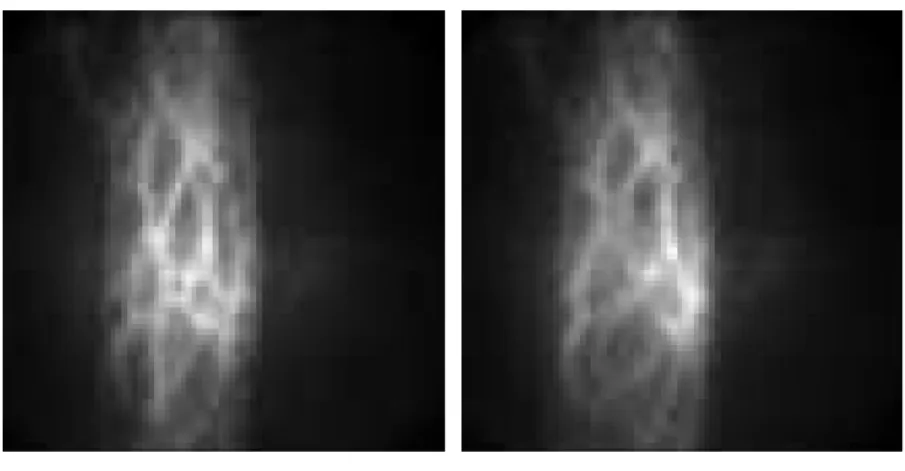
The aim of this study was to verify the presence of mechanosensitive neurons in the MP of the guinea pig ileum, to study their response to the application of a defined stimulus and to characterize these cells. Identification of activity in any network, in particular in the gut, is experimentally very challenging as recordings from individual sensory cells are unlikely to reflect behaviour of the network. Since we understood the importance to have an overview on one entire ganglion of the MP to comprehend the complex interactions of the different neurons within the enteric network, we carried on our study using the Neuro Imaging technique. With this technique it was possible to record action potentials simultaneously in a large number of neurons with high spatial and temporal resolution and this allows us to understand the differential activation of enteric populations in response to a mechanical stimulation within the ganglion.
Identification of sensory neurons is of central importance to understand sensory transmission under normal conditions and in gut diseases associated with sensorimotor dysfunctions, such as Irritable Bowel Syndrome (IBS) or Inflammatory Bowel Disease (IBD). Many of the symptoms prominent in the functional GI disorders are indeed consistent with dysfunction of the sensory and/or motor apparatus of the digestive tract. Identification and characterization of sensory neurons will be an important step to identify novel targets that help to normalise sensory function.
Material and methods
Tissue preparations
For our experiments we used male guinea pigs “BFA-Bunt” and “Dunkin Hartley” (Charles River laboratories, Kisslegg, Germany; Harlan GmbH, Borchen, Germany). The experimental animals were kept under standardized conditions in airflow cabinets (Ehret Uniprotect, Emmendingen, Germany). The diet consisted of a standard diet for guinea pigs (Rohfaserpellets, Altromin, Germany). Water was available ad libidum. The animals had two weeks for acclimatisation. The daily rhythm was set by a timer to 14 hours of light (from 7 am to 9 pm).
Guinea pigs were killed by cervical dislocation followed by exsanguination. This method was approved by the local animal ethical committee and is according to the Germans law for animal protection and animal welfare guidelines. At slaughter the animals had an average weight of 395 ± 60 (mean ± standard deviation).
After killing the animals the abdomen was opened with barb forceps (FST # 11023-10, Fine Science Tools, Heidelberg, Germany) and rough scissors (FST # 14001-13, Fine Science Tools). Forceps with flat corrugated tips (FST # 11000-14, Fine Science Tools) were used to hold the abdominal wall. The ileum was quickly removed with a pair of scissors with rounded tips (FST # 14010-15, Fine Science