Facoltà di Medicina e Chirurgia
Dipartimento di Medicina Sperimentale e Scienze Biochimiche
Cattedra di Virologia
Tesi di Dottorato in
Microbiologia Medica e Immunologia
(XXII ciclo)
Impact of Integrase Polymorphisms and Minor Quasispecies
in HIV-1 Infected Individuals Naive or Treated with
Strand-transfer Integrase Inhibitors: a Refined Analysis by Cloning
and 454-Pyrosequencing Techniques.
Candidato:
Relatori
Prof. Carlo-Federico Perno
Dott. Daniele Armenia Dott.ssa Francesca Ceccherini-Silberstein
3
Table of Contents
Abstarct/Riassunto
4
1 Introduction
8
1.1 HIV
8
1.1.1 Morphology
10
1.1.2 Genome
10
1.1.3 Replication
12
1.1.4 Pathogenesis
18
1.1.5 Epidemiology
20
1.2 Antiretroviral therapy
21
1.2.1 Protease inhibitors (PIs)
25
1.2.2 Reverse transcriptase inhibitors (NRTIs/NNRTIs)
28
1.2.3 Integrase inhibitors (INIs)
33
1.2.4 Fusion inhibitors (FIs)
40
1.2.5 CCR5 inhibitors
51
1.3 Drug resistance
42
1.3.1 Mechanism of drug resistance
43
1.3.1a PIs
43
1.3.1b NRTIs
44
1.3.1c NNRTIs
45
1.3.1d INIs
47
1.3.1e FIs
50
1.3.1f CCR5 inhibitors
51
1.4 Resistance test
51
1.4.1 Clinical importance of performing resistance test
51
1.4.2 Phenotypic test
53
1.4.3 Genotipic test
54
1.4.4 Interpretation of genotypic resistance profile
59
1.4.4a PIs
59
1.4.4b NRTIs
63
1.4.4c NNRTIs
71
1.4.4d INIs
73
1.4.5e FIs
75
1.4.5f CCR5 inhibitors
76
1.5 Rational of work
78
2 Methods
80
2.1 Clonal analysis
80
2.1.1 RNA isolation, cDNA synthesis and PCR
80
2.1.2 Cloning of RT-IN amplicons into HXB2D-based RT-IN deleted backbone
80
2.1.3 IN site-directed mutant (SDM) plasmids
81
2.1.4 Production of replication-competent recombinant viruses
81
2.1.4 Drug susceptibility testing of recombinant viruses (phenotyping)
81
2.1.5 IN sequencing
81
2.1.6 Mutations
82
2.1.7 Replication capacity assays
82
2.1.8 Statistical analysis
83
2.2 Population sequencing
84
2.2.1 Patients
84
2.2.2 IN Sequencing
85
2.2.3 Phylogenetic analyses
86
2.2.4 Mutations
87
2.2.5 Genotypic sensisitive score
87
2.2.5 Statistical analysis
87
2.3 Ultra-Deep-454 Pyrosequencing
88
4
2.3.2 Massively parallel sequencing
89
2.3.3 Mutations
89
2.3.4 Phylogenetic analyses
89
2.3.6 Cloning of IN amplicons into HXB2D-based IN deleted backbone
90
2.3.7 IN site-directed mutant (SDM) plasmids
90
2.3.8 Statistical analysis
90
3 Results
92
3.1 Secondary HIV-1 Integrase Resistance Mutations, Found as Minority Quasispecies
in Integrase Therapy Naive Patients, Have Little or no Effect on Susceptibility to Integrase
Inhibitors
92
3.1.1 Production of clonal recombinant virus stocks
92
3.1.2 Determination of raltegravir and elvitegravir susceptibility of clonal RT-IN recombinant
viruses
95
3.1.3 Genotypic characterization of mutations associated with resistance to raltegravir and
Elvitegravir
97
3.1.4 Phenotypic characterization of mutations associated with resistance to raltegravir and
Elvitegravir
100
3.1.5 Susceptibility to raltegravir and elvitegravir of site direct mutant viruses carrying
primary and secondary resistance IN mutations
101
3.1.6 Genotypic characterization of RT mutations in RT-IN recombinant viruses
3.1.7 Replication capacity of recombinant clonal viruses
103
3.2 Impact of baseline HIV-1 integrase polymorphisms with virological outcome
in patients starting a Raltegravir-containing regimen
105
3.2.1 Efficacy of raltegravir containing regimen at 24 weeks of treatment
105
3.2.2 Prevalence of integrase baseline polymorphisms according with subtype
106
3.2.3 Prevalence of integrase polymorphisms and raltegravir resistance mutations
according with subtype and virologic response
107
3.2.4 Independent predictors of virologic success at 24 weeks of raltegravir treatment
by univariate and multivariate logistic regression.
109
3.2.5. Prevalence of T125A mutations according with raltegravir treatment
112
3.3. Primary Mutations Associated with Resistance to Raltegravir are not Detectable by
Pyrosequencing in Integrase-Inhibitors Naïve Patients
114
3.3.1 UDPS coverage and sample size results
114
3.1.2 Prevalence of baseline raltegravir resistance associated mutation detected
by Population-sequencing
114
3.1.3 Comparison of mutation detectability with population sequencing and UDPS
115
3.1.4 Baseline prevalence of raltegravir resistance mutations detected
by UDPS according to virologic response at 24 weeks of raltegravir containing regimen.
117
3.1.5 Prevalence of raltegravir resistance mutations over time by UDPS
118
3.1.6 Phylogenetic analyses of haplotypes evolution during raltegravir treatment
121
of 4 representative failing patients
3.1.7 Phenotypic resistance associated with resistance mutations harbored at failure
125
3.1.8 Phenotypic resistance profile and replication capacity of viruses carrying
Y143C/R and/or N155H mutations.
127
4 Discussion
129
5 Summary and conclusions
136
6 Acknowledgments
140
5
Abstract
Background: Raltegravir (RAL) is a very potent and effective strand-transfer integrase-inhibitor (InSti), recently
FDA-approved for use also in first-line highly active antiretroviral therapy (HAART) regimen. Nowadays, by the available literature, the knowledge about the role either of integrase (IN) polymorphisms or HIV-1 minor quasispecies on virologic response and development of resistance to raltegravir is still poor. Therefore, the aim of this study is to explore the presence of InSti resistance mutations in HIV-1 quasispecies present in InSti-naïve patients and to evaluate their impact on in vitro phenotypic susceptibility to InSTIs, on replication capacities and on virologic response to raltegravir, by 3 different approaches: Cloning, population sequencing and ultra-deep 454 pyrosequencing (UDPS).
Methods: For the clonal approach, the RT-RNase H-IN region was PCR amplified from plasma viral RNA obtained
from 49 HIV-1 subtype B-infected patients (21 drug naïve and 28 failing HAART not containing InSTIs) and recombined with an HXB2-based backbone with RT and IN deleted. Recombinant viruses were tested against raltegravir and elvitegravir and for replication capacity. Three-hundred forty-four recombinant viruses from 49 patients were successfully analyzed both phenotypically and genotypically.
For the population sequencing approach, we analyzed 206 multi-experienced patients that received raltegravir plus optimized background therapy (OBT) from eight clinical centres within Italy and France. HIV-1 RNA and IN genotypes were assessed at baseline and at failure. For 177 patients, viremia values at 24-week were available. The prevalence of baseline integrase mutations was calculated in the overall population, and in the responding and non-responding patients at 24 weeks. For polymorphisms with prevalence >5%, the codon usage of mutated amino acids were also considered. Logistic regression analyses (uni- and multivariate) were performed to investigate if baseline integrase polymorphisms and other variables (such as: baseline HIV-1 RNA, drugs in co-usage and/or subtype) were independent predictors of virologic response.
For the UDPS approach a sub-group of 27 patients treated with raltegravir were genotyped by UDPS at baseline and at failure. IN phenotyping was also performed at baseline and during treatment for failing patients.
In all three approaches, all IN mutations, with particular attention to known InSti resistance associated mutations, have been analyzed. The cut-off limit of reliable detection for UDPS was considered >0.1% (≥50 reads).
Results: Regarding clonal analysis, the majority of clones were not phenotypically resistant to InSTIs: 0/344 clones
showed raltegravir resistance, and only 3 (0.87%) showed low-level elvitegravir resistance. No primary resistance mutations for raltegravir and elvitegravir were found as major or minor species. Secondary mutations, such as T97A and G140S, found rarely and only as minority quasispecies, were present in the elvitegravir-resistant clones. A novel mutation, E92G, although rarely found in minority quasispecies, showed elvitegravir resistance.
Regarding the analyses of baseline IN mutations, among the 206 patients genotyped by population sequencing, 186 (90.3%) patients were infected by HIV-1 subtype B versus 20 (9.7%) infected by non-B viruses (4A, 1C, 2D, 5F, 2G, 5CRF_02AG, 1CRF_12BF2). At week 24, 70% of patients achieved virologic response (71.3% [114/160] and 58.8% [10/17] infected by B and non-B viruses, respectively, p=NS). At baseline, all major raltegravir resistance mutations were completely absent, and secondary mutations (L74M, T97A, G140A, V151I, N155S, G163R) were present at very low frequency (≤1%). The presence at baseline of these secondary resistance mutations, as well as all other polymorphisms (with the exception of T125A, specific codon GCA, ―see below‖) did not statistically influence the virologic response among patients starting raltegravir (Fisher test, Benjamini-Hockberg correction). By multivariate logistic regression, the independent predictors of worse virologic response were: baseline viremia (OR=0.42 [CI:0.3-0.7], p=0.0003), AZT or D4T co-usage (OR=0.31 [CI:0.1-0.9], p=0.04) and baseline presence of polymorphism T125A (specific GCA codon, that is consensus sequence for subtypes A, C, D, G and for CRF02_AG) (OR=0.30 [CI: 0.1-0.7], p=0.006). Such prevalence of T125A (specific GCA codon) was higher in patients infected with non-B subtype (13/20 [65%]) vs B subtype (35/186 [19%]) (OR=0.12 [CI:0.05-0.33], P=0.00003), with a greater consistence among failing patients ( 6/7 [86%] non-B subtype vs 14/46 [30%] B subtype, OR=0.07 [CI:0.01-0.52],p=0.009).
Regarding UDPS analyses, among >200,000 IN sequences analyzed, no minor variants of primary raltegravir mutations with a prevalence of >0.1% were found at baseline. The secondary mutations such as T97A, F121Y and V151I secondary mutations, were rarely found at baseline, in both failing- and success-group of patients, with a frequency ranging from 0.3 to 99% of viral species. Independently of the sequencing method, the presence of secondary-resistant species at baseline was not associated, at failure, with evolution at the same amino acid position or to specific primary raltegravir resistance mutations. Raltegravir phenotypic resistance has never been observed at baseline. At failure, all patients carrying primary mutations N155H, Q148H/R or Y143R, in presence of secondary (L74M, T97A, E92Q, G140S, V151I, E157Q, G163R, S230R) and novel (E92A, T112A) mutations, showed fold changes on susceptibility to RAL >30-100. Interestingly, in 1 patient, we found the combination of two primary mutations at failure, Y143C and N155H, by both population sequencing and UDPS. These mutations appeared at failure for >80% on same haplotypes, and showed a very high phenotypic resistance, particularly for raltegravir (FC raltegravir = 1255.3; FC elvitegravir = 625.3).
Conclusion: By classic and ultra sensitive genotyping (and phenotyping) methods, pre-existing raltegravir resistance is
a rare event in InSti-naïve patients, and when present, is confined to a restricted minority of secondary variants only. At baseline, only T125A mutation (specific GCA codon), higher prevalent in non-b subtype viruses, was associated with
6
poorer virologic response to raltegravir. This finding in non-B subtypes is intriguing and further research is warranted. The clinical implications and relevance of this polymorphism is still to be determined.
Overall, this study suggests that at this point IN genotyping in all patients before raltegravir treatment may not be cost-effective and should not be recommended until evidence of transmitted drug resistance to InSTIs or the clinical relevance of IN minor variants/polymorphisms is determined.
7
Riassunto
Introduzione: Raltegravir è un potente ed efficace inibitore dell‘ integrasi (IN) di HIV-1, recentemente approvato
dall‘FDA anche nei regimi HAART di prima linea. Dagli studi attualmente disponibili in letteratura, il ruolo dei polimorfismi naturali e delle quasi specie minoritarie dell‘integrasi sul responso virologico agli InSti e sullo sviluppo di resistenza durante il fallimento è ancora poco chiaro. Pertanto questo lavoro mira a verificare la presenza di mutazioni di resistenza agli InSti nelle quasispecie naturali di HIV-1 in pazienti naive a tali inibitori e a valutare l‘impatto sulla suscettibilità fenotipica in vitro, sulla capacità replicativa virale e sul responso virologico a raltegravir utilizzando tre diversi approcci: il metodo clonale, il sequenziamento di popolazione e il pirosequenziamento massivo 454 (Ultra-deep 454 Pyrosequencing [UDPS]).
Metodi: Per l‘approccio clonale, le sequenze di RT-RNAse H-IN sono state amplificate tramite PCR da campioni di
plasma da 49 individui infetti da HIV-1 sottotipo B (21 naive al trattamento e 28 in fallimento a regimi antiretrovirali non includenti gli InSti) e ricombinate con un vettore di espressione contenente lo stipite di HIV-1 HXB2D deleto della regione RT-IN. I virus ricombinanti ottenuti sono stati testati per la suscettibilità a raltegravir ed elvitegravir e per la capacità replicativa. Da 49 pazienti sono stati ottenuti 344 cloni ricombinanti testati genotipicamente e fenotipicamente
in vitro.
Per l‘analisi con il sequenziamento di popolazione, sono stati analizzati 206 pazienti multi-trattati, provenienti da 8 diversi centri clinici italiani e francesi, che iniziavano il trattamento con un regime contenente raltegravir. Il genotipo dell‘IN e la viremia sono stati effettuati prima e durate l‘inizio della terapia. Alla 24esima settimana di trattamento erano disponibili valori di viremia per 177 pazienti. La prevalenza delle mutazioni è stata calcolata nella popolazione totale, nei pazienti che hanno raggiunto il successo virologico e nei i pazienti che hanno fallito alla 24esima settimana di trattamento. Per i polimorfismi con una prevalenza maggiore del 5% è stato anche considerato l‘uso specifico dei codoni codificanti le mutazioni. Per valutare se i polimorfismi ed altre variabili (la viremia, i farmaci co-somministrati e il sottotipo) fossero predittori indipendenti di successo virologico, è stata effettuata un‘analisi di regressione logistica (uni e multivariata).
Per l‘analisi con l‘UDPS un sottogruppo di 27 pazienti trattati con raltegravir è stato genotipizzato al baseline e al fallimento. Inoltre è stato effettuato il test fenotipico delle popolazioni virali al fallimento.
Per tutti e tre gli approcci sono state analizzate tutte le mutazioni dell‘ integrasi con particore attenzione alle mutazioni di resistanza note. In particolare per l‘UDPS, il rilevamento delle mutazioni è stato considerato attendibile osservando una prevalenza >0.1%(>50 varianti).
Risultati: Dall‘approcio clonale, la maggior parte dei cloni testati non ha mostrato resistenza fenotipica agli InSti:
0/344 cloni hanno mostrato resistenza per raltegravir e solo 3 cloni (0.87%) hanno mostrato bassi livelli di resistenza per elvitegravir. Non è stata osservata alcuna mutazione di resistenza primaria per raltegravir e/o elvitegravir. Nei cloni resistenti a elvitegravir sono state trovate alcune mutazioni secondarie, come la T97A e la G140S. Inoltre è stata osservata una nuova mutazione, E92G, anch‘essa in quasispecie minoritaria, associata a resistenza fenotipica a elvitegravir.
Tra i 206 pazienti analizzati con l‘approccio di sequenziamento di popolazione, 186 (90.3%) sono risultati infetti da sottotipo B mentre 20 (9.8%) sono risultati infetti da sottotipi non B (4A, 1C, 2D, 5F, 2G, 5CRF_02AG, 1CRF_12BF2). Alla 24esima settimana di trattamento con raltegravir il 70% dei pazienti ha raggiunto il successo virologico (il 71.3% [114/160] e il 58.8% [10/17] infetti da virus di sottotipo B e non-B rispettivamente, p=NS). Al basale non è stata trovata alcuna mutazione di resistenza primaria mentre le secondarie (L74M, T97A, G140A, V151I, N155S, G163R) hanno mostrato una bassa frequenza (≤1%). La presenza al basale di tali mutazioni secondarie, come di altri polimorfismi (con l‘eccezione della T125A, codone GCA, ―vedi sotto‖) non hanno influenzato statisticamente il responso virologico dei pazienti che hanno iniziato raltegravir (Fisher test, correzione di Benjamini-Hockberg). Dall‘analisi di regressione logistica multivariata, i predittori indipendenti di negativo responso virologico erano: la viremia al basale (OR=0.42 [CI:0.3-0.7], p=0.0003), la co-somministrazione di AZT or D4T (OR=0.31 [CI:0.1-0.9], p=0.04) e la presenza al basale del polimorfismo T125A (specifico codone GCA, che è sequenza di riferimento per i sottotipi A, C, D, G and for CRF02_AG) (OR=0.30 [CI: 0.1-0.7], p=0.006). La prevalenza di questa mutazione, T125A(specifico codone GCA) risulta più alta nei pazienti infetti da sottotipi non-B (13/20 [65%]) vs B subtype (35/186 [19%]) (OR=0.12 [CI:0.05-0.33], P=0.00003) con una maggiore discrepanza tra i pazienti in fallimento ( 6/7 [86%] non-B subtype vs 14/46 [30%] B subtype, OR=0.07 [CI:0.01-0.52],p=0.009).
Dall‘approccio UDPS, al baseline, tra più di 200000 sequenze dell‘IN analizzate, non è stata trovata alcuna variante minoritaria con resistenza primaria a raltegravir con una frequenza > 0.1%. Le mutazioni secondarie T97A, F121Y e V151I sono state trovate raramente e indifferentemente in pazienti in successo e/o in fallimento, con una frequenza compresa tra lo 0.3-99% delle specie virali. Indipendentemente dal metodo di sequenziamento utilizzato, la presenza di varianti resistenti secondarie al basale non correlava al fallimento, né con l‘evoluzione alla stessa posizione amminoacidica, né con lo sviluppo di mutazioni primarie. Al basale non è stata osservata resistenza fenotipica a raltegravir, tuttavia al fallimento, i pazienti che hanno sviluppato le mutazioni primarie N155H, Q148H/R o Y143R, associate ad altre mutazioni secondarie (L74M, T97A, E92Q, G140S, V151I, E157Q, G163R, S230R) o non note (E92A, T112A) hanno mostrato un ridotta suscettibilita a raltegravir (Fold change >30-100). Di particolare interesse, in 1 paziente in fallimento, è stata trovata la combinazione delle mutazioni primarie N155H e Y143C, sia utilizzando il sequenziamento di popolazione sia l‘UDPS. Queste mutazioni, apparse al fallimento nell‘80% degli aplotipi del
8
paziente, sono associate ad alta resistenza fenotipica particolarmente spiccata per raltegravir (FC raltegravir = 1255.3; FC elvitegravir = 625.3).
Conclusioni: Dai saggi fenotipici e di genotipizzazione classici e ultra sensibili, la resistenza pre-esistente a raltegravir
nei pazienti naive agli InSti è un evento raro e quando presente risulta confinato soltanto in quasi specie minoritarie secondarie. Al basale, solo la presenza della mutazione T125A(GCA), più prevalente nei sottotipi non-B, è risultata associata a un inferiore responso virologico a raltegravir. Questa osservazione nei sottotipi non-B è intrigante e necessita di ulteriori investigazioni. L‘impatto clinico e la rilevanza di questo polimorfismo devono comunque ancora essere determinati.
In conclusione, questo studio suggerisce che, allo stato attuale, effettuare il genotipo dell‘integrasi in tutti i pazienti prima dell‘inizio di raltegravir, potrebbe avere un rapporto costo-beneficio spostato verso il costo e non dovrebbe essere raccomandato, almeno fino a quando non si abbiano evidenze di resistenza trasmessa agli InSti o sia chiarita la rilevanza clinica dei polimorfismi e quasispecie minoritarie naturali.
9
Transcription Translation Replication1. Introduction
1.1 HIV
The central dogma of molecular biology states that in biological cells the information flow follows
the scheme
DNA
RNA
Protein
(Crick, 1958; Crick, 1970). Replication (DNA to DNA), transcription (DNA to RNA), and
translation (RNA to protein) occur in all living cells, while reverse transcription occurs only in cells
infected with retroviruses or hepadnaviruses (Hepatitis B virus). Retroviruses carry their genome
information in the form of two positive sense (5‘→3‘ direction) RNA copies. The diploid nature of
their genome is unique among viruses. Replication can be accomplished only in host cells by
converting their RNA to DNA and incorporating the viral genes into the host genome.
Retroviruses were traditionally divided into three subfamilies, based mainly on pathogenicity rather
than on genome relationship (oncoviruses which cause neoplastic disorders, spumaviruses which
give cytopathic effect in tissue culture but apparently not associated with any known disease, and
lentiviruses which induce slowly progressing inflammatory, neurological and immunological
diseases). In the last decade, the international committee on the taxonomy of viruses has recognized
seven distinct genera in the Retroviridae family (Table 1.1)(Fields, et al., 1996).
10
Table 1.1. Retroviruses genera
The retrovirus family is divided in 7 genera: the Alpharetroviruses, Betaretroviruses, Gammaretroviruses, Deltaretroviruses and Epsilonviruses (all of which used to be classified as one genus, the oncoviruses), the Lentiviruses (which includes HIV) and the Spumaviruses.
The human immunodeficiency virus (HIV), identified in 1983, is a member of the Lentivirus genus
which is exogenous, non-oncogenic retrovirus causing persistent infections leading to chronic
diseases with long incubation periods (lenti for slow). Like the human T-cell leukemia virus
(HTLV) family of primate onco-retroviruses, lentiviruses are complex retroviruses(Cullen, 1991).
The significant characteristic of the complex retroviruses is the ability to regulate their own
expression via virally encoded protein factors not found in other retroviruses. This property has
been proposed to be essential for the long-term association of the complex retroviruses with the host
and the generation of chronic active infections. The lentiviral complexity is reflected in their
replication cycle, which reveals intricate regulatory pathways, unique mechanisms for viral
11
1.1.1 Morphology
The HIV virion is a spherical virus particle of about 100 nm in diameter (Fig. 1.1). The viral
envelope consists of a lipid bilayer derived from the host cell membrane during release of the newly
produced particles from an infected cell. Embedded in the viral envelope are proteins from the host
cell as well as viral protein complexes composed of the transmenbrane glycoprotein gp41 (TM) and
the surface glycoprotein gp120 (SU). These trimeric TM-SU complexes constitute the
characteristics spike of the virion that are involved in cell recognition and entry.
A matrix shell compromising ca. 2000 copies of the matrix p17 (MA) lines the inner surface of the
viral membrane. In the center of a mature HIV particle resides the cone-shaped capsid (or cone).
The capsid is made of ca. 2000 copies of the viral capsid protein p24 (CA). It encloses two single
strands of the HIV RNA genome stabilized as a ribonucleoprotein complex with ca. 2000 copies of
the nucleocapsid protein p7 (NC). Additionally, the capsid contains the three virally encoded
enzymes, reverse transcriptase, protease, and integrase as well as accessory proteins such as nef, vif,
vpr. There are three additional accessory proteins rev, tat, vpu, that are not packaged into the virion.
High resolution three-dimensional information is available for all HIV proteins (Frankel, et al.,
1998; Turner, et al., 1999)
1.1.2 Genome
The genome of HIV has a length of approximately 9.2 kbp. Like all retroviruses it contains the
characteristics:
5‘- gag – pol – env - 3‘
motif consisting of the three structural genes gag, pol, and env (Fig. 1.2). The Gag (group antigen)
gene encodes the large precursor polyprotein p55 that is cleaved in four proteins: the matrix p17,
the "core" capsid p24, the nucleocapsid p7 and the p6(Freed, 1998). The pol (polymerase)gene
12
complex p51 and p66, integrase p32. The env (envelope) gene directs the production of an envelope
precursor protein gp160, which undergoes cellular proteolytic cleavage into the outer envelope
glycoprotein gp120 and the transmembrane glycoprotein gp41.
The RNA genome is flanked by two short redundant (R) sequences at both termini with adjacent
unique sequences, U5 and U3, found at the 5‘ and 3‘ ends, respectively. In addition, HIV has at
least six more genes encoding viral proteins with regulatory functions (tat and rev) or accessory
functions (nef, vif, vpr and vpu) (for reviews see(Cullen, 1991; Emerman, et al., 1998; Kleim, et al.,
1996; Piguet, et al., 1999; Pollard, et al., 1998; Trono, 1995).
Fig.1.1. The immature and mature forms of HIV-1.
Typical lentivirus particles are spherical, about 80-110 nm in diameter, and consist of a lipid bilayer membrane surrounding a conical core. The two identical single-stranded RNA (ssRNA) molecules, of about 9.2kB each, are associated with the nucleocapsid proteins p7gag (NC). They are packed into the core along with virally encoded enzymes: reverse transcriptase (RT), integrase, and protease. P24gag comprises the inner part of the core, the capsid (CA). The p17gag protein constitutes the matrix (MA) which is located between the nucleocapsid and the virion envelope. The viral envelope is produced by the cellular plasma membrane and contains the protruding viral Env glycoproteins: gp120 surface glycoprotein (SU) and gp41 transmembrane protein (TM).
(from:
http://tolomeo.files.wordpress.com/ 2008/11/hiv.gif )
13
Fig. 1.2. HIV genomic organization. Like all other retroviruses, HIV has three structural genes gag, pol and env(heavily shaded), which are flanked by the long terminal repeats (LTR‘s). In addition it has six more genes, including two regulatory genes tat and rev (stippled) and four accessory genes nef, vif, vpr and vpu (white).
1.1.3 Replication
The HIV replication cycle begins with the recognition of the target cell by the mature virion. The
major targets for HIV infection are cells bearing the HLA class II receptor, CD4, on their cell
surfaces. These include T-helper lymphocytes and cells of the monocyte/macrophage lineage
including microglia cells in the brain. The virus-CD4 binding occurs via specific interactions
between the viral outer envelope glycoprotein gp120 and the amino-terminal immunoglobulin like
domain of CD4(Dalgleish, et al., 1984; Klatzmann, et al., 1984). These interactions are sufficient
for binding but not for infection. Subsequently the virus glycoprotein gp120 interacts with
additional cell-surface proteins to promote fusion of the viral and cellular membranes. These
coreceptors have recently been identified to be members of the chemokine receptor family and
include CXCR4 and CCR5(Alkhatib, et al., 1996; Deng, et al., 1996; Moore, 1997). The initial
binding of HIV to the CD4 receptor is mediated by conformational changes in the gp120 subunit,
followed by a conformational change in the gp41 subunit, induced by the chemokine receptors, that
allows fusion and subsequent entry of HIV. Various strains of HIV differ in their use of chemokines
coreceptors. There are strains of HIV known as T-tropic strains, which selectively interact with the
CXCR4 chemokine coreceptor of lymphocytes, while M-tropic strains of HIV interact with the
14
(Littman, 1998; Moore, 1997). HIV-1 infection of CD4 negative cells, such as neural cells, has also
been reported (Clapham et al., 1989; Harouse et al., 1989; Kozlowski et al., 1991; Kunsch et al.,
1989) but the mechanisms of HIV entry are still unclear. Membrane fusion is followed by an
uncoating event that allows the intracellular reverse transcription. The viral RNA is transcribed in
the cytosol into double stranded DNA by the reverse transcriptase (Hansen, et al., 1987; Muesing, et
al., 1985)This enzyme have three enzymatic activities: RNA-dependent DNA polymerase,
DNA-dependent DNA polymerase, and ribonuclease H (RNase H). The reverse transcription process
takes place within a large nucleic acid-protein complex known as the preintegration complex (PIC)
by the assistance of the accessory protein Vif (Schwedler, et al., 1993)and the nucleocapsid protein
NC(Allain, et al., 1994). Once synthesized, the viral DNA is transported to the nucleus of the
infected cell as part of the PIC that appears to include tightly condensed viral nucleic acids and the
integrase, p17, reverse transcriptase, and Vpr proteins. In contrast to other retroviruses, that require
cell division and concomitant breakdown of the nuclear envelope to gain access to the nuclear
compartment, the lentiviral PIC is actively imported into the nucleus during the interphase
(Bukrinsky, et al., 1992; Lewis, et al., 1994). Nuclear import of the PIC seems to be directed by the
accessory protein Vpr (Fouchier, et al., 1997; Heinzinger, et al., 1994), the Gag matrix protein p17
(Bukrinsky, et al., 1993; Schwedler, et al., 1994) and the integrase(Gallay, et al., 1997). Vpr does
not contain a conventional nuclear localization signal but appears to function by connecting the PIC
to the cellular nuclear import machinery (Fouchier, et al., 1998; Popov, et al., 1998). The ability of
lentiviruses such as HIV-1 to utilize active transport mechanisms for translocation of the PIC into
the nucleus, allows these viruses to infect non-dividing cells such as differentiated macrophages,
quiescent T lymphocytes and possibly neurons. In the nucleus, integrase catalyzes covalent
integration of the viral DNA into the host genome, where it resides permanently as a provirus. An
important modification as a result of reverse transcription and integration is the duplication of the
U5 and U3 sequences in the LTR, such that the provirus now is flanked by tandemly repeated
15
involves a complex interplay between cis-acting DNA and RNA elements present within the
chromatin-associated proviral LTRs, cellular transcription factors and the viral regulatory protein
Tat (transcriptional transactivator).
The regulation of the HIV transcription involves a complex interplay between cis-acting DNA and
RNA elements present within the chromatin-associated proviral LTRs, cellular transcription factors
and the viral regulatory protein Tat (transcriptional transactivator). In an arrangement similar to that
of several inducible cellular promoters, the HIV-1 promoter, which is located in the U3 region of
the 5‘LTR, contains a TATA box and binding sites for several cellular DNA-binding transcription
factors, such as NF-kB, Sp1 and TBP(Jones, et al., 1994). It is highly inducible and responds to the
activation status of the infected cell. NF-kB is the major inducible cellular activator. It is well
established that many cells in the lymphoid tissue of infected individuals are latently infected
(Pantaleo, et al., 1993), even though the viral replication in the body is always active. In resting
T-cells, the activity of the HIV promoter is minimal, leading to viral quiescence in infected primary
cells. Therefore, viral activation is associated with cell activation. The transcription of the provirus
by the cellular RNA polymerase II results in a primary transcript that may serve three distinct
functions: 1) it constitutes genomic RNA that is incorporated into the virion; 2) it serves as template
for translation (Gag and Gag-Pol); 3) it functions as the precursor RNA for the production of
diverse subgenomic mRNAs (Fig 1.3).
As mentioned before, HIV encodes two essential regulatory proteins Tat and Rev, which increase
viral gene expression at the transcriptional and post-transcriptional levels, respectively. HIV mRNA
expression is biphasic and can be divided into early (Rev-independent) and late (Rev-dependent)
stages (Kim, et al., 1989). First, shortly after the infection of cells, multiply spliced (~ 2kb) RNA
species are formed from the primary transcript and three proteins are produced: Tat, Rev and Nef,
therefore referred as early gene products(Schwartz, et al., 1990). Tat [for reviews see (Cullen, 1998;
Emerman, et al., 1998; Rubartelli, et al., 1998), greatly increases transcription from the HIV
16
which is located at the 5‘ end of the nascent viral RNA transcript (Berkhout et al., 1989; Dingwall
et al., 1989). Tat recruits two cellular factors to this complex: cyclin T and cyclin-dependent protein
kinase-9 (Cdk9). Cyclin T is proposed to bind directly Tat and to increase its affinity for the TAR
RNA(Wei, et al., 1998). Cdk9 phosphorylates the RNA polymerase II transcription complex and
thus stimulates transcriptional elongation (Wei, et al., 1998). Rev (regulator of expression of the
virion), which accumulates during the early phase of expression, initiates late gene expression by
binding a unique RNA element located in the env coding region of HIV-1, the so called
Rev-responsive element (RRE). This interaction promotes the stability and transport of unspliced (~ 9
kb) and partially spliced (~ 4 kb) HIV-1 mRNAs out of the nucleus. These mRNAs are responsible
for the production of the viral enzymes and structural proteins (Daly, et al., 1989; Felber, et al.,
1989; Hadzopoulou-Cladaras, et al., 1989; Hammarskjold, et al., 1989; Malim, et al., 1989).
Therefore Gag, Pol, Env, Vif, Vpr, and Vpu proteins are referred to as late HIV-1 proteins.
The Nef (negative factor) protein play various functions. In particular, it enhances viral expression
in quiescent cells and mediates lymphocyte chemotaxis and activation at sites of virus replication
(Kestler, et al., 1991; Koedel, et al., 1999; Miller, et al., 1994; Swingler, et al., 1999). The Env
precursor polyprotein (gp160) is synthesized in the endoplasmatic reticulum (ER) where it is
glycosylated and appears to oligomerize to a trimeric structure posttranslationally (Wyatt, et al.,
1998; Wyatt, et al., 1998). Thereafter, it is cleaved to produce the non-covalently associated (gp41
TM - gp120 SU)3 trimeric glycoprotein complex, which is transported to the cell membrane for
virus assembly. Vpu is thought to enhance this process and inhibit a premature trapping of CD4 to
Env in the ER by binding CD4 molecules, which are also synthesized in the ER, and directing them
to the ubiquitin-proteasome degradation pathway (Margottin, et al., 1998; Schubert, et al., 1998;
Strebel, et al., 1988; Willey, et al., 1992; Willey, et al., 1992). Similarly, the accessory protein Nef
facilitates the routing of CD4 from cellsurface and Golgi apparatus to lysosomes, resulting in
endosomal degradation and preventing inappropriate interaction with Env (Aiken, et al., 1994). In
17
downregulation of CD4 and MHC class I molecules on the surface of infected cells also helps
infected cells to evade immune responses of the host, such as killing by cytotoxic T lymphocytes
(Collins, et al., 1998; Kerkau, et al., 1997).
During synthesis of the Gag polyprotein by ribosomes, a translational frameshift may occur,
resulting in generation of smaller amount of Gag-Pol precursor polyproteins, which associate with
the Gag polyprotein at the cellular membrane. The N-terminally myristoylated MA domain of the
Gag/GagPol polyproteins directs insertion of the Gag precursors into the cellular membrane and
interacts with the cytoplasmic tail of gp41 resulting in the anchoring of Env to the viral particle
(Dorfman, et al., 1994). Approximately 1200 to 2000 copies of Gag precursor bud to form an
immature particle, which encapsidates two copies of the unspliced viral RNA genome, by the ability
of NC to interact with nucleic acids. Vif and Vpu proteins have been reported to play a role in
packaging of the nucleoprotein core and in virion release, respectively (Höglund, et al., 1994;
Lamb, et al., 1997). Concomitantly or immediately following the external budding, the cleavage of
the Gag/Gag-Pol polyproteins by the virally encoded PR produces the structural proteins MA, CA,
NC as well as the independent enzymes PR, RT and IN. This final step primes new virus particles
18
Figure1.3. Replication cycle of HIV-1. Each fundamental step is presented in bold. Names in italic refer to viral geneproducts involved in the specific steps. HIV-1 gene expression is stimulated by HIV-1 Tat and Rev, which act at transcriptional and post-transcriptional levels, respectively, and can be divided into two phases. The early phase is Rev-independent and the later phase is Rev-dependent (text in gray). Rev stabilizes and mediates export of singly spliced and unspliced RNA transcripts out of the nucleus into the cytoplasm.
Modified from Ceccherini-Silberstein, 2001 (http://edoc.ub.Muenchen.de/archive/00000533/01/Ceccherini-Silberstein_Francesca.pdf).
19
1.1.4 Pathogenesis
AIDS
HIV infection has been associated with the acquired immunodeficiency syndrome (AIDS). A
diagnosis of AIDS is made whenever a person is HIV-positive and have:
CD4+ T cell count below 200 cells/mms
CD4+ T cells account for fewer than 14% of all lymphocytes
Diagnosis with one or more of the 25 AIDS defining illness, including various opportunistic
infection, brain and nerve disease, certain cancers, and wasting syndrome
Approximately 10% of HIV-infected patients progress to AIDS within the first 2 to 3 years of
infection, while for approximately 40% this progression is observed over a period of 10 years. 10%
to 17% of HIV-infected patients may be AIDS free, some with no evidences of disease progression.
These variations in responses may be due to differences in the degree of stimulation of the immune
system by infection with the other pathogens as well as to viral factor, such as deletions in the nef
gene or altered cell tropism (Kupfer, et al., 1998).
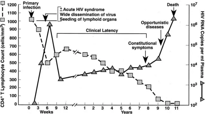
Course of infection
Schematically, the course of infection can be divided into an acute, an asymptomatic, and
symptomatic phase (Fig. 1.4). The acute phase accounts for the first 5-10 weeks of infection and is
characterized by high virus production, and activation of lymphocytes in lymphonodes. Up to 5x10
3infectious particles per ml of blood plasma may be found in the first days after infection. This
viremia is curtailed within a few weeks and level off at the beginning of the asymptomatic phase to
the so-called virological set point, that is a predictor of disease progression. During this CD4+ cells
numbers decrease at a low steady rate, while virus replication remains constant at a low rate. The
duration of the asymptomatic phase may last between 2 and 20 years. The end stage of disease,
when the patient develops AIDS, is characterized by CD4+ cells count below 200 copies/ml and
increased quantities of the virus. The number of CD8+ cytotoxic lymphocytes also decreases and
20
CD4+T cell depletion
The hypothesis that CD4+ cell depletion is caused the lysis of infected cells during viral replication
has been supported by the observation of an immediate and large increase of CD4+ count after the
initiation of antiretroviral therapy that blocks viral replication (Ho, et al., 1995; Wei, et al., 1995).
This hypothesis has not withstood more detailed analyses of T cell dynamics (Roederer, 1998). In
fact, it has been turned out that in HIV-infected patients all T cell subset are progressively
destroyed, irrespective of CD4+ expression, and AIDS appear to be a disease of perturbed
homeostasis. Many pathogenetic mechanisms have been proposed, including viral gene products,
syncitium formation, direct virus killing of cell, apoptosis, autoimmunity, cytokine and chemokines
expression, superantigens, virus directed cell mediated cytolysis and disruption of lymphoid
architecture.
21
1.1.5 Epidemiology
Several African primates harbour lentiviruses and HIV is believed to be entered the human
population in Africa by zoonotic transmission of SIV
cpzfrom chimpanzee population. The first
cross species transfer has been estimated to have occurred between 1915 and 1941(Korber, et al.,
2000). Two types of HIV are known: the most common HIV-1, which is responsible to the
world-wide AIDS epidemic and the immunologically distinct HIV-2 (Clavel, et al., 1986), which is much
less common and less virulent (Ariyoshi, et al., 1999; Ariyoshi, et al., 2000), but produces clinical
findings similar to HIV-1 (Wilkins, et al., 1993). The HIV-1 type itself includes four groups M, N,
O, P which have different geographic distributions but all produce similar clinical symptoms (Fig.
1.5). The M group is further divided into 9 pure subtypes (A, B, C, D, F, G, H, J, K), 4 sub-subtype
(A1, A2, F1, F2) and 45 circulating recombinant forms on the basis of phylogenetic analysis.
Almost all subtypes are present in Africa, while in Europe, North America, and Australia subtype B
is more dominant, and subtype C is more common in Asia (Robertson, et al., 2000; Mc Cutchan,
2000; Plantier, et al., 2009).
Figure 1.5. Phylogenetic relationship of primate lentiviruses. Phylogenetic tree derived from the alignment of pol gene sequences of HIV-1 and SIV strain ( SIVcpz and SIVgor). Reproduced from Plantier 2009
22
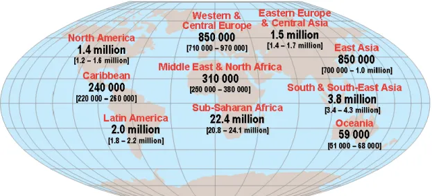
At the end of 2009, 33.4 million adults and children have been estimated to live with HIV/AIDS,
most of them in Sub-Saharan Africa and South East Asia (Fig. 1.6). Only a minority of
HIV-infected individuals live in the industrialized countries and has access to the anti-HIV drugs and
professional health care.
Figure 1.6. Geografical distribution of HIV/AIDS cases. From UNAIDS 2009
1.2 Antiretroviral therapy
The drugs currently used to treat HIV-1 infection are directed against the four viral enzymes, an
envelope glycoprotein and a human receptor: protease (PR), reverse transcriptase (RT), the
transmembrane glycoprotein gp41 and more recently, also against Integrase (IN) and human CCR5
receptors. In table 1.2 all anti-HIV compounds currently approved for clinical use by the U.S. Food
and Drug Administration (FDA) [Division of AIDS, National Institute of Allergy and Infectious
Diseases, National Institutes of Health] are listed. In figure 1.7 the available drugs in clinic by
23
Fig. 1.7 HIV replication cycle according with ARV’s available by today.Initial entry of HIV into a target cell can be blocked by use of the entry inhibitor maraviroc, which prevents viral interaction with the CCR5 coreceptor. Fusion of the viral membrane with the target cell membrane can be blocked by the peptidic inhibitor enfuvirtide, which prevents a conformational change in the viral Env protein needed to bring the two membranes into close proximity. Reverse transcription of the viral RNA into DNA can be blocked by nucleoside/tide reverse transcriptase inhibitors (NRTIs) which are incorporated into the viral DNA and act to chain terminate DNA synthesis. Non-nucleoside reverse transcriptase inhibitors (NNRTIs) are non-competitive inhibitors of reverse transcriptase. Integrase strand transfer inhibitors (INSTIs), such as raltegravir, are active site inhibitors of the viral integrase enzyme and prevent the strand transfer reaction, the final ligation of the 3_-processed viral DNA into the host genome. Protease inhibitors (PIs) prevent the proteolytic processing of translated viral proteins by the viral protease enzyme, resulting in defective virions. Combinations of drugs from two or more of these classes when combined together form the basis of highly active antiretroviral therapy (HAART).(From:D.J. McColl, X. Chen. 2008. Antiviral Research)
InStis
RAL
24
Table 1.2. Antiretroviral drugs in clinical use.
Multi-class combination products
Brand
Name
Generic Name
Manufacturer Name
Approval Date
Time to Approval
Atripla
efavirenz,
emtricitabine and
tenofovir disoproxil
fumarate
Bristol-Myers Squibb
and Gilead Sciences
12-July-06
2.5 months
Nucleoside(tide) Reverse Transcriptase inhibitors (NRTIs)
Brand
Name
Generic Name
Manufacturer Name
Approval Date
Time to Approval
Combivir
lamivudine and
zidovudine
GlaxoSmithKline
27-Sep-97
3.9 months
Emtriva
emtricitabine, FTC
Gilead Sciences
02-Jul-03
10 months
Epivir
lamivudine, 3TC
GlaxoSmithKline
17-nov-95
4.4 months
Epzicom
abacavir and
lamivudine
GlaxoSmithKline
02-Aug-04
10 months
Hivid
zalcitabine,
dideoxycytidine, ddC
(no longer marketed)
Hoffmann-La Roche
19-Jun-92
7.6 months
Retrovir
zidovudine,
azidothymidine, AZT,
ZDV
GlaxoSmithKline
19-mar-87
3.5 months
Trizivir
abacavir, zidovudine,
and lamivudine
GlaxoSmithKline
14-nov-00
10.9 months
Truvada
tenofovir disoproxil
fumarate and
emtricitabine
Gilead Sciences, Inc.
02-Aug-04
5 months
Videx EC
enteric coated
didanosine, ddI EC
Bristol Myers-Squibb
31-Oct-00
9 months
Videx
didanosine,
dideoxyinosine, ddI
Bristol Myers-Squibb
9-Oct-91
6 months
Viread
tenofovir disoproxil
fumarate, TDF
Gilead
26-Oct-01
5.9 months
Zerit
stavudine, d4T
Bristol Myers-Squibb
24-Jun-94
5.9 months
Ziagen
abacavir sulfate, ABC
GlaxoSmithKline
17-Dec-98
5.8 months
Non-nucleoside Reverse Transcriptase inhibitors (NNRTIs)
Brand
Name
Generic Name
Manufacturer Name
Approval Date
Time to Approval
Intelence
etravirine
Tibotec Therapeutics
18-Jan-08
6 months
Rescriptor
delavirdine, DLV
Pfizer
04-apr-97
8.7 months
Sustiva
efavirenz, EFV
Bristol Myers-Squibb
17-Sep-98
3.2 months
Viramune
nevirapine, NVP
Boehringer Ingelheim
21-Jun-96
3.9 months
25
Protease inhibitors (PIs)
Brand
Name
Generic Name
Manufacturer Name
Approval Date
Time to Approval
Agenerase
amprenavir, APV
GlaxoSmithKline
15-apr-99
6 months
Aptivus
tipranavir, TPV
Boehringer Ingelheim
22-Jun-05
6 months
Crixivan
indinavir, IDV,
Merck
13-mar-96
1.4 months
Fortovase
saquinavir (no longer
marketed)
Hoffmann-La Roche
07-nov-97
5.9 months
Invirase
saquinavir mesylate,
SQV
Hoffmann-La Roche
6-Dec-95
3.2 months
Kaletra
lopinavir and
ritonavir, LPV/RTV
Abbott Laboratories
15-Sep-00
3.5 months
Lexiva
Fosamprenavir
Calcium, FOS-APV
GlaxoSmithKline
20-Oct-03
10 months
Norvir
ritonavir, RTV
Abbott Laboratories
01-mar-96
2.3 months
Prezista
darunavir
Tibotec, Inc.
23-Jun-06
6 months
Reyataz
atazanavir sulfate,
ATV
Bristol-Myers Squibb
20-Jun-03
6 months
Viracept
nelfinavir mesylate,
NFV
Agouron
Pharmaceuticals
14-mar-97
2.6 months
Fusion inhibitors
Brand
Name
Generic Name
Manufacturer Name
Approval Date
Time to Approval
Fuzeon
enfuvirtide, T-20
Hoffmann-La Roche
& Trimeris
13-mar-03
6 months
Entry inhibitors - CCR5 co-receptor antagonist
Brand
Name
Generic Name
Manufacturer Name
Approval Date
Time to Approval
Selzentry
maraviroc
Pfizer
06-August-07
8 months
HIV integrase strand transfer inhibitors
Brand
Name
Generic Name
Manufacturer Name
Approval Date
Time to Approval
Isentress
raltegravir
Merck & Co., Inc.
12--Oct-07
6 months
Approval dates are taken from the FDA web site
(
http://www.fda.gov/forconsumers/byaudience/forpatientadvocates/hivandaidsactivities/ucm118915
.htm
)
26
1.2.1. Protease inhibitors
Structure and function of protease
The HIV protease (PR) (HIV-1 and HIV-2) is a homodymeric aspartyl protease consisting of 99
amino acids per monomer. Three domains of the PR are frequently referred to in the scientific
literature: the active site cavity, the dimerization domain, and the flaps (see Fig. 1.8). The main
contribution of the HIV PR to the viral life cycle is in the maturation of the assembled viral particle.
The PR recognizes a series of heptamers in the Gag (p55) and Gag-Pol (p160) polyproteins and
cleaves them at 9 distinct sites releasing the constitutive components of the viral matrix (MA/p17)
capsid (CA/p24) and nucleocapsid (NC/p7) as well as the functional enzymes reverse transcriptase
(RT), PR and integrase (IN) (Kohl, et al., 1988; Jacks, et al., 1988) . At the core of the HIV PR, two
aspartic acid residues (one in each monomer) stabilize the addition of water across the amide bond
of a susceptible polypeptide to create a tetrahedral transition state intermediate. This intermediate
form is then broken generating
the C-terminal carboxylic acid
and N-terminal amine, thereby
resulting in cleavage of the
substrate(Navia, et al., 1989;
Wlodawer, et al., 1989).
Fig. 1.8 Three-dimensional structure of HIV PR dimer depicting the primary (major) and secondary (minor) mutations associated with resistance to protease inhibitors (Johnson et al., 2009). Mutated residues are represented
with their Cα atoms (spheres) and colored red and blue for major and minor mutations, respectively. Active site aspartates and darunavir bound to the active site are represented in sticks. The figure was generated using the structure of highly mutated patient derived HIV PR (Saskova et al., 2009) [PDB code 3GGU, doi:10.1128/JVI.00451-09] and program PyMol [DeLano Scientific LLC, San Carlos, CA, USA.; http://www.pymol.org].
27
Protease inibitors
Detailed knowledge of the structure of HIV protease and its substrate has led to the development of
specific protease inhibitors (PIs). They have been designed to bind the viral protease with high
affinity but tend to occupy more space than the natural substrates. Currently, there are nine PIs
approved for clinical use: saquinavir, ritonavir, indinavir, nelfinavir, amprenavir, lopinavir,
atazanavir, tipranavir and darunavir (Fig. 1.9, Table 1.2). Most of them are prescribed with a
concomitant low dose of ritonavir as boosting agent. All of them, with the exception of tipranavir,
are competitive peptidomimetic inhibitors, mimicking the natural substrate of the viral protease.
The peptidomimetic inhibitors contain a hydroxyethylene core, which prohibits cleavage of the
protease inhibitor by the HIV-1 protease (Craig, et al., 1991; Kempf, et al., 1995; Koh, et al., 2003;
Partaledis, et al., 1995; Patick, et al., 1996; Robinson, et al., 2000; Sham, et al., 1998; Vacca, et al.,
1994) (Fig.1.9). Instead of a peptidomimetic hydroxyethylene core, tipranavir contains a
28
Fig. 1.9 Chemical structures of the nine HIV-1 protease inhibitors approved for clinical use. (A) Peptidomimeticprotease inhibitors, characterized by a hydroxyethylene core, indicated with dashed-line boxes. (B) Non-peptidomimetic protease inhibitor characterized by a dihydropyrone ring, as indicated with a dashed-line box.
29
1.2.2 Reverse transcriptase inhibitors
Structure and function of reverse transcriptase
Reverse transcriptase is the replicative enzyme of HIV and other retroviruses (Fig. 1.10). Reverse
transcriptase copies the single-stranded viral genomic RNA into double-stranded DNA, which is
consequently integrated into host cell genome. Reverse transcriptase has two enzymatic activities: a
polymerase that can copy either RNA or DNA and an RNase H that degrades the RNA strand of
RNA–DNA intermediates formed during viral DNA synthesis. HIV-1 reverse transcriptase is
composed of two subunits, p66 and p51; p51 and p66 have the same N terminus. p66 has 560 amino
acid residues; p51 has 440 residues (Telesnitsky, et al., 1997). Crystallographic studies of HIV-1
reverse transcriptase revealed important features of the enzyme‘s structure and function
(Kohlstaedt, et al., 1992; Jacobo-Molina, et al., 1993). p66 contains two domains: polymerase and
RNase H. p51 lacks the RNase H domain. The polymerase domain of p66 and p51 contains four
common subdomains, termed ‗fingers‘, ‗palm‘, ‗thumb‘ and ‗connection‘. The folding of the
individual subdomains is similar in p66 and p51, but the spatial arrangement of the subdomains
differs markedly. p66 contains the active sites for both polymerase and RNase H; p51 primarily
plays a structural role. Highly conserved regions in the fingers and palm subdomains of p66,
together with two helices of the thumb subdomain, act as a clamp that helps position the template–
primer. One of these regions (part of the palm subdomain) is the DNA ‗primer grip‘. The primer
grip is responsible for the appropriate placement of the primer terminus at the polymerase active
site and is involved in translocation of the template–primer following nucleotide incorporation
(Jacobo-Molina, et al., 1993; Ding, et al., 1998; Ghosh, et al., 1996). Appropriate
binding/positioning of the template–primer is also important for appropriate cleavage of the RNA–
DNA substrate by the RNase H activity of reverse transcriptase
(Sarafianos, et al., 2001; Julias, et
al., 2002; Julias, et al., 2003). HIV-1 reverse transcriptase inhibitors currently available as
30
Figure 1.10. Reverse transcriptase structure. The representation is based on a crystal structure with PDB code 1rtd.Nucleoside/tide reverse transcriptase inhibitors (NRTIs)
The mechanism of action of NRTIs is based on competitive inhibition of reverse transcription. After
tryphosphorylation by cellular kinases, NRTIs compete with the natural deoxynucleoside
triphosphates (dNTPs) for the incorporation into the nascent chain of viral DNA thus acting as
chain terminator of the DNA chain elongation during reverse transcription. To date 8 NRTIs are in
clinical use (Table 1.2, Figure 1.11).
HN O CH3 O O HO N
Zidovudine; ZDV
HN N O O CH3 HO O N3Analog of T
Stavudine; d4T
fingers
palm
thumb
thumb
palm
connection
RNase H
connection
p66
p51
31
Figure 1.11. Nucleoside/tide reverse transcriptase inhibitors (NRTIs)
*Nucleotide Reverse transcriptase inhibitors (NRTIs)
Non-Nucleoside reverse transcriptase inhibitors (NNRTIs)
NNRTIs (Fig. 1.12) bind at the NNRTI-binding pocket (NNIBP), a hydrophobic pocket adjacent to
the polymerase active site (∼10 Å) (Fig. 1.13).
Didanosine; ddI
Tenofovir;TDF*
Analog of G
Lamivudine; 3TC
O S HO N N O NH2Zalcitabine; ddC
O HO N N O NH2Analog of C
Analog of A
HN N N O O HO N N N N N NH2 CH3 O P OH O OH HO N N N N HN NH2Abacavir; ABC
Emcitrabine; FTC
32
Figure 1.12. Non Nucleoside Reverse Transcriptase Inhibitors (NNRTIs)The NNRTI-binding pocket consists of residues L100, K101, K103, V106, T107, V108, V179,
Y181, Y188, V189, G190, F227, W229, L234, and Y318 of p66 and E138 of p51.
Biochemical data have shown that NNRTIs are noncompetitive inhibitors and do not interfere
directly with the binding of either the dNTP or the nucleic acid substrates of RT. Pre-steady state
kinetic analysis of single nucleotide addition in the presence of NNRTIs has shown that binding of
NNRTI interferes with the chemical step of DNA synthesis.29,30 (Kati, et al., 1992; Zang, et al.,
33
Fig. 1.13. Ribbon representation of the NNRTI-binding pocket, showing the residues where NNRTI-resistance34
1.2.3. Integrase inhibitors
Structure and function of Integrase
HIV-1 integrase is a 32 kDa protein of 288 amino acids, comprising three functional domains: the
N-terminal domain (amino acids 1-49), the catalytic core domain (amino acids 50-212), and the
C-terminal domain (amino acids 213-288) 23(Engelman, et al., 1992) (Fig 1.14-1.15). The N-C-terminal
domain contains a highly conserved zinc-binding H
12H
16C
40C
43motif 22,24 (Rice, et al., 1996;
Polard, et al., 1995) involved in the stabilization folding and proper multimerization of the integrase
subunits 25-27 (Burke, et al., 1992; Zheng, et al., 1996). The catalytic core domain, which plays a
critical role in integrase enzymatic activity, contains the catalytic D
64D
116E
152motif that is
conserved in all retroviral integrase, as well as in retro-transposons from plants, animals and fungi
and in some bacterial transposases (Rice, et al., 1996; Polard, et al., 1995; Avidan, et al., 2008;
Kulkosky, et al., 1995). It also contains other functional domains and residues such as the nuclear
localization signal, a critical sequence mediating the nuclear import of the integrase in the context
of the preintegration complex (Bouyac-Bertoia, et al., 2001); the K
186-R
187-K
188multimerization
motif at the dimer:dimer interface ,(Wang, et al., 2001; Berthoux, et al., 2007); and several
important residues (H12, L102, A128, A129, C130, W131, W132, I161, R166, Q168, E170, H171,
T174, M178, Q214L) involved in the chemical bond and hydrophobic contacts with the human lens
epithelium-derived growth factor (LEDGF/p75), which is an essential cellular cofactor for HIV
integration, linking the integrase to chromatin (Busschots, et al., 2007; Cherepanov, et al., 2005;
Hombrouck, et al., 2007; Maertens, et al., 2003; Rahman, et al., 2007). The C-terminal domain has
strong but nonspecific DNA-binding activity and is involved in the binding with viral and cellular
DNA with the minimal nonspecific DNA binding region (MDBD 220-270 aa)(Engelman, et al.,
1994; Lutzke, et al., 1998; Lutzke, et al., 1994) . This domain, required for the integration reaction,
is involved also in protein oligomerization and interactions with the reverse transcriptase (Lutzke, et
35
reactions: the first is a cleavage of two conserved nucleotides from the 3’ ends of both long
terminal repeat (LTR) strands of the viral cDNA (3‘processing)(Engelman, et al., 1991). This
reaction takes place in the cytoplasm within a nucleoprotein complex, referred to as the
pre-integration complex (Miller, et al., 1997). The pre-pre-integration complex is transported through the
nuclear pore to the nucleus where the second step (strand transfer) occurs. This consists of the
insertion and covalent ligation of the viral cDNA into the host genome (Engelman, et al., 1991).
Gap filling of the interfaces between the viral and host genomic DNA is then completed using the
host DNA repair machinery via a mechanism that is not yet fully understood (Yoder, et al., 2000).
Since there is no human homolog of this enzyme, the HIV integrase represents a rational and
important target for treating HIV infection and preventing AIDS (Fig 1.16). All integration steps
can potentially be inhibited and each step can be considered a possible drug target. Multiple
integrase inhibitors have been in different phases of development and can be divided into five
classes: (i) DNA-binding inhibitors, (ii) 3‘ processing inhibitors, (iii) nuclear translocation/import
inhibitors, (iv) strand transfer inhibitors, and (v) gap repair inhibitors (Fig 1.16)(Lataillade, et al.,
2006; Pommier, et al., 2005; Semenova, et al., 2008).
Fig.1.14.Structural domains of HIV integrase.
Schematic of the domain structure of HIV integrase. Three structural and functional domains have been identified. The N-terminal domain (residues 1–50, NTD) contains a HH-CC zinc finger motif and is required for dimerization and binding of cellular factors. The catalytic core domain (residues 51–212, CCD) contains the conserved residues forming a catalytic triad (Asp64, Asp116, and Glu152) that are required to coordinate essential divalent metal ions (Mn2+ or Mg2+). The C-terminal domain (residues 213–288, CTD) shares homology with the SH3 DNA-binding domains and binds DNA non-specifically. (From:D.J. McColl, X. Chen. 2010. Antiviral Research)