53
PART II
Introducció
55
En el marc del projecte EuroImage i per l’interès existent al departament en la regió q24-q26 del cromosoma 15 humà aquest segon apartat del treball està centrat en l’anàlisi transcripcional d’aquesta regió, en l’identificació de nous gens mapats entre q24 i q26, i en una caracterització preliminar dels mateixos a nivell de patró d’expressió, seqüència, estructura genòmica i exploració del seu paper potencial en funció de la seqüència aminoacídica predita. A més a més, l’anàlisi de gens a 15q24-q26 ha comportat l’observació de l’existència de paralogia amb la regió p13.3-p12 del cromosoma 19 humà. S’inclou l’anàlisi d’aquestes dues regions cromosòmiques amb l’objectiu d’obtenir una caracterització més profunda de les mateixes i de la seva relació des del punt de vista de l’evolució del genoma.
I. Cromosoma 15 humà. Reordenaments cromosòmics.
Inestabilitat genòmica
Una de les característiques evidenciades els darrers anys és l’existència d’una freqüència significativament elevada de reordenaments i alteracions citogenètiques a nivell del braç llarg del cromosoma 15 humà. Alteracions d’aquest tipus poden donar lloc al que s’anomenen patologies o transtorns d’origen genòmic (Lupski, 1998b; 2003; Stankiewicz & Lupski, 2002) (Taula 5). És a dir, malalties causades per la pèrdua, guany o disrupció de l’integritat genòmica d’un gen o nombre de gens. Aquest tipus de desordres d’origen genòmic, ja sigui per deleció, duplicació, translocació o inversió, es distingeixen de les tradicionals patologies mendelianes en que el seu origen no són mutacions puntuals sinó que s’originen per reordenaments genòmics que afecten fragments relativament grans de DNA. Generalment, aquests reordenaments es produeixen per mecanismes de recombinació, en contraposició amb les mutacions puntuals, les quals usualment procedeixen d’errors de replicació o reparació. En el cas dels transtorns d’origen genòmic, s’han associat determinades estructures i seqüències genòmiques amb punts concrets de trencament i reordenament, suggerint l’existència d’una predisposició a reordenaments per la presència de patrons redundants de seqüència genòmica. Aquestes regions d’homologia creuada actuarien com a responsables de crear inestabilitat en el genoma i afavorir l’aparició de reorganitzacions genòmiques (Shaw & Lupski, 2004).
56
Taula 5. Alguns exemples de transtorns genòmics humans i el reordenament cromosòmic al qual han estat associats (adaptat de (Emanuel & Shaikh, 2001)).
Transtorn genòmic Reordenament Localització
Tamany reordenament (Mb) Referències Charcot-Marie-Tooth tipus 1A (CMT1A) Duplicació intersticial 17p12 1.5 ( C h a n c e et al., 1994; Lupski, 1998a) Neuropatia hereditària (HNPP) Deleció 17p12 1.5 ( C h a n c e et al., 1994; Lupski, 1998a) Síndrome de Smith-Magenis Deleció 17p11.2 5 ( C h e n et al., 1997) Duplicació 17p11.2 Duplicació intersticial 17p11.2 5 ( P o t o c k i et al., 2000) Neurofibromatosi tipus I (NF1)
Deleció 17q11.2 1.5 (Dorschner et al., 2000) Síndrome Prader-Willi (PWS) Deleció 15q11-15q13 4 (Amos-Landgraf et al., 1999; Christian et al., 1999) Síndrome d’Angelman (AS) Deleció 15q11-15q13 4 (Amos-Landgraf et al., 1999; Christian et al., 1999) Duplicació invertida 15 (Huang et al.) Cromosoma marcador extranumerari 15q11-15q14 4 ( H u a n g et al., 1997) Síndrome de Williams-Beuren (WBS)
Deleció 7q11.23 1.6 (Perez Jurado et
al., 1998)
S í n d r o m e d e D i G e o r g e i velocardiofacial (DGS/VCFS)
Deleció 22q11.2 3 (Edelmann et al., 1999)
Síndrome ull de gat (CES) Cromosoma marcador extranumerari 22q11.2 3 (McTaggart et al., 1998) Ictiosi lligada al cromosoma X Deleció Xp22 1.9 ( B a l l a b i o & Andria, 1992) Hemofília A Inversió Xq28 0.5 ( N a y l o r et al.,
1996)
En el cas específic del cromosoma 15 humà existeixen diversos exemples de malalties o síndromes que es troben associades a alteracions a nivell genòmic. Són destacables les delecions a nivell de la regió 15q11-q13 presents en individus amb les síndromes de Prader-Willi i Angelman (PWS/AS) (Amos-Landgraf et al., 1999; Christian et al., 1999; Khan & Wood, 1999). De forma semblant, els cromosomes 15 dicèntrics identificats en certs casos de PWS constitueixen un exemple de la capacitat de reordenament d’aquest cromosoma (Webb et al., 1995). La duplicació invertida [inv dup(15)] és el
57
segon reordenament més comú que afecta el cromosoma 15 i dóna lloc a un cromosoma 15 extranumerari (Blennow et al., 1995; Huang et al., 1997). S’han identificat duplicacions proximals de 15q en casos d’autisme i individus amb graus variables de retard mental (Cook et al., 1997). A la regió més proximal de 15q, s’han observat triplicacions intersticials en fenotips caracteritzats per alteracions mentals i motores (Schinzel et al., 1994). S’han detectat també triplicacions, duplicacions i translocacions entre el cromosoma 15 i el cromosoma 7 en pacients afectats de dismorfologia lleu i atacs de tipus epilèptic (Bettelheim et al., 1998; Jewett et al., 1998). A 15q també s’han descrit tetrasomies distals associades a retard mental, hipotonia i alteracions morfològiques lleus (Blennow et al., 1994; Rowe et al., 2000). Delecions i duplicacions intersticials a nivell distal de 15q també han estat publicades (Browne et al., 2000; Han et al., 1999; Verma et al., 1996). Finalment, un exemple clar i concret d’alteració a 15q associada a patologia és la translocació entre 15q25 i 12p13 present en pacients amb fibrosarcoma congènit (CFS) (Knezevich et al., 1998).
Durant les darreres dècades el desenvolupament tecnològic ha possibilitat optimitzar la resolució de les anàlisis de la seqüència genòmica humana. L’ús d’eines citogenètiques com el bandejat cromosòmic per a l’identificació de reordenaments genòmics s’ha vist substituïda per tècniques més específiques i de més resolució com l’hibridació in situ fluorescent (FISH) o el pintat cromosòmic. Més recentment la tecnologia d’arrays-CGH (hibridació genòmica comparada sobre microarrays) s’ha començat a implementar amb èxit per identificar delecions i duplicacions genòmiques amb resolucions encara majors (Bruder et al., 2001; Shaw et al., 2004). Aquests estudis han evidenciat que la majoria de reordenaments genòmics no tenen lloc a l’atzar, sino que es tracta d’errors inherents als processos de manteniment d’un genoma tan complex com l’humà.
II. Origen i significació dels fenòmens de paralogia
La presència de gens paràlegs o de regions de paralogia en una mateixa espècie i en un moment de temps és un reflex de la història evolutiva del genoma de l’organisme. La paralogia entre seqüències acostuma a originar-se per duplicació o amplificació seguida d’un procés de divergència successiva
58
més o menys dràstica en funció de la pressió selectiva que va sent exercida sobre aquelles seqüències. En canvi, es parla d’ortologia quan es fa referència a l’existència d’homologia entre seqüències després d’un procés d’especiació, per tant homologia entre seqüències corresponents al mateix gen en espècies diferents.
Els mecanismes principals per generar fenòmens de paralogia en vertebrats són les duplicacions regionals i els processos de tetraploidització (a partir de dues duplicacions del genoma sencer) (Ohno et al., 1968). Els parells de gens paràlegs derivats d’un gen ancestral comú, estan sotmesos a diferents pressions selectives que determinaran la progressiva divergència entre ells. Els grups de gens paràlegs acaben formant algunes de les nombroses famílies i subfamílies de gens que s’han anat descobrint amb la disponibilitat de la seqüència genòmica dels organismes. El grau de conservació i similitud entre gens d’un mateix grup paràleg és variable, i la seva identificació i classificació pot basar-se en les identitats de seqüència, o fins i tot en alguns casos, en dades funcionals. Les dificultats principals per a identificar grups paràlegs
Figura 5. Paralogia entre els cromosomes humans 1, 6, 9, 15 i 19. Adaptat de (Lundin et al., 2003)
59
recauen en els fenòmens de silenciament diferencial de gens per donar lloc a pseudogens (gens no actius que poden retenir durant cert temps la seva seqüència i estructura original, i patir divergència com a conseqüència d’una pressió selectiva diferenciada) i en els reordenaments genòmics (Lundin, 1979; 1993).
Els estudis portats a terme fins ara que han analitzat regions paràlogues suggereixen que hi ha hagut un mínim de dues duplicacions genòmiques en les etapes inicials de l’evolució dels vertebrats. Els grans canvis morfològics a nivell d’òrgans, com en el cas del sistema nerviós, per exemple, coincideixen amb increments en bloc del nombre de gens al genoma. La presència de regions de paralogia al genoma humà és un reflexe d’aquest augment (Lundin et al., 2003). Un exemple descrit de grup paràleg està constituït per regions dels cromosomes 1, 6p, 9, 15 i 19p humans (Figura 5) (Lundin et al., 2003). En aquest cas s’hi impliquen nombrosos gens pertanyents al complexe major d’histocompatibilitat (MHC) (Katsanis et al., 1996). Un altre exemple de paralogia al genoma humà inclou els cromosomes 2q, 7, 12q13 i 17q, on hi destaquen els grups de gens Hox entre d’altres (Lundin, 1993). L’existència de paralogia ha estat descrita pels segments dels cromosomes humans 11, 15 i 19. S’ha proposat que han derivat d’un cromosoma ancestral comú conjuntament amb regions sintèniques en els cromosomes 7 i 9 de ratolí (Seldin et al., 1991).
En qualsevol cas, l’estudi de regions paràlogues conjuntament amb les corresponents regions ortòlogues en espècies allunyades evolutivament en major o menor grau, pot permetre conèixer la història evolutiva del genoma i dels cromosomes dels vertebrats.
III. Duplicacions segmentàries
L’observació relativament recent de que patologies específiques com algunes mencionades en els apartats anteriors són causades per reordenaments cromosòmics recurrents, ha indicat la possibilitat de que l’inestabilitat genòmica i predisposició a reorganitzacions del DNA d’aquestes regions estigui directament relacionada amb l’estructura i seqüència de les mateixes (Emanuel & Shaikh, 2001; Ji et al., 2000; Lupski, 1998b). Els reordenaments que
60
impliquen regions grans del genoma poden dividir-se en funció de la complexitat i tamany de les regions flanquejants. En qualsevol dels casos, els reordenaments poden comportar que els nivells d’expressió de nombrosos gens estiguin afectats i produeixin determinats fenotips.
Taula 6. Presència de duplicacions segmentàries al genoma humà. Dades basades en la seqüència genòmica del Juny del 2002. S’analitzen duplicacions segmentàries de més de 5 kb i amb identitats de seqüència majors al 90%. Adaptat de (Cheung et al., 2003).
Cromoso
ma Tamany (pb) Duplicacionsintracromosòmiques
(pb)
Duplicacions intercromosòmiques (pb)
Duplicacions
totals (pb) Errors %cromoso
mes 1 246,874,334 5,278,549 2,854,898 7,056,274 4,369,406 1.8 2 240,681,600 4,917,160 3,298,723 6,892,585 2,311,522 1.0 3 194,908,136 2,128,493 1,654,201 3,146,570 3,979,610 2.0 4 192,019,378 2,599,650 2,164,382 4,061,432 2,482,740 1.3 5 180,966,400 3,519,480 1,464,945 4,530,406 2,297,998 1.3 6 170,309,517 2,358,252 743,875 2,877,392 569,918 0.3 7 157,432,793 8,636,434 2,614,326 10,139,669 205,130 0.1 8 143,874,322 2,318,984 1,125,241 2,612,280 3,956,756 2.8 9 132,438,756 7,248,232 4,801,871 8,341,767 1,589,734 1.2 10 134,416,750 5,279,301 1,375,341 6,334,458 1,250,157 0.9 11 137,442,545 3,622,080 1,670,412 4,363,619 2,028,875 1.5 12 131,300,572 1,894,547 971,490 2,816,187 3,383,730 2.6 13 113,446,104 918,255 1,202,102 1,855,806 146,198 0.1 14 104,324,908 531,219 820,880 1,335,177 13,814 0.0 15 99,217,355 4,593,233 2,344,618 5,634,201 1,739,894 1.8 16 81,671,585 4,917,218 2,228,116 6,012,178 2,113,843 2.6 17 80,052,782 4,775,137 646,968 5,274,195 2,145,614 2.7 18 77,516,809 525,636 700,654 1,226,290 1,443,775 1.9 19 60,013,307 2,700,984 704,757 3,156,687 335,190 0.6 20 62,842,997 592,441 873,152 1,052,248 147,940 0.2 21 44,626,493 481,879 1,303,776 1,504,333 0 0.0 22 47,748,585 1,741,766 1,374,363 2,770,386 0 0.0 X 14,924,9818 2,625,206 2,927,714 5,518,712 2,185,046 1.5 Y 58,368,225 5,959,836 3,524,276 8,461,355 56,204 0.1 Sense mapar 1,391,854 179,709 378,110 407,013 116,923 8.4 Total 3,043,135,925 80,343,681 43,769,191 107,381,220 4,369,406 1.8
La complexitat dels fenotips observats associats a anomalies genòmiques relativament grans suggereix un paper directe pels gens inclosos en la regió problema, però alhora un efecte genòmic global degut a alteracions en la regulació d’altres gens relacionats. Observacions més recents han detectat la presència de seqüències repetides complexes d’1 a 500 kb a nivell de punts de trencament de reordenaments genòmics i també immerses en les pròpies seqüències que pateixen el reordenament. Les duplicacions segmentàries
61
poden resultar de la duplicació de regions del genoma representant gens, pseudogens, grups de gens contigus o altres fragments cromosòmics. La freqüència amb la que es troben seqüències d’aquestes característiques al genoma humà s’estima entre el 3’5 i 5% (Cheung et al., 2001; Samonte & Eichler, 2002). Es distribueixen de forma no uniforme, apareixent significativament concentrades a nivell de regions concretes del genoma, especialment a les regions pericentromèriques i subtelomèriques (Shaw & Lupski, 2004) (Taula 6). Les identitats entre aquestes seqüències superen el 95% i poden arribar a ser del 99%, fet que constitueix una de les principals dificultats per a aconseguir caracteritzar-les i determinar amb exactitud la seva seqüència i localització (Chen et al., 1997; Lupski, 1998b; Shaw & Lupski, 2004).
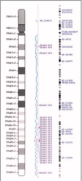
Els estudis sobre la presència de reordenaments cromosòmics a nivell del braç llarg del cromosoma 15 han identificat seqüències repetides de tamanys de fins a 60 kb anomenades LCR15. Han estat localitzades a 15q11-q13, 15q22, 15q24, 15q25 i 15q26, i presenten identitats significatives entre elles i amb seqüències presents a altres cromosomes (6q, 7p i 12p) (Gratacos et al., 2001; Pujana et al., 2001) (Figura 6). S’ha postulat que existeixen com a mínim 30 copies d’aquestes seqüències a 15q. En el cas concret d’aquestes seqüències al cromosoma 15 es parla de LCR15 (low copy repeats 15).
Els duplicons LCR15 presenten una mida variable entre 13 i 60 kb, amb unes identitats de seqüència superiors al 90% i contenen seqüències amb similaritat significativa a tres gens humans ja descrits: gens golgina-like (GLP), a un gen codificant per una proteïna amb dominis SH3 (SH3P18) i al gen de la dinamina 1 (DNM1) (Gratacos et al., 2001; Pujana et al., 2001). La presència de gens o pseudogens ha estat descrita en moltes de les duplicacions segmentàries estudiades fins al moment, són agents potenciadors de fenòmens de recombinació. L’eucromatina que constitueix el DNA que s’expressa habitualment (gens, pseudogens) es troba menys condensada, més oberta i per tant, presenta una major predisposició a patir reorganitzacions. Aquest fet afegit a una pressió selectiva determinada i a fenòmens de conversió gènica afavorint la conservació de seqüència dels gens que s’hi troben inclosos comporta que aquestes regions genòmiques siguin considerades punts calents
62
de recombinació genòmica (Chen et al., 1997; Lupski, 1998b). La localització d’aquestes seqüències a 15q suggereix que poden tenir un paper en la generació de reordenaments cromosòmics associats a anomalies genòmiques del cromosoma 15 humà com, per exemple, l’autisme entre d’altres (Han et al., 1999; Silva et al., 2002).
Figura 6. Distribució de duplicons identificats al braç llarg del cromosoma 15 humà. Adaptat de (Gratacos et al., 2001)
63
IV. Transtorn d’ansietat associat a 15q24-q26
L’ansietat en humans és una resposta de protecció enfront l’adversitat. Es caracteritza per una sèrie de respostes específiques del sistema nerviós autònom i per comportaments d’autodefensa. Una reacció ansiosa excessiva pot esdevenir un desavantatge i comprometre greument la qualitat de vida de l’individu. Es distingeixen sis formes de transtorn d’ansietat: el transtorn de pànic, l’ansietat generalitzada, la fòbia social, la fòbia específica, el transtorn per estrès post-traumàtic i els transtorns obsessivo-compulsius (American Psychiatric Association. & American Psychiatric Association. Task Force on DSM-IV., 2000).
Nombrosos estudis han demostrat l’existència de factors familiars en la manifestació dels transtorns d’ansietat (Fyer et al., 1995; Noyes et al., 1987). L’existència d’un cert grau d’heretabilitat s’ha evidenciat mitjançant
comparacions entre bessons. S’ha aconseguit detectar una major
concordància de fenotip en parelles de bessons monozigòtics en comparació amb els bessons dizigòtics (Kendler et al., 1995; Perna et al., 1997). En referència al risc de patir un transtorn d’ansietat, aquests estudis han establert que els factors genètics expliquen entre el 30% i 50% de la variabilitat que existeix entre individus (Hettema et al., 2001; Kendler, 2001). La variabilitat restant ha de ser explicada per factors ambientals específics de cada individu. A més a més, els estudis de risc han confirmat l’existència d’heretabilitat en aquest tipus de transtorn atribuïnt valors de risc a patir transtorns d’ansietat 4 a 10 vegades majors en individus amb familiars afectats respecte a individus sense antecedents familiars de la malaltia (Crowe et al., 1983; Hettema et al., 2001; Starcevic et al., 1993; Weissman et al., 1997).
Els estudis de lligament global del genoma per tal d’identificar gens o regions genòmiques implicades en l’evolució d’aquest tipus de transtorn no han obtingut resultats concrets o concloents (Crowe et al., 2001; Knowles et al., 1998; Weissman, 1993). Tot i això, alguns estudis han identificat regions de susceptibilitat al cromosoma 7p (Crowe et al., 2001; Logue et al., 2003) i al cromosoma 9q (Thorgeirsson et al., 2003). S’ha detectat cosegregació del transtorn de panic amb síndromes que inclouen cistitis, desordres tiroideus o
64
migranya crònica (Weissman et al., 2004). S’han obtingut dades d’associació significativa entre el transtorn de pànic i marcadors del cromosoma 13 (Weissman et al., 2004), així com amb el receptor d’adenosina 2A del cromosoma 22 (Hamilton et al., 2004).
Als anys 90, l’observació d’una major incidència dels transtorns d’ansietat en un estudi de famílies afectades per laxitud articular va suggerir que podia existir un mateix locus o regió cromosòmica responsable per a les dues afectacions (Bulbena et al., 1993; Martin-Santos et al., 1998). L’anàlisi de set genealogies en les quals es detectava cosegregació de transtorns d’ansietat i laxitud articular va donar lloc l’any 2001 a l’identificació d’associació entre els transtorns d’ansietat i la presència d’una duplicació de la regió q24-q26 del cromosoma 15 humà (Gratacos et al., 2001). Aquest polimorfisme genòmic va ser identificat mitjançant tècniques citogènetiques (FISH), i va permetre distingir diferents tipus de duplicacions (centromèriques, telomèriques, invertides, directes). L’ocurrència de mutacions de novo i la presència de mosaicisme en graus variables van fer proposar un model no-mendelià com a patró d’herència d’aquest factor de susceptibilitat. La duplicació inicialment reportada es va anomenar DUP25 i també es va detectar en el 7% de la població general (Gratacos et al., 2001). S’ha estimat que aquesta regió té una mida d’aproximadament 17 Mb, que conté de 50 a 60 gens i que es caracteritza per un elevat grau d’inestabilitat genòmica degut a la presència de duplicacions segmentàries flanquejants i al llarg de tot 15q, tal com s’ha mencionat anteriorment (Pujana et al., 2001).
L’observació d’associació entre 15q24-q26 i els transtorns d’ansietat fou de gran interès ja que es tractava de la primera associació identificada entre un polimorfisme genòmic d’aquest estil i una malaltia psiquiàtrica amb una incidència poblacional tan significativa. A més a més, en aquest cas s’aconseguia identificar un nou tipus de mutació genòmica associada a malaltia que no estaba lligada a cap dels loci contigus. L’importància i rellevància dels resultats obtinguts per Gratacòs i col.laboradors va impulsar diversos estudis d’associació entre 15q24-q26 i transtorns mentals en poblacions diferents. S’han aplicat tècniques de FISH, PCR quantitativa i hibridació (MAPH) per tal de dur a terme els mateixos estudis en altres poblacions i famílies afectades de
65
transtorn d’ansietat (Hollox & Armour, 2003; Schumacher et al., 2003; Tabiner et al., 2003; Weiland et al., 2003; Zhu et al., 2004). Fins aquest moment, cap dels estudis publicats ha estat capaç de replicar en altres poblacions els resultats obtinguts a la població catalana usada en el primer estudi per Gratacòs et al. El per què de la no replicació de resultats pot ser indicatiu de la dificultat en la detecció de la duplicació mitjançant FISH, de l’existència de diversos graus de mosaicisme entre individus i línies cel.lulars, dels patrons d’herència no mendelians, de les observacions d’inestabilitat genòmica en la regió i de la possibilitat de que no es tracti d’una duplicació per se (veure Discussió).
De tota manera, ja sigui per la potencial associació a transtorns d’ansietat, per la naturalesa inestable de la regió o com a contribució imprescindible a l’obtenció de la seqüència completa del genoma humà i els gens que hi són continguts, la regió q24-q26 ha esdevingut una zona d’alt interès per a ser caracteritzada a nivell transcripcional i de contingut gènic. És per això que, com a part del Consorci EuroImage i per la nostra implicació en l’identificació de gens nous humans (veure part I), part dels esforços del nostre grup es van adreçar cap a l’identificació de gens mapats prèviament a la regió 15q24-q26 segons les bases de dades públiques.
67
PART II: Objectius
• En el marc del consorci EuroImage, identificació de nous gens humans continguts en la regió q24-q26 del cromosoma 15 humà i conseqüent caracterització a nivell de seqüència nucleotídica, patró d’expressió i homologia en altres espècies
• Identificació i anàlisi de paralogia entre les regions q24-q26 del cromosoma 15 i p13.3-p12 del cromosoma 19 humans
• Identificació i caracterització de nous gens humans localitzats a la regió p13.3-p12 del cromosoma 19
69
PART II
Resultats
71
Resultats
Com a membres del consorci EuroImage i com a resultat de l’enfoc i concentració en una regió genòmica específica, la regió q24-q26 del cromosoma 15 humà, es va avançar significativament en l’identificació i caracterització de diversos gens, la majoria dels quals es presenten a les publicacions que constitueixen els apartats següents d’aquest treball. L’anàlisi transcripcional de la regió ha permès identificar l’existència de paralogia entre 15q24-q26 i la regió p13.3-p12 del cromosoma 19, així com aprofundir en la seva naturalesa i la seva significació a nivell evolutiu. Aquest últim punt queda reflectit en els articles sobre gens del cromosoma 15 amb paràlegs al 19 i els treballs identificant gens nous a la mencionada regió del cromosoma 19.
73
I. Identificació, expressió i mapatge del gen humà C15orf4
En aquest cas es presenta l’identificació i caracterització d’un nou gen humà amb similitud de seqüència a la proteïna YmL30 dels ribosomes mitocondrials de llevat. Les dades obtingudes de mapatge del gen C15orf4 confirmen la seva presència dins de la regió 15q24-q26. S’identifiquen els gens ortòlegs en altres espècies confirmant l’existència real d’aquest gen amb una funció biològica predita conservada al llarg de l’evolució. L’expressió de C15orf4 presenta uns nivells basals ubicus i un enriquiment a testicle, suggerint una possible funció específica a nivell de teixit.
Ref. DNA Seq. 2001;12(2):91-6 Cloning, mapping and expression analysis of C15orf4, a novel human gene homologous to the yeast mitochondrial ribosomal protein
YmL30 gene
Laura Carim, Lauro Sumoy, Nuria Andreu, Xavier Estivill and Mònica Escarceller
Medical and Molecular Genetics Center, Institut de Recerca Oncològica,
Hospital Duran i Reynals, Autovia de Castelldefels km 2,7 L'Hospitalet de Llobregat, 08907 Barcelona, Spain
Correspondence: Mònica Escarceller
Phone: 34-93-260-7775
Fax: 34-93-260-7776
e-mail: [email protected]
Abstract
We have identified C15orf4, a novel human gene showing homology to the yeast mitochondrial ribosomal protein YmL30. C15orf4 encodes a transcript of 1,006 nt with an ORF of 279 amino acids and a predicted protein size of 31.7 kDa. Expression of C15orf4 is enriched in testis. C15orf4 was positioned to chromosome 15q24 by radiation hybrid mapping. We have identified the C15orf4 mouse orthologue as well as homologues in other species.
Keywords: C15orf4, EUROIMAGE, chromosome 15q24, ribosomal YmL30.
Introduction
Within the EUROIMAGE full-length cDNA sequencing project underway in our laboratory (Adams et al 1991; Deloukas et al. 1998; Lennon et al. 1996; Schuler et al. 1998) we sequenced cDNA clones corresponding to EST clusters with the aim of identifying new genes. Clusters were selected on the basis of clone size, chromosomal localization and tissue distribution of transcripts. The EST contigs were built and analyzed in silico and representative clones were chosen for sequencing.
Using this approach, we have characterized a new gene, C15orf4, which shows a significant similarity to a putative ORF in Drosophila melanogaster, to the “decoy” gene in Arabidopsis thaliana and to the mitochondrial ribosomal protein (MRP) YNL252c gene in Saccharomyces cerevisiae.
The yeast ORF YNL252c gene is the synonym of YmL30 mitochondrial ribosomal protein of the large subunit (Graack and Wittmann-Liebold, 1998; Kitakawa et al, 1997; Sen-Gupta et al, 1997). Mitochondrial ribosomal proteins (MRPs) are the counterparts in that organelle of the cytoplasmic ribosomal proteins in the host. MRPs fulfill similar functions in protein biosynthesis but they are distinct in number, features and primary structures from the cytoplasmic riboproteins. To date, most of the information about MRPs has been obtained from the yeast S. cerevisiae and 50 different MRPs have been determined, although biochemical data and mutational analysis propose a total number substantially higher. Ribosomes of all species contain a core of structurally and functionally conserved riboproteins. Additional non-conserved proteins are considered to maintain functions specific for the respective species. The function of the MRPs may go beyond the mere biosynthesis of the small number of proteins encoded by the mitochondrial DNA. The
characterization of the complete set of human MRPs and the elucidation of their role is a process at its beginning (Graack and Wittmann-Liebold, 1998).
We present here the cloning, mapping and expression analysis of C15orf4, a novel human gene homologous to the yeast MRP YmL30 gene.
Material and Methods.
cDNA isolation and sequence analysis
ESTs from UniGene cluster Hs.14018 (http://www.NCBI.nlm.nih.gov/UniGene) were assembled using the EST
CAP assembly program (http://gcg.tigem.it/cgi-bin/uniestass.pl) and Sequencher (GeneCodes) sequence assembly software. Clones were obtained from the EUROIMAGE distribution centers. Sequence was determined by primer walking using the Perkin-Elmer BigDye reagents on an ABI PRISM-377 fluorescent automated sequencer and custom synthesized sequencing primers (LifeTech).
Full-length cDNA sequence was obtained using the rapid amplification of cDNA ends (RACE) method on SMART™ RACE cDNA from adult human placenta (Clontech), according to the manufacturer's kit instructions. The following primers were used: G1 (5’ TGTTCAGGGATGTTCGGTCA 3’), G2 (5’ CTTTCTTCTTTGCCAGTCGC 3’) and G3 (5’ GCTGTTAGGGGTGGCGG 3’) for 5’ C15orf4 extension. PCR extended products were subcloned into the pGEM-T easy vector (Promega) and sequenced as above. We generated nine independently generated extended clones to determine the cDNA ends.
Sequence comparisons were performed using ClustalW 1.7 (http://dot.imgen.bcm.tmc.edu:9331/multi-align/multi-align.html).
Boxed multiple sequence alignments were obtained with the BOXSHADE 3.21 program (http://www.ch.embnet.org/software/BOX_form.html). To search for known motifs or functional domains, protein pattern and domain databases consulted were Prosite, SMART and Pfam (http://www.hgmp.mrc.ac.uk/GenomeWeb/prot-domain.html).
The human C15orf4 nucleotide and protein sequences are available in GenBank under Acc. No. AF210056 and the mouse C15orf4 othologue ones under Acc. No. AF217090. The C15orf4 name has been approved by the
Human Gene Nomenclature Committee (http://www.gene.ucl.ac.uk/nomenclature/).
Northern blot analysis
A multiple-tissue northern blot (MTN II blot, Clontech) was hybridized to a 1-kb EcoRI-NotI restriction product corresponding to the cDNA insert from IMAGE clone 259218; and to a 2-kb ß-actin cDNA supplied commercially (Clontech) as control for quantification. Probes were labeled using a random primer DNA labeling kit (Amersham Pharmacia). Blots were hybridized overnight at 65ºC in ExpressHyb solution (Clontech) and washed at 68ºC in 0.2XSSC/0.5%SDS.
C15orf4 radiation hybrid mapping
To precisely localize the C15orf4 gene we used the Stanford TNG4 whole genome radiation hybrid panel (Stewart et al. 1997). Two point linkage analysis was performed using the RHMAP-2.0 on the RH Server at the Stanford Human Genome Center (http://www-shgc.stanford.edu/RH/index.html). We used primers corresponding to STS marker SHGC-15202 (D15S1261): F (5’
TCTAATCCCAGACTTGTCTGAGC 3’) and R (5’ TGTGGGTCACTAAGGATGAGC 3’). The PCR conditions were 1 cycle at 94ºC for 1 min 30 s; 30 cycles at 94ºC for 15 s, 62ºC for 23 s and 72ºC for 30 s; and 1 cycle at 72ºC for 5 min.
BAC assignment was obtained through BLAST searching against the
BAC ends database at TIGR (http://www.tigr.org/tdb/humgen/bac_end_search/bac_end_search.ht
ml) and contiguous BACs were determined from the ctc.ace clone tracking database (http://genome.wustl.edu/gsc/cgi-bin/ace/ctc_choices/ctc.ace).
Results and Discussion Cloning of C15orf4
In our effort to identify new genes, we constructed and analyzed in silico unique gene EST clusters on the basis of clone size, chromosomal localization and tissue expression. A partial human cDNA sequence with a single open reading frame (ORF) named C15orf4 was identified during the analysis of EST clusters within the physical region in 15q24.
The UniGene cluster representative of the clones was Hs.14018. Human IMAGE cDNA clones whose ESTs extended most 5’ and 3’ in the cDNA were chosen for sequencing: 1963245 (GenBank Acc.No. AI355098), 1723436 (GenBank Acc.No. AI188252) and 259218 (GenBank Acc.No. N29438).
Since the clones did not cover the entire transcript, the full-length cDNA sequence was obtained by 5' RACE extension (see Methods). The assembly of both IMAGE and RACE clones gave as a result a total transcript length of 1,006 bp (including the polyA tail), with an ORF (from
nt 12 to 849) encoding a 279 amino acid product with a calculated mass of 31.7 kDa and a pI=6.6. A polyadenylation signal (AATAAA) was observed at nt 967 and a polyA tail at the end (987 nt).
Analysis with protein domain identification software did not reveal the presence of any important feature with the exception of a coiled coil domain from amino acids 66th to 88th.
At the protein level, the most significative hit obtained after BLAST homology searching against “non redundant” databases (TBLASTN program at NCBI (http://www.ncbi.nlm.nih.gov/cgi-bin/BLAST/); Altschul et al. 1997) was a Drosophila translated genomic fragment in chromosome 3L, region 62A10-62B5 (GenBank Acc. No. AC005557). Further search in the Drosophila databases to find out if the genomic region could correspond to a characterized gene gave no positive result
(Berkeley Drosophila Genome Project, http://www.fruitfly.org/;http://www.sanger.ac.uk/Projects/D_melano
gaster/). We propose that the level of homology between both proteins (39% identity and 57% similarity within the 230 amino acid alignment) is suggestive of the existence of a distant C15orf4 gene homologue in the fruit fly.
The next significative BLAST homology hits were the A. thaliana “decoy” mRNA (GenBank Acc.No. U87586) and the S. cerevisiae ORF YNL252c gene (GenBank Acc.No. Z71528). Within the regions aligning with the human C15orf4 gene, the Arabidopsis and yeast ORFs showed 42% and 31% identity; 55% and 45% similarity, respectively.
The Arabidopsis “decoy” gene was described by Zhang and Somerville when analyzing the phenotypical defects in early embryogenesis caused by the twn2-1 mutation (Zhang and Somerville, 1997). This new gene encoded a protein of 19.1 kDa with low sequence homology to the yeast mitochondrial ribosomal protein, YmL30. The
authors pursued no further research on the gene and at present the function of the protein remains unknown.
The yeast ORF YNL252c gene is the synonym of YmL30 ribosomal protein of the large subunit (Munich Information Center for Protein Sequences; http://www.mips.biochem.mpg.de/proj/yeast/). Its complete ORF had an estimated molecular mass of 30 kDa and the mature form is calculated to be 16 kDa by electrophoresis. It localizes in the mitochondria and null mutants show slow growth and slightly increased thiabendazole sensitivity (Graack and Wittmann-Liebold, 1998; Kitakawa et al, 1997; Sen-Gupta et al, 1997). It has not been determined whether its function is essential or not for mitochondrial function (Graack and Wittmann-Liebold, 1998).
BLAST searches against “mouse” and “other” EST databases identified possible C15orf4 homologues in other species. We selected two murine IMAGE clones which once sequenced, completed the entire cDNA transcript in mouse: 1891319 (GenBank Acc.No. AI266903) and 2123770 (GenBank Acc.No. AI930359). The mouse protein showed a remarkably high level of homology with its human counterpart: 80% identity and 87% similarity (Fig. 1).
We were also able to identify the partial sequence of close C15orf4 homologues in other species: Rattus sp. (83% identity, 90% similarity in the aligned regions); the zebrafish Danio rerio (58% identity, 68% similarity); the trematode Schistosoma mansoni (33% identity, 52% similarity); and a single EST in Drosophila corresponding to the genomic clone above described (GenBank Acc.No. AI387313) (Fig.1).
The identification of the mouse homologue, as well as the possibility of the protein being represented in a broad range of species seems to be in agreement with it holding an essential role in eukaryotic cells. One
attractive possibility is that C15orf4 could be a novel MRP member of the mitochondrial ribosome.
Expression of C15orf4
Expression studies of C15orf4 with northern blots of human tissues (MTN II blot, Clontech), were carried out by hybridizing with a specific probe (see Methods). In adult tissues, basal expression was largely ubiquitous (Fig. 2), showing a 1.1-kb mRNA species. Remarkably, C15orf4 transcript signal was highly enriched in testis. The localized expression pattern suggests a tissue specific role in humans for C15orf4.
Mapping of C15orf4
Chromosomal localization of the human C15orf4 gene was determined by radiation hybrid mapping using the Stanford TNG4 panel. The gene was linked to STS SHGC-101328 with a lod score of 8.2 at an approximate distance of 120 kb. This STS is contained in RPCI-11 BAC clones 97O12 and 1127D9, in agreement with the fact that D15S1261, the marker contained in UniGene cluster Hs.14018, is contained in BAC 173D20 which overlaps with BACs 97O12 and 1127D9, in a physical contig mapped in 15q24 (D15S151-D15S202).
Acknowledgments
We are grateful to A. Puig and D. Otero for technical support with DNA sequencing. We wish to thank the HGMP Resource Center in Hinxton, UK, and the RZPD in Berlin, Germany, for supplying us with IMAGE cDNA clones. This work has been supported by EU Biomed Project No. BMH4-CT97-2284 to X. E and by CICYT-IN95-0347. M.E. is funded by the Spanish Ministry of Education (CIDYT contract FPI-070-97)
and L.S. by the Catalan autonomous government (CIRIT-RED contract 1998-64).
References
Adams MD, Kelley JM, Gocayne JD, Dubnick M, Polymeropoulos MH, Xiao H, Merril CR, Wu A., Olde B, Moreno RF, et al.: Complementary DNA sequencing: expressed sequence tags and human genome project. Science 252:1651-6 (1991).
Altschul SF, Maden TL, Schaffer AA, Zhang J, Zhang Z, Miller W, Lipman, DJ: Gapped BLAST and PSI-BLAST: a new generation of protein database search program. Nucleic Acids Res 25, 3389-402 (1997).
Deloukas P, Schuler GD, Gyapay G, Beasley EM, Soderlund C, Rodriguez-Tome P, Hui L, Matise TC, McKusick KB, Beckmann JS, Bentolila S, Bihoreau M, Birren BB, Browne J, Butler A, Castle AB, Chiannilkulchai N, Clee C, Day PJ, Dehejia A, Dibling T, Drouot N, Duprat S, Fizames C, Bentley DR, et al.: A physical map of 30,000 human genes. Science 282: 744-746 (1998).
Graack HR and Wittmann-Liebold B. Mitochondrial ribosomal proteins (MRPs) of yeast. Biochem J 329: 433-48 (1998). Rewiew.
Kitakawa M, Graack HR, Grohmann L, Goldschmidt-Reisin S, Herfurth E, Wittmann-Liebold B, Nishimura T, Isono KSen-Gupta. Identification and characterization of the genes for mitochondrial ribosomal proteins of Saccharomyces cerevisiae. Eur J Biochem 245:449-56 (1997 ).
Lennon G, Auffray C, Polymeropoulos M, Soares MB: The I.M.A.G.E. Consortium: an integrated molecular analysis of genomes and their expression. Genomics 33:151-2 (1996).
Schuler GD, Boguski MS, Stewart EA, Stein LD, Gyapay G, Rice K, White RE, Rodriguez-Tome P, Aggarwal A, Bajorek E, Bentolila S, Birre BB, Butler A, Castle AB, Chiannilkulchai N, Chu A, Clee C, Cowles S, Day PJ, Dibling T,
Drouot N, Dunham I, Duprat S, East C, Hudson TJ, et al.: A gene map of the human genome. Science 274: 540-6 (1996).
Sen-Gupta M, Guldener U, Beinhauer J, Fiedler T, Hegemann JH. Sequence analysis of the 33 kb long region between ORC5 and SUI1 from the left arm of chromosome XIV from Saccharomyces cerevisiae. Yeast 13(9):849-60 (1997 ).
Stewart, EA, McKusick, KB, Aggarwal, A, Bajorek, E, Brady, S, Chu, A, Fang, N, Hadley, D, Harris, M, Hussain, S, Lee, R, Maratukulam, A, O'Connor, K, Perkins, S, Piercy, M, Qin, F, Reif, T, Sanders, C, She, X, Sun, WL, Tabar, P, Voyticky, S, Cowles, S, Fan, JB, Cox, DR, and et al.: An STS-based radiation hybrid map of the human genome. Genome Res 7:422-33 (1997).
Zhang JM and Somerville CR. Suspensor-derived polyembryony caused by altered expression of valyl-tRNA synthetase in the twn2 mutant of Arabidopsis. Proc Natl Acad Sci U S A 94:7349-55 (1997).
Figure legends.
Figure 1. Multiple sequence alignments of human C15orf4 protein and its homologues in mouse, rat (partial ORF), zebrafish (partial), Drosophila, the trematode S. mansoni (partial), the A. thaliana “decoy” protein and the yeast YmL30. Partial sequences are indicated by arrows. Identical residues are printed in reverse type and similar residues are shaded. Consensus sequence is shown at the bottom, identical amino acids are marked with asterisks and similar amino acids, in at least three species, with dots.
Figure 2. Multiple-tissue northern blot analysis of C15orf4. The 1-kb EcoRI-NotI restriction product was used as a probe revealing a testis enriched 1.1-kb mRNA species. C15orf4 and β−actin control transcripts are labeled.
C 1 5 o r f 4 1 M A A P VRR T L L GVAGG W RR F ERLW AG SL S S RSLAL A A AP S S N GS P W R LL G AL C L Q R P P VVS KPLTP LQ E EM ASL L Q QIE- - - IE R S L m o u s e 1 M A A P VGR T L L GLARG W RQLDRFW AG - -S S RGL S LEA A SS SSRS P W R LS G AL C L Q R P PLITKALTP LQ E EM AGL L Q QIE- - - VE R S L r a t 1 z e b r a f i s h 1 D r o s o p h i l a 1 - - - MLRRI V Q V G - - AREL SRAQ SST ASA E KWDLY AGV L V ERLP VVS KSLNP L E KQ F QD L LW RV E- - - YENS L t r e m a t o d e 1 WN I F SG L CI RR PAV IA P ELKP L E KQ VA D L LG KV E- - - -F E R SH d e c o y 1 - - - M P R S SLRLL A KPL L E - S R R G F C T S S D K I V A SVL - - - -F E RL R Y m L 3 0 1 - - - M K V NLM L K R G L A TAT A T ASS APP K I K VGV LLSRIPIIK S ELN EL E KK Y Y E Y Q S E LEK R L M W T F P A Y F YFK K G T c o n s e n s u s 1 . . . . . . . . . . . . . . . . . . . . . . . . . . . . . . . . . . . . . . . . . . . . . . C 1 5 o r f 4 8 4 YS D H E L RAL D EN -Q R L A K K KAD- - - L HDEE D E QDI LLA Q D L E D M W E QK F LQ- - - -FK L G AR I T E A DE KN D RTS LNRKL D RNLVL m o u s e 8 2 YS D H E L RAL D E A-Q R L A K K KAD- - - L YDEEQE QGIT LA Q D L E D M W E QA F LQ- - - -FRPG ARET E A D KKN D RTS LHRKL D RNLVL r a t 1 z e b r a f i s h 1 IV TA Q D L E DVW E QN LK Q- - - -FQPA PRL Q G D G E - TDM SS L E RCLA D SLVL D r o s o p h i l a 6 5 KS D H E LK H E REI - V Q A E L IKQ G K I - - - QV DLE DA G S K Q TA Q D LKDA Y V E E LKK - - - -FQ L G SRTTP DDQ ANR T TST DRCL DD TL Y L t r e m a t o d e 4 0 LSAH E L RH E TET - KRIAS A L S K G - - - -VG K S AEE S L I TAR E AEIM W EL E A EQ- - - Y KPA ERLT END KS E N L KSA WRVL DK PL Y L d e c o y 3 8 V V I P K P D P A V YA- F Q E FKF N W Q Q - - - Q F R R R Y P D E FLD I A K N R A K GEY Q M D - - - Y VPA PR I T E A D KNN D RKS LYRAL DK KL Y L Y m L 3 0 7 4 V A EHK F L SLQ K G P I S K K N G I W F P R G I P D I K H G R E R S T KQE V K L S DDS T V A F S N N QKE Q S K D D V N R P V IPN DR I T E A DR SN DM KS L E RQLSRTL Y L c o n s e n s u s 9 6 . . . . . . . . . . . . . . . . . . . . . . . . . . . . . . . . . . . . . C 1 5 o r f 4 1 6 0 L VR -E KFG D Q D- - -V WIL P QAE W-Q P G E T L RGT A E RTL ATL SEN N M E A K F L G N A P C G H YTF K F PQAMR T E SN LG A K V F F F K A L L L T G D FSQAGNK m o u s e 1 5 8 L VR -E K L G D Q D- - -V WML P QVE W-Q P G E T L RGT A E R I L ATL SEN N M E A K F L G N A P C G H Y K F K F P K AI QT E S DLGVK V F F F K A L L L T G D F V QAG K K r a t 1 L VR -E K L G D Q D- - - LWML P QVE W-Q P G E T L RGT A E R I L ATL SEN N M E A K F L G N A P C G H Y K F K F P K AIR T E S DLGVK V F F F K A L L L T G D F V QTG K K z e b r a f i s h 4 6 L VQ - K D VGSQK - - - IWLL P QIE W-QTG E T L RQT A E RAL ASLPGA D L KATF L G N A P C GFY KYKYP KD V Q KEG L VG A K V F F F K AV M S S Q K H L P L EKN D r o s o p h i l a 1 4 3 L VQ - QK L GQQE - - - H L IL P QG K R - E EG ES MRQT A E RVLR E S CGQ E L Q V LFYG N A PVGF HKYKYPR N QR T ET - VG A K V F FY RASLR SGQ V P E N L TK t r e m a t o d e 1 1 6 L VQ - S P N - V S S - - - GWNL PI A P I - S DGK NL RQ VAD SIA T SLL P S R A K W C I FG NTP d e c o y 1 1 4 LI F G K P FGA T S D K PV WP FPE K V Y - D S E PT L RK CA ES ALK S V VGD L T H T YFVG N A PM AHM A I Q P T E E M P D L P - - S YKRF F F KC S V V A A S K Y D I S T A Y m L 3 0 1 6 9 L VK - DKS - - - G - - - TWK FPN F D L S D E S K PLH V HA EN ELK LL S GD Q I Y T W S V S A TPIGV L Q D E R - - N R T AEF I V K S H I L A GKF DLV A S - - - K N D A F c o n s e n s u s 1 9 1 * . . . . . . . . . . * . . . . . . . . * . . . * . . . . . . . C 1 5 o r f 4 2 5 0 GH HV WV TKDE L GDY LK -P K Y L A Q V R R FV SD L -m o u s e 2 4 8 SRHV WA SK E E L GDY LQ -P K Y L A Q V R R F L L DS D G L S C L r a t 9 1 G RHV WA SK E E L GDY LQ -P K Y L A Q V R R F L L D L -z e b r a f i s h 1 3 6 - T F AWV KKDE LQ E FLK -PEY LKQ V R R FI M TL -D r o s o p h i l a 2 3 2 - - F EWL PK EALN E KLT N T AYA Q SVK KF L -t r e m a -t o d e d e c o y 2 0 6 R I L C G -Y m L 3 0 2 5 2 E D F AWL TKGEI S EYV P - K DYF N K T E F LLADN -c o n s e n s u s 2 8 6 . . . . . . . . . .
Figura 1 (Carim-Todd et al., 2001)
75
II. Identificació i caracterització dels gens HMG20A i HMG20B
La publicació següent demostra l’existència d’una nova classe de proteïnes amb dominis HMG (high mobility group) amb una potencial funció en la regulació de la transcripció i conformació de la cromatina. L’aïllament del gen HMG20A mapat a la regió q24 del cromosoma 15 humà porta a l’identificació d’un gen paràleg, HMG20B, al cromosoma 19. Tots dos transcrits presenten una distribució generalitzada a nivell de teixit. La disponibilitat en aquell moment de seqüència genòmica provisional a les bases de dades públiques va permetre establir l’estructura exó-intró de tots dos gens i analitzar la seva conservació a aquest nivell.
Cytogenet Cell Genet 88:62–67 (2000)
HMG20A and HMG20B map to human
chromosomes 15q24 and 19p13.3 and
constitute a distinct class of HMG-box genes
with ubiquitous expression
L. Sumoy, L. Carim, M. Escarceller, M. Nadal, M. Gratacòs, M.A. Pujana, X. Estivill, and B. Peral
Medical and Molecular Genetics Center, Institut de Recerca Oncològica, Hospital Duran i Reynals, Barcelona (Spain)
Supported by EU Biomed Project BMH4-CT97-2284 to X.E. (which also funded L.C.) and by grants for research equipment and infrastructure (CICYT-IN95-0347 and CIRIT-SGR00444). M.E. and B.P. are funded by the Ministerio de Educacio´n, Spain (MEC-CIDYT CT97-2284 and CT96-1364, respectively). L.S. is supported by CIRIT, Generalitat de Catalunya (CIRIT-RED Contract CT97-2284). B.P.’s present address is Neurology Unit, Fundacio´n Jiménez Dı´az, Avda. Reyes Cato´licos, 2, 28040 Madrid (Spain).
Received 22 June 1999; revision accepted 20 October 1999.
Request reprints from Dr. Lauro Sumoy, Medical and Molecular Genetics Center, Institut de Recerca Oncològica, Hospital Duran i Reynals,
Autovia de Castelldefels Km 2,7, L’Hospitalet de Llobregat, 08907 Barcelona (Spain); telephone: 34-93-260-7775; fax: 34-93-260-7776; e-mail: [email protected].
ABC Fax + 41 61 306 12 34 E-mail [email protected] www.karger.com
© 2000 S. Karger AG, Basel 0301–0171/00/0882–0062$17.50/0
Accessible online at: www.karger.com/journals/ccg
Abstract. The HMG box encodes a conserved DNA binding domain found in many proteins and is involved in the regula-tion of transcripregula-tion and chromatin conformaregula-tion. We describe HMG20A and HMG20B, two novel human HMG box-con-taining genes, discovered within the EURO-IMAGE Consor-tium full-length cDNA sequencing initiative. The predicted proteins encoded by these two genes are 48.4 % identical (73.9 % within the HMG domain). The HMG domain of both HMG20 proteins is most similar to that of yeast NHP6A (38 %
to 42 %). Outside of this domain, HMG20 proteins lack any significant homology to other known proteins. We determined the genomic structure and expression pattern of HMG20A and HMG20B. Both genes have several alternative transcripts, expressed almost ubiquitously. HMG20A maps to chromo-some 15q24 (near D15S1227) and HMG20B to 19p13.3 (be-tween D19S209 and D19S216). The HMG20 genes define a distinct class of mammalian HMG box genes.
Copyright © 2000 S. Karger AG, Basel
Completion to full length of the sequences of unique cDNA clones represented in dbEST is a key step toward the character-ization of all human genes (Auffray et al., 1995; Lennon et al., 1996; Collins et al., 1998). With this aim and working within the Euro-IMAGE Consortium, we have discovered a novel family of genes that have an HMG domain as their most out-standing feature.
The basic signature of the HMG domain, originally defined in high-mobility-group protein 1, HMG-1 (Jantzen et al., 1990), comprises approximately 70 amino acid residues form-ing three ·-helices which mediate the interaction of HMG
pro-teins with the minor groove of DNA. HMG domains recognize irregular DNA structures, such as non-B DNA, cruciforms, and cisplatin adducts, and are capable of bending DNA (Landsman and Bustin, 1993; Laudet et al., 1993; Baxevanis and Lands-man, 1995).
There are two major types of HMG box genes based on ami-no acid sequence and DNA binding specificity. Members of the HMG1/2 class of proteins have low sequence target specificity, are expressed in many tissues, and usually have more than one HMG domain; they regulate nucleosome assembly. The TCF/ SOX class includes sequence-specific DNA-binding proteins, with a single HMG domain, which regulate tissue-specific tran-scription (Grosschedl et al., 1994; Soullier et al., 1999).
We describe here the sequence, genomic structure, expres-sion pattern, and chromosome location of two novel human HMG box genes, which we have named HMG20A and HMG20B. Based on sequence conservation criteria, HMG20 genes constitute a distinct class of mammalian HMG genes.
Materials and methods
cDNA isolation and sequencing
Expressed sequence tags (ESTs) from Unigene clusters Hs.69594 and Hs.32317 (http://www.NCBI.nlm.nih.gov/UniGene), corresponding to HMG20A and HMG20B respectively, were assembled using the EST CAP
Cytogenet Cell Genet 88:62–67 (2000) 63 assembly program (http://gcg.tigem.it/cgi-bin/uniestass.pl) and Sequencher
(GeneCodes) sequence assembly software. Additional ESTs were found by BLAST searching dbEST. IMAGE cDNA clones chosen for sequencing were: 548013, 270903, 140081, and 147037 for human HMG20A; 532078, 587808, 308007, 267627, 179729, and 1695848 for human HMG20B; and 522081 and 834855 for mouse Hmg20B. None of the IMAGE cDNA clones corresponding to mouse Hmg20A was available for sequencing. Sequence was determined by primer walking using the Perkin-Elmer BigDye reagents on an ABI PRISM-377 fluorescent automated sequencer and custom-synthe-sized oligonucleotides (LifeTech).
Full-length cDNA sequence was obtained using the rapid amplification of cDNA ends (RACE) method on Marathon-Ready cDNA from adult human heart (Clontech). Primers were: G4 (5)-CAG TAG TGG CGT GGA TTG TTG GT-3)) and G5 (5)-GCC TCT GTT CAT TGC CTT CTG CT-3)) for 5) HMG20A extension; and C2F (5)-ATG AAG TTA CAG GCT AGC AC-3)), G1 CAG TGA GGA GGC AGT AAA TGA AG-3)), and G2 (5)-AAG TTG TTG CCT ATT CAG TGT TAC-3)) for HMG20A 3) extension. PCR-extended products were subcloned into the pGEM-T-easy vector (Promega) and sequenced as above. Nucleotide sequences for the cDNAs of HMG20A, HMG20B, and Hmg20B are available from GenBank under Accession Nos. AF146222, AF146223, and AF146224, respectively. Official gene symbols are ISGN approved.
Protein sequence analysis
Protein sequences were aligned using the PILEUP and GAP programs (GCG). Boxed multiple sequence alignments shown in the figures were obtained with the BOXSHADE program (http://www.isrec.isb-sib.ch:8080/ software/BOX_form.html). To detect conserved protein domains, we used Pfam, PROSITESCAN, PRINTS, PRODOM, BLOCKS, PROFILESCAN, TOPITS, SMART, and PREDICTPROTEIN, available at http://www. hgmp.mrc.ac.uk/GenomeWeb/prot-domain.html, http://coot.embl-heidel-berg.de/SMART, and http://www.embl-heidelberg.de/predictprotein.
Genomic cloning of HMG20A
Genomic sequence of the HMG20A gene was obtained from clones gen-erated in the process of establishing a physical map of the 15q24 chromosom-al region. A high-density human bacterichromosom-al artificichromosom-al chromosome (BAC) libra-ry (CITB, Research Genetics) was screened by nucleic acid hybridization using STS WI-5695 (D15S739). Four positive BAC clones were obtained (80J7, 121A10, 194E21, and 204M2) that contained inserts of approximately 130 kb. After restriction analysis and Southern blotting, we confirmed that the four BAC clones were almost identical and contained the entire HMG20A gene. Exon/intron structure was determined by direct sequencing of BAC DNA or PCR products obtained using exonic primers (Big Dye, Per-kin-Elmer/Applied Biosystems). Thermocycling parameters for BAC se-quencing were 5 min at 95 ° C, 35 cycles of 30 s each at 95 ° C, and 4 min at 60 ° C.
We determined the exon/intron structure of the HMG20B gene by direct comparison between its cDNA and genomic sequences using the GAP pro-gram (GCG).
Northern blot hybridization
Commercial human Multiple Tissue Northern blots (Clontech) were hybridized to random primed cDNA probes. Probes for HMG20A were inserts from IMAGE clones 140081 (EcoRI-HindIII) and 548013
(BamHI-HindIII); for HMG20B the insert from clone 532078 (EcoRI-BamHI); and
for ß-actin a 2-kb cDNA supplied commercially (Clontech). Blots were hybridized overnight at 65 ° C in ExpressHyb hybridization solution (Clon-tech) and washed at 68 ° C in 0.2 × SSC, 0.5 % SDS.
HMG20A radiation hybrid mapping
To precisely localize the HMG20A gene, we used the Stanford G3 and TNG whole genome panels (Stewart et al., 1997). Two-point linkage analysis was performed using the RHMAP-2.0 on the RH Server at the Stanford Human Genome Center (http://www-shgc.stanford.edu/RH/index.html). We used primers C2F (used also for RACE) and C2R (5)-AGT TCC AAA CAC ATG TAC AC-3)). The PCR conditions were 1 cycle at 94 ° C for 2 min, 35 cycles of 30 s each at 94 ° C, 58 ° C for 30 s, and 74 ° C for 30 s, and 1 cycle at 74 ° C for 7 min.
Fluorescence in situ hybridization (FISH) analysis of HMG20A
BAC clone 80J7 containing the HMG20A gene was used as a probe for FISH on metaphase chromosome spreads as described (Nadal et al., 1997). The only modification was in the last three post-hybridization washes, which were in 2 × SSC at 42° C instead of 0.1 × SSC at 60 ° C.
Results
Isolation of the HMG20A gene
In the process of characterizing the q24 region in chromo-some 15, we screened a human genomic BAC library using STS WI-5695. The end sequence of a 12-kb BamHI single-copy sub-clone, contained in all four BACs obtained from the screen, was found to have sequence similarity to two dbEST entries (Acces-sion Nos. AA113373 and AA919704, from human and mouse origin, respectively). Human EST AA113373 was found to belong to a Unigene cluster; cDNA clones corresponding to ESTs from the same cluster were sequenced within the EURO-IMAGE full-length cDNA sequencing project underway in our laboratory. The longest cDNA, IMAGE clone 548013, con-tained a complete open reading frame. The full-length cDNA sequence was obtained by 5) and 3) RACE extension. Assembly of the different clones gave as a result a total transcript length of 3,773 bp.
Analysis of the protein sequence encoded by this cDNA showed the presence of an HMG box, which was the only ami-no acid region of significant conservation. Because of this, we named this gene HMG20A, conforming with international human gene nomenclature rules (Shows et al., 1979). The human HMG20A gene encodes a predicted 347 amino acid protein (Fig. 1) with an expected molecular weight of 40.1 kDa. The amino acid sequence of the mouse Hmg20A gene was par-tially deduced from EST sequences (Accession Nos. AI574467, AI574369, AA823250, AI119468, AI119138, and AA144479 for the N-terminal portion; AA919704 for the internal C-termi-nal portion; and AA249948 for the C-termiC-termi-nal portion; Fig. 1) and found to be very well conserved (100 % within the HMG domain).
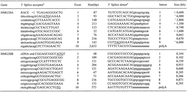
The entire transcript for HMG20A spans 10 exons (Fig. 2). All the junctions between exons and introns are in accordance with the rule that introns begin with a GT dinucleotide and end with AG (Table 1). The first exon encodes a 5) untranslated region (UTR), and the last one encodes a very long 3) UTR with five non-canonical polyadenylation signals within 200 nucleo-tides upstream of the polyA tail in the longest RACE-extended cDNA clone.
Finding of a second HMG20 gene
A second mammalian gene very closely resembling HMG20A was found by EST sequence database searching. Human ESTs similar, but nonidentical, to HMG20A were found, identical to a partial cDNA (Accession No. AF072836, unpublished data). The longest clone, IMAGE clone 532078, contained an open reading frame encoding a 317 amino acid protein (Fig. 1), 35.8 kDa in predicted size and smaller than HMG20A. The sequence of four other clones was also deter-mined and found to be mostly identical. Protein sequence com-parison, with a 70.7 % overall similarity and 48.4 % overall
64 Cytogenet Cell Genet 88:62–67 (2000)
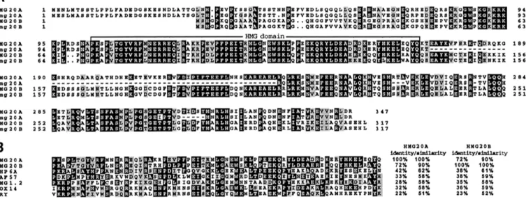
Fig. 1. (A) Amino acid sequence comparison between human HMG20A and HMG20B and murine Hmg20A and Hmg20B. Amino acids that are identical in all genes are boxed in black, while similar amino acids are boxed in gray. The HMG domain is bracketed. Dots indicate gaps introduced in the sequences for optimal alignment; dashes in the mouse Hmg20A putative sequence are unknown residues (due to lack of ESTs covering the region or to poor EST sequence). (B) Multiple sequence alignment of the HMG domains of HMG20 proteins compared to the prototype HMG classes SRY and HMG-1 and to the best matches obtained by BLAST comparison, yeast NHP6A, and mammalian BAF57 and SOX14. Black and gray boxes indicate identities and similari-ties, respectively, between at least three of the seven sequences. Numbers on the right indicate percent identity and similarity to HMG20A and HMG20B.
Fig. 2. Genomic structures of the HMG20A gene, based on the sequencing and Southern blot mapping data, and of HMG20B gene, based on cDNA-genomic sequence alignment comparison (see Table 1). The respective open reading frames are shown as boxes immediately beneath the cDNA sequences; the HMG domain is shaded. Exons are depicted as numbered boxes in the genomic DNA and cDNA. An alternative splicing isoform of HMG20B includes exon 3 (RACE38).
identity (with 91.3 % similarity and 73.9 % identity within the HMG domain) suggests that this second gene is a close homolog of HMG20A, and thus we have named it HMG20B (Fig. 1).
The 1,524-bp-long 532078 clone probably represents the predominant HMG20B gene product. A few rare EST se-quences and a single PCR product obtained in one of the RACE experiments indicated that the HMG20B gene may have alter-natively spliced transcripts expressed at very low levels or in very specific cell types (not shown).
Two partial mouse cDNA clones derived from ESTs match-ing human HMG20B were sequenced, which overlapped to construct a 1,632-bp transcript cDNA. The open reading frame is the same size and shares 93.7 % identity with the human HMG20B protein (94.2 % identity within the HMG domain).
The genomic sequences of the human HMG20B and mouse
Hmg20B genes have been determined by others (GenBank
Accession Nos. AC005786 and AF067430, respectively; un-published data). The human HMG20B genomic structure was obtained directly by comparison between the respective cDNA and genomic sequences (Fig. 2) and has at least 9 exons. The exact number of exons could not be determined, since there appear to be minor splice variants.
HMG20A gene expression analysis
HMG20A Northern blot hybridization showed two major transcripts of approximately 4 and 9 kb and a barely detectable 1.5-kb mRNA (Fig. 3). The 3.7-kb full-length cDNA sequence may appear as 4 kb on Northern blots due to extensive
polyade-Cytogenet Cell Genet 88:62–67 (2000) 65
Fig. 3. Northern blot hybridization analysis of HMG20A and HMG20B. cDNA probes corresponding to clone 548013 (for HMG20A), 532078 (for HMG20B), and a commercially supplied clone for ß-actin, used as a loading control, were hybridized to polyA RNA from multiple human tissues blotted onto a nylon membrane. Three size mRNA transcripts can be detected for HMG20A of approximately 9, 4, and 1.5 kb in size. Two bands are detected with the HMG20B probe at 2.6 and 1.5 kb. The positions of the detected bands are marked by the triangles on the right.
66 Cytogenet Cell Genet 88:62–67 (2000)
Fig. 4. Mapping of the human HMG20A gene by FISH. The 80J7 BAC clone containing the human HMG20A gene was labeled with biotin and hybridized to human metaphase chromosomes. Hybridization of 80J7 local-izing HMG20A to chromosome region 15q24 is indicated by arrows.
nylation or a sizing imprecision. The 4-kb form is the most abundant HMG20A mRNA and is expressed almost ubiqui-tously–spleen, testis, and heart having the highest levels and peripheral blood leukocytes lower levels. Brain expression is mostly uniform, with higher levels being found in the cerebel-lum and lower levels in the spinal cord and the subthalamic nucleus. The 9-kb HMG20A transcript is differentially ex-pressed in spleen, testis, leukocytes, and brain and is barely detectable in the remaining tissues.
Expression of HMG20B
HMG20B is also expressed among a wide variety of tissues, with Northern blot hybridization analysis showing two major mRNA forms of approximately 2.6 and 1.5 kb in size. The 2.6-kb form is expressed at lower levels and can be detected in thy-mus, prostate, placenta, liver, kidney, and pancreas. The 1.5-kb mRNA corresponds with the size of the full-length HMG20B cDNA clones we have sequenced. The highest expression levels of the 1.5-kb form are seen in the prostate, testis, heart, and kidney, while brain, spleen, lung, skeletal muscle, and leuko-cytes show lower levels. Within the brain, the 1.5-kb form of HMG20B is expressed almost uniformly, with increased levels in the corpus callosum and hippocampus.
Chromosome location of HMG20A and HMG20B
FISH analysis localized human HMG20A to 15q24 (Fig. 4). The Unigene EST cluster had been mapped between D15S114 and D15S989 (Deloukas et al., 1998). Our own radiation hybrid mapping, using the G3 panel, determined that HMG20A was linked to STS SHGC-15284 (close to D15S1227), with a lod score of 9.46. Using the TNG panel, HMG20A was found at an estimated distance of 180 kb from SHGC-20921 (D15S984) with a lod score of 5.79.
The chromosome map position of HMG20B was inferred from genomic clone sequence annotation to be in 19p13.3, between D19S209 and D19S216 (Deloukas et al., 1998).
Discussion
We have found two new genes, HMG20A and HMG20B, which add to a list of over 250 different genes coding for pro-teins with an HMG domain, a protein motif capable of binding to DNA (Baxevanis and Landsman, 1995; Soullier et al., 1999). Protein sequence comparison indicates that the HMG20A and HMG20B proteins have a much higher resemblance to each other than to any other HMG-box gene. The conservation in HMG20 gene structure in exons with the strongest amino acid similarity is consistent with the two genes deriving from a com-mon ancestor.
HMG20 protein sequence comparisons show conservation with other known proteins only within the HMG domain. The closest match to both HMG20 proteins is the yeast NHP6A nonhistone chromatin binding protein (Kolodrubetz and Bur-gum, 1990) (Fig. 1B). NHP6A has a single HMG domain with non–sequence-specific DNA binding properties and is in-volved in potentiating the transcriptional activation (Paull et al., 1993). Among mammalian HMG-box genes most similar to HMG20 (Fig. 1B) is BAF57, encoding a subunit of the SWI/ SNF complex (Wang et al., 1998). However, the level of simi-larity is not high enough to establish homology to HMG20A and HMG20B.
Parsimony, maximum likelihood, and protein distance phy-logenetic comparisons excluded the HMG20 genes from the TCF/SOX subfamily (Laudet et al., 1993) and placed them along with the remaining subgroups (including NHP6 and BAF57) (Baxevanis and Landsman, 1995). This information would suggest that HMG20 genes belong to the HMG1/2 type of non–sequence-specific HMG proteins. However, the lack of conservation outside of the HMG domain suggests that the HMG20 genes constitute a distinct class of HMG genes.
Both HMG20A and HMG20B are transcribed with a wide tissue distribution (Fig. 3). At first, the wide expression pattern of HMG20A and HMG20B suggests that they could be per-forming a housekeeping role as nonhistone components of chromatin, like HMG-1. The other possibility is that, although they have a generalized pattern of expression, they could act locally through interaction with tissue-specific transcription factors.
The HMG20A gene was confirmed to map to 15q24 by FISH and radiation hybrid mapping techniques. Hereditary conditions known to map in the 15q24 region include noctur-nal frontal lobe epilepsy (Phillips et al., 1998) and severe men-tal retardation with spasticity and pigmentary tapetoretinal degeneration (Mitchell et al., 1998). The corresponding synten-ic region in mouse chromosome 9 lacks mutations for whsynten-ich the gene remains unknown (Blake et al., 1999).
HMG20B is located in chromosome 19p13.3, between the markers D19S209 and D19S216. No known human diseases have been mapped to this region (McKusick, 1998). In mice,
chro-Cytogenet Cell Genet 88:62–67 (2000) 67 mosome 10, which is syntenic to human 19p13.3 (by neighbor
gene reference). Mutations in this region include jittery (jt), a recessive sublethal mutation affecting neuromotor coordina-tion and male fertility (Kapfhamer et al., 1996), and grizzled
(gr), a recessive mutation causing hair pigmentation and tail
defects with reduced viability (Bloom and Falconer, 1966). It will be very interesting to determine whether Hmg20B is asso-ciated with either of these two mutations.
Acknowledgements
We wish to thank A. Puig and D. Otero for technical support, M. Riutort for advice on phylogenetic analysis, and S. de la Luna for helpful criticisms to this manuscript. The HGMP Resource Center in Hinxton (UK) and the RZPD in Berlin (Germany) supplied us with IMAGE cDNA clones.
References
Auffray C, Behar G, Bois F, Bouchier C, Da Silva C, Devignes MD, Duprat S, Houlgatte R, Jumeau MN, Lamy B, et al.: IMAGE: molecular integration of the analysis of the human genome and its expres-sion. CR Acad Sci III 318:263–272 (1995). Baxevanis AD, Landsman D: The HMG-1 box protein
family: classification and functional relationships. Nucl Acids Res 23:1604–1613 (1995).
Blake JA, Richardson JE, Davisson MT, Eppig JT: The Mouse Genome Database (MGD): genetic and ge-nomic information about the laboratory mouse. The Mouse Genome Database Group. Nucl Acids Res 27:95–98 (1999).
Bloom JL, Falconer DS: “Grizzled”, a mutant in linkage group X of the mouse. Genet Res 7:159–167 (1966).
Collins FS, Patrinos A, Jordan E, Chakravarti A, Geste-land R, Walters L: New goals for the U.S. Human Genome Project: 1998–2003. Science 282:682– 689 (1998).
Deloukas P, Schuler GD, Gyapay G, Beasley EM, Sod-erlund C, Rodriguez-Tome P, Hui L, Matise TC, McKusick KB, Beckmann JS, Bentolila S, Biho-reau M, Birren BB, Browne J, Butler A, Castle AB, Chiannilkulchai N, Clee C, Day PJ, Dehejia A, Dibling T, Drouot N, Duprat S, Fizames C, Bent-ley DR, et al: A physical map of 30,000 human genes. Science 282:744–746 (1998).
Grosschedl R, Giese K, Pagel J: HMG domain pro-teins: architectural elements in the assembly of nucleoprotein structures. Trends Genet 10:94–100 (1994).
Jantzen HM Admon A, Bell SP, Tjian R: Nucleolar transcription factor hUBF contains a DNA-bind-ing motif with homology to HMG proteins. Nature 344:830–836 (1990).
Kapfhamer D, Sweet HO, Sufalko D, Warren S, John-son KR, Burmeister M: The neurological mouse mutations jittery and hesitant are allelic and map to the region of mouse chromosome 10 homolo-gous to 19p13.3. Genomics 35:533–538 (1996). Kolodrubetz D, Burgum A: Duplicated NHP6 genes of
Saccharomyces cerevisiae encode proteins
homolo-gous to bovine high mobility group protein 1. J Biol Chem 265:3234–3239 (1990).
Landsman D, Bustin M: A signature for the HMG-1 box DNA-binding proteins. Bioessays 15:539–546 (1993).
Laudet V, Stehelin D, Clevers H: Ancestry and diversi-ty of the HMG box superfamily. Nucl Acids Res 21:2493–2501 (1993).
Lennon G, Auffray C, Polymeropoulos M, Soares MB: The I.M.A.G.E. Consortium: an integrated molec-ular analysis of genomes and their expression. Ge-nomics 33:151–152 (1996).
McKusick VA: Mendelian Inheritance in Man: Cata-logs of Human Genes and Genetic Disorders. (Johns Hopkins University Press, Baltimore 1998).
Mitchell SJ, McHale DP, Campbell DA, Lench NJ, Mueller RF, Bundey SE, Markham AF: A syn-drome of severe mental retardation, spasticity, and tapetoretinal degeneration linked to chromosome 15q24. Am J hum Genet 62:1070–1076 (1998). Nadal M, Moreno S, Pritchard M, Preciado MA,
Esti-vill X, Ramos-Arroyo MA: Down syndrome: char-acterisation of a case with partial trisomy of chro-mosome 21 owing to a paternal balanced transloca-tion (15;21)(q26;q22.1) by FISH. J med Genet 34:50–54 (1997).
Paull TT, Haykinson MJ, Johnson RC: Yeast HMG proteins NHP6A/B potentiate promoter-specific transcriptional activation in vivo and assembly of preinitiation complexes in vitro. Genes Dev 7:1521–1534 (1993).
Phillips HA, Scheffer IE, Crossland KM, Bhatia KP, Fish DR, Marsden CD, Howell SJ, Stephenson JB, Tolmie J, Plazzi G, Eeg-Olofsson O, Singh R, Lopes-Cendes I, Andermann E, Andermann F, Berkovic SF, Mulley JC: Autosomal dominant noc-turnal frontal-lobe epilepsy: genetic heterogeneity and evidence for a second locus at 15q24. Am J hum Genet 63:1108–1116 (1998).
Shows TB, Alper CA, Bootsma D, Dorf M, Douglas T, Huisman T, Kit S, Klinger HP, Kozak C, Lalley PA, Lindsley D, McAlpine PJ, McDougall JK, Meera Khan P, Meisler M, Morton NE, Opitz JM, Partridge CW, Payne R, Roderick TH, Rubinstein P, Ruddle FH, Shaw M, Spranger JW, Weiss K: International system for human gene nomencla-ture (1979): ISGN (1979). Cytogenet Cell Genet 25:96–116 (1979).
Soullier S, Jay P, Poulat F, Vanacker JM, Berta P, Lau-det V: Diversification pattern of the HMG and SOX family members during evolution. J molec Evol 48:517–527 (1999).
Stewart EA, McKusick KB, Aggarwal A, Bajorek E, Brady S, Chu A, Fang N, Hadley D, Harris M, Hus-sain S, Lee R, Maratukulam A, O’Connor K, Per-kins S, Piercy M, Qin F, Reif T, Sanders C, She X, Sun WL, Tabar P, Voyticky S, Cowles S, Fan JB, Cox DR, et al: An STS-based radiation hybrid map of the human genome. Genome Res 7:422–33 (1997).
Wang W, Chi T, Xue Y, Zhou S, Kuo A, Crabtree GR: Architectural DNA binding by a high-mobility-group/kinesin-like subunit in mammalian SWI/ SNF-related complexes. Proc natl Acad Sci, USA 95:492–498 (1998).