DOI 10.1212/WNL.0000000000001030
published online October 29, 2014
Neurology
Alessia Giossi, Marco Ritelli, Paolo Costa, et al.
artery dissection
Connective tissue anomalies in patients with spontaneous cervical
This information is current as of October 29, 2014
http://www.neurology.org/content/early/2014/10/29/WNL.0000000000001030.full.html
located on the World Wide Web at:
The online version of this article, along with updated information and services, is
Neurology. All rights reserved. Print ISSN: 0028-3878. Online ISSN: 1526-632X.
since 1951, it is now a weekly with 48 issues per year. Copyright © 2014 American Academy of
® is the official journal of the American Academy of Neurology. Published continuously
Neurology
Alessia Giossi, MD
Marco Ritelli, PhD
Paolo Costa, MD
Andrea Morotti, MD
Loris Poli, MD
Elisabetta Del Zotto,
MD, PhD
Irene Volonghi, MD
Nicola Chiarelli, PhD
Massimo Gamba, MD
Paolo Bovi, MD
Giampaolo Tomelleri,
MD
Monica Carletti, MD
Nicoletta Checcarelli,
MD
Giorgio Meneghetti, MD
Michele Morra, MD
Mauro Chinaglia, MD
Valeria De Giuli, MD
Marina Colombi, PhD
Alessandro Padovani,
MD, PhD
Alessandro Pezzini, MD
Correspondence to Dr. Pezzini: [email protected]Supplemental data
at Neurology.org
Connective tissue anomalies in patients
with spontaneous cervical artery dissection
ABSTRACT
Objective:
To investigate the prevalence of connective tissue abnormalities in patients with
spon-taneous cervical artery dissections (sCeAD).
Methods:
We systematically assessed clinically detectable signs of connective tissue aberration
in a series of consecutive patients with sCeAD and of age- and sex-matched patients with
ische-mic stroke unrelated to CeAD (non-CeAD IS) by a standard examination protocol including 68
items, and performed extensive molecular investigation for hereditary connective tissue
disor-ders in all patients with sCeAD.
Results:
The study group included 84 patients with sCeAD (mean age, 44.5
6 7.8 years; 66.7%
men) and 84 patients with non-CeAD IS. None of the patients with sCeAD met clinical or
molec-ular diagnostic criteria for established hereditary connective tissue disorder. Connective tissue
abnormalities were detected more frequently in the group of patients with sCeAD than in the
group of those with non-CeAD IS (mean number of pathologic findings, 4.5
6 3.5 vs 1.9 6 2.3;
p , 0.001). Eighty-one patients (96.4%) in the sCeAD group had at least one detectable sign
compared with 55 patients (66.7%) in the group with non-CeAD IS (p , 0.001). Skeletal, ocular,
and skin abnormalities, as well as craniofacial dysmorphisms, were the clinical signs more
strongly associated with sCeAD. Signs suggesting connective tissue abnormality were also more
frequently represented in patients with sCeAD than in patients with traumatic CeAD (28.6%,
p ,
0.001; mean number of pathologic findings, 1.7
6 3.7, p 5 0.045).
Conclusions:
Connective tissue abnormalities are frequent in patients with sCeAD. This
reinfor-ces the hypothesis that systemic aberrations of the connective tissue might be implicated in
the pathogenesis of the disease.
Neurology
®2014;83:1
–6
GLOSSARY
HCTD5 hereditary connective tissue disorder; IS 5 ischemic stroke; sCeAD 5 spontaneous cervical artery dissection.
The pathogenesis of spontaneous cervical artery dissection (sCeAD), the most frequent cause of
ischemic stroke in young and middle-aged adults, is still unclear. The finding of composite
col-lagen fibrils and fragmented elastic fibers on electron microscopic examination of skin biopsy
specimens in more than half of patients with sCeAD is an argument in favor of the hypothesis
that connective tissue aberrations might have a causative role.
1,2However, whether such
morphologic alterations represent subclinical phenotypes of a generalized disorder of the
con-nective tissue remains to be determined. Hereditary concon-nective tissue disorders (HCTDs) have
been rarely diagnosed among patients with CeAD and external stigmata of connective tissue
abnormalities inconsistently detected.
3,4These observations cast doubts on the assumption of
a primary disorder of the connective tissue. Therefore, although the hypothesis of an
under-lying arteriopathy leading to structural instability of the vessel wall is likely and generally
accepted, the exact nature of this disorder is still matter of debate.
1To further investigate this
From the U.O. Neurologia (A.G.), Istituto Clinico S. Anna, Brescia; Dipartimento di Scienze Cliniche e Sperimentali (P.C., A.M., L.P., I.V., V.D.G., A. Padovani, A. Pezzini), Clinica Neurologica, Università degli Studi di Brescia; U.O. di Recupero e Rieducazione Funzionale (E.D.Z.), IRCCS Fondazione Don Gnocchi, Milano; Stroke Unit (M.G.), Neurologia Vascolare, Spedali Civili di Brescia; U.O. Neurologia (P.B., G.T., M. Carletti), Azienda Ospedaliera–Universitaria Borgo Trento, Verona; U.O.C. Neurologia (N. Checcarelli), Ospedale Valduce, Como; Diparti-mento di Neuroscienze (G.M.), Università di Padova; U.O.C. Neurologia (M.M.), Ospedale di Arzignano; S.O.C. Neurologia (M. Chinaglia), Ospedale di Rovigo; and Sezione di Biologia e Genetica (M.R., N. Chiarelli, M. Colombi), Dipartimento di Medicina Molecolare e Traslazionale, Università degli Studi di Brescia, Italia.
Go to Neurology.org for full disclosures. Funding information and disclosures deemed relevant by the authors, if any, are provided at the end of the article.
Table 1 Items included in the standard examination protocol
Skeletal features
1. Pectus carinatum: protrusion of the sternum/adjacent ribs
2. Reduced upper to lower segment ratio or arm span to height ratio.1.05
3. Walker-Murdoch sign (wrist sign): instruct the patient to grip his wrist with his opposite hand. If thumb and fifth finger of the hand overlap with each other, this represents a positive sign.
4. Steinberg sign (thumb sign): instruct the patient to hold the thumb across the palm of the same hand. If the entire thumb nail protrudes beyond the ulnar border of hand, this represents a positive sign
5. Scoliosis: radiographically defined as a lateral curvature of the spine.20° in the coronal plane 6. Spondylolysis/spondylolisthesis: radiographically defined
7. Reduced extension of the elbow (,170°)
8. Pes planus (flat foot): medial displacement of the medial malleolus so that the instep of the foot comes in contact with the ground when standing
9. Talipes equinovarus (clubfoot): the foot is rotated internally at the ankle 10. Pectus excavatum: depression of the sternum/adjacent ribs
11. Joint hypermobility: Beighton score$5
12. Complications of joint hypermobility (sprains, dislocations/subluxations)
13. Small joints hypermobility: hyperextensibility of the joints of the forefinger and middle finger (so-called“telescoping”) 14. Tendon/muscle ruptures
15. Arachnodactyly: long slender fingers and toes. It is scored when the middle finger length exceeds the palm length 16. Camptodactyly: fixed flexion deformity of the proximal interphalangeal joints
17. Polydactyly (hyperdactyly)
18. Increased bone fragility (multiple bone fractures) 19. Chronic joint/limb pain
20. Joint contractures: stiffness of the joint preventing full extension 21. Congenital dislocation of the hips
22. Beighton score
Thumb: ability to passively touch the forearm with the thumb, while flexing the wrist Fifth finger (Gorling sign): ability to passively extend the fifth finger to 90° or more Elbow recurvatum: hyperextension of the elbow beyond 10°
Genu recurvatum: hyperextension of the knee beyond 10°
Forward flexion of the trunk with knees fully extended so that the palms of the hands rest flat on the floor 23. Marfan-like habitus: dolichostenomelia, long slender fingers, pectus deformity
Ocular features
24. Enophthalmos: the posterior displacement of the eye within the orbit
25. Down-slanting palpebral fissures: when the outer canthus is positioned lower than usual 26. Hypoplastic iris or ciliary muscle causing decreased miosis
27. Exotropia
28. Proptosis: the forward displacement of the eye 29. Blue sclera
30. Myopia
31. Epicanthus: a fold of skin extending from the upper eyelid to or over the inner canthus of the eye 32. Deep-set eyes
Cutaneous features
33. Striae atrophicae (not associated with marked weight loss or pregnancy or repetitive stress) 34. Thin, translucent skin (especially noticeable on the chest/abdomen)
35. Easy bruising: (1) reported by patient, but no visible hematoma, (2) fewer than 5 visible skin hematomas, and (3) more than 5 visible skin hematomas. Spontaneous ecchymoses, frequently recurring in the same areas and causing characteristic brownish discoloration
Continued
issue, we systematically searched for clinical
signs of connective tissue anomalies in a
cohort of patients with CeAD and control
subjects.
METHODS
We undertook a hospital-based case-control study
of consecutive patients with CeAD and patients with non-CeAD
ischemic stroke (non-CeAD IS) prospectively recruited during a
44-month period (between March 2010 and November 2013).
Patients in the CeAD group were those whose diagnosis was
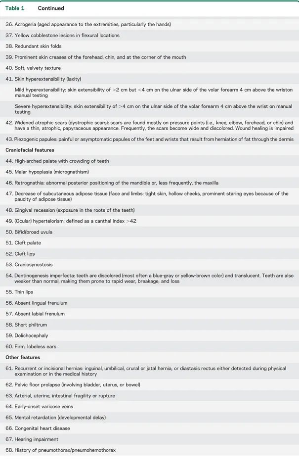
Table 1 Continued
36. Acrogeria (aged appearance to the extremities, particularly the hands) 37. Yellow cobblestone lesions in flexural locations
38. Redundant skin folds
39. Prominent skin creases of the forehead, chin, and at the corner of the mouth 40. Soft, velvety texture
41. Skin hyperextensibility (laxity)
Mild hyperextensibility: skin extensibility of.2 cm but ,4 cm on the ulnar side of the volar forearm 4 cm above the wriston manual testing
Severe hyperextensibility: skin extensibility of.4 cm on the ulnar side of the volar forearm 4 cm above the wrist on manual testing
42. Widened atrophic scars (dystrophic scars): scars are found mostly on pressure points (i.e., knee, elbow, forehead, or chin) and have a thin, atrophic, papyraceous appearance. Frequently, the scars become wide and discolored. Wound healing is impaired 43. Piezogenic papules: painful or asymptomatic papules of the feet and wrists that result from herniation of fat through the dermis Craniofacial features
44. High-arched palate with crowding of teeth 45. Malar hypoplasia (micrognathism)
46. Retrognathia: abnormal posterior positioning of the mandible or, less frequently, the maxilla
47. Decrease of subcutaneous adipose tissue (face and limbs: tight skin, hollow cheeks, prominent staring eyes because of the paucity of adipose tissue)
48. Gingival recession (exposure in the roots of the teeth) 49. (Ocular) hypertelorism: defined as a canthal index.42 50. Bifid/broad uvula
51. Cleft palate 52. Cleft lips 53. Craniosynostosis
54. Dentinogenesis imperfecta: teeth are discolored (most often a blue-gray or yellow-brown color) and translucent. Teeth are also weaker than normal, making them prone to rapid wear, breakage, and loss
55. Thin lips
56. Absent lingual frenulum 57. Absent labial frenulum 58. Short philtrum 59. Dolichocephaly 60. Firm, lobeless ears Other features
61. Recurrent or incisional hernias: inguinal, umbilical, crural or jatal hernia, or diastasis rectus either detected during physical examination or in the medical history
62. Pelvic floor prolapse (involving bladder, uterus, or bowel) 63. Arterial, uterine, intestinal fragility or rupture 64. Early-onset varicose veins
65. Mental retardation (developmental delay) 66. Congenital heart disease
67. Hearing impairment
confirmed
by
MRI/magnetic
resonance
angiography
or
conventional angiography,
5with or without stroke.
Dissections occurring as an immediate consequence of a
major trauma were labeled
“traumatic” (for definition, see
e-Methods on the Neurology
®Web site at Neurology.org). The
group of control subjects was selected from a cohort of patients
with first-ever cerebral ischemia, after exclusion of the subgroup
with CeAD-related infarction. Non-CeAD IS patients were
frequency-matched with CeAD patients on sex and age (in 3-year
intervals). Clinically detectable signs of connective tissue
abnor-malities were systematically investigated in each subject by 2
raters, a neurologist (A.G.) and a geneticist (M. Colombi), expert
in the assessment of connective tissue signs, to ensure a
homoge-neous evaluation. This standardized examination included the
assessment of 68 signs reflecting the spectrum of the clinical
features observed in the Ehlers-Danlos syndrome vascular type,
Marfan syndrome, pseudoxanthoma elasticum, osteogenesis
im-perfecta, arterial tortuosity syndrome, and Loeys-Dietz syndrome
(table 1).
e3–e13Each sign was counted as an all-or-none variable
(present vs absent), resulting in an individual connective score
and a mean number of pathologic findings for each group (mean
score). The 2 raters were blinded to the status of the patients.
Interrater reliability was assessed by having the 2 examiners
cat-egorize the same set of 68 signs in all patients and was evaluated
using the
k statistic, according to the method described by
Cohen.
6We categorized these signs according to the specific
organ or system involved into (1) skin abnormalities, (2) ocular
abnormalities, (3) skeletal abnormalities, (4) craniofacial
dysmor-phisms, and (5) abnormalities involving other organs.
All patients with CeAD underwent direct sequencing of
TGFBR1 and TGFBR2 genes,
762 of SMAD3 and TGFB2 genes,
while only in selected cases, COL3A1 (n
5 10), SLC2A10 (n 5 3),
FBN1 (n
5 2), and ACTA2 (n 5 2) genes were analyzed, based on
individual characteristics. To test the hypothesis that connective
tissue disorders have a major role also in the pathogenesis of sporadic
CeAD cases, we planned to exclude from the present analysis those
patients whose clinical features had eventually fulfilled the diagnostic
criteria for one of the monogenic disorders reported above and/or in
whom molecular screening revealed causative mutations.
Standard protocol approvals, registrations, and patient
consents.
The study was approved by relevant local authorities.
Written informed consent was obtained from all patients (or next
of kin).
RESULTS
The study group was composed of 84
pa-tients with sCeAD (mean age, 44.5
6 7.8 years;
66.7% men) and 84 patients with non-CeAD IS
(mean age, 44.0
6 8.7 years; 66.7% men). Compared
to patients with non-CeAD IS, patients with sCeAD
had a lower prevalence of hypercholesterolemia, a
higher prevalence of migraine, particularly without
aura, tended to be taller and thinner, and less
frequently hypertensive, smokers, and diabetics (table 2).
Short-lasting triggering factors preceding the
occurrence of the vascular event were also detected
more often in the group of patients with sCeAD than
in the group of patients with non-CeAD IS (table
e-1). None of the patients with sCeAD met clinical
diagnostic criteria or carried causal mutations for
established HCTD. Interrater agreement for the
assessment of connective signs was high with 80 of
the 84 patients rated identically by the 2 raters
(
k 5 0.84). Signs suggesting connective tissue
abnor-malities were detected more frequently in the group
of patients with sCeAD than in the group of those
with non-CeAD IS (mean score, 4.5
6 3.5 vs 1.9 6
2.3; p
, 0.001; figure e-1). Eighty-one patients
(96.4%) in the sCeAD group had at least one
detect-able sign compared with 55 patients (66.7%) in the
group with non-CeAD IS (p
, 0.001). Overall,
skel-etal, ocular, and skin abnormalities, as well as
Table 2 Demographic and clinical characteristics of the study groupCeAD (n5 84) non-CeAD IS (n5 84) p Value Age, y 44.56 7.8 44.06 8.7 0.718 Sex, male 56 (66.7) 56 (66.7) 1.000 Height, cm 171.66 8.9 169.36 8.5 0.085 Weight, kg 70.36 13.3 74.66 14.7 0.052 Hypertension 19 (22.6) 28 (33.3) 0.122 Diabetes mellitus 1 (1.29) 5 (6.0) 0.210 Hypercholesterolemia 11 (13.1) 29 (34.5) 0.001 Smoking 22 (26.2) 30 (35.7) 0.182 Any migraine 36 (42.8) 13 (15.5) ,0.001 MO 31 (36.9) 8 (9.5) ,0.001 MA 5 (6.0) 5 (6.0) 1.000 Dissected vessel
Internal carotid artery 48 (57.1) Vertebral artery 18 (21.4) Multiple vesselsa 15 (17.8) Fibromuscular dysplasia 4 (4.8) 0 (0.0) 0.121 Presenting symptom Stroke 62 (73.8) TIA 6 (7.1) Local symptomsb 43 (51.1) Retinal ischemia 0 (0.0) SAH 0 (0.0) Cause of strokec
Large vessel disease 8 (9.5) Cardiac embolism 35 (41.7) Small vessel disease 4 (4.8) Other determined etiology 52 (83.9) 9 (10.7) Undetermined origin
Multiple possible etiologies 10 (16.1) 1 (1.1) Complete evaluation 27 (32.1) Incomplete evaluation 0 (0.0)
Abbreviations: CeAD5 cervical artery dissection; IS 5 ischemic stroke; MA 5 migraine with aura; MO5 migraine without aura; SAH 5 subarachnoid hemorrhage.
Data are mean6 SD or n (%). See e-Methods for risk factor definition.
aMore than one vessel involved.
bIncluding Horner syndrome, cranial nerve palsies, head or neck pain, or pulsatile tinnitus. cAccording to TOAST (Trial of Org 10172 in Acute Stroke Treatment) criteria.
craniofacial dysmorphisms, were more frequently
observed in the group of patients with sCeAD, as
opposed to the abnormalities involving other organs,
which were equally distributed in the 2 groups. Facial
appearance of patients with sCeAD was more often
characterized by tight skin, hollow cheeks, and
prom-inent staring eyes as a consequence of paucity of
adi-pose tissue. The most prominent skeletal features
were scoliosis and mild pectus excavatum, while the
most prominent skin feature was mild
hyperexten-sion. Hypermobility/laxity of some joints was also
more frequently observed in the group of patients
with sCeAD (Beighton score
8$2 in 16.7% vs
3.6% [p
5 0.009]; mean score, 0.6 6 1.1 vs 0.1 6
0.5 [p
5 0.001]; table 3).
We also studied 7 patients whose CeAD was
obvi-ously due to a major trauma, during the same study
period. Signs suggesting connective tissue
abnormal-ity were less frequently represented in this group
com-pared to the group of patients with sCeAD (traumatic
CeADs with at least one detectable sign, 28.6%, p
,
0.001; mean score, 1.7
6 3.7, p 5 0.045).
DISCUSSION
Our findings indicate that clinically
detectable connective tissue abnormalities, frequently
observed in patients with HCTD, are also highly
prevalent in patients with sCeAD. This provides
indi-rect support to the
“connective hypothesis” of the
disease. Structural deviations in the main components
of connective tissue, collagen and elastic fibers, may
lead to functional impairment of the mechanical
sta-bility and elasticity of the arterial wall, predisposing to
dissection at given points of minor resistance. Similar
to other previous studies, we did not diagnose a
def-inite HCTD in any of the patients included in our
series, despite the extensive clinical and molecular
investigation.
9Whether this implicates the existence
of a milder form of connective tissue disorder
predis-posing to CeAD remains unknown, even though it
seems plausible. The low prevalence of clinically
rec-ognizable connective signs in patients with traumatic
CeAD, as opposed to spontaneous cases, is a further
argument in favor of this view. Overall, our findings
are in line with the prevailing theory that sCeAD
represents a multifactorial disease and the end result
of a synergistic interplay between an underlying
consti-tutional arteriopathy and short-lasting environmental
factors. These triggering factors, normally insufficient
to induce arterial wall rupture alone, could transiently
facilitate dissection in a fragile, previously asymptomatic,
vessel wall. Obviously, we cannot but speculate on the
relation between the clinical signs of connective tissue
abnormality we detected and a generalized disorder of
the connective tissue. In this regard, our findings need to
be validated by a study that correlates patient phenotype
with pathologic data. Notably, the only study in which
clinical signs of connective tissue abnormalities were
systematically assessed by standard examination found
no preponderance of these stigmata in patients with
CeAD and argued against a connective tissue disorder
underlying the disease. However, the 25 clinical items
used in that analysis reflect the spectrum of signs mainly
found in Marfan and Ehlers-Danlos syndromes, and it is
unclear why authors did not include specific features of
other HCTDs in their standardized examination
protocol.
4Difference in sample size and the lack of
patients’ molecular characterization should also be
considered when comparing the results of that study
with ours, because they might contribute to explain
some discrepancies.
The most relevant shortcoming of both analyses, as
well as of others previously reported,
2,3is that they rely
heavily on the identification and subjective
interpreta-tion of signs that are qualitative or semiquantitative
(i.e., skin extensibility, joint hypermobility, tissue
fra-gility, and bruising), and for which, therefore, normal
standards are unavailable. However, the excellent
in-terrater agreement in the assessment of these items
makes this drawback unlikely in our analysis. Clinically
detectable signs of connective tissue abnormalities are
subtle in most patients with sCeAD and, therefore,
Table 3 Distribution of connective tissue anomalies in patients with sCeAD andnon-CeAD IS Connective abnormalities sCeAD (n5 84) non-CeAD IS (n5 84) p Value At least 1 81 (96.4) 55 (66.7) ,0.001 Sum score 4.56 3.5 1.96 2.3 ,0.001 Organ involvement
Bone and jointsa 57 (67.9) 28 (33.3) ,0.001
Gorling sign 8 (9.5) 1 (1.2) 0.034 Elbow recurvatum 14 (16.7) 5 (6.0) 0.049 Scoliosis 19 (22.6) 4 (4.8) 0.001 Pectus excavatum 14 (16.7) 1 (1.2) ,0.001 Beighton scoreb Mean6 SD 0.66 1.1 0.16 0.5 0.001 ‡2 14 (16.7) 3 (3.6) 0.009 Marfan-like habitus 15 (17.9) 5 (6.0) 0.030 Skina 35 (41.7) 20 (23.8) 0.014
Mild skin hyperextension 16 (19.0) 6 (7.1) 0.038 Eyea 39 (46.4) 25 (29.8) 0.026
Craniofacial dysmorphismsa 41 (48.8) 20 (23.8) 0.001
Decrease of subcutaneous adipose tissue 16 (19.0) 4 (4.8) 0.07 Thin lips 9 (10.7) 2 (2.4) 0.05 Other organsa 14 (16.7) 7 (8.3) 0.160
Abbreviations: IS5 ischemic stroke; sCeAD 5 spontaneous cervical artery dissection. Data are mean6 SD or n (%).
aNo. of patients with at least one sign involving the specific organ or system. bIndividual score values range, 0–9 (0–1 point for each item).
they might remain undetected because they can only
be recognized by experienced examiners. This
repre-sents an obvious limitation to the possibility of
apply-ing such a standardized approach in everyday clinical
practice when trying to detect patients at higher risk of
dissection. Nevertheless, in the absence of rigorous
his-topathologic studies, the demonstration of a distinctive
connective tissue phenotype in patients with sCeAD
reinforces the hypothesis that the disorder implicated
in the pathogenesis of the disease is probably not
lim-ited to the vascular bed, and that the connective tissue
is the most likely target organ.
AUTHOR CONTRIBUTIONS
Dr. Giossi: manuscript drafting/revising, study design, data analysis and interpretation, data acquisition. Marco Ritelli, Paolo Costa, Andrea Morotti, Loris Poli, Elisabetta Del Zotto, Irene Volonghi, Nicola Chiarelli, Massimo Gamba, Paolo Bovi, Giampaolo Tomelleri, Monica Carletti, Nicoletta Checcarelli, Giorgio Meneghetti, Michele Morra, Mauro Chinaglia, Valeria De Giuli, Marina Colombi: manuscript drafting/revising, data acquisition. Alessandro Padovani: manuscript drafting/revising, study supervision. Dr. Pezzini: manuscript drafting/revising, study design, data anal-ysis and interpretation, data acquisition, statistical analanal-ysis, study supervision.
STUDY FUNDING
No targeted funding reported.
DISCLOSURE
The authors report no disclosures relevant to the manuscript. Go to Neurology.org for full disclosures.