Department of Life and Environmental Sciences
XV PhD cycle in Life and Environmental Sciences
Curriculum Biomolecular Sciences (SB)
α-Hydrazido acids for the synthesis
of bioactive amphiphilic compounds
PhD candidate: Supervisor:
Paolo Amabili
Samuele Rinaldi, PhD
with the aim to obtain new foldamers (oligomers with a predictable folding). Thus, we modified pirrolidin-2-one tethered -amino acids, previously used in our laboratory to prepare hexamers, devised a new imidazolidinone-tethered -hydrazido acid (AOPIC) suitable to give oligomers that were analysed using spectroscopic (NMR and CD) and computational (MD) techniques. The computational analysis, besides furnishing the theoretical proof of the 8-helix as the only stable structure, strongly evidenced a “hydrazido-turn” sequence, where additional 5-membered H-bonded cycles were enclosed within the 8-membered ones.
In the search for simpler and cheaper analogues, we directed our attention towards analogues of the previously employed oligomers, where constrictions and chirality were missing, with the aim to obtain a secondary structure with minor synthetic effort and increased versatility in side chains substitution. In fact, an appropriate disposition of side chains could be a good starting point for the synthesis of amphiphilic foldamers to be tested as antibacterial agents.
Since many difficulties are connected with the use of peptide drugs, synthetic mimics of AMPs (SMAMPs) are receiving ever more interest and importance as new drug candidates, owing to the rapid and widespread development of antibiotic resistances. Thus, within this topic, we carried out the synthesis of amphiphilic α-hydrazido acid derivatives, which could be novel lead compounds for developing a new class of SMMAMPs.
The Minimum Inhibitory Concentration (MIC) was evaluated for a few properly derivatized α-hydrazido acid monomers, and the preliminary results showed a promising antimicrobial activity suitable for biological applications, eventually leading to a structure optimization for improvement of the pharmacological properties.
I
1.1 Introduction ... 1
1.2 β-Peptide Foldamers ... 3
1.3 Foldamers of β-Peptide Mimetics ... 8
2. IMIDAZOLIDINONE-BASED α-HYDRAZIDOPEPTIDES ... 12
2.1 Introduction ... 12
2.2 Results and Discussion ... 13
2.2.1 Synthesis of New Imidazolidinone-based -Hydrazidopeptides ... 13
2.2.1 Investigation by NMR and CD Spectra ... 15
2.2.2 Computational Investigation ... 23
2.2.3 ROESY analysis ... 31
2.3 Conclusions ... 38
2.4 Experimental ... 40
2.4.1 Materials and methods ... 40
2.4.2 Synthetic procedures and characterizations ... 40
2.5 Supplementary session ... 47
2.5.1 Logical Analysis of Possible H-bonding Patterns for α-hydrazidopeptides ... 47
2.5.2 Determination of Intermolecular H-bonding from Dilution Experiments ... 51
2.5.3 Titrations with DMSO-d6. ... 60
2.5.4 Computational details ... 62
2.5.5 Backbone Resonances Assignment for Compounds 4 and 5 ... 63
3. ACHIRAL α-HYDRAZIDOPEPTIDES ... 67
3.1 Introduction ... 67
3.2 Results and Discussion ... 68
3.2.1 Synthesis of -hydrazido acid monomers and oligomers ... 68
3.2.2 Preliminary assignment of secondary structure of oligomers ... 76
3.2.3 Preliminary NMR and DFT analysis of Boc-MONOMER-NH2 conformers ... 86
3.3 Preliminary Conclusions ... 92
3.4 Experimental ... 92
II
4. THE NEED FOR NEW ANTIBACTERIAL AGENTS: AN OVERVIEW ... 105
4.1 Introduction: The “Post-Antibiotic Era” ... 105
4.2 Natural AntiMicrobial Peptides (AMPs): are they promising candidates? ... 106
4.2.1 Problems related to the use of natural antimicrobial peptides ... 110
4.3 Synthetic Mimics of AntiMicrobial Peptides (SMAMPs) ... 113
4.3.1 Antimicrobial foldamers ... 114
4.3.2 Antimicrobial polymers ... 117
4.3.3 Small Molecular Mimics of AMPs: SMMAMPs ... 121
5. α-HYDRAZIDO ACID-BASED SMMAMPS ... 130
5.1 Introduction ... 130
5.2. Results and discussion ... 131
5.2.1 Synthesis of compounds ... 131
5.2.2 Antimicrobial activity ... 135
5.3 Conclusions ... 143
5.4 Experimental ... 144
5.4.1 General methods and materials for the synthesis of products ... 144
5.4.2 Procedures for the evaluation of MICs ... 145
5.4.3 Synthetic procedures and characterizations ... 145
CHAPTER 1:
1. FOLDAMERS: AN OVERVIEW 1.1 Introduction
In all the proteins, as well as in every peptidic structure, a correct folding is essential for the formation of the “active sites” where the functional groups, often separated along the amino-acidic chain, are rearranged in a predetermined three-dimensional structure, necessary for their activity. For understanding the close relationship between structure and activity and also the principles that are at the basis of a correct folding in an amino-acidic backbone, we could perform a de novo design of biomimetic polymers with unusual properties never seen in nature. Those syntheses could have important practical applications in pharmaceutical and material sciences. This represent the “Foldamers” field of study.1
“A foldamer is any oligomer that folds into a conformationally ordered state in solution, the structures of which are stabilized by a collection of non-covalent interactions between nonadjacent monomer units”.1 This definition has important implications:
A foldamer is an oligomer, and this entail the presence of a regular repetitive structural motif (a monomer) in the backbone
Foldamers are oligomeric and not polymeric structures
The fact that the ordered conformation is “in solution”, imply that the solvent covers an important role in the structure of these molecules and in the fluctuations of atomic coordinates in the preferred conformation
The molecule has to organize with a dynamic process in (a) prevalent conformation(s): oligomers where there isn’t such a possibility are not considered foldamers
When the molecule is folded, it adopts a single set (or few of them) of superimposable conformations, that is to say a single secondary structure
The secondary structure is stabilized by non-covalent interaction between non-adjacent monomeric units: if the potential energy surface of intramonomer covalent bonds and the steric effects between adjacent residues are the only reasons for the adopted secondary structure, an oligomer cannot be considered as a foldamer (e.g. polyproline, polyaldehydes, polymethacrylate, etc.). Furthermore, we cannot consider a foldamer the structure that present a secondary structure stabilized by hydrogen bonds between adjacent residues.
This last point is relevant, because it makes the definition of foldamer consistent with the “synthetic analogue of a natural secondary structure”. In fact, every natural secondary structure
corresponds to this definition. Obviously, the torsional preferences for the dihedral angles and the steric effects between adjacent residues will contribute to determine the secondary structure, but they will not be sufficient in itself, exactly as in the natural α-peptides, where many other factors can influence the folding (e.g. H-bonding).
The pathway to create a new foldamer follows several steps:
i. Identification of new oligomeric backbones with a high tendency to fold into a well defined secondary structure. This imply the ability to predict the relationship between the conformational properties of the monomer and the corresponding oligomers. ii. It’s necessary to insert in the monomer suitable and well oriented chemical
functionalities, in order to improve the folding propensities of oligomers (preorganization).
iii. The foldamers have to be obtained in a synthetically efficient way, through both a practical and stereoselective synthesis of enantiomerically pure monomers and a high-yielding oligomerization process.
In the last decades, several research groups have devoted their studies towards the development of new monomers to build novel foldamers.1 According to the features of the
monomeric units, all these oligomers could be grouped into two main principal classes (Figure 1): “aliphatic” and “aromatic” foldamers.
Figure 1. Examples of foldameric backbones.
The huge number of building monomers brought to a wide number of secondary and higher-order structures, some of which are reported in Figure 2.
Figure 2. Some secondary and supersecondary structures adopted by foldamers.
1.2 β-Peptide Foldamers
Among aliphatic foldamers, the β-peptides family is the most interesting and the most studied for several reasons:
They are the littlest structural variation with respect to the natural α-peptides. Generally, they are very stable towards proteases.
Even though they have an extra C-C bond with free rotation, with the proper substitutions they can form secondary structures, especially helices, more stable than the ones of α-peptides.
For both acyclic and cyclic monomers, there are many different techniques that allow the easy synthesis in enantiomerically pure form and with all the desired side chains/functionalities.
With the β-amino acids it is possible to have much more substitution patterns in comparison to -amino acids (Figure 3), thus offering an enhanced opportunity to control the conformation of the corresponding oligomers.
Figure 3. Substitution and cyclization scheme of α- and β-amino acids.
The conformations of β-peptides can be analysed in term of backbone torsional angles ω, φ, θ e ψ, as.2 The effects of substituents on the conformation of -peptides were investigated in a
plethora of experimental studies,1 and a generic example of how different substitution patterns
Figure 4. Effect of the substituents on the torsional dihedral angle θ.
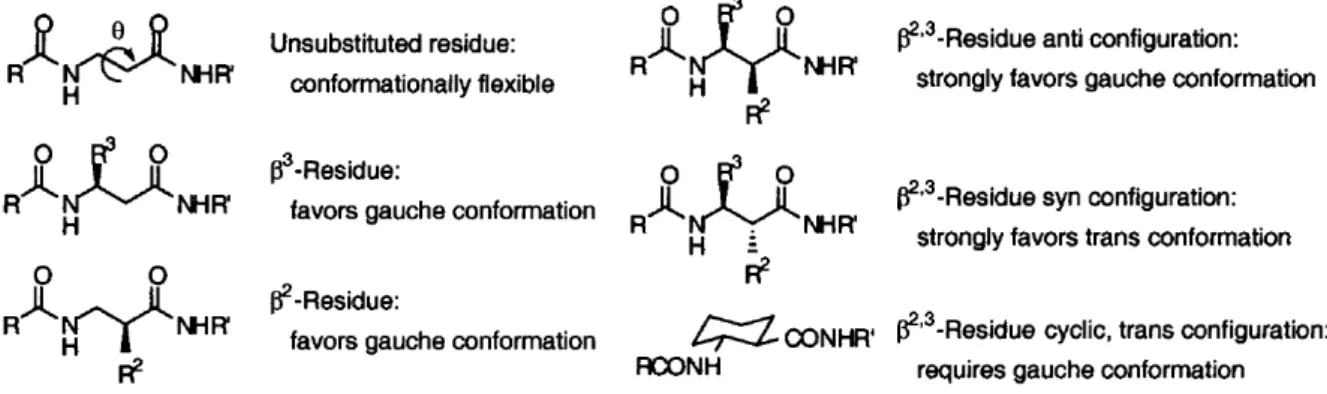
Therefore, different substitution patterns influence the torsional angles, bringing to several stables conformations as helices, sheets or turns, even if the effect of all the noncovalent interactions must be accurately taken into account. For example, it was evidenced that syn and anti-β2,3-peptides, obtained from -lactams with cis and trans configuration, have the intrinsic
conformational tendency to give different secondary structures due to steric hindrance between the substituents (Figure 5).4
Figure 5. Intrinsic conformational preferences of syn- and anti-β2,3-peptides.
In particular, an anti substitution leads to a preference for the -strand (Figure 6), while a syn substitution facilitates the formation of helices.
Figure 6. Preferential formation of -sheets for anti- 2,3-peptides.
The nomenclature of the helical conformations is very variable in literature.3,5,6 The one
reported in Figure 7, and used throughout the entire discussion, relies simply on the number of the atoms occurring between the C=O and N-H groups involved in the H-bond (Gellman’s nomenclature).3 Other nomenclatures will be used only when needed.
Figure 7. Different helix conformations for -peptides.
The most common structure for various acyclic β2-, β3- and β2,3-substituted amino acids is,
by far, the 14-helix, where there is a C=O(i)•••H-N(i-2).1 Anyway, exploiting a suitable
substitution scheme in the monomers and a proper placement of differently substituted monomers in the peptidic sequence, other secondary structures such as 10/12-helices, 10-membered turns and β-sheets were obtained (Figure 8).7
Figure 8. Examples of helices for substituted β-amino acids. (a) 14-helix of a dodecamer made of β2,3-disobstituted-amino acids; (b) 10/12 helix of a peptide with an alternate placement of
monosubstituted β2- and β3-amino acids. Only H-bonded hydrogens were displayed for clarity.
On the contrary, β-peptides containing the conformationally constrained cyclic amino acid trans-2-amino-cyclopentanecarboxylic acid (ACPC) adopt a 12-helix conformation, stabilized by H-bonding between amide carbonyl groups at position i and the amide proton at position i+3. On the other hand, when the monomer is the trans-2-aminocyclohexanecarboxylic acid (ACHC), the β-peptide folds in a 14-helix conformation, stabilized by H-bonding between the amide proton at position i and the carbonyl at position i+2 (Figure 9).8
Figure 9. (a) 14-helix conformation for ACHC; (b) 12-helix for an oligopeptide of trans-ACPC. Only H-bonded hydrogens were displayed for clarity.
1.3 Foldamers of β-Peptide Mimetics
The same reasons seen above, mainly related to the search for new secondary structures and an improved biological activity/stability, which had previously led to the expansion of β-peptides field, have subsequently also led to the expansion of the field of their mimetics.
In α-aminoxy acids, the β carbon is replaced with an oxygen atom, and the tendency to give 8-membered cycles by intramolecular hydrogen bonds (N-O turns) is already evident in suitably derivatized monomers (Figure 10).9
Figure 10. Structural determination of the N-O turns in α–aminoxy acid monomers (left: NOEs in solution; right: X-rays of the crystal structure).
The intramolecular hydrogen bonds formed by oligomers are very strong, as evidenced by the impressive chemical shift values of the protons involved, that is all the NHs except the one belonging to the first residue. Moreover, the striking overall stability of the resulting 8-helix structure is proven by the observations that all the intramolecularly H-bonded NHs are almost completely insensitive to unfolding processes caused by both intermolecular hydrogen bonding and competition of a strong H-bond-acceptor solvent, such as DMSO-d6 (Figure 11).
Figure 11. (a) δ of amide protons for the hexamer shown (n = 6) in CDCl3 at 25 °C, as a function
of logarithm of concentration. (b) δ of amide protons when increasing amounts of DMSO-d6 were
added to a 5 mM solution of the hexamer in 0.5 mL of CDCl3 at 25 °C.
On the contrary, in the case of the 1st residue NH, which is the only one not involved in the
formation of intramolecular hydrogen bonds, the expected substantial increase of δ due to either the H-bond-driven self-association or the formation of a strong hydrogen bond with DMSO-d6
is readily evident. The most stable secondary structure deduced for those α-aminoxy-peptides, which was actually computed at the level HF/6-31G* on a simplified model tetramer (all the substituents are Me) with a C-terminal amide derivatization, is an 8-helix with a pitch very close to 2 residues, precisely a 1.88-helix, using the helix nomenclature.6 In such a slightly twisted
28-helix, the sequence of N-O turns arranges the side chains alternate on opposite sides of the
helix, in a β-strand-like fashion, with a pattern reminiscent of a twisted parallel β-sheet (Figure 12).
Figure 12. The 8-helix structure calculated at the HF/6-31G* level for Ri = Me.
Mimetics of -peptides based on the insertion of a sp3 nitrogen atom in the place of the Cβ
atom, that is α-hydrazino-peptides, were also synthesized (Figure 13). Even though the secondary structures adopted in solution strongly vary depending on the particular substitution, and they are sometimes random coils, the presence of the basic and pyramidalized nitrogen atom led to the discovery of a peculiar folding, that is a new type of 8-helix.10
Figure 13. α-Hydrazino acids with Cα- and Nβ-substitutions.
In fact, at least in the crystalline state or in absence of acid and competing solvents, secondary structures consisting of a series of “hydrazino-turns” were obtained. Such structures show both the 8-helix H-bonding pattern, that is CO(i-2)-NH(i), and the additional N(i-1)-NH(i) H-bonds enclosed in the 8-membered cycles (Figure 14).
Figure 14. Left: general structure of a hydrazino-turn. Right: X-rays structure of the 8-helix composed of a hydrazino-turns series, obtained from the crystal of the hexamer shown (side chains and unnecessary hydrogens were omitted for clarity).
CHAPTER 2:
Imidazolidinone-based
α-hydrazidopeptides
2. IMIDAZOLIDINONE-BASED α-HYDRAZIDOPEPTIDES 2.1 Introduction
As reported in the previous chapter, -peptides are probably the most widely investigated peptide mimics and promising candidates for drug design, due to their protease resistance and their ability to adopt stable secondary structures with as few as six residues or less. In the framework of the synthesis of oligomers with a predictable folding, we have already reported the synthesis of hexamers based on conformationally constrained -amino acids, in which the C and C atoms are tethered in a pirrolidin-2-one ring (Figure 15).11 NMR studies and
molecular dynamics simulations showed the ability of trans-AOMPC ((3R,4S)-4-amino-5-oxo-1-((S)-1-(4-methoxyphenyl)ethyl)pyrrolidine-3-carboxylic acid) hexamer to fold into a 12-helix when R1 = H,11a similarly to others cyclic -peptides based on trans 2-aminocyclopentane
or 2-aminopyrrolidine carboxylic acids.8
The systematic methylation of the C atoms along the backbone induced a gradual transition from the 12-helix to the 8-helix, due to the steric hindrance of the methyl groups and their effect on dihedrals. In fact, when the third AOMPC residue was changed for AMOMPC (R1 = Me,
(3R,4S)-4-amino-3-methyl-5-oxo-1-((S)-1-(4-methoxyphenyl)ethyl)pyrrolidine-3-carboxylic acid), local 8-helix structures perturbed the predominant 12-helix in the most stable conformers, while when two AMOMPC were inserted as the second and fourth residues, only local 12-helices were present in the predominant 8-helix. Finally, the AMOMPC hexamer showed a stable 8-helix,11b which is a rare structure for -peptides and has been so far observed only
employing peculiar conformationally constrained monomers (Figure 15).12,13
Figure 15. (a) Pyrrolidinone-based β-amino acids used as monomers in the previous works. (b) Secondary structures assumed by AOMPC and AMOMPC hexamers.
In an effort to expand the field of peptide mimics and explore more folding properties, the synthesis of new scaffolds is required. For instance, the replacement of the C and C atoms of the β-amino acid residues by heteroatoms is an attractive design strategy. The substitution of the C atom with nitrogen leads to hydrazino peptides, also called aza- 3-peptides, composed
of -hydrazino acids. As a result, additional H-bonds via the basic sp3 N are possible, thus
originating secondary structures made of 8-membered pseudocycles whit internal 5-membered cycles (“hydrazino turns”) that cannot be formed by “normal” -peptides.10
The overall effect can be so strong that a single residue prone to form such a structure can direct the switch from the natural 12-helix of trans-2-aminocyclobutanecarboxylic acid oligomers to the 8-helix, at least up to a length of 6 residues.14
2.2 Results and Discussion
2.2.1 Synthesis of New Imidazolidinone-based -Hydrazidopeptides
In this contest, we designed a cyclic -hydrazido acid by substituting the stereogenic C atom on our previous -amino acid monomer with nitrogen, switching from a pirrolidin-2-one-tethered -amino acid (AOMPC) to an imidazolidin-2-one-pirrolidin-2-one-tethered -hydrazino acid, AOPIC ((R)-3-amino-2-oxo-1-((S)-1-phenylethyl)imidazolidine-4-carboxylic acid). It should be noted that, despite the structural resemblance with the -hydrazino acids already reported in literature, in this case the N atom should be much less basic, due to its delocalization onto the imidazolidinone carbonyl, thus AOPIC has to be considered more appropriately as a N -acylated -hydrazino acid, or a -hydrazido acid (Figure 16).
Figure 16. (a) Switch from pyrrolidinone-based monomers (AOMPC and AMOMPC) to the imidazolidinone-based monomers (AOPIC) by formal C-H → N substitution. The red color highlights the -amino acid and -hydrazido acid moieties in AOMPC and AOPIC, respectively. (b) A generic -hydrazino acid, with a basic sp3 N atom. (c) A generic -hydrazido acid, with a
We therefore synthesized conformationally constrained 2,3-oligoazapeptides of different
lengths, with the aim to investigate their folding patterns.
All the synthetic procedures reported here refers to previous works carried in our research group.15 Anyway, in this thesis, all the methodologies were deeply reviewed and optimized,
with the aim of obtaining the oligomers with high purity and better yields. Most importantly, a complete spectroscopic and computational investigation was performed in order to ascertain the preferred folding.
Methyl aziridine-2-carboxylate 1 was the readily available starting material16 for the
synthesis of Boc-AOPIC-OMe monomer used in the oligomers’ synthesis. To introduce the hydrazine functionality, 1 was ring-opened by nucleophilic addition of t-butyl carbazate, catalysed by boron trifluoride etherate. This reaction proceeded with a complete conversion but low regioselectivity towards 2, probably due to steric hindrance effects. Finally, the imidazolidinone ring in the monomer, Boc-AOPIC-OMe 3, was created by a high-yielding cyclization of 2 with phosgene.
Scheme 1. Reagents and conditions: (i) t-butyl carbazate, BF3∙Et2O, dry THF, reflux; (ii) COCl2,
MeOH, DCM, rt; (v) EDC, TEA, dry DCM, 0 °C; (vi) PyBroP, DIPEA, dry DCM, -18 C to rt; (vii) NH3/NH4Cl, dry MeOH, rt; (viii) CH3COCl, pyridine, dry DCM, rt; THF = tetrahydrofuran,
DCM = dichloromethane, EDC = 1-ethyl-3-(3-dimethylaminopropyl)carbodiimmide hydrochloride, TEA = triethylamine, PyBroP = bromotripyrrolidinophosphonium hexafluorophosphate, DIPEA = diisopropylethylamine.
All the oligomers were synthesized exploiting the usual Boc removal/ester hydrolysis/peptide coupling sequence in liquid phase (Scheme 1), but the Boc-deprotected intermediates showed a somewhat unexpected behaviour. In fact, while they demonstrated a reduced nucleophilicity, in comparison to common amino acids, during the peptide couplings, they also gave traces of the corresponding N -trifluoroacetylated products when submitted to Boc removal, using both commercial and redistilled trifluoroacetic acid. Thus, in order to obtain quantitatively the almost pure Boc-deprotected intermediates, suitable to be used in the following coupling without any purification, all the Boc removals were carried out using anhydrous HCl in MeOH/DCM mixtures. Moreover, the coupling reactions had to be carried out in quite concentrated solutions, using 1 mL or less of dichloromethane per mmol of N-deprotected intermediate, to achieve good (Boc-(AOPIC)2-OMe 4) or moderate yields
(Boc-(AOPIC)4-OMe 5 and Boc-(AOPIC)6-OMe 6) (Scheme 1). C-terminal amide monomer
Boc-AOPIC-NH2 7 was synthesized to test the possible subsistence of an inherent tendency towards
the formation of 8-membered H-bonded cycles, as evidenced by related -aminoxy acids.9
Eventually, N-acetyl monomer Ac-AOPIC-OMe 8 was synthesized because its NH, which was further demonstrated to be intramolecularly non-H-bonded, should be the right reference for any hydrogen atoms belonging to peptide bonds in oligomers 4, 5 and 6 that do not participate in hydrogen bonding. In the same way, the NH of Boc-AOPIC-OMe 3 should be the right reference to which compare the Boc-NHs belonging to monomer 7 and to the first residues of oligomers 4-6.
2.2.1 Investigation by NMR and CD Spectra
Although the complete assignment of NH resonances was possible only for monomers and
oligomers up to tetramer 5, due to broad peaks with an extended overlap in hexamer 6, the simple analysis of their values in diluted solutions gave some important indications (Figure 17 and starting points in Figure 18 and Figure 19). In fact, the simple C-terminal amide monomer
7 showed a striking difference, about 2.7 ppm, between the intramolecularly bonded and
8membered Hbonded cycles reported for a monomeric Cterminal amide derivative of an -aminoxy acid, that lead to 8-helix structures in oligomers.9 Moreover, a similar difference
between bonded and unbonded hydrogens was also observed for a closely related C-terminal amide -hydrazino acid,10c which actually showed the proclivity to form an 8-membered
pseudocycle with an enclosed additional 5-membered cycle, that is a hydrazino turn, in which the sp3 basic hydrazinic nitrogen acts as an acceptor.
Figure 17. Chemical shifts for hydrazidic protons of Boc-AOPIC-OMe 3, Boc-(AOPIC)2-OMe 4,
Boc-AOPIC-NH2 7 and Ac-AOPIC-OMe 8 (1 mM solutions in dry CDCl3 at 25 °C). The
8-membered H-bonded cycles are reported with continuous lines, the possible additional 5-membered cycles with dashed lines.
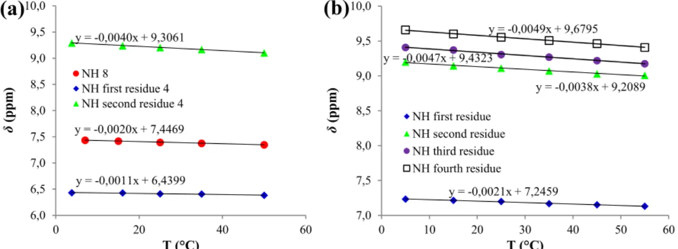
Taking into account oligomers 4-6, the NH chemical shifts in diluted CDCl3 solutions
evidence the possibility for a reinforcing, cooperative effect as the length increases. Starting from 9.20 ppm for second residue of dimer 4, the NHs for the three intramolecularly H-bonded
NHs become 9.11, 9.31 and 9.56 ppm for tetramer 5, with values raising from the second to the fourth residue. The strengthening effect is even larger for hexamer 6, with NH = 9.24-9.62 ppm
for the five H-bonding NHs (residues 2-6, values not assigned).
Moreover, in all compounds, hydrazidic Boc- or Ac-derivatized NHs belonging to the first residues are not involved even in weak intramolecular H-bonds, neither by a 6-membered cycle with the intraresidue carbonyl oxygen of the -hydrazido acid moiety, nor by a 5-membered
cycle with the imidazolidinonic carbonyl. This can be easily inferred by the comparison of values obtained for all the first residue hydrazidic hydrogens in Figures 17-19, especially the N-acetyl derivative 8 (7.39 ppm), with the ones reported for the simple N,N′-dibenzoyl-hydrazide, whose 5-membered H-bonds lead to NH = 9.23 ppm, or other symmetric and
asymmetric benzohydrazides, where the intervention of additional (bifurcated) 6-membered H-bonds gives NH up to 11.25 ppm.17 This is very likely due to the possible repulsive interactions
that could arise between the carbonyl oxygen onto N and the imidazolidinonic urea moiety, or the substituted C , if the hydrazidic NH was H-bonded to the -hydrazido acid carbonyl or the imidazolidinone carbonyl, respectively.
It should be noted at this point that such a H-bonding pattern, where the first residue NHs are always non-H-bonded and the number of intramolecular hydrogen-bonds is in any cases equal to the number of residues minus one, rules out from a logical point of view the most common structures obtained for the related -peptides,the 12- and 14-helices,1 as well as the
uncommon 10/12-7 and 10-helices.18 In fact, from a logical point of view, in the case of a
homogeneous secondary structure these first experimental observations point strongly towards the helix folding, or its variant made of bifurcated H-bonds that form contemporaneously 8-terms cycles with enclosed 5-8-terms cycles (see the detailed discussion in Paragraph 2.5.1 Logical Analysis of Possible H-bonding Patterns for α-hydrazidopeptides).
Anyway, it was evident from NMR experiments with more concentrated solutions that, even in the case of the intramolecularly H-bonded NHs, a contribution originating from self-association was also present in all cases. Then, before proceeding with all the other structural investigations, it was necessary to safely determine if the intermolecular H-bonding was reasonably negligible at the concentrations used. In fact, in an effort devoted to the synthesis of self-assembling hydrazides, Zhao et al. obtained an equilibrium constant Kinter of 112 M-1 for a
weak H-bond-driven intermolecular homodimerization, when just two reciprocal C=O···H-N interactions arranged in a -sheet-like disposition were present between the molecules, but values up to 4.7 × 10⁴ M-1 for quadruply H-bonding heterodimers.19 Thus, due to the fact that,
in line of principle, our compounds could behave as multiply hydrogen-bonding structures and dimerize, or even oligomerize, the tendency to the intermolecular H-bonding was quantitatively evaluated by dilution experiments in dry CDCl3 on the reference monomer Ac-AOPIC-OMe 8
and the simplest oligomer, dimer Boc-(AOPIC)2-OMe 4 (Figure 18). Anhydrous CDCl3 was
chosen to determine rigorously only the self-association, eliminating the effect of residual monomeric water, because a comparison with a 1 mM solution of 4 in wet deuterochloroform
furnished a negligible increase in chemical shift for the intramolecularly H-bonded NH belonging to the second residue (+0.01 ppm), but a quite pronounced increment for the BocNH (+0.06 ppm). The non-linear regression of the observed chemical shift ( obs) vs the total
substrate concentration data for 8 allowed to obtain an intermolecular association constant Kinter
= 4.7±0.6 M-1, which is in agreement with just a weak tendency to the homodimerization by
formation of intermolecular H-bonds (for a detailed discussion see Paragraph 2.5.2 Determination of Intermolecular H-bonding from Dilution Experiments).
In effect, this value is very small if compared to the ones reported by Zhao, while it is identical to the one obtained for N-methylacetamide in the less polar CCl4, which becomes only
0.8 M-1 in the more polar CD
2Cl2 (dielectric constants: carbon tetrachloride, 2.24; chloroform
4.81; methylene chloride, 8.93).20,21 The computed percent molar fractions for the
intermolecularly H-bonded homodimer of compound 8, fD, are 0.93%, 7.99% and 37.2% at a
total substrate concentration of 1 mM, 10 mM, and 0.1 M, respectively (Table 6 in Paragraph 2.5.2 Determination of Intermolecular H-bonding from Dilution Experiments), indicating that, even for such a weak intermolecularly H-bonding substrate, the self-association becomes no more negligible if experiments are not carried out in very diluted solutions. Using the data for the first residue NH (Boc-NH) of Boc-(AOPIC)2-OMe 4, a larger intermolecular association
constant Kinter of 8.1±0.6 M-1 was calculated, with percent molar fractions for the homodimer
due to intermolecular H-bonding of Boc-NH, fD, that raise to 1.57%, 12.4% and 46.5% at a total
substrate concentration of 1 mM, 10 mM, and 0.1 M, respectively.
Figure 18. Variation of NH proton chemical shifts of reference monomer Ac-AOPIC-OMe 8 and dimer Boc-(AOPIC)2-OMe 4 as a function of total substrate concentration from dilution
experiments in dry CDCl3 at 25 °C. 6,0 6,5 7,0 7,5 8,0 8,5 9,0 9,5 10,0 0,00 0,01 0,02 0,03 0,04 0,05 0,06 0,07 0,08 0,09 0,10 δob s (p pm ) Conc (M) NH 8 NH first res. 4 NH second res. 4
Using the same approach, but taking also into account the presence of the intramolecular H-bond for the second residue NH of compound 4, the equilibrium constants for both the intermolecular and the intramolecular hydrogen-bonded species could not be computed exactly, because it is impossible to know precisely the limiting chemical shifts for the completely non-H-bonded ( M) and the completely intramolecularly H-bonded NH ( intra). Anyway, a limiting
D = 9.6017 ppm could be safely extrapolated for the completely intermolecularly H-bonded 4,
while the chemical shift of 7.3742 ppm obtained for the non-hydrogen-bonded reference compound 8, where the NH is acylated as in the second residue of Boc-(AOPIC)2-OMe 4, can
represent a good approximation for M. Thus, we used the highest values available for
intramolecularly hydrogen-bonded NHs in tetramer Boc-(AOPIC)4-OMe 5 (9.5553 ppm) and
hexamer Boc-(AOPIC)6-OMe 6 (9.6148 ppm), in diluted solutions of anhydrous CDCl3, as
estimates for intra, and then calculated approximate intramolecular and intermolecular
association constants, Kintra and Kinter, of the second residue NH in Boc-(AOPIC)2-OMe 4 (see
detailed discussion in 2.5.2 Determination of Intermolecular H-bonding from Dilution Experiments). The plausible ranges for the equilibrium constants related to the NH belonging to the second residue of compound 4 were estimated as Kintra = 4.3-5.0 and Kinter = 18-21 M-1,
thus indicating a quite strong tendency to the intramolecular hydrogen-bonding, even in a simple dimer without the possibility for a cooperative effect, and again a weak, but not negligible, propensity to the intermolecular self-association, at least in a non-hydrogen-bonding solvent. In fact, in this case the percent molar fractions for the homodimer are 2.24%, 16.4% and 52.6% at a total substrate concentration of 1 mM, 10 mM, and 0.1 M, respectively, then we preferred to carry out all the subsequent NMR and CD analysis at concentrations lesser than 2 mM, with the exception of bidimensional NMR spectra, without extending the analysis of the competition between intra- and intermolecular H-bonding to longer oligomers.
The titration with DMSO-d6, which is a strong H-bond acceptor, of diluted solutions of
reference monomer Ac-AOPIC-OMe 8, dimer Boc-(AOPIC)2-OMe 4, tetramer Boc-(AOPIC)4
-OMe 5 and hexamer Boc-(AOPIC)6-OMe 6 in CDCl3, is coherent with a progressive unfolding
as the percentage of DMSO-d6 added increases. However, although the limiting values in only
DMSO-d6, which are always in the range 9.98-10.25 ppm for all the hydrazide NHs and
8.84-8.86 ppm for the carbazate Boc-NHs, are indicative of a probably complete disruption of the folding observed in chloroform, the ability to resist to unfolding is strikingly different among oligomers 4-6 (Figure 19 and Tables 3-6 in Paragraph 2.5.3 Titrations with DMSO-d6.).
Figure 19. Variation of NH proton chemical shifts (ppm) of (a) reference monomer Ac-AOPIC-OMe 8, (b) dimer Boc-(AOPIC)2-OMe 4, (c) tetramer Boc-(AOPIC)4-OMe 5 and (d) hexamer
Boc-(AOPIC)6-OMe 6 as a function of increasing percentages of DMSO-d6 added (v/v) to 1 mM (4 and
8) or 2 mM solutions (5 and 6) in CDCl3 at 25 °C.
As it can be easily deduced by inspection of Figure 19b, the unfolding process of dimer is already almost complete at 10% added DMSO-d6. In fact, for the NH of second residue in dimer
Boc-(AOPIC)2-OMe 4, the observed chemical shift at 10% DMSO-d6, 10%DMSO-d6 = 10.04 ppm,
is very close to the value observed in only DMSO-d6, DMSO-d6 = 10.19 ppm, the difference
being only 0.15 ppm. The same difference become 0.30, 0.28 and 0.30 ppm for the second, third and fourth residues NHs, respectively, of tetramer Boc-(AOPIC)4-OMe 5, indicating an
increased resistance to unfolding, which is apparent even by the monotonic increase of chemical shifts up to 30% added DMSO-d6 (Figure 19c). The behaviour of hexamer 6 deserves a careful
analysis because, even if values are almost constant for percentages of added DMSO-d6
between 10% and 30% (Figure 19d), they are not indicative of a complete unfolding. This can
7,0 7,5 8,0 8,5 9,0 9,5 10,0 10,5 11,0 0 10 20 30 δ (p pm ) % DMSO-d6 NH
(a)
Ac-AOPIC-OMe 8 δ in DMSO-d6: 9.9836 ppm 6,0 6,5 7,0 7,5 8,0 8,5 9,0 9,5 10,0 10,5 11,0 0 10 20 30 δ (p pm ) % DMSO-d6 NH first residue NH second residue(b)
δ in DMSO-d6: 8.8438 ppm Boc-(AOPIC)2-OMe 4 δ in DMSO-d6: 10.1921 ppm 7,0 7,5 8,0 8,5 9,0 9,5 10,0 10,5 11,0 0 10 20 30 δ (p pm ) % DMSO-d6 NH first residue NH second residue NH third residue(c)
δ in DMSO-d6: 10.2479 and 10.2264 ppm δ in DMSO-d6: 9.9787 ppm δ in DMSO-d6: 8.8370 ppm Boc-(AOPIC)4-OMe 5 7,0 7,5 8,0 8,5 9,0 9,5 10,0 10,5 11,0 0 10 20 30 δ (p pm ) % DMSO-d6 NH NH NH NH NH NH(d)
δ in DMSO-d6: 10.2440 ppm δ in DMSO-d6: 10.0501 ppm δ in DMSO-d6: 10.0178 ppm δ in DMSO-d6: 8.8644 ppm Boc-(AOPIC)6-OMe 6 δ in DMSO-d6: 10.270 ppm (2 peaks)be inferred by the differences between 10%DMSO-d6 and DMSO-d6 values, which range from 0.22
to 0.74 ppm (Table 9), indicating that the secondary structure maintains some stability at least up to a 30% added DMSO-d6, and that the complete unfolding must occur at higher percentages.
The reduced temperature coefficient of chemical shift, / T, is a parameter commonly used in the conformational analysis of proteins in solution,22 although its predictive use as an H-bond
indicator gave many times poor or unreliable results.23 Anyway, for many oligopeptides and
their mimics, / T values more negative than -2.6 ppb/K have been associated to a dynamic equilibration between an H-bonded and a non-H-bonded structure.24Conversely, smaller values
have been generally interpreted as a completely H-bonded or a completely non-H-bonded NH, even if simple diamides have shown that small values can also derive from a complex balance among the many factors affecting the difference in stability between intramolecularly bonded and unbonded NHs.21
In the present case, the BocNHs of dimer 4 and tetramer 5 and the AcNH of monomer 8, which have been previously proven to be intramolecularly non-H-bonded, show / T values of -1.1, -2.1 and -2.0 ppb/K, respectively (Figure 20). On the contrary, the second residue NH of dimer 4 has a / T = -4.0 ppb/K, and the second, third and fourth residue NHs of tetramer
5 have, respectively, values of -3.8, -4.7 and -4.9 ppb/K, which reflect the cooperativity of
H-bonds and their order of strength revealed by chemical shifts.
Figure 20. Variation of NH proton chemical shifts as a function of temperature. (a) Ac-AOPIC-OMe, 8 and Boc-(AOPIC)2-OMe, 4 (1 mM solutions in dry CDCl3). (b) Boc-(AOPIC)4-OMe, 5 (2
mM solution in dry CDCl3).
A further indication about the existence of a H-bonded-driven secondary structure and the possibility for a reinforcing cooperative effect due to elongation was obtained from analysis of CD spectra (Figure 21). Oligomers 4-6 were dissolved in methanol at concentrations where
y = -0,0020x + 7,4469 y = -0,0011x + 6,4399 y = -0,0040x + 9,3061 6,0 6,5 7,0 7,5 8,0 8,5 9,0 9,5 10,0 0 20 40 60 δ (p pm ) T (°C) NH 8 NH first residue 4 NH second residue 4
(a)
y = -0,0021x + 7,2459 y = -0,0038x + 9,2089 y = -0,0047x + 9,4323 y = -0,0049x + 9,6795 7,0 7,5 8,0 8,5 9,0 9,5 10,0 0 10 20 30 40 50 60 δ (p pm ) T (°C) NH first residue NH second residue NH third residue NH fourth residue(b)
self-association was previously demonstrated to be negligible by NMR, even in a much less hydrogen-donating/accepting solvent (CDCl3). We did not undertake a comparison between
experimental and theoretically computed spectra, although CD spectra were sometimes successfully used to determine the preferred conformation, or the equilibration between different folding patterns.25 In fact, it was demonstrated that it can sometimes be difficult, or at
least misleading, to derive the secondary structure from CD spectra, even in the case of much more extensively studied foldamers, such as the -peptidic ones.26
The per-residue molar ellipticity of monomer Boc-AOPIC-OMe 5 differs not only quantitatively, but also qualitatively, from the ones of oligomers, in that the intensity increases up to 195 nm, with the exception of a secondary maximum at 220 nm, while for the oligomers there is a main peak at 202 nm (Figure 6). The CD spectra of dimer 4, tetramer 5 and hexamer
6 are similar to the ones of the robust 8-helix structures formed by related -aminoxy acids,27
although they also resemble the one of a disordered hexapeptide made of N benzylated -hydrazino acids,10a both in the peak at about 200 nm and in the lack of a significant Cotton
effect. It is noteworthy that, contrarily to this latter case, where the corresponding trimer had a different maximum and an increased intensity compared to the hexamer, in the present case there is no change in max passing from the dimer to the hexamer, while there is an evident
increase in per-residue molar ellipticity as the length increases. Moreover, the unfolded structure of those benzylated -hydrazino peptides was mainly deduced on the basis of the missing Cotton effect, but this was demonstrated to be not always true. In fact, despite the evident Cotton effect in the typical CD spectra of 14-helices formed by many -peptides,28 the
same feature is not present in the highly stable 14-helix formed by trans-2-aminocyclohexanecarboxylic acid (ACHC) oligomers,29 and it is also absent in the case of the
Figure 21. CD spectra of 1 mM Boc-AOPIC-OMe 3 (green line), 1 mM Boc-(AOPIC)2-OMe 4
(purple line), 0.1 mM Boc-(AOPIC)4-OMe 5 (red line) and 0.1 mM Boc-(AOPIC)6-OMe 6 (black
line) solutions in MeOH at 25 °C. Data normalized for concentration and number of residues.
The analysis of CD spectra in methanol, which is a much stronger H-bond donor/acceptor in comparison to chloroform, makes the gain in stability of the ordered secondary structure as the length increases even more evident than it could be derived from NMR spectra. In fact, it is obvious from the appearance of the maximum at 202 nm that dimer 4 is already partly folded, while the continuous increase in intensity passing from dimer to hexamer shows that the proportion of ordered conformation enlarges simultaneously with the elongation, thus proving the cooperativity in the folding stabilization.
2.2.2 Computational Investigation
Our experimental findings were plenty confirmed by an extensive molecular dynamics simulation on the hexamer, followed by cluster analysis and hydrogen bond lifetimes analysis. This computational approach gave also additional information and shed some light on the preference between the two variant of the preferred structure, namely the “pure” 8-helix and its counterpart where there is an additional 5-terms cycle inside every 8-membered cycle, that is to say a sequence of the so-called hydrazino turns (note: in this case they are actually hydrazido turns).
MD simulations were carried out with the AMBER 11.0 suite of programs.30 The peptide is
parametrized using GAFF (general AMBER force field) force field.31 The standard RESP
procedure is carried out to assign charges to atoms by Antechamber.32 The peptide was built
and immersed in a solvent box of explicit chloroform molecules, reproducing the conditions in
-10000 0 10000 20000 30000 40000 50000 195 205 215 225 235 245 255 265 275 285 295 [θ] T (de g cm 2dm ol -1) λ (nm)
which NMR spectra were registered. Periodic boundary conditions were used (see Paragraph 2.5.4 Computational for more details)
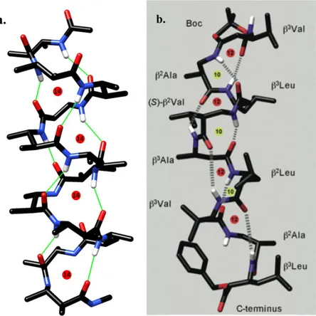
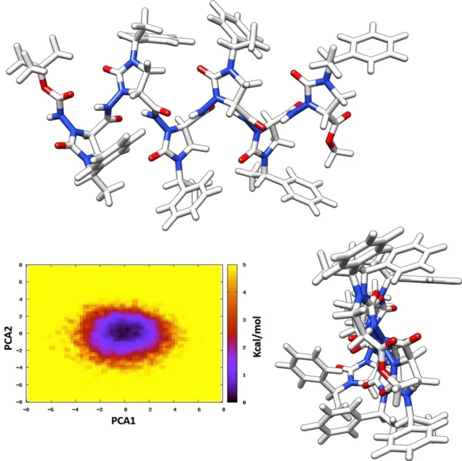
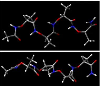
The molecular dynamics simulation for 100 ns highlighted the existence of an 8-helix-type folding as the sole stable secondary structure, as it is readily apparent from the results of the principal component analysis (PCA) (Figure 22).33 The right-handed helix has a pitch of ≈6.3
Å and 1.9 residues per turn then, using the more complete helix nomenclature, it can be named as a (P)-1.98-helix. Moreover, the same results indicate that the preferred folding is very stiff,
whit backbone dihedral angles poorly dispersed around the mean values and the phenylethyl chains always alternatively directed towards opposite sides with respect to the helix axis, in a β-strand-like fashion (Figure 22).
Figure 22. Results of conformational analysis of Boc-(AOPIC)6-OMe 6. Representative views of
PCA1 and PCA2 are the two eigenvectors with the lowest eigenvalues of the principal component analysis calculated from the analysis of the MD trajectory.
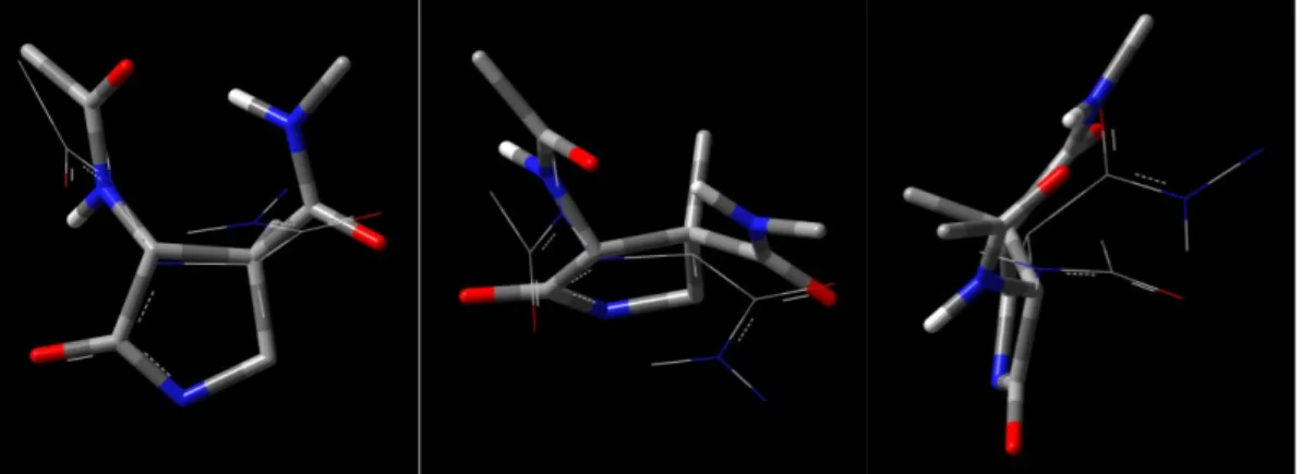
A deeper insight is necessary to establish, at least from a computational point of view, the preference between the “pure” and the “hydrazido-turn-series” variants of 8-helix, which relies on subtle geometric difference. As depicted in Figure 23 for the global minimum structure, the hydrazidic nitrogen atoms in the imidazolidinone rings are slightly pyramidalized and participate to 5-membered H-bonded cycles. Their arrangement is similar to the one reported for many non-conformationally constrained α-hydrazino-peptides,10 and even closer to the one
evidenced for an Nβ-acylated aza-β3-cyclohexapeptide.10f
The H-bonds lifetime analysis furnished other important indications. In fact, in the system taken in consideration (room temperature and chloroform as the solvent), the great contribution from the C=O(i)•••H-N(i+2) and the Nβ(i)•••H-N(i+1) is strikingly evident, these H-bonds being occupied for 98.4-99.9% of the time (Figure 23). This also means that the computational results strongly corroborate the experimental deduction of an almost completely H-bonded folding of hexamer in chloroform at room temperature.
Figure 23. Conventional H-bonds (heteroatom•••H-N) characterizing the hydrazido-turn series in Boc-(AOPIC)6-OMe 6, depicted in the global minimum structure. The inset is a close view of the
Moreover, an additional stabilization comes from unconventional (non-standard) H-bonds (Figure 24). Among all the contributions, the four C=O•••H-C bonds between imidazolidinone carbonyl of residue i and phenyl orto-hydrogen of residue i-2 are, by far, the most energetically important and are occupied for a substantial percent of time. Eventually, a minor contribution comes also from the C=O•••H-C bonds between the second residue imidazolidinone carbonyl and the t-butyl hydrogens of the N-terminal Boc protecting group.
Figure 24. Unconventional H-bonds (C=O•••H-C) in Boc-(AOPIC)6-OMe 6, depicted in the global
minimum structure. In red: C=O•••H-C bonds between imidazolidinone carbonyls and phenyl orto-hydrogens. In green: C=O•••H-C bonds between imidazolidinone carbonyl of second residue and t-butyl hydrogens.
At this point,some peculiar features of the preferred folding should be emphasized. First, as it is evident from the inspection of Figure 22-Figure 24, due to the directionalities of H-bond donors and acceptors, the hydrogen-bonding functionalities are solvent-exposed and they also appear to be available for bimolecular interactions leading to self-association. This could partly explain, at least in a qualitative way, the fact that even in presence of strong intramolecular hydrogen bonds (evidenced for all the oligomers), some tendency to both bimolecular association and competition by DMSO was experimentally ascertained by NMR analysis (see Paragraph 2.2.1 Investigation by NMR and CD Spectra). Moreover, both these proclivities were almost completely absent in the structurally related 8-helix-forming pyrrolidinone-based β-hexapeptide already synthesized in our laboratory, Boc-(AMOMPC)6-OMe (see Paragraph
2.1 Introduction).11b Anyway, obviously hydrogen bonds non-shielded from the solvent, or
from bimolecular association, are not per se sufficient to explain the above reported experimental findings, which must be related to some energetic factor leading to a little destabilization in comparison to the very stable and stiff 8-helix of pyrrolidinone-based β-peptide parent compounds. Unfortunately, at the moment no compelling hypothesis can be made about the reason(s) of the observed behaviour, but these explanations are beyond the scope of this thesis and will be the topic of a future study. Anyway, on the basis of the impressive amount of studies available on foldamers, a general observation can at least be made. In fact, among the many factors that can affect the stability of an oligomer, the most important was demonstrated to be by far the preorganization of the monomers, that is the inherent tendency to arrange firmly the H-bonding functionalities in the proper positions and with the correct directionalities.1 Thus, the little instability in comparison with the 8-helix
formed by our previously reported β-peptide hexamer, Boc-(AMOMPC)6-OMe, which was
based on a pyrrolidinone-tethered monomer, could be tentatively ascribed to a generic “less than optimal” preorganization of the imidazolidinone-tethered -hydrazido acids, which would lead to some tensions during the formation of H-bonds.
In this regard, another interesting feature of the computed 8-helix folding of the hexamer Boc-(AOPIC)6-OMe deserve some comments. Using the more precise conformer-based
nomenclature of Perczel,6 the close inspection of the 8-helices reported in literature for
β-peptides reveals that the most part of the 8-helices obtained so far, based on trans-β-amino acids tethered on peculiar cycles,11b,12b-d,14 exists in the helical (H8) conformation (Figure 25). The
only exception are the 8-helices show by oligopeptides derived from the achiral 1-(aminomethyl)cyclopropanecarboxylic acid12a and the cis-aminocyclobutane-1-carboxylic acid
(ACBA), the latter being actually able to form both the zigzag structures Z8, leading to the 8-helix, and Z6, leading to another β-strand-like secondary structure (Figure 25).12e
Figure 25. Conformer-based description of the two types of arrangements leading to an 8-helix folding (H8 and Z8) and another arrangement (Z6) leading to a strand-like structure, reported for a generic β-amino acid diamide.
On the contrary, considering the 8-helices formed by -aminoxy-peptides9 and properly
derivatized -hydrazino-peptides,10b it is easy to deduce that, even if they are not in a “perfect”
Z8 conformation, their folding is much more similar to a zigzag disposition than to an helical (H8) one. Moreover, the average backbone torsional angles for the computed Z8-like minimum energy conformation of -aminoxy-tetramer9 reported in Figure 26 (ϕ = -125°, θ = 80°, ψ =
25°), and for the crystal structure of -hydrazino-pentamer in Figure 27 (ϕ = -120°, θ = 75°, ψ = 15°),10b are the only ones quite close to the values of backbone dihedrals computed for the
global minimum of our hexamer Boc-(AOPIC)6-OMe, 6 (Table 1). The close inspection of
global minimum structure for hexamer Boc-(AOPIC)6-OMe and -hydrazino acid pentamer
crystal structure highlights also the expected differences in H-bonds lengths and in pyramidalizations of nitrogen atoms participating to the 5-membered cycles, which are both due to the reduced basicity of sp2 nitrogens in hydrazido-turns, compared to sp3 nitrogens in
hydrazino-turns. In fact, while for -hydrazino-pentamer the C=O•••H-N (2.03-2.44 Å) and the Nβ•••H-N (2.18-2.39 Å) distances are in all cases quite close, for Boc-(AOPIC)6-OMe the
computed C=O•••H-N lengths (1.89-2.00 Å) are always much shorter than the Nβ•••H-N ones (2.38-2.43 Å). Furthermore, the analysis of improper dihedrals angles confirms that all Nγ atoms are strongly out-of-plane (54-62°) when they are attached to sp3 Nβ atoms in
-hydrazino-pentamer, but they are in a quasi-planar arrangement (5-16° out-of-plane bending) when they are bonded to sp2 Nβ atoms in Boc-(AOPIC)
6-OMe. These observations confirm that
the overall contribution of the additional 5-membered cycles to the hydrazido-turn stability is much lesser than the same contribution in the case of hydrazino-turns.
Figure 26. Minimum energy conformer computed at HF/6-31G* level for an -aminoxy acid tetramer (taken from Ref. 9).
Figure 27. X-ray crystal structure of an -hydrazino acid pentamer (taken from Ref. 10b). Thus, as reported above, there are completely different backbone conformations that can lead to 8-helices, even if they have always the same strand-like global structure and arrangement of side chains.
This is even more evident comparing the two 8-helices obtained from the present hexamer, Boc-(AOPIC)6-OMe 6, and its formal parent compound, that is our previously synthesized
pyrrolidinone-based hexamer, Boc-(AMOMPC)6-OMe, 11b (Figure 28 e Table 1). Due to the
fact that Boc-(AMOMPC)6-OMe was constructed starting from a monomer having an opposite
absolute configuration at C , thus leading to a secondary structure with opposite helicity, during the comparison the signs of dihedral angles in Table 1 must be inverted.
It is evident from data in Table 1 that, despite the illusory resemblance of the two 8-helices, the in-depth analysis confirms that Boc-(AMOMPC)6-OMe is well described as a H8
conformation. On the contrary, the arrangement of Boc-(AOPIC)6-OMe is more resembling to
a Z8 conformation, even if the values of backbone torsional angles are not in a perfect agreement with the ones computed by Perczel for β-peptides.6
Table 1. Comparison of dihedral angles in minimum energy conformers of 8-helices of imidazolidinone-based Boc-(AOPIC)6-OMe and pyrrolidinone-based Boc-(AMOMPC)6-OMe.a
Residue
number Boc-(AOPIC)6-OMe Boc-(AMOMPC)6-OMe
a ϕ θ ψ ϕ θ ψ 1 -108.5 68.4 18.4 89.9 -104.4 45.5 2 -109.4 68.5 15.1 90.9 -101.4 46.7 3 -107.9 68.4 14.5 95.8 -98.3 48.7 4 -110.5 62.3 11.1 96.5 -101.9 49.3 5 -111.8 65.0 7.9 91.7 -101.3 49.3 6 -99.4 61.0 -88.8 88.8 -99.1 -50.8
a Taken from Ref. 11b.
Eventually, the striking differences between the two 8-helices of Boc-(AOPIC)6-OMe and
Boc-(AMOMPC)6-OMe can be better evidenced by the visual inspection of both backbone
conformations in the global minima, using the mirror image in the case of Boc-(AMOMPC)6
-OMe (Figure 28).
Figure 28. Top: fragments of structures of Boc-(AOPIC)6-OMe and mirror images of
Boc-(AOMPC)6-OMe and Boc-(AMOMPC)6-OMe considered in the comparison. Middle: H8
structure of Boc-(AOMPC)6-OMe (left), H8 structure of Boc-(AMOMPC)6-OMe (centre) and
Z8-like structure of Boc-(AOPIC)6-OMe (right). Bottom: three different views of the superimposed
structures of Boc-(AOPIC)6-OMe and Boc-(AMOMPC)6-OMe (wireframe = Boc-(AOPIC)6-OMe,
stick = Boc-(AMOMPC)6-OMe). Unnecessary hydrogens and phenylethyl side chains omitted for
clarity.
In Figure 28, it is also evident the even more remarkable difference between the folding of the present imidazolidinone-based hexamer, Boc-(AOPIC)6-OMe, and its closer analogue, the
C -unmethylated version of the previous pyrrolidinone-tethered hexamer, Boc-(AOMPC)6
-OMe.11a Although the different conformation of H-bonding functionalities is obvious, due to
the fact that the secondary structure of Boc-(AOMPC)6-OMe was ascertained to be a 12-helix,
it is very interesting to note how dramatically the formal change C → N affects the disposition of the groups involved, even if the cyclic fragments have very similar structures. Anyway, the elucidation of the deep reasons, that is to say the energetic factors related to all the possible intra- and inter-residue interactions, are beyond the scope of this work and will be analysed further.
2.2.3 ROESY analysis
In the case of hexamer Boc-(AOPIC)6-OMe 6, the extensive overlap and the broad peaks
prevented the residue-specific assignment of 1H and 13C backbone resonances, even after many
attempts with prolonged acquisition times, different solvent mixtures and temperatures. On the contrary, 1H and 13C resonances could be assigned for dimer Boc-(AOPIC)
2-OMe 4
and tetramer Boc-(AOPIC)4-OMe 5, with the exception of phenylethyl side chains. In
particular, for dimer 4 the analysis of a concentrated sample (0.1 M) in CDCl3 at 35 °C allowed
us to obtain resolved 1H and 13C signals and easily detectable HSQC and HMBC crosspeaks,
at 19 °C, in order to narrow 1H and 13C resonances and then detect the requested heteronuclear
couplings. The assignments for dimer 4 (Table 2) and tetramer 5 (Table 3) were obtained as follows:
- from the tBu protons, the Boc carbonyl group resonance was identified by HMBC spectrum;
- from the Boc carbonyl carbon, the NH(A) was assigned by HMBC correlation;
- NH(A) did not show the expected crosspeak with C4(A), but furnished an evident 3J coupling
with the imidazolidinonic carbonyl C2(A);
- from C2(A), both H5 and H5’ of residue A were assigned by HMBC correlation;
- from H5(A) and H5’(A), C5(A) was identified by HSQC spectrum;
- from C5(A), 2J coupling allowed to assign H
4(A) resonance;
- from H4(A), the resonances of C4(A) and carbonyl group (peptidic CO) of residue A were
assigned by HSQC and HMBC correlations, respectively;
- from the peptidic CO(A), HMBC spectrum revealed the 2J coupling with the NH(B)
resonance;
- from NH(B), continuing to exploit the same step-by-step approach, all the backbone resonances were sequentially assigned.
Table 2. Assignment of 1H and 13C backbone resonances of compound Boc-(AOPIC)
2-OMe 4 (0.1 M in CDCl3 at 35 °C). A B Peptidic CO 169.7 --- C4 59.3 57.4 NHCO-OtBu 156.3 --- NHCO2-C(CH3)3 82.6 --- NHCO2-C(CH3)3 28.2 ---
C5 41.1 39.9 Imidazolidinone N1-(CO)-N3 (C2) 158.9 157.7 CO2CH3 --- 52.7 CO2CH3 --- 170.1 NH 6.90 9.41 H4 4.17 4.47 H5 + H5’ 3.44-3.46 3.27-3.29 NHCO2-C(CH3)3 1.50 --- CO2CH3 --- 3.75
Table 3. Assignment of 1H and 13C backbone resonances of compound Boc-(AOPIC)
4-OMe, 5 (20
mM in CDCl3:DMSO-d6 9:1 at 19 °C, with powdered 4 Å m.s.).
A B C D Peptidic CO 169.95 170.66 168.54 --- C4 58.46 58.07 58.76 56.77 NHCO-OtBu 154.74 --- --- --- NHCO2-C(CH3)3 81.04 --- --- --- NHCO2-C(CH3)3 28.11 --- --- --- C-5 41.02 40.70 41.22 39.39
Imidazolidinone N1-(CO)-N3 (C2) 158.40 157.36 157.82 157.50 CO2CH3 --- --- --- 52.17 CO2CH3 --- --- --- 169.00 NH 8.18 9.48 9.90 9.92 H4 4.44 4.45 4.17 4.30 H5 3.25 3.17 3.06 2.93 H5’ 3.83 3.76 3.70 3.54 NHCO2-C(CH3)3 1.42 --- --- --- CO2CH3 --- --- --- 3.40
Some portions of the ROESY spectrum are reported in Figure 29, while a full correlation table is reported in Table 4.
In particular, the portions in Figure 29 are related to the cross-peaks which are decisive in order to determine the precise folding, that is to distinguish between the two variants of the 8-helix folding.
Figure 29. Representative portions of the ROESY spectrum of Boc-(AOPIC)4-OMe, 5, showing
Table 4. Intensities of ROESY peaks (arbitrary units) of tetramer Boc-(AOPIC)4-OMe, 5 (20 mM
in CDCl3:DMSO-d6 9:1 at 19 °C, with powdered 4 Å m.s.).
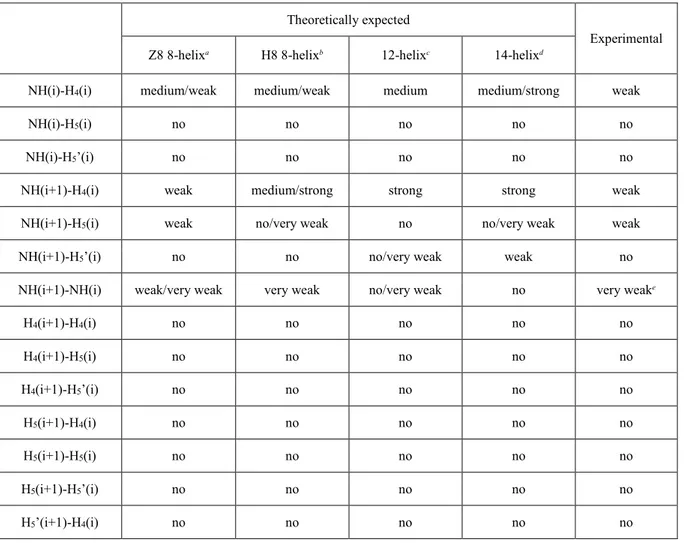
With the aim to confirm the computationally evident preference for the zig-zag 8-helix (Z8) over its helical (H8) counterpart, the experimental intensities of ROESY cross-peaks obtained for tetramer Boc-(AOPIC)4-OMe, 5, were compared to the theoretically expected ones for both
the variants of the 8-helix secondary structure (Table 5and Figure 30). In the case of Z8 secondary structure, the average backbone dihedrals computed for the lowest energy conformer of hexamer Boc-(AOPIC)6-OMe, 6, were used in order to determine the relevant interatomic
distances and then the expected ROESY cross-peaks. Due to the fact that no H8 folding was evidenced for Boc-(AOPIC)6-OMe during molecular dynamics simulation, the backbone was
forced to take such a conformation. To this end, we used the average dihedrals computed for the minimum energy conformer of the related pyrrolidinone analogue Boc-(AMOMPC)6-OMe,
which was demonstrated to fold into a very stable H8 8-helix.11b Even if all the other secondary
structures were already discarded, the expected cross-peaks for the 12- and 14-helices, obtained again imposing the suitable backbone torsional angles to our imidazolidinonic structure, were also added in Table 5 in order to better appreciate the striking difference in the expected ROESY signals.
Table 5. Comparison between experimental and theoretically expected cross-peaks intensities for various secondary structures of tetramer Boc-(AOPIC)4-OMe, 5.
Theoretically expected
Experimental
Z8 8-helixa H8 8-helixb 12-helixc 14-helixd
NH(i)-H4(i) medium/weak medium/weak medium medium/strong weak
NH(i)-H5(i) no no no no no
NH(i)-H5’(i) no no no no no
NH(i+1)-H4(i) weak medium/strong strong strong weak
NH(i+1)-H5(i) weak no/very weak no no/very weak weak
NH(i+1)-H5’(i) no no no/very weak weak no
NH(i+1)-NH(i) weak/very weak very weak no/very weak no very weake
H4(i+1)-H4(i) no no no no no H4(i+1)-H5(i) no no no no no H4(i+1)-H5’(i) no no no no no H5(i+1)-H4(i) no no no no no H5(i+1)-H5(i) no no no no no H5(i+1)-H5’(i) no no no no no H5’(i+1)-H4(i) no no no no no
H5’(i+1)-H5(i) no no no no no
H5’(i+1)-H5’(i) no no no no no
NH(i+2)-H4(i) no no no no no
NH(i+2)-H5(i) weak medium/strong no no weak
NH(i+2)-H5’(i) no no no no no
H4(i+2)-H4(i) no no no no no
H4(i+2)-H5(i) no no very weak no no
H4(i+2)-H5’(i) no no no no no H5(i+2)-H4(i) no no no no no H5(i+2)-H5(i) no no no no no H5(i+2)-H5’(i) no no no no no H5’(i+2)-H4(i) no no no no no H5’(i+2)-H5(i) no no no no no H5’(i+2)-H5’(i) no no no no no NH(i+3)-H4(i) no no no no no
NH(i+3)-H5(i) no no very weak/no no no
NH(i+3)-H5’(i) no no no no no H4(i+3)-H4(i) no no no no no H4(i+3)-H5(i) no no no no no H4(i+3)-H5’(i) no no no no no H5(i+3)-H4(i) no no no no no H5(i+3)-H5(i) no no no no no H5(i+3)-H5’(i) no no no no no H5’(i+3)-H4(i) no no no no no H5’(i+3)-H5(i) no no no no no H5’(i+3)-H5’(i) no no no no no
a Evaluated considering the most stable conformers computed in molecular dynamics simulations. b Evaluated considering the hypothetical H8
structure obtained by using the backbone torsional angles of the H8 folding computed for the absolute minimum of the related pyrrolidinone
analogue Boc-(AMOMPC)6-OMe (Ref. 11b). c Evaluated considering the hypothetical 12-helix structure obtained by using the backbone
torsional angles of the folding computed for the absolute minimum of the related pyrrolidinone analogue Boc-(AOMPC)6-OMe (Ref. 11a). d
Evaluated considering the hypothetical 14-helix structure obtained by using the typical backbone torsional angles for β-peptides (Ref. 34). e
Although the overall arrangement of both Z8 and H8 variants is very similar, the close inspection of interatomic nearest-neighbor distances involving NH(i+1) and either H4(i) or
H5(i), as well as distances between NH(i+2) and H5(i), rules out the helical (H8) conformation
(Figure 30). In fact, in the case of the H8 folding, medium/strong intensities are expected for both the NH(i+1)-H4(i) and NH(i+2)-H5(i) through-space couplings, while only extremely
weak (or no) cross-peaks should be visible for the NH(i+1)-H5(i) interactions. On the contrary,
in the case of the zig-zag (Z8) folding, all the NH(i+1)-H4(i), NH(i+1)-H5(i) and NH(i+2)-H5(i)
cross-peaks should be weak and of comparable intensities, exactly as determined experimentally.
Figure 30. Representative views of crucial NH(i+1)-H4(i), NH(i+1)-H5(i) and NH(i+2)-H5(i)
distances in (A) the minimum energy Z8 8-helix structure computed for hexamer Boc-(AOPIC)6
-OMe, 6, and (B) the hypothetical H8 8-helix structure obtained for the same compound by using the backbone torsional angles computed for the minimum energy H8 8-helix structure of the related pyrrolidinone analogue Boc-(AMOMPC)6-OMe (Ref. 11b).
2.3 Conclusions
In searching for new foldamers, novel mimics of β-peptides based on the formal substitution Cβ → Nβ(acyl) have been synthesized. Thus, starting from our previously reported hexamers based on pirrolidin2onetethered trans amino acids, namely AOMPC and its C -methylated counterpart AMOMPC, the dimer, tetramer and hexamer based on a new imidazolidinone-tethered -hydrazido acid (3-amino-2-oxo-imidazolidine-4-carboxylic acid
(AOPIC) were constructed and analysed using spectroscopic (NMR and CD) and computational (MD) techniques.
The formal C → N substitution changed dramatically the folding in comparison to the parent AOMPC hexamer, which showed a stable 12-helix in chloroform. In fact, all the oligomers formed 8-helices, with as few as two residues, and even a simple C-terminal-amide monomer highlighted the same pronounced tendency. The stability of the secondary structure was demonstrated to be a function of the length, thus proving the existence of a synergistic effect, and of the presence of hydrogen-bond-accepting/donating solvents, the effect being more pronounced for shorter oligomers.
The computational analysis, besides furnishing the theoretical proof of the 8-helix as the only stable structure, allowed to distinguish between the two possible variants of such a folding, strongly pointing towards the “hydrazido-turn” sequence, where additional 5-membered H-bonded cycles are enclosed within the 8-membered ones. Molecular dynamics simulations also underlined the substantial participation of unconventional hydrogen-bonds, involving imidazolidinone carbonyls and C-H belonging to phenyl and t-butyl groups. Moreover, the in-depth analysis of the backbone conformation pointed out that the arrangement of H-bond-donating and accepting functionalities is strikingly different from the H8 structure observed predominantly in literature for the 8-helices, and assumed by our previously reported hexamer of a pyrrolidinone-based C -methylated trans- -amino acid, AMOMPC. Indeed, it is more resembling to a Z8 conformation, as plenty confirmed by the ROESY spectrum, even if the backbone torsional angles are distorted in comparison to the preferred values for such a conformation in β-peptides. The sole Z8-like structures described with quite similar values are the Z8 8-helices of -aminoxy- and -hydrazino-peptides but, in spite of the likeness in backbone dihedrals between the latter and our AOPIC foldamers, the sequences of hydrazino- and hydrazido-turns evidenced important differences in terms of relative contributions from the 8-membered C=O•••H-N and the 5-membered Nβ•••H-N H-bonds, due to differences in both basicity and geometrical arrangements between sp2 and sp3 Nβ nitrogens.
These results contribute to our knowledge and understanding of the growing family of foldamers, especially the ones based on peptide mimics. A series of studies, which will be devoted to investigate deeply all the factors that may govern the conformational preferences for both the imidazolidinone-tethered -hydrazido-peptides and their acyclic versions, will be reported in due course.