DOI: 10.1016/j.athoracsur.2007.07.007
2007;84:1862-1869
Ann Thorac Surg
http://ats.ctsnetjournals.org/cgi/content/full/84/6/1862
located on the World Wide Web at:
The online version of this article, along with updated information and services, is
Print ISSN: 0003-4975; eISSN: 1552-6259.
Southern Thoracic Surgical Association. Copyright © 2007 by The Society of Thoracic Surgeons.
is the official journal of The Society of Thoracic Surgeons and the
The Annals of Thoracic Surgery
Two-Year Improvement in Multidimensional Body
Mass Index, Airflow Obstruction, Dyspnea, and
Exercise Capacity Index After Nonresectional Lung
Volume Reduction Surgery in Awake Patients
Eugenio Pompeo,
MD,
and Tommaso C. Mineo,
MD
Thoracic Surgery Division, Emphysema Center, Tor Vergata University, Rome, Italy
Background. This study analyzed the comprehensive
2-year outcome of nonresectional lung volume reduction surgery (LVRS) in awake patients, including calculation of the multidimensional BODE index (body mass index, degree of airflow obstruction assessed by spirometry, modified Medical Research Council dyspnea grade, and 6-minute walking distance), which has proved a useful predictor of survival in patients with chronic obstructive pulmonary disease.
Methods. The study cohort included 42 patients
un-dergoing LVRS while awake within a staged bilateral program entailing unilateral LVRS, followed by con-tralateral treatment performed at the reappearance of disabling symptoms. Outcome measures included hospi-tal stay, procedure-related costs, calculation of the mul-tidimensional BODE index, actuarial survival, and free-dom from contralateral LVRS. Results were compared with those of a control group undergoing resectional LVRS under general anesthesia.
Results. The groups were well matched in demographics
and baseline measures. There was no operative mortality. Median hospital stay was significantly shorter in the awake group (6 days versus 9 days, p < 0.0001); median procedure-related costs were significantly lower in the awake group (€5220 versus €8580; p < 0.0001). At intergroup comparisons of awake versus control group of clinical results, the BODE index improved postoperatively in both groups (ⴚ2.24 ⴞ 1.0 versusⴚ1.95 ⴞ 1.0, intergroup p ⴝ 0.35) and remained improved for up to 2 years (ⴚ1.95 ⴞ 1.3 versus ⴚ1.37 ⴞ 1.4, intergroup p ⴝ 0.1); 2-year survival and freedom from contralateral LVRS rates were 87% versus 91% (pⴝ 0.52) and 74% versus 73% (pⴝ 0.71), respectively.
Conclusions. A significant improvement in the BODE
index, satisfactory survival, and high rate of freedom from contralateral LVRS occurred both in the awake and control group, although the awake procedure proved more cost-effective.
(Ann Thorac Surg 2007;84:1862–9) © 2007 by The Society of Thoracic Surgeons
T
here is increasing scientific evidence that resectional lung volume reduction surgery (LVRS) can induce long-lasting clinical improvements in selected patients with upper-lobe predominant emphysema and that clin-ical benefit and survival are better than those achieved with maximized medical treatment [1–5]. The most widely used surgical techniques entail unilateral[6 – 8]or bilateral staple resection of the most emphysematous lung tissue performed under general anesthesia through open or thoracoscopic approaches.Unfortunately, even the use of video-assisted thoraco-scopic surgery (VATS) [9]did not modify the consider-able procedure-related morbidity, which can be mainly attributed to general anesthesia and surgical trauma deriving from resection of emphysematous lung tissue. After resectional LVRS, expected mortality is 5.5% and
pulmonary morbidity is 30%[10]. Time spent for postop-erative recovering is often prolonged, and about 30% of patients are still hospitalized or in rehabilitation facilities 1 month after LVRS [11]. As a result, the cost-effectiveness of LVRS continues to be questioned.
In recent years, new surgical and bronchoscopic lung volume reduction techniques have been proposed in an attempt to reduce the typical shortcomings of resectional LVRS[12–15]. We have developed a nonresectional LVRS technique in awake patients that respects the basic con-cepts of resectional LVRS but adds some theoretic ad-vantages and can be performed solely with thoracic epidural anesthesia.
After an initial pilot study to assess feasibility and early results [16], we have now analyzed the comprehensive 2-year outcome of nonresectional LVRS in awake pa-tients, including the multidimensional body mass index, airflow obstruction, dyspnea, and exercise capacity (BODE) index, which has proved a useful predictor of survival[17]in patients with chronic obstructive pulmo-nary disease (COPD) and has recently shown to improve significantly after LVRS[18].
Accepted for publication July 5, 2007.
Presented at the Forty-third Annual Meeting of The Society of Thoracic Surgeons, San Diego, CA, Jan 29 –31, 2007.
Address correspondence to Dr Pompeo, Cattedra di Chirurgia Toracica, Policlinico Università Tor Vergata, V.le Oxford 81, Rome, 00133, Italy; e-mail: [email protected].
Material and Methods
This analysis included 42 patients undergoing unilateral awake LVRS at the Tor Vergata University School of Medicine between January 2001 and March 2005. This time span was chosen to assure a minimum follow-up of 2 years to adequately assess clinical outcome, need of contralateral treatment, and survival in the patient co-hort. All patients gave written informed consent for the procedure, and the Tor Vergata Ethical Committee ap-proved the study. Eligibility criteria for LVRS have been already described[19]and included the finding of severe emphysema with radiologic evidence of distinct hetero-geneity of disease within the lung associated with severe disability, despite maximized medical care, postbron-chodilator forced expiratory volume in 1 second (FEV1) less than 40% predicted, and residual volume (RV) of more than 180% predicted. All patients were former smokers and had quit smoking at least 4 months before the operation. No patient was homozygous for ␣1-antitrypsin deficiency.
Unilateral LVRS was done within our standard treat-ment strategy entailing initial unilateral LVRS performed on the most diseased lung, followed by eventual con-tralateral treatment delayed until the clinical benefit achieved with the first procedure was lost, as indicated by deterioration of FEV1, dyspnea index, exercise capacity back to baseline values, or a combination of these. Contraindications for awake LVRS included any of the following: radiologic evidence of extensive pleural adhe-sions with pleural scarring and calcifications, a contrain-dication for thoracic epidural anesthesia (including pa-tient refusal or noncompliance), unfavorable anatomy, previous surgery of the cervical or upper thoracic spine, compromised coagulation (thromboplastin time ⬍ 80%, prothrombin time ⬎ 40 seconds, or platelet count ⬍ 100/nL), or a bleeding disorder.
Static lung volumes were determined by plethysmog-raphy, and diffusing capacity for carbon monoxide was assessed by the single-breath technique. Pulmonary function tests were performed before and after adminis-tration of two puffs of aerosolized salbutamol.
Exercise tolerance was assessed by standard 6-minute walk test (SMWT) and maximal incremental treadmill
test. Dyspnea was rated according to the modified Med-ical Research Council Score. Quality of life was assessed with the self-administered Medical Outcomes Study 36-Item Short-Form Health Survey questionnaire (SF-36).
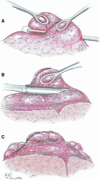
Outcome measures included satisfaction with anesthe-sia scored into 4 grades (from 1⫽ unsatisfactory to 4 ⫽ excellent), hospital stay, calculation of the multidimen-sional BODE index, which integrates measures of body mass index (BMI), degree of airflow obstruction indicated by FEV1, modified Medical Research Council dyspnea index, and exercise capacity assessed by the SMWT; actuarial survival, and freedom from contralateral LVRS. Fig 1. Main operative steps of awake nonresectional lung volume reduction surgery. (A) The most emphysematous lung region targeted for plication is depressed by a cotton swab while the lung edges are grasped by two ring forceps. (B) Afterwards, both lung edges are grasped by a single ring forceps, and a “no knife” endoscopic stapler is fired at the base of the plicated area. (C) The same maneuver is repeated three times along a single ideal line to create a linear but interrupted suture line crossing the lung apex from the ventral seg-ment of the upper lobe back to the dorsal one.
Table 1. Variables and Point Values Assigned for the Computation of the Multidimensional Body Mass Index, Airflow Obstruction, Dyspnea, and Exercise Capacity Index [17]
Variable
Points on BODE Index
0 1 2 3
FEV1(% predicted) ⱖ65 50–64 36–49 ⱕ35
SMWT distance (m) ⱖ350 250–349 150–249 ⱕ149
MMRC dyspnea scale 0–1 2 3 4
Body mass index ⬎21 ⱕ21
BODE⫽ body mass index, airflow obstruction, dyspnea, and exercise
capacity; FEV1⫽ forced expiratory volume in 1 second; MMRC⫽
modified Medical Research Council; SMWT⫽ 6-minute walking test.
GENERAL
The BODE index was calculated according to the criteria proposed by Celli and colleagues [17], which entails assignment of a score from 0 to 3 for dyspnea index, FEV1and SMWT, and values of 0 and 1 for BMI. The points for each variable were totaled so that the BODE index ranged from 0 to 10 points, with higher scores indicating a more severe disability (Table 1).
Also calculated were procedure-related costs, includ-ing devices, routine laboratory assessment, dressinclud-ing ma-terials, medications, surgical instrumentation operative time-related costs, and hospital stay. All outcome mea-sures, except the anesthesia satisfaction score, were com-pared with those of an equivalent control group consist-ing of the last 42 patients undergoconsist-ing unilateral resectional LVRS under general anesthesia.
Anesthesia and Surgical Technique
Anesthesia and surgical technique have been already described in detail [16]. Briefly, thoracic epidural anes-thesia was initiated to achieve somatosensory and motor block at the T1 to T8 level while preserving diaphrag-matic respiration. The thoracic epidural catheter was inserted at the T4 level. In the operating room, patients received a continuous infusion of 0.5% ropivacaine and 1.66-g/mL sufentanil into the epidural space. During the procedure, patients breathed oxygen through a Ven-turi face mask to keep oxygen saturation above 90%. During wound closure, the anesthetic regimen was changed to 0.16% ropivacaine and 1-g/mL sufentanil at 2 to 5 mL/h.
In patients undergoing resectional LVRS with general anesthesia, a thoracic epidural catheter was inserted between T5 and T8 and a continuous infusion of ropiva-caine was initiated. General anesthesia was induced with intravenous propofol (1.5 to 2 mg/kg), fentanyl (0.1 mg),
and vecuronium (0.1 mg/Kg) and maintained using a continuous infusion of propofol, fentanyl, and vecuro-nium. A left-sided double-lumen tube was routinely used. In all patients, the epidural catheter was removed 48 hours after the procedure.
All operations were done with VATS. The patient was placed in full lateral decubitus position. A four-trocars access was used for a 30° 10-mm camera and instrumentation. Whenever severe lung hyperinflation persisted despite induction of the surgical pneumotho-rax, thus jeopardizing adequate visualization, an endo-paddle lung retractor was used to increase the operat-ing space. Carbon dioxide insufflation was not used. The most emphysematous target areas were visualized and depressed with a cotton swab while the redundant lung edges were gently grasped by two ring forceps. Both lung edges were then grasped together with a single ring forceps, and a 45-mm, “no knife” endosta-pler (Endopath 45NK, Ethicon Endosurgery, Pomezia, Italy) was applied on the plicated lung region, starting at the apex of the upper lung lobe and continuing, applying two other cartridges in the ventral and dorsal side of the targeted area to perform a linear, inter-rupted suture line (Fig 1).
In the control group, staple resection of target areas was performed in the standard manner, excising a re-versed U-shaped single strip of emphysematous lung tissue to reduce the upper lobe by about 50%. No suture line buttress was used in either group.
Whenever conversion to general anesthesia was deemed necessary, this was done after insertion of a chest tube and one-layer closure of the thoracic incisions, placement of the patient in supine position, intravenous induction of anesthesia with propofol, and double-lumen tube intubation for single-lung ventilation.
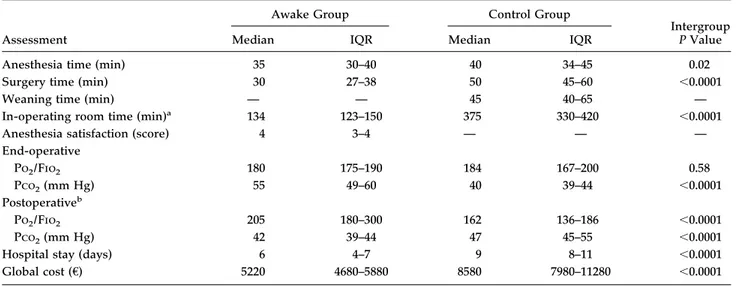
Table 2. Perioperative Data
Assessment
Awake Group Control Group
Intergroup P Value
Median IQR Median IQR
Anesthesia time (min) 35 30–40 40 34–45 0.02
Surgery time (min) 30 27–38 50 45–60 ⬍0.0001
Weaning time (min) — — 45 40–65 —
In-operating room time (min)a 134 123–150 375 330–420 ⬍0.0001
Anesthesia satisfaction (score) 4 3–4 — — —
End-operative Po2/Fio2 180 175–190 184 167–200 0.58 Pco2(mm Hg) 55 49–60 40 39–44 ⬍0.0001 Postoperativeb Po2/Fio2 205 180–300 162 136–186 ⬍0.0001 Pco2(mm Hg) 42 39–44 47 45–55 ⬍0.0001
Hospital stay (days) 6 4–7 9 8–11 ⬍0.0001
Global cost (€) 5220 4680–5880 8580 7980–11280 ⬍0.0001
aIncludes anesthesia time, surgery time, weaning time, and time spent in the recovery room. bPostoperative assessment was performed 24 hours
after the operation.
Fio2⫽ fraction of inspired oxygen volume; IRQ⫽ interquartile range; Pco2⫽ carbon dioxide tension; Po2⫽ oxygen tension.
Statistical Analysis
Group descriptive statistics are presented as median with interquartile range. Owing to the small sample size, nonparametric Wilcoxon rank sum and Mann-Whitney tests were used for paired and unpaired data, respec-tively. Frequencies were compared with the Fisher exact test. Survival and risk of contralateral treatment were assessed by the Kaplan-Meier method, and intergroup differences were assessed by the log-rank test. The out-come variables were analyzed on an intention-to-treat
basis. All reported p values are two-sided. No interim analysis was done during the course of the study.
Results
There was no difference between the awake and control group in median age (64.5 years versus 66 years, p⫽ 0.63) and sex ratio (female/male, 3:39 versus 4:38, p⬎ 0.99).
Conversion to general anesthesia was necessary in 2 patients in the awake group because of panic attack in 1 and hypercapnia exceeding 80 mm Hg in another. No patients in either group required conversion to thoracotomy.
Partial failure of epidural analgesia occurred in 3 patients, who required an additional perioperative local injection of anesthetic. During the operation, intermittent in-expiratory lung motion was easily controlled by the endopaddle, and in no instances did the maintained diaphragmatic motion jeopardize surgical maneuvers.
Perioperative results are detailed in Table 2, which shows that anesthesia time, operative time and global in-operating room time were shorter in the awake group. Oxygenation remained satisfactory throughout the pro-cedure in both groups, although end-operative partial pressure of carbon dioxide (Pco2) was significantly higher in the awake group. However, postoperative par-tial pressure of oxygen/fraction of inspired oxygen and Pco2measured 24 hours after surgery were better in the awake group.
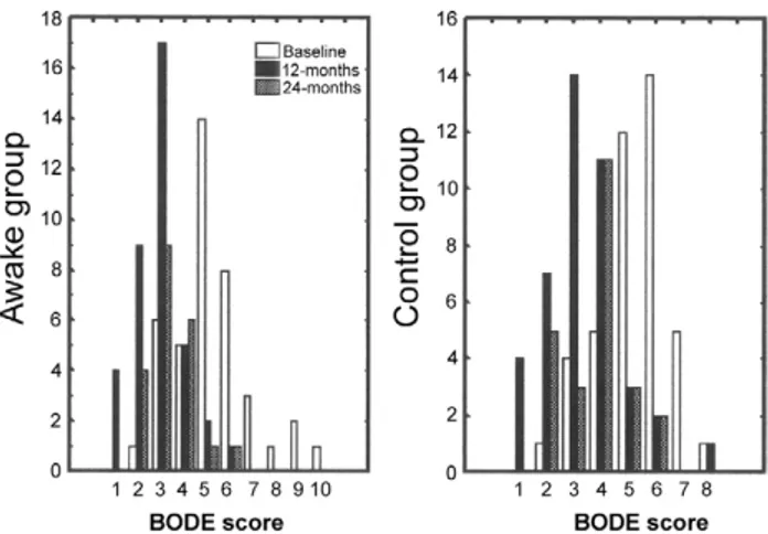
Satisfaction with the epidural anesthesia was scored as excellent by 31 patients, good by 9, and satisfactory by 2. All patients in the awake group were allowed to drink, Fig 2. Distribution of preoperative and postoperative BODE index
(body mass index, airflow obstruction, dyspnea, and exercise capac-ity) in awake and control patients. (Baseline, clear bars; 12 months, black bars; 24 months, patterned bars.)
Table 3. Clinical Results
Indicator
Baseline, Median (IQR) 12 Months, Median (IQR) 24 Months, Median (IQR) Awake Group Control Group Awake Group Control Group Awake Group Control Group BODE score 5.0 (4.0–6.0) 5.0 (5.0–6.0) 3.0a(2.0–3.0) 3.0a(2.0–4.0) 3.0b(3.0–4.0) 4.0b(3.0–4.0)
Body mass index 23 (22–24) 23 (21–25) 23b(22–25) 23 (22–25) 24.6c(23–27) 25b(22–26)
FEV1(L) 0.84 (0.7–1.0) 0.82 (0.69–0.98) 1.17a(1.0–1.37) 1.12a(0.96–1.3) 1.05a(0.93–1.24) 0.96a(0.85–1.10) FEV1(%) 31 (26–37) 27 (24–35) 40a(38–51) 39.5a(32–46) 37a(32–45) 34a(29–39) FVC (L) 2.5 (2.0–2.9) 2.62 (2.10–2.88) 2.9a(2.6–3.25) 3.0a(2.35–3.23) 2.8b(2.4–3.2) 2.96a(2.33–3.23) FVC (%) 66 (59–77) 71.3 (55.4–79.5) 83a(70–90) 79a(62–89) 78c(62–86) 77a(62–85) RV (L) 5.28 (4.8–5.7) 4.86 (4.5–5.5) 4.1a(3.5–4.7) 4.0a(3.5–4.4) 4.4b(3.8–5.1) 4.13b(3.55–4.4) RV (%) 228 (195–256) 218 (183–243) 189a(144–209) 174a(152–198) 200b(169–211) 169b(175–204) TLC (L) 7.7 (7.1–9.0) 8.0 (7.46–8.73) 6.9b(6.5–8.1) 7.36b(6.9–8.1) 7.1c(7.0–7.2) 7.54c(6.91–8.21) TLC (%) 135 (124–145) 129 (115–138) 118b(103–126) 117b(103–130) 109c(103–129) 119c(113–125) SMWT (m) 385 (350–420) 375 (346–410) 480a(435–520) 460a(420–490) 460a(410–500) 450a(420–470)
MITT (Bruce class) 0.50 (0.50–1.0) 0.50 (0.50–1.0) 1.50a(1.5–2.5) 1.50a(1.0–2.0) 1.5b(1.5–2.0) 1.5b(1.0–2.5)
Pao2(mmHg) 71.5 (64–76) 68 (66–70) 75a(70–80) 70a(68–73) 72 (68–75) 69 (66–71)
Paco2(mm Hg) 39 (38–42) 40 (39–42) 38 (36–40) 39 (38–41) 38 (38–41) 40 (39–41)
Dyspnea index (score) 3 (3–4) 3.0 (3.0–3.0) 2a(2–2) 2a(1.0–2.0) 2a(2–2) 2a(2.0–2.0)
PF (SF-36 score) 25 (15–40) 25 (10–35) 50a(50–70) 50a(45–65) 50b(45–67.5) 50b(45–55)
Preoperative to postoperative changes:ap⬍ 0.0001; bp⬍ 0.0008; cp⬍ 0.05.
BODE⫽ body mass index, airflow obstruction, dyspnea, and exercise capacity; IQR⫽ interquartile range; FEV1⫽ forced expiratory volume in
1 second; FVC⫽ forced vital capacity; IRQ⫽ interquartile range; MITT⫽ maximal incremental treadmill test; Paco2⫽ arterial carbon
dioxide tension; Pao2⫽ arterial oxygen tension; PF (SF-36 score) physical functioning domain score of the Medical Outcomes Study Short-Form
36; RV⫽ residual volume; SMWT⫽ 6-minute walk test; TLC⫽ total lung capacity.
GENERAL
eat, and walk within a few hours after the operation, and in most instances, physiotherapy was started the same day of the procedure.
There was no difference in awake versus control group median visual analogue scale scores at 24 hours (2 versus 2, p⫽ 0.23) nor in their need of additional analgesia at the fourth postoperative day (4 versus 5 patients). Temporary catheterization was required for urinary retention in 2 patients in the awake group and 3 in the control group. Ninety-day mortality was nil in both groups. The most relevant nonfatal complications were prolonged air leaks (⬎ 7 days), which occurred in 9 patients (21%) in the
awake group versus 19 patients (45%) in the control group (p ⫽ 0.03). Other less frequent complications included transient arrhythmias and pneumonia, which occurred in 4 patients and 1 patient, respectively, in the awake group and in 5 and 3 patients, respectively, in the control group. Finally, hospital stay was shorter in the awake group than in the control group, and overall medical costs were lower (Table 2).
No patients were lost to follow-up, which was 41 months in the awake group and 43 months in the control group. At 12 months, significant preoperative to postop-erative improvements occurred in the BODE index (Fig 2) and other outcome measures, including FEV1, FVC, and RV, which remained significantly improved for up to 2 years (Table 3). BMI increased due to a change in median body weight, which increased from 65 to 67 kg in the awake group (p ⫽ 0.0005) and from 68 to 69 kg in the control group (p ⫽ 0.0002). Intergroup comparison showed that FEV1and RV improved more in the awake group (p⬍ 0.04 and p ⬍ 0.03, respectively) whereas FVC improved more in the control group (p⫽ 0.006).
During follow-up, 10 patients (24%) in the awake group and 14 patients (29%) in the control group underwent completion of the bilateral treatment. Reasons for need of contralateral treatment were FEV1 decline to baseline value in 15 patients (7 in the awake group; 8 in the control group) and reappearance of incapacitating dyspnea in 9 patients (4 patients in the awake group; 5 patients in the control group).
Overall, 6 patients in the awake group died of respira-tory failure (n⫽ 5) or a cancer-related cause (n ⫽ 1), and 9 patients in the control group died of respiratory failure (n⫽ 6), cardiac disease (n ⫽ 2), or cancer-related causes (n ⫽ 1). The study groups did not differ in actuarial survival and freedom from contralateral treatment (Fig 3).
Comment
Nonresectional LVRS in awake patients is the culmina-tion of 10 years’ experience with comprehensive emphy-sema treatment at the Tor Vergata University and con-stitutes one further step within our adventure of minimizing both surgical and anesthesiologic trauma without jeopardizing the benefit achievable by resec-tional LVRS.
The main finding of this study is that nonresectional LVRS in awake patients resulted in a significant improve-ment in the multidimensional BODE index, which lasted for more than 2 years. Moreover, clinical results, freedom from contralateral treatment, and survival were highly satisfactory and comparable with those achieved in the control group, although hospitalization and overall costs were significantly reduced in the awake group.
A recent analysis of the National Emphysema Treat-ment Trial (NETT) results [20] has shown that air leak occurs in 90% of patients undergoing LVRS, is often prolonged, and is associated with a more complicated and protracted hospital course. Yet in the same study, surgical approach, buttressing, stapler brand, and intra-operative adjunctive procedures were not associated Fig 3. Kaplan-Meier curves show predicted freedom from (A)
con-tralateral lung volume reduction surgery (log-rank test p⫽ 0.71)
and (B) survival (log-rank test p⫽ 0.52) in control (dotted line) and
awake patients (solid line).
with fewer or less prolonged air leaks. We believe that the smooth postoperative course observed after LVRS in awake patients reflected a more rapid recovery, with prompt resumption of main activities of daily life and a relatively low rate of prolonged air leaks, which com-pared favorably with the control group’s results and other series data[2, 8, 20].
Nonresectional LVRS maintains the basic concepts outlined by Brantigan and colleagues[21]and refined by Cooper and colleagues[22], including a reduction of 20% to 30% of the lung volume, suturing performed along a single ideal line, and use of stapling devices. Yet, it adds some conceptual differences that might have contributed to reduce the incidence and duration of air leaks. First, peripheral suturing minimizes the interruption of sub-segmental bronchi and vessels. Second, a linear but interrupted suture line is likely to be more flexible than a continuous one, theoretically reducing lung surface ten-sion during lung reexpanten-sion. Furthermore, our plication technique might result in a more uniform distribution of the lung expansion forces around the suture line due to the inlay buttress created by the plicated bullous tissue itself.
Celli and colleagues[17]proposed the BODE index as a simple multidimensional grading system that predicted the risk of death among patients with COPD better than each of the individual components. Among BODE com-ponents, FEV1was chosen because it can reliably predict health status, the rate of exacerbations of COPD, the pharmacoeconomic costs, results of LVRS, and the risk of death. The MMRC dyspnea scale and SMWT investigate other aspects of the disease and have been widely used in the assessment of LVRS results. Finally, BMI has been found to be inversely correlated with survival and, in our opinion, constitutes a further interesting outcome vari-able. Indeed, we have already reported that fat-free mass and body weight increased 6 months after LVRS, and this improvement correlated with a reduction in residual volume[23].
Imfeld and coworkers[18], recently reported that the postoperative BODE index improved from 7.2 to 4.0 at 3 months after bilateral resectional LVRS, and the postop-erative BODE score predicted the risk of death better than each single component.
In a multicenter study by Miller and colleagues [5], surgical mortality was 16% at 2 years, with no 30-day mortality and two deaths at 90 days; the increase in FEV1 was 30% or 265 mL, and improvement in SMWT was 78 m. The same authors have also found that LVRS costs an additional Can$28,119 compared with best medical care. Their conclusions were that cost of LVRS is high but in keeping with other treatment modalities currently available. In the NETT trial, cost-effectiveness analysis
[11]showed direct medical costs of LVRS versus medical therapy were significantly higher in the surgical arm ($61,145 versus $15,738, respectively). Conclusions of this study were that LVRS is costly relative to medical ther-apy, although the procedure may be cost-effective if benefits are maintained over time.
Obviously, comparisons between our study and the
aforementioned series are inappropriate because of ma-jor differences between countries in health care organi-zation and costs of materials, hospitaliorgani-zation, and human resources. Nonetheless, our preliminary results seems to suggest that nonresectional LVRS in awake patients might dramatically reduce medical costs of this surgical procedure. This might be due at least in part to reduced in-operating room times, which in our study reflected an almost immediate postoperative transfer of the awake patients to the ward. This contrasted with the weaning time and recovery room time that were always required in general anesthesia patients to achieve a stable clinical condition.
In a recent update of the NETT trial[4], patients with upper-lobe-predominant emphysema and low exercise capacity demonstrated better 5-year survival, improved 3-year exercise capacity, and 5-year relief of dyspnea than medically treated patients. In our series, 38 of 42 patients (90%) had upper-lobe-predominant emphy-sema, and all patients had impaired exercise capacity, although this latter figure is not perfectly matched with NETT data owing to differences in exercise testing. None-theless, we had no 90-day mortality, and the 2-year survival rate was 87%, a figure that is in line with the best results in the literature[1, 2, 18].
Physiopathologic Considerations
In LVRS done under general anesthesia, one-lung venti-lation is deemed necessary. However, several adverse effects can derive from this type of anesthesia, including an increased risk of pneumonia, impaired cardiac perfor-mance, neuromuscular problems, and damage related to mechanical ventilation, which includes barotrauma, vo-lutrauma, and atelectrauma [24]. Furthermore, general anesthesia with instrumentation of the airways can elicit bronchospasm and life-threatening complications. Most of these adverse effects could be avoided by using epidural anesthesia in awake patients; however, the use of thoracic epidural anesthesia in patients with compromised respiratory function raises several theo-retical concerns:
1. The motor blockade induced by epidural anesthe-sia might lead to respiratory failure.
2. The sympathetic blockade related to the epidural anesthesia can lead to an increased bronchial tone and airway hyperreactivity[25].
3. An open pneumothorax could have compressed the dependent lung, eventually resulting in further deterioration of patient’s functional compromise. Our preliminary experience with LVRS in awake pa-tients suggests that most of these concerns were un-founded. In fact, simple administration of oxygen through a Venturi mask prevented hypoxemia, although permissive hypercapnia developed frequently. The pathophysiology of this event is not fully understood, but hypoventilation due to partial collapse of the operated lung and a rebreathing effect seem reasonable hypothe-ses. Nonetheless, the perioperative raise in carbon diox-ide was well tolerated by the patients and resolved more
GENERAL
rapidly than in the control group. We attribute this feature to the better synchronization of rib-cage– abdominal motion, which was immediately evident after awake LVRS. A further effect that could have contributed to keep respiratory function satisfactory throughout the procedure is the maintained diaphragmatic motion, which might have decreased the detrimental effect of the abdominal pressure leading the paralyzed diaphragm to compress the dependent lung during general anesthesia.
Limitations
Our study has some limitations. First, although preoper-ative and postoperpreoper-ative patient data were prospectively stored in a database, the overall analysis has to be considered retrospective because BODE index results have been stored in a prospective manner since April 2004 and retrospectively in patients operated on before that date. Second, follow-up is still too short to draw definitive statements about advantages of nonresectional LVRS in awake patients versus the resectional procedure. Finally, the validity and cost-effectiveness of initial uni-lateral awake LVRS with completion of biuni-lateral treat-ment at the reappearance of disabling symptoms must still be considered investigational and merits further investigation.
Conclusions
We have shown that sustained improvement in multidi-mensional BODE index, a high rate of freedom from contralateral LVRS, and satisfactory survival occurred after nonresectional LVRS in awake patients. These re-sults compared with those achieved in a historical cohort undergoing resectional LVRS under general anesthesia, although the procedure in awake patients proved more cost-effective owing to a shorter hospitalization and lower costs. A randomized study is welcome to corrobo-rate, implement, or contradict our encouraging findings.
The study was supported by a grant from Teleflex Medical s.r.l.
References
1. Gelb AF, McKenna RJ Jr, Brenner M, Epstein JD, Zamel N. Lung function 5 yr after lung volume reduction surgery for emphysema. Am J Respir Crit Care Med 2001;163:1562– 6. 2. Ciccone AM, Meyers BF, Guthrie TJ, et al. Long-term
out-come of bilateral lung volume reduction in 250 consecutive patients with emphysema. J Thorac Cardiovasc Surg 2003; 125:513–25.
3. National Emphysema Treatment Trial Research Group. A randomized trial comparing lung-volume-reduction surgery with medical therapy for severe emphysema. N Engl J Med 2003;248:2059 –73.
4. Naunheim KS, Wood DE, Moshenifar Z, et al. Long-term follow-up of patients receiving lung-volume-reduction sur-gery versus medical therapy for severe emphysema by the National Emphysea Treatment trial Research Group. Ann Thorac Surg 2006;82:431– 43.
5. Miller JD, Malthaner RA, Goldsmith CH, et al; for the Canadian Lung Volume Reduction Surgery Study. A ran-domized clinical trial of lung volume reduction surgery versus best medical care for patients with advanced
emphy-sema: a two-year study from Canada. Ann Thorac Surg 2006;81:314 –21.
6. Keenan RJ, Landreneau RJ, Sciurba FC, et al. Unilateral thoracoscopic surgical approach for diffuse emphysema. J Thorac Cardiovasc Surg 1996;111:308 –16.
7. Naunheim KS, Keller CA, Krucylak PE, Singh A, Ruppel G, Osterloh RN. Unilateral video-assisted thoracic surgical lung reduction. Ann Thorac Surg 1996;61:1092– 8.
8. Mineo TC, Pompeo E, Mineo D, Rogliani P, Leonardis C, Nofroni I. Results of unilateral lung volume reduction sur-gery in patients with distinct heterogeneity of emphysema between-lungs. J Thorac Cardiovasc Surg 2005;129:73–9. 9. National Emphysema Treatment Trial Group. Safety and
efficacy of median sternotomy versus video-assisted thoracic surgery for lung volume reduction surgery. J Thorac Cardio-vasc Surg 2004;127:1350 – 60.
10. Naunheim KS, Wood DE, Krasna MJ, et al. Predictors of operative morbidity and mortality in the National Emphy-sema Treatment Trial. J Thorac Cardiovasc Surg 2006;131: 43–53.
11. National Emphysema Treatment Research Group. Cost ef-fectiveness of lung-volume-reduction surgery for patients with severe emphysema. New Engl J Med 2003;348:2092–102. 12. Toma TP, Hopkinson NS, Hillier J, et al. Bronchoscopic volume reduction with valve implants in patients with severe emphysema. Lancet 2002;361:931–33.
13. Rendina EA, De Giacomo T, Venuta F, et al. Feasibility and safety of the airway bypass procedure for patients with emphysema. J Thorac Cardiovasc Surg 2003;125:1294 –9. 14. Yim APC, Hwong TMT, Lee TWL, et al. Early results of
endoscopic lung volume reduction for emphysema. J Thorac Cardiovasc Surg 2004;127:1564 –73.
15. Wood DE, McKenna RJ, Yusen RD, et al. A multicenter trial of an intrabronchial valve for treatment of severe emphy-sema. J Thorac Cardiovasc Surg 2007;133:65–73.
16. Mineo TC, Pompeo E, Mineo D, Tacconi F, Marino M, Sabato AF. Awake nonresectional lung volume reduction surgery. Ann Surg 2006;243:131– 6.
17. Celli BR, Cote CG, Marin JM, et al. The body-mass index, airflow obstruction, dyspnea, and exercise capacity index in chronic obstructive pulmonary disease. New Engl J Med 2004;350:1005–12.
18. Imfeld S, Bloch KE, Weder W, Russi EW. The BODE index after lung volume reduction surgery corrrelates with sur-vival. Chest 2006;129:873– 8.
19. Pompeo E, Marino M, Nofroni I, Matteucci G, Mineo TC and the Pulmonary Emphysema Research Group. Reduction pneumoplasty versus respiratory rehabilitation: a random-ized trial. Ann Thorac Surg 2000;70:948 –54.
20. DeCamp M, Blackstone EH, Naunheim KS, et al; for the NETT Research Group. Patients and surgical factors influ-encing air leak after lung volume reduction surgery: lessons learned from the National Emphysema Treatment Trial. Ann Thorac Surg 2006;82:197–207.
21. Brantigan OC, Mueller E, Kress MB. A surgical approach to pulmonary emphysema. Am Rev Respir Dis 1959;80:194 – 202.
22. Cooper JD, Trulock EP, Triantafillou AN, et al. Bilateral pneumectomy (volume reduction) for chronic obstructive pulmonary disease. J Thorac Cardiovasc Surg 1995;109: 106 –19.
23. Mineo TC, Ambrogi V, Pompeo E, Bollero P, Mineo D, Nofroni I, for the Pulmonary Emphysema Research Group. Body weight and nutritional changes after reduction pneu-moplasty for severe emphysema: a randomized study. J Tho-rac Cardiovasc Surg 2002;124:660 –7.
24. Whitehead T, Slutsky AS. The pulmonary physician in critical care. 7: Ventilator induced lung injury. Thorax 2002; 57:635– 42.
25. Groeben H. Epidural anesthesia and pulmonary function. J Anesth 2006;20:290 –9.
DISCUSSION
DR G. ALEXANDER PATTERSON(St. Louis, MO): I’d say that the short period of follow-up is a real problem with respect to analyzing a need for intervention on the other side. I gather by intervention you mean LVRS on the contralateral side; is that what you mean?
DR POMPEO:Well, actually, median follow-up was something more than 40 months for each group, then maybe is not too small to analyze the need for contralateral treatment. Anyway, you have seen that there are few patients needing a contralateral treatment within the first 24 months. Maybe there were some-what more than 10 in each group. Many patients do not need any contralateral treatment for many years, indeed.
DR MALCOLM M. DECAMP(Boston, MA): I enjoyed that very much. We have all been searching for surrogates that would predict either favorable or poor outcomes for the management of these sick patients. Just a word of caution about the use of the BODE index. BODE has been validated across a larger spectrum of emphysema than we are talking about for volume reduction surgery. BODE is a predictor for bad outcomes as it declines. It has not been concretely validated in terms of an improving BODE correlating with improved survival.
You had excellent outcome in terms of survival in all your patients. It is a nice goal to come up with one of these composite indices of both adverse outcome and benefit, but we have to recognize that with LVRS, we are taking care of a fairly narrow spectrum of emphysema. When changing BODE in the positive direction, we need a larger study to make sure that the BODE index is really measuring what we think contributes to enhanced survival.
DR POMPEO:Thank you for your comments. I believe the most interesting thing in this issue is that BODE index includes some of the most employed outcome measures employed for lung volume reduction surgery, like the FEV1, the modified Medical
Research Council dyspnea index and the 6-minute walking test. And you have seen that even the body mass index is signifi-cantly modified by lung volume reduction surgery, as we have already shown in a paper published in the Journal of Thoracic and
Cardiovascular Surgery, and mainly due to a net increase in
fat-free mass and body weight after the procedure.
DR MICHAEL S. MULLIGAN(Seattle, WA): It is a little intim-idating for some us to think about having one of these critically ill patients be wide awake while we make holes in their chest and operate on them. And what was the incidence wherein one of your patients would decompensate and then they’d need urgent airway control? And so if you think of that being done proactively, we consider that to be more safe. But did you have any occasion wherein you had to intervene, somebody was retaining too much CO2, his oxygenation was failing, and you
then had to urgently intubate, and isolating that lung was not that practical or easy in that context perhaps?
DR POMPEO:We did have 2 patients who needed the conver-sion to general anesthesia due to panic attack more than respiratory failure. Because as you have seen, oxygenation can be maintained quite easily during an awake procedure, the problem is that the rise in Pco2can become a problem if the
procedure is going to be too long.
GENERAL