Deoxyribonucleic Acid Damage
in Human Lymphocytes After
Percutaneous Transluminal Coronary Angioplasty
Maria Grazia Andreassi, BSC, Nicoletta Botto, BSC, Antonio Rizza, MD, Maria Giovanna Colombo, BSC, Cataldo Palmieri, MD, Sergio Berti, MD, Samantha Manfredi, BSC, Serena Masetti, BSC,
Aldo Clerico, MD, Andrea Biagini, MD Massa, Italy
OBJECTIVES We investigated the presence of oxidative deoxyribonucleic acid (DNA) damage in the peripheral lymphocytes of patients undergoing percutaneous transluminal coronary angio-plasty (PTCA) by using the micronucleus test and comet assay, which are sensitive biomarkers of DNA damage.
BACKGROUND Although it has recognized that ischemia-reperfusion can induce oxidative DNA damage, its occurrence in patients undergoing PTCA has not yet been demonstrated.
METHODS Three groups of patients were enrolled: 30 patients with documented coronary heart disease who underwent elective PTCA (group I); 25 patients who underwent elective coronary angiography for diagnostic purpose (group II); and 27 healthy, age- and gender-matched subjects (group III). For each subject, the frequency of micronucleated binucleated (MNBN) cells, DNA single-strand breaks (SSBs), endonuclease III-sensitive sites, and sites sensitive to formamidopy-rimidine glycosylase (FPG) were analyzed before and after diagnostic procedures.
RESULTS The mean basal values of MNBN cells (p⫽ 0.04), DNA-SSBs (p ⫽ 0.001), endonuclease III-sensitive sites (p⫽ 0.002), and FPG sites (p ⬍ 0.0001) were significantly higher in groups I and II than in group III. A high significant increase of MNBN cell frequency was observed in group I after the PTCA procedure (11.0⫾ 1.3 vs. 19.8 ⫾ 1.6, p ⬍ 0.0001), whereas no significant difference was observed in group II (10.2 ⫾ 1.3 vs. 12.9 ⫾ 1.4, p ⫽ 0.18). A significant positive correlation was observed between the increase in the MNBN cell rate and total inflation time during PTCA (R⫽ 0.549, p ⫽ 0.0017). The levels of DNA-SSBs (11.7 ⫾ 1.4 vs. 26.5 ⫾ 3.0, p ⫽ 0.0003) and FPG sites (13.8 ⫾ 1.8 vs. 22.5 ⫾ 2.4, p ⫽ 0.01) were also higher after PTCA.
CONCLUSIONS Our results provide evidence for oxidative DNA damage after PTCA, likely related to ischemia-reperfusion injury. (J Am Coll Cardiol 2002;40:862– 8) © 2002 by the American College of Cardiology Foundation
Reactive oxygen species, such as superoxide anion, hydrogen peroxide, peroxynitrite, and hydroxyl radical, seem to play a pivotal role in the pathogenesis of ischemia-reperfusion injury (1). Free radicals exert their cytotoxic effects by producing several metabolic dysfunctions, including the peroxidation of polyunsaturated fatty acids in membrane or plasma lipoproteins, direct inhibition of mitochondrial re-spiratory chain enzymes, inactivation of membrane sodium channels, and other protein modifications (2,3). Moreover, it is well known that deoxyribonucleic acid (DNA) damage frequently occurs in cells exposed to oxidative stress (4).
The presence of free radicals has been demonstrated after percutaneous transluminal coronary angioplasty (PTCA) (5– 8), but at the present time, the induction of DNA damage by free radicals in the circulating cells of patients undergoing PTCA has not yet been demonstrated.
Therefore, this study was designed to investigate, by using the micronucleus test and comet assay in the human lymphocytes of patients undergoing PTCA, whether this
procedure, considered to be a model of ischemia-reperfusion injury, may cause genetic damage.
We have to consider that the induction of micronuclei in circulating human lymphocytes is a well-known cytogenetic test widely employed to assess the genetic effects of spon-taneously and mutagenically induced DNA damage (9).
The comet assay is considered a sensitive marker of oxidative stress and DNA damage (10). Indeed, the use of lesion-specific enzymes, such as endonuclease III and for-mamidopyrimidine glycosylase (FPG), which create DNA strand breaks at sites of oxidative damage, is a fast approach for measuring of oxidative DNA damage (11,12).
Our study was performed in a group of patients who underwent elective PTCA for the presence of coronary stenosis. Moreover, to exclude a hypothetical direct effect of X-ray exposure and contrast media on DNA damage, a second group of patients who underwent diagnostic coro-nary angiography for the presence of myocardial ischemia on effort was also included.
METHODS
Patients. Two groups of patients characterized by the
documentation of myocardial ischemia on a stress
echocar-From the Laboratory of Cellular Biology, CNR Institute of Clinical Physiology, G. Pasquinucci Hospital, Massa, Italy. This study was entirely supported by grants from the Italian National Research Council.
Manuscript received March 26, 2001; revised manuscript received May 2, 2002, accepted May 24, 2002.
diogram with dipyridamole, performed according to the standard protocol of use in our institute (13), were consec-utively recruited: 30 patients (25 men and 5 women; mean age 61.3⫾ 2.0 years) undergoing elective PTCA (group I) and 25 patients (19 men and 6 women; mean age 64.4⫾ 2.5 years) undergoing diagnostic coronary angiography (group II). In addition, 27 healthy subjects (19 men and 8 women; mean age 60.5 ⫾ 1.4 years) matched for age and gender with the patients of groups I and II were also included as control subjects (group III).
The severity of coronary artery disease (CAD) was expressed simply by the number of affected vessels (one-, two-, or three-vessel disease). Moreover, the extent and severity of CAD were also evaluated by using the Duke scoring system (14), with a grade scale ranging from 0 to 100 (0⫽ no disease, 100 ⫽ most severe disease).
Patients with episodes of angina within the last 24 h before the procedure and myocardial infarction within the last three months were excluded. All patients in whom the procedure was performed on an urgency or emergency basis were also excluded.
For the procedures, the patients received nonionic con-trast media (Ipromid, Ultravist 370, Schering, Germany).
All patients gave written, informed consent to be
in-cluded in the present study, which was approved by our local ethical committee.
Venous blood samples were obtained the day before and three days after the clinical and interventional procedures. This interval between the two samples was chosen to perform the micronucleus test and comet assay in peripheral lymphocytes after a relatively extended period of time from the procedure.
The demographic and clinical data, angiographic find-ings, and procedural variables of the study patients are reported in Table 1.
Cytokinesis block assay and lymphocyte cultures. This
technique employs cytochalasin B to interrupt the division of each cell before cytokinesis. Therefore, cells that have completed only one nuclear division can be recognized by their binucleated appearance (9).
The micronucleus is a small chromatinic particle visible in interphasic cells close to the main nucleus. It can come from an acentric chromosomal fragment or from a whole chro-mosome not correctly incorporated in one of the two daughter cells at the end of the mitotic division. Thus, micronuclei are a reliable biomarker of exposure to both clastogen and aneuploidogen agents that affect the spindle apparatus.
Two separate cultures from each sample were set up by mixing 0.3 ml of whole blood with 4.7 ml RPMI 1640 medium (ICN Pharmaceuticals Inc., Irvine, California), supplemented with 10% fetal calf serum (ICN), 1.5% phytohemagglutinin (Wellcome, Uxbridge, UK), and anti-biotics (100 IU/ml penicillin and 100 mg/ml streptomycin; Sigma, St. Louis, Missouri). Cultures were maintained for 72 h at 37°C in a humidified atmosphere of 5% carbon dioxide in air, and cells were blocked in cytokinesis by adding 6g/ml cytochalasin B (Sigma) after 44-h
incuba-Abbreviations and Acronyms CAD ⫽ coronary artery disease DNA ⫽ deoxyribonucleic acid
FPG ⫽ formamidopyrimidine glycosylase MNBN⫽ micronucleated binucleated PTCA ⫽ percutaneous transluminal coronary
angioplasty SSB ⫽ single-strand breaks
Table 1. Demographic and Clinical Characteristics Group I (nⴝ 30) Group II (nⴝ 25) Group III (nⴝ 27) p Value Age (yrs) 61.3⫾ 2.0 64.4⫾ 2.5 60.5⫾ 1.4 NS Males 25 19 19 NS Smokers 21 13 10 0.04 Duke score 36.9⫾ 1.7 30.5⫾ 6.1 NS Ejection fraction (%) 53.7⫾ 1.4 49.9⫾ 2.5 NS Risk factors Hypertension 16 15 NS Diabetes 7 5 NS Dyslipidemia 22 15 NS Medications Oral nitrate 21 12 NS Calcium antagonist 16 8 NS Beta-blocker 12 10 NS Aspirin 17 9 NS Procedural variables
Total inflation time (s) 115.6⫾ 12.0 —
Fluoroscopy exposure rate (Gy/cm2) 144.1⫾ 18.1 55.5⫾ 8.7 0.0001
Fluoroscopy exposure time (min) 18.3⫾ 1.9 9.1⫾ 1.2 0.0003
Data are presented as the mean value⫾ SEM or number of patients or control subjects. NS⫽ not significant.
tion. Harvesting of cells, hypotonic treatment, fixation, and slide preparation were performed according to the standard method of use in our laboratory (15). For each sample, 1,000 binucleated cells were scored blindly under an optical microscope (final magnification ⫻400) for analysis of mi-cronuclei, following the criteria for acceptance of micronu-clei (9). We evaluated the micronucleated binucleated (MNBN) cell frequency as the number of micronucleated cells per 1,000 cells.
Comet assay. PRIMARY DNA DAMAGE. The alkaline single-cell gel-electrophoresis assay was performed in 10 patients in group I, 10 patients in group II, and 11 subjects in group III (healthy control subjects). In its alkaline version, the formation of DNA single-strand breaks (SSBs) becomes apparent, and the amount of DNA migration, a “comet-like tail,” indicates the amount of DNA damage in the cell.
The assay was performed according to the method previously described (10 –12). Briefly, isolated leukocytes were suspended in a solution of 0.5% low-melting agarose and pipetted on a microscope slide precoated with 1% normal-melting agarose. Then, the slides were immersed in a lysis solution containing Triton X-100 and 2.5 mol/l NaCl to lyse the cells and allow DNA unfolding. The resulting nucleoids were electrophoresed for 25 min at 25 V (300 mA) in alkaline buffer (1 mmol/l Na2EDTA, 300 mmol/l
NaOH, pH⬎13).
OXIDATIVE DNA DAMAGE. The measurement of oxidative DNA damage was performed by using specific repair enzymes that induce breaks in the DNA sites of oxidative damage. Endonuclease III was used to detect oxidized pyrimidines, and formamidopyrimidine glycosylase (FPG) was used to detect damaged purines, including 8-oxo-guanine (11,12). After lysis, the slides were incubated at 37°C for 45 min with endonuclease III and 30 min with FPG. Then, the slides were washed in a neutralizing buffer (0.4 mol/l Tris-HCl, pH 7.5) and stained with 50 g/ml ethidium bromide and covered with a cover slip.
For each subject, 50 comets were analyzed under a fluorescence microscope. The percentage of DNA in the tail was taken as a measure of DNA break frequency. The net amount of damage represented by endonuclease III-sensitive or FPG-III-sensitive sites was obtained by subtracting the percent DNA in the tail without enzyme (i.e., strand breaks) from the percent DNA in the tail with enzyme incubation.
Comets in each gel were analyzed in a blinded manner, using the Komet 3.0 image program (Kinetic Imaging Ltd., Liverpool, U.K.).
Statistics. The results are expressed as the mean value ⫾
SEM. The data for the three independent groups (I, II, and III) were analyzed by analysis of variance, and significant differences between the pairs of mean values were tested by the Scheffe´ test. The Scheffe´ test was chosen for multiple comparisons because it is very impermeable to violations of the assumptions typically associated with the multiple
com-parisons procedure. Statistical evaluations of differences before and after PTCA were performed by using the paired Student t test. The association between an increase in DNA and the effect of each procedural variable (e.g., total inflation time needed for PTCA, radiation dose, fluoroscopy expo-sure time) was evaluated by simple regression analysis. Multiple regression analysis was also applied to investigate the effect of each variable (e.g., age, gender, smoking status, atherogenic risk factors, total inflation time, radiation dose, fluoroscopy exposure time) in determining the MNBN cell increase. Statistical analyses were performed using the statistical package Statview, version 5.0 (SAS Institute, Abacus Concepts, Inc., Berkeley, California).
RESULTS
Relationship between oxidative DNA damage and CAD.
Typical examples of DNA alterations observed in patients are reported in Figure 1.
When analyzing the DNA damage by the micronucleus test, the number of basal MNBN cells was higher in groups I and II than in group III (11.0⫾ 1.3 and 10.2 ⫾ 1.3 vs. 7.0 ⫾ 0.9, p ⫽ 0.04).
The mean values of DNA-SSBs (p ⫽ 0.001), endonu-clease III-sensitive sites (p ⫽ 0.002), and FPG sites (p ⬍ 0.0001) were significantly higher in groups I and II (Fig. 2). Moreover, the mean of FPG-sensitive sites increased with the number of affected vessels (17.2⫾ 2.9, 13.9 ⫾ 2.4, and 35.8 ⫾ 0.5 for one-, two-, and three-vessel disease, respectively; p ⫽ 0.004). A significant relationship was found between FPG-sensitive sites and the extent of CAD evaluated by means of the Duke scoring system (r⫽ 0.663, p⫽ 0.01).
DNA damage and PTCA. Significant differences were
observed in the radiation dose (144.1⫾ 18.1 vs. 55.5 ⫾ 8.7 Gy/cm2, p⫽ 0.0001) and fluoroscopy exposure time (18.3 ⫾
1.9 vs. 9.1⫾ 1.2 min, p ⫽ 0.0003) between groups I and II. A high significant MNBN cell frequency increase was observed in group I after PTCA (11.0⫾ 1.3 vs. 19.8 ⫾ 1.6, p⬍ 0.0001), whereas no significant difference was observed in group II after coronary angiography (10.2⫾ 1.3 vs. 12.9 ⫾ 1.4, p ⫽ NS) (Fig. 3). A significant positive correlation was observed between the increase in the MNBN cell rate and the total inflation time during PTCA (r⫽ 0.549, p ⫽ 0.0017) (Fig. 4), although no significant relationships were found between the MNBN cell increase and radiation dose or fluoroscopy exposure time. Finally, multiple regression analysis showed that the total inflation time appeared to be the only determining factor in the increase of MNBN cell frequency (p ⫽ 0.02).
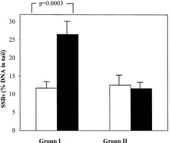
Oxidative DNA damage and PTCA. The mean levels of
DNA strand breaks, measured as the percentage of DNA in the tail, were significantly higher in group I after PTCA (11.7 ⫾ 1.4 vs. 26.5 ⫾ 3.0, p ⫽ 0.0003), although no significant difference was observed in group II after coronary angiography (12.5⫾ 2.8 vs. 11.6 ⫾ 1.5, p ⫽ NS) (Fig. 5).
Moreover, the FPG-sensitive sites were strongly increased after PTCA (13.8 ⫾ 1.8 vs. 22.5 ⫾ 2.4, p ⫽ 0.01), and a trend toward an increase, although not significant, was observed for the levels of endonuclease III (18.7⫾ 3.4 vs. 27.1 ⫾ 3.1, p ⫽ 0.08). No significant differences were observed in the levels of FPG-sensitive sites and endonu-clease III-sensitive sites in group II after coronary angio-graphy.
DISCUSSION
This study shows that patients with CAD, in the basal condition, have a higher level of DNA damage, as compared with normal subjects, according to our previous observation (15). Moreover, to the best of our knowledge, this study provides the first evidence of possible oxidative DNA damage in the peripheral lymphocytes of patients undergo-ing PTCA.
Comparison with other studies. A number of previous
studies have shown an increase in cardiac oxidative stress induced by an injury incurred during primary angioplasty (5– 8). Recently, it has also been demonstrated that lipid peroxidation and F2-isoprostane—stable end products of oxygen free radical–mediated lipid peroxidation—are
signif-icantly increased during the reperfusion after PTCA (8,16). Although DNA represents a potential target for free radical attack, no evidence is yet available demonstrating DNA damage as a result of ischemia-reperfusion injury after PTCA.
Indeed, it has been shown that oxidation is a major cause of DNA damage in humans, and the number of oxidative hits on DNA may be ⬎103/cell per day (17). Reactive oxygen species cause extensive DNA damage, including SSBs, the formation of modified bases, chromosomal dam-age, and mutations in mammalian cells (4,18). Several experimental models of ischemic injury have also shown the influence of reperfusion-associated oxygen radicals on DNA damage. In fact, some studies have shown that ischemia-reperfusion induces DNA damage in animal studies and isolated tissue (19 –21).
Recent studies have reported that the induction of apo-ptosis is an initial cellular event induced by balloon angio-plasty (22,23). Indeed, it is well known that apoptosis is an effective mechanism for eliminating cells with genetic le-sions, thus reducing the potential for genetic instability and cellular dysfunction (24). In addition, the induction of chromosomal and DNA damage has been demonstrated in
human peripheral leukocytes after ischemia-reperfusion in-jury (25,26).
Value and limitations of the study. Our study
demon-strates the presence of DNA damage in the lymphocytes of patients with CAD, who represent an ideal situation of increased production of oxygen free radicals. These results are in agreement with another study reporting higher DNA adduct levels in the cardiac tissue of patients with severe CAD (27), as well as with other recent observations suggesting that alterations at the DNA level may contribute significantly to the development of atherosclerotic disease (28 –30).
Our results show that PTCA induces an increase in oxidative DNA damage in the circulating cells of patients. These effects were seen in human lymphocytes of the systemic circulation. Peripheral lymphocytes are
advanta-geous for exposure analysis because they circulate through-out the body and thus provide an estimate of average whole-body exposure. In fact, the conceptual basis for this approach is the hypothesis that the extent of genetic damage in peripheral blood lymphocytes reflects critical events for mutagenic and carcinogenic processes in target tissues.
Similarly, specific genetic changes may occur in smooth muscle cells of the wall, leading to restenosis, as suggested by an increased mutation rate of microsatellites in both primary and secondary restenotic atherosclerotic plaques (31). It is also possible that the observed DNA damage may increase apoptosis (22,23). However, a recent study sug-gested that restenotic intimal cells may be resistant to apoptosis (32), and increased DNA synthesis has been observed after balloon angioplasty in an atherosclerotic rabbit model (33). This evidence suggests that intimal hyperplasia of smooth muscle cells may be a result of their clonal growth in response to a mutagenic event,
underscor-Figure 2. Deoxyribonucleic acid (DNA) damage, as demonstrated by single strand breaks (SSBs), formamidopyrimidine glycosylase (FPG) sites, and endonuclease III (E III) sites, in the lymphocytes of patients classified into three study groups, represented by the mean percent DNA in the tail for 50 comets from each patient.
Figure 3. Frequency of micronucleated binucleated cells in patients under-going percutaneous transluminal coronary angiography (group I) and coronary angiography (group II). Open bars ⫽ before procedure; solid bars⫽ after procedure.
Figure 4. Relationship between micronucleated binucleated (MNBN) cell increase after percutaneous transluminal coronary angiography and total inflation time. y⫽ 1.123 ⫹ 0.072x; r ⫽ 0.549, p ⫽ 0.002.
ing the similarity between atherosclerotic and carcinogenic processes (28 –30).
The hypothesis that oxygen free radicals may be respon-sible for these alterations is plaurespon-sible (4,34,35); however, some limitations of this study should also be considered. First and foremost, the possibility that X-ray exposure and contrast media could be responsible for increased DNA damage should be considered (36), even if the observed increase in the DNA alteration rate is not significantly related to radiation exposure.
No direct correlation between oxidative DNA damage and oxidative stress, as biochemical markers (e.g., malonal-dehyde), was examined in this study. However, these limitations do not deprecate the main observation of our study, and additional work should be done in this area. Our results provide clear evidence that oxidative DNA damage, particularly the level of 8-oxo-guanine, in the peripheral lymphocytes of patients with CAD is significantly enhanced as compared with that of normal subjects. Further oxidative DNA damage is produced after PTCA, likely related to ischemia-reperfusion injury. Larger studies are indicated to determine whether the increased DNA damage may be an additional prognostic factor in CAD, as well as to investi-gate the association between oxidative DNA damage and restenotic coronary lesions.
Acknowledgments
We acknowledge the help given by the paramedical and technical staff of our hemodynamic laboratory.
Reprint requests and correspondence: Dr. Maria Grazia Andre-assi, CNR Institute of Clinical Physiology, G. Pasquinucci Hos-pital, Via Aurelia Sud-Montepepe 54100, Massa, Italy. E-mail: [email protected].
REFERENCES
1. Zweier JL, Tanerty JT, Weisfeldt ML. Direct measurement of free radical generation following reperfusion of ischemic myocardium. Proc Natl Acad Sci USA 1987;84:1404 –7.
2. Gardner HW. Oxygen radical chemistry of polynsaturated fatty acids. Free Radic Biol Med 1989;7:65–9.
3. Kowaltowski AJ, Vercesi AE. Mitochondrial damage induced by conditions of oxidative stress. Free Radic Biol Med 1999;26:463–71. 4. Halliwell B, Aruoma O. DNA damage by oxygen-derived species: its mechanism and measurement in mammalian systems. FEBS Lett 1991;281:9 –19.
5. Roberts MJ, Young IS, Trouton TG, et al. Transient release of lipid peroxides after coronary artery balloon angioplasty. Lancet 1990;336: 143–5.
6. Coghlan JG, Flitter WD, Holley AAE, et al. Detection of free radicals and cholesterol hydroperoxides in blood taken from the coronary sinus of man during percutaneous transluminal coronary angioplasty. Free Radic Biol Med 1991;14:409 –17.
7. Blann A, Midgley H, Burrows G, et al. Free radicals, antioxidants, and endothelial cell damage after percutaneous transluminal coronary angioplasty. Coron Artery Dis 1993;4:905–10.
8. Buffon A, Santini S, Ramazzotti V. Large, sustained cardiac lipid peroxidation and reduced antioxidant capacity in the coronary circu-lation after brief episodes of myocardial ischemia. J Am Coll Cardiol 2000;35:633–9.
9. Fenech M. The cytokinesis-block micronucleus technique: a detailed description of the method and its application to genotoxicity studies in human populations. Mutat Res 1993;285:35–44.
10. Gedik C, Wood S, Collins A. Measuring oxidative damage to DNA: HPLC and the comet assay compared. Free Radic Res 1998;29:609–15. 11. Collins A, Duthie S, Dobson V. Direct enzymic detection of endog-enous oxidative base damage in human lymphocyte DNA. Carcino-genesis 1993;14:1733–5.
12. Collins A, Dusinska M, Gedik C, Stetina R. Oxidative damage to DNA: do we have a reliable biomarker? Environ Health Perspect 1996;104 Suppl 3:465–9.
13. Picano E, Lattanzi F, Masini M, Distante A, L’Abbate A. High dose dipyridamole echocardiography test in effort angina pectoris. J Am Coll Cardiol 1986;8:848 –54.
14. Smith LR, Harrel FE, Rankin JS, et al. Determinants of early versus late cardiac death in patients undergoing coronary artery bypass graft surgery. Circulation 1991;84 Suppl III:III245–53.
15. Botto N, Rizza A, Colombo MG, et al. Evidence for DNA damage in patients with coronary artery disease. Mutat Res 2001;493:23–30. 16. Iuliano L, Pratico` D, Greco C, et al. Angioplasty increases coronary
sinus F2-isoprostane formation: evidence for in vivo oxidative stress during PTCA. J Am Coll Cardiol 2001;37:76 –80.
17. Ames BN. Endogenous DNA damage as related to cancer and ageing. Free Radic Res Commun 1989;7:121–8.
18. Floyd RA, Schneider JE. Hydroxy free radical damage to DNA. In: Vigo-Pelfrey C, editor. Membrane Lipid Oxidation, Vol. 1. Boca Raton, FL: CRC Press, 1990:69 –90.
19. Li Y, Chopp M, Jiang N, Zhang ZG, Zaloga C. Induction of DNA fragmentation after 10 to 120 minutes of focal cerebral ischemia in rats. Stroke 1995;26:1252–8.
20. Murakami K, Kondo T, Chan PH. Reperfusion following focal cerebral ischemia alters distribution of neuronal cells with DNA fragmentation in mice. Brain Res 1997;751:160 –4.
21. Cordis GA, Maulik G, Bagchi D, Riedel W, Das DK. Detection of oxidative DNA damage to ischemic reperfused rat hearts by 8-hydroxydeoxyguanosine formation. J Mol Cell Cardiol 1998;30: 1939 –44.
22. Perlman H, Maillard L, Krasinski K, Walsh K. Evidence for the rapid onset of apoptosis in medial smooth muscle cells after balloon injury. Circulation 1997;95:981–7.
23. Pollman MJ, Hall JL, Gibbons GH. Determinants of vascular smooth muscle cell apoptosis after balloon angioplasty injury: influence of redox state and cell phenotype. Circ Res 1999;84:113–21.
24. Carson DA, Ribeiro JM. Apoptosis and disease. Lancet 1993;341: 1251–4.
25. Fabiani JN, Farah B, Vuilleminot A, et al. Chromosomal aberrations and neutrophil activation induced by reperfusion in the ischaemic human heart. Eur Heart J 1993;14 Suppl G:12–7.
Figure 5. Deoxyribonucleic acid (DNA)-single strand breaks (SSBs) in patients undergoing percutaneous transluminal coronary angiography (group I) and coronary angiography (group II). Open bars ⫽ before procedure; solid bars⫽ after procedure.
26. Willy C, Dahouk S, Starck C, Kaffenberger W, Gerngrob H, Plappert U. DNA damage in human leukocytes after ischemia/reperfusion injury. Free Radic Biol Med 1998;25:245–51.
27. Van Schooten FJ, Hirvonen A, Maas LM, et al. Putative susceptibility markers of coronary artery disease: association between VDR geno-type, smoking, and aromatic DNA adduct levels in human right atrial tissue. FASEB J 1998;12:1409 –17.
28. De Flora S, Izzotti A, Walsh A, Degan P, Petrilli GL, Lewtas J. Molecular epidemiology of atherosclerosis. FASEB J 1997;11:1021–31. 29. Andreassi MG, Botto N, Colombo MG, Biagini A, Clerico A.
Genetic instability and atherosclerosis: can somatic mutations account for the development of cardiovascular disease? Environ Mol Mutagen 2000;35:265–9.
30. Ross J, Stagliano N, Donovan M, Breitbart R, Ginsburg G. Athero-sclerosis and cancer: common molecular pathways of disease develop-ment and progression. Ann NY Acad Sci 2001;947:271–92.
31. McCaffrey TA, Du B, Consigli S, et al. Genomic instability in the type II TGF-beta1receptor gene in atherosclerotic and restenotic vascular cells. J Clin Invest 1997;100:2182–8.
32. Buariels G, Schluckebier S, Hutter R, et al. Apoptosis in restenosis versus stable angina atherosclerosis: implication for the pathogenesis of restenosis. Arterioscler Thromb Vasc Biol 1998;18:1132–9. 33. Kamenz J, Seibold W, Wohlfrom M, et al. Incidence of intimal
proliferation and apoptosis following balloon angioplasty in an athero-sclerotic rabbit model. Cardiovasc Res 2000;45:766 –76.
34. Bennett MR. Reactive oxygen species and death: oxidative DNA damage in atherosclerosis. Circ Res 2001;88:648 –50.
35. Lee SH, Blair IA. Oxidative DNA damage and cardiovascular disease. Trends Cardiovasc Med 2001;11:148 –55.
36. Norman A, Cochran ST, Sayre JW. Meta-analysis of increases in micronuclei in peripheral blood lymphocytes after angiography or excretory urography. Radiat Res 2001;155:740 –3.