An alternative method for neonatal cerebro-myocardial perfusion
Giovanni Battista Luciani*, Fabrizio De Rita, Giuseppe Faggian and Alessandro Mazzucco
Division of Cardiac Surgery, University of Verona, Verona, Italy* Corresponding author. Division of Cardiac Surgery, University of Verona, OCM. Piazzale Stefani 1, 37126 Verona, Italy. Tel: +39-045-8122485; fax: +39-045-8123308; e-mail: [email protected] (GB Luciani).
Received 29 August 2011; received in revised form 6 November 2011; accepted 14 November 2011
Abstract
Several techniques have already been described for selective cerebral perfusion during repair of aortic arch pathology in children. One method combining cerebral with myocardial perfusion has also been proposed. A novel technique is reported here for selective and independent cerebro-myocardial perfusion for neonatal and infant arch surgery. Technical aspects and potential advantages are discussed.
Keywords:Cerebral protection• Hypoplastic left heart syndrome • Interrupted aortic arch • Myocardial protection • Neonate • Perfusion (cerebral, myocardial)
Aortic arch repair in the neonate is traditionally accomplished during a period of deep hypothermic circulatory arrest. In order to reduce the potential damage of brain ischaemia, selective, regional low-flow perfusion (RLFP) techniques have been intro-duced, suggesting favourable neurological outcome [1,2]. More recently, combined perfusion strategies of cerebral and myocar-dial circulation, using a Y connector on the arterial line, have been proposed in an attempt to reduce cardiac morbidity and mortality of complex arch surgery [3–5]. An alternative and novel strategy for cerebro-myocardial protection in the neonate is presented here, where RLFP is combined with controlled and independent coronary perfusion.
TECHNIQUE
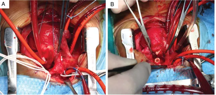
Via midline sternotomy, the ascending aorta, aortic arch, des-cending aorta, neck vessels and ductus are dissected and mobi-lized to allow cannulation and tension-free anastomosis: in ductal-dependent systemic circulation, the branch pulmonary ar-teries are also isolated (Fig. 1A). An arterial cannula 6 Fr (Medtronic, Inc., Minneapolis, MN, USA) for neonates or 8 Fr for infants is directly inserted into the innominate artery or through a Polytetrafluoroethylene (PTFE) graft (for neonates <2.0 kg), pre-viously anastomosed in end-to-side fashion to the innominate artery. A cardioplegia delivery system is inserted into the aortic root: in ductal-dependent descending aortic circulation, the car-dioplegia line is initially inserted into the ductus arteriosus (Fig.1A) and then relocated into the aortic root after ductus ex-cision. A single-stage atrial cannula is used for venous return and a left ventricular vent (Edwards Lifesciences LLC, Irvine, CA, USA) is then introduced through the right upper pulmonary vein. When concomitant repair of intracardiac anomalies is required, bicaval cannulation (Edwards Lifesciences LLC) is used for venous
return. In the latter cases, the aortic root perfusion system is switched to deliver cardioplegia. A schematic of the cardio-pulmonary bypass circuit is presented in Fig.2. Briefly, a blood-based priming solution for bypass is prepared in order to maintain Hb level > 8.5 g/dl. An α-stat acid–base management strategy is used. Under moderate systemic hypothermia (25°C nasopharynx) and low-flow perfusion (25–50 ml/kg/min), left common carotid and left subclavian arteries are snared down to start selective cerebral perfusion. The descending aorta is cross-clamped and a second aortic cross-clamp is placed immediately above the cardioplegia cannula to start selective, controlled and independent coronary perfusion (Fig. 1B and Supplementary Video 1). During selective coronary perfusion, the left heart is vented (non-working, beating heart). Mean blood pressure in the right radial artery is maintained between 40 and 45 mmHg, cerebral bloodflow rate, via the arterial line, regulated at base-line levels of 30–40 ml/kg/min, while myocardial flow rate, via the rotor of the cardioplegia line through the delivery system, at 15–20 ml/kg/min [5]. Aortic arch repair is performed with beating heart and working brain: in cases of severe arch hypo-plasia, extensive and lengthy patch augmentation can be carried out accurately (Supplementary Video 2). During rewarming, the flow rate is progressively increased (upto 150 ml/kg/min). After cardiopulmonary bypass weaning, modified ultrafiltration is rou-tinely applied (Fig.2).
RESULTS
The technique herein has been successfully applied in nine neonates and infants (median age 11 days), with body weights ranging from 1.6 to 10 kg (median 3.2 kg), having aortic arch patch augmentation (five patients: severe arch hypoplasia in three, Norwood stage I in two) and end-to-side anastomosis (four patients: IAA+VSD in three, arch hypoplasia and aortic
© The Author 2012. Published by Oxford University Press on behalf of the European Association for Cardio-Thoracic Surgery. All rights reserved.
CO N G E N IT A L
Interactive CardioVascular and Thoracic Surgery 14 (2012) 645–647
BRIEF COMMUNICATION
stenosis in one). Peak serum troponin I averaged 16 ± 42μmol/l. Four patients, body weight <3.0 kg, needed delayed sternal closure (average 7.4 ± 2 days, range 5–10). On early (day 1) post-operative echocardiographic assessment, all but one patient, with hypoplastic left heart syndrome and severe preoperative tri-cuspid regurgitation, showed preserved left or systemic ventricu-lar function. Average duration of mechanical ventilation was 3.3 ± 5.6 days (range 1–14). One neonate, with IAA+VSD and severe pulmonary hypertension required inhaled nitric oxide adminis-tration for 6 days. Average length of ICU stay was 4.8 ± 4.4 days (range 1–13). All patients survived to discharge, but one with
hypoplastic left heart syndrome weighing 2.0 kg at stage I palli-ation, who succumbed to the sequelae of acute limb ischaemia.
COMMENT
The main goal of the current technique is to achieve a separate and controlled management of myocardial and cerebral flows during RLFP. Unlike the dual arterial Y-connected technique previ-ously reported, where relative perfusion properties of brain and heart cannot be regulated [3–5], a novel set-up of simultaneous
Figure 1.(A) Extracardiac anatomy in newborn with severe arch hypoplasia and coarctation: hypoplastic ascending aorta is also visible. Extensive dissection of ascending and descending aorta, arch vessels and branch pulmonary arteries: snares around the vessels are evident. Innominate artery cannula for systemic (cere-bral) perfusion, right atrial for venous return and right superior pulmonary vein for left heart drainage are in place. Cardioplegia delivery system is positioned in ductus arteriosus. Snares around the arch vessels and descending aorta are visible. Branch pulmonary arteries are snared. (B) The innominate artery cannula is advanced distally and snared proximally. An aortic cross-clamp is placed distal to the aortic root cannula, allowing regional myocardial perfusion. An additional aortic cross-clamp is placed in the mid-descending aorta. During selective, independent cerebro-myocardial perfusion, the distal arch has been opened and the ductal tissue resected from the descending thoracic aorta.
Figure 2.A schematic view of cardiopulmonary bypass circuit for cerebro-myocardial perfusion.
G.B. Lucianiet al. / Interactive CardioVascular and Thoracic Surgery 646
cerebro-myocardial perfusion using two different sources of arter-ialflow is described here: the cerebral, dependent on the arterial line, and the myocardial, dependent on the cardioplegia delivery system. This method allows independent and controlled regulation of each flow by varying the speed of the individual pump. Advantages of this alternative technique for cerebro-myocardial perfusion include: (i) ability to adjust coronary perfusion in case of signs of ischaemia evident on continuous EKG monitoring; (ii) prompt conversion to cardiac arrest, by switching the perfusate from blood to blood cardioplegia via the same delivery line. This property is particularly advantageous in low body weight (less than 2.0 kg) newborn with hypoplastic ascending aorta (HLHS, IAA), where multiple cannulation of the diminutive arterial vessel may be traumatic, causing adventitial haematoma or even dissec-tion; (iii) facile interface with modified ultrafiltration.
FUNDING
Supported by Institutional Funds from Cariverona Project.
SUPPLEMENTARY MATERIAL
Supplementary material is available atICVTS online.
Conflict of interest: none declared.
REFERENCES
[1] Pigula FA, Nemoto EM, Griffith BP, Siewers RD. Regional low-flow perfu-sion provides cerebral circulatory support during neonatal aortic arch reconstruction. J Thorac Cardiovasc Surg 2000;119:331–9.
[2] Visconti KJ, Rimmer D, Gauvreau K, del Nido P, Mayer JE Jr, Hagino I et al. Regional low-flow perfusion versus circulatory arrest in neonates: one-year neurodevelopmental outcome. Ann Thorac Surg 2006;82: 2207–13.
[3] Kostelka M, Walther T, Geerdts I, Rastan A, Jacobs S, Dähnert Iet al. Primary repair for aortic arch obstruction associated with ventricular septal defect. Ann Thorac Surg 2004;78:2233–9.
[4] Lim HG, Kim WH, Park CS, Chung ES, Lee CH, Lee JRet al. Usefulness of regional cerebral perfusion combined with coronary perfusion during one-stage total repair of aortic arch anomaly. Ann Thorac Surg 2010;90: 50–7.
[5] Oppido G, Pace Napoleone C, Turci S, Davies B, Frascaroli G, Martin-Suarez Set al. Moderately hypothermic cardiopulmonary bypass and low-flow antegrade selective cerebral perfusion for neonatal aortic arch surgery. Ann Thorac Surg 2006;82:2233–9.
eComment. Using selective myocardial perfusion for interrupted aortic arch surgery
Author:Leo A. Bockeria
Bakoulev Scientific Center for Cardiovascular Surgery, Moscow, Russia doi:10.1093/icvts/ivs120
© The Author 2012. Published by Oxford University Press on behalf of the European Association for Cardio-Thoracic Surgery. All rights reserved.
I have read the article by Luciani et al. [1] on an alternative method for neonatal cerebro-myocardial perfusion with great interest. Despite the small number of patients, it is an impressive and stimulating paper, but there are a few points to be considered. The main novelty of this method is the use of the selective pump for independent myocardial perfusion. At the same time, as I understood, in Figure 2, there is an inaccuracy in the direction of bloodflow in cerebral perfusion line.
In our opinion, the presented method is convenient and justified only for blood to blood cardioplegia. In all other cases, namely when using crystalloid solutions such as“Custodiol”, this only complicates the perfusion circuit and has no visible benefits. In our experience with interrupted aortic arch surgery for neonatals and infants, the traditional method with selective cerebral perfusion and circulatory arrest has been applied. At the same time, complications associated with trauma, dissection, or haematoma of vessels (as described in this article) were not observed.
References
[1] Luciani GB, De Rita F, Faggian G, Mazzucco A. An alternative method for neonatal cerebro-myocardial perfusion. Interact CardioVasc Thorac Surg 2012;14:644–6.
Supplementary Video1: Heart is slowly beating and non-working: during
cerebro-myocardial perfusion, the hypoplastic distal aortic arch is opened and ductal tissue is carefully resected.
Supplementary Video2:During cerebro-myocardial perfusion, on a beating
heart, extensive pericardial homograft patch augmentation of the diffusely hypoplastic arch is carefully carried out.
CO N G E N IT A L