!
SCUOLA NORMALE SUPERIORE
Pisa
!
!
CLASSE DI SCIENZE MATEMATICHE, FISICHE E NATURALI
CORSO DI PERFEZIONAMENTO IN NEUROBIOLOGIA
Triennio 2008-2010
!
!
Tesi di perfezionamento
!
“Fluoxetine treatment promotes functional recovery in
a rat model of cervical spinal cord injury”
!
!
!
Candidata:
Relatori:
Manuela Scali
Prof. Lamberto Maffei
Dott. Alessandro Sale
Index
!
Preface
6
Introduction
8
Chapter 1: The spinal cord
8
Spinal cord anatomy 8
Spinal cord injury 13
General pathophysiology 13
Experimental models of SCI 18
Contusion and compression models 18 Transection models 20 Ischemia models 20
Chapter 2: Plasticity
22
Plasticity in the spinal cord 24
Chapter 3: Experimental therapies for spinal cord injury
29
Cell transplantation therapies 30
Peripheral nerve grafts 30
Schwann cells 30
Olfactory ensheathing glia 31
Bone marrow stromal cells 32
Neural progenitors 33
Autologous macrophages 34
Gene therapy 35
Neuroprotective treatments 38
Channel blockers 43
Modulators of myelin associated inhibitors 46
Targeting the Glial Scar 50
Modulation of intrinsic regenerative response 52
Modulation of serotoninergic tone 53
Rehabilitative training 56
Chapter 4: Aim of the thesis
59
Materials and Methods
62
Animal treatment 62
Spinal cord injury 62
Behavioral assessment 64
Montoya Staircase reaching task 64
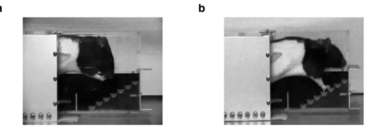
Horizontal ladder 64
Footprint analysis of gait 65
Corticospinal tracing and histological assessment 66
Immunohistochemistry 67
Western blot 67
Enzyme Linked ImmunoSorbent Assay (ELISA) 69
Neurotransmitter quantification 69
Synaptosome purification 69
Release experiments 70
Results
72
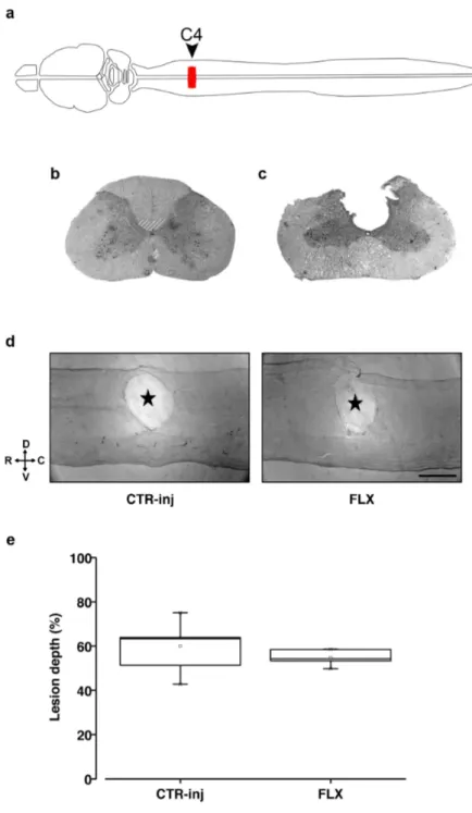
Assessment of the lesion after SCI 72
Fluoxetine promotes motor recovery in skilled-task after SCI 74
Montoya staircase task 74
Horizontal ladder 77
Fluoxetine induces a faster recovery of gait coordination in lesioned rats 79
Fluoxetine promotes sprouting in the injured spinal cord 81
Corticospinal tract (CST) plasticity 81
Serotoninergic fibers 83
Excitation/inhibition balance is modulated by fluoxetine 85
Fluoxetine effect on neurotrophin expression in the intact spinal cord 88
Discussion
90
Fluoxetine enhances motor recovery after spinal cord injury 90
Fluoxetine induces plastic rearrangements in the spinal cord 92
Anatomical plasticity 92
Involvement of the excitatory/inhibitory balance 94
Neurotrophins 96
Conclusions and future directions 97
Appendix: A rich environmental experience reactivates visual cortex
plasti-city in aged rats.
99
!
!
!
!
!
!
!
Preface
!
This Thesis consists of two parts: in the first part, I studied the efficacy of fluoxetine on the recovery from spinal cord injury in rats; in the second part, I evaluated the effects of envi-ronmental enrichment on visual cortex plasticity in the aging rat. Both of the studies resulted in peer-reviewed publication (Scali et al., 2012; 2013).
!
When I started my PhD program, spinal cord injury was a completely new field in my lab. Under my Supervisor, Prof. Lamberto Maffei's, suggestion I moved to Cambridge (UK) to learn the necessary skills to bring the technique of the lesion model in Pisa laboratory, and after a necessary training I was able to start the experiments.
I chose to focus the main part of my dissertation on spinal cord injury because I devoted to this project most of my time. The study related to visual cortex plasticity in the aging rat is illustrated in the Appendix.
!
!
!
!
!
!
!
!
!
!
“When you examine a man with a dislocation of a vertebra of his neck, and you find him un-able to move his arms, and his legs. His penis is erect; urine drips from his penis unknowing-ly. Then you have to say: A disease one cannot treat.”
Introduction
Chapter 1: The spinal cord
Spinal cord anatomy
The spinal cord is the part of the central nervous system that controls limb and trunk move-ments and which receives sensory information from these areas; moreover, the spinal cord has an important role in viscera and blood vessel function of thorax, abdomen and pelvis. It looks like a dorsoventrally flattened cylinder coated by the meninges (dura mater, arachnoid and pia mater) and it lies inside the vertebral canal, where it extends from the base of the skull to the first lumbar vertebra. When observed in a transverse section, the cord appears in the shape of a butterfly constituted by central grey matter (which contains nerve cell bodies) surrounded by external white matter (formed by the axons of ascending and descending pathways and glial cells). The central grey locally organizes motor behavior and regulates sensory signals: the dorsal horns of the butterfly contain the cell bodies of ascending sensory fibers, while the ventral horns contain the motoneurons (Barson and Sands, 1977). The central region, which connects the dorsal and ventral horns, is called intermediate grey matter and surrounds the central canal (which is remnant of the embryological ventricular system and is continuous with the fourth brain ventricle; Figure 1).
The human spinal cord can be divided in 31 segments along its rostral-caudal axis, grouped by anatomists in five sections: cervical (C1-C8), thoracic (T1-T12), sacral (S1-S5), lumbar (L1-L5) and coccygeal (Co1). In other species the number of segments is different, as in the rat, which has 34 segments (C1-C8; T1-T13; S1-S6; L1-L4; Co1-Co3). Each segment is associated with a pair of sensory (arising from the dorsal horns) and motor (arising from the ventral horns) nerves that relay signals between periphery and the central nervous system; the nerve rootlets arising from the cord are bundled together so that one pair of spinal nerves
emerges from each segment. Because of the higher necessity for neural computation in limb regions, the grey matter is larger in cervical and lumbar areas, where the brachial and lum-bosacral plexuses arise.
The central grey of was divided by Bror Rexed in ten layers, based on their cytoar-chitercture (Rexed, 1952; 1954). The layers, called laminae, are organized in a dorsal-ventral fashion, except for lamina X, which is the neuroglial tissue that surrounds the central canal (Figure 1).
Figure 1. Transverse section of human spinal cord. Grey matter is indicated in grey color. Numbers indicate
lam-inae. Adapted from Watson et al., 2009.
!
The dorsal horn is formed by laminae I to VI: laminae I-IV are the recipient of sensory input associated with cutaneous stimuli; lamina V is the thickest layer of the dorsal horn and receives afferents from viscera, skin and muscles, while lamina VI is primarily concerned with proprioceptive sensations that signal limb position and movement and is found only in the segments corresponding to cervical and lumbar enlargements. Lamina VII occupies the intermediate grey matter and the dorsal part of the ventral horn; it is constituted by interneu-rons that connect to motoneuronal pools and is involved in the control of posture and move-ment. In lamina VIII, propriospinal interneurons modulate motor activity, connecting mo-toneurons on the same and opposite side of the spinal cord. Lamina IX lies within the ventral
and innervate muscle fibers. Lastly, lamina X, which surrounds the central canal, receives no-ciceptive information form skin and viscera (Grau et al., 2006; Watson et al., 2009).
The white matter is formed by longitudinally running fibers and glia. Groups of fibers can be classified based on the anatomical position and function: small bundles of axons locat-ed in a given area are definlocat-ed fasciculi, while a group of fasciculi sharing common features is called funiculus; when a group of nerve fibers have the same origin, course, termination and function, the group is named tract. A group of tracts with a related function constitutes a pathway.
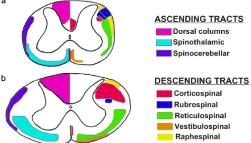
Grey matter horns divide the white matter in three distinct regions: dorsal, lateral and ventral columns (Figure 1). Dorsal columns are located between dorsal horns and are separat-ed by the dorsal mseparat-edian sulcus; they are principally constitutseparat-ed by the central processes of dorsal root ganglion cells, which form the direct pathway conveying skin sensation and pro-prioception from the limbs and trunk to the brain. The direct pathway is constituted by the cuneate and gracile fasciculi, that ascend ipsilaterally and synapse respectively in the cuneate and gracile nuclei in the medulla oblongata; here, the fibers originating from these nuclei cross the midline and constitute the medial lemniscus pathway, which terminates in the ven-troposterolateral nucleus of the thalamus. The gracile fasciculus contains the afferent fibers arising from lower trunk and extremities below the T6 spinal cord segment. In C1-T6 spinal cord segments, the gracile fasciculus is located medially with respect to the cuneate fascicu-lus, which constitutes the afferent pathway ascending from the upper trunk and extremities. These pathways are involved in transmitting sensations of discriminatory touch, deep pres-sure, proprioception, sense of position of joints, stereognosis and vibration. In rodents and many non-primate mammals, the ventral-most part of the dorsal column contains the dorsal corticospinal tract (CST), a bundle of fibers that originates primarily from the layer V of pri-mary motor, premotor and somatosensory cortical areas; the axons of pyramidal neurons then
pass through the internal capsule, cerebral peduncle, longitudinal fibers of the pons, and medullary pyramid, to reach the caudal end of the brain stem, where most of them cross to the opposite side in the pyramidal decussation and then lie in the ventral part of the dorsal column of the spinal cord. In most mammals, the majority of CST fibers terminate on the neurons of the medial parts of the base of the dorsal horn and the intermediate grey matter (Rexed’s lam-inae III-VI), which then send connections to lamina IX motoneurons. Differently from ro-dents, in primates and carnivores the major CST bundle is found in the dorsal part of the lat-eral column (see Figure 2); moreover, some of its fibers make direct synaptic connections with motoneurons in lamina IX (up to 20% in humans): this is correlated with the develop-ment of ‘skilled’ motor capacities, such as an increased dexterity of distal musculature (Heffner and Masterton, 1983; Watson et al., 2009); it is important to underline that in ro-dents, CST axons mediate fine movements of the limbs, whereas in humans the corticospinal tract is much more involved in almost all aspects of voluntary motor control (Blesch and Tuszynski, 2009).
The lateral and ventral columns are formed by a variety of ascending and descending fiber groups. The principal ascending tract is the spinothalamic tract, which transmits noci-ceptive, thermal, non-discriminative touch and pressure sensations to the somatosensory areas of the thalamus; another important ascending tract is the spinocerebellar one, which conveys informations from Golgi tendons and muscle spindles to the cerebellum, for the control of movement coordination. The descending tracts, besides CST tract (in primates), include: the rubrospinal tract, that arises form the red nucleus and is involved in the control of skilled movements (Whishaw et al., 1998); the reticulospinal tract, which originates in the reticular formation of the romboencephalon and is involved in postural control and modulation of some sensory and autonomic functions (Tracey, 2004); the vestibulospinal tract, that
repre-peiano, 1972); the raphespinal tract, that descends from raphe in medulla oblongata and has neuromodulatory influences on motor, autonomic, reproductive and excretory function and is also involved in modulating perception to pain (Tracey, 2004).
As well as these long ascending and descending projections, in the white columns there are many fibers, called propriospinal, that connect one spinal cord segment with another. These fibers lie very close to the grey matter and participate in a variety of physiological and behavioral processes, such as modulation of afferent and descending inputs to the central pat-tern generators (CPG) for locomotion and respiration, as well as autonomic functions like vis-ceroreception and pain perception. The largest propriospinal pathways connect the brachial and lumbosacral enlargements to coordinate limb movements (Watson et al., 2009).
Figure 2. Approximate location of some ascending and descending tracts in rat and human spinal cord. (a) rat;
(b) human. The diagrams are not drawn to scale. Dotted lines indicate areas where tracts appear to overlap. Left side: sensory; right side: motor. Adapted from Watson et al., 2009.
Spinal cord injury
General pathophysiology
Spinal cord injury (SCI) is the most common cause of invalidity in young adults. This condi-tion affects 2.5 million people in the world, with an incidence of 130000 new cases every year (Thuret et al., 2006). The high majority of injuries have a traumatic origin (motor vehicle crashes, falls, violence and sport accidents) and affect principally young adults in the 15-29 years old age group. Non-traumatic injuries, as those caused by cancer, ischemia and multiple sclerosis, seem to be more related with old age. Moreover, the prevalence is 3.8 folds higher in males than females (Wyndaele and Wyndaele, 2006; van den Berg et al., 2010). Since quite half of injured patients is represented by young men that become unable to continue their job career and start to need assistance and special medical cares, social and economic costs of SCI are enormous.
Spinal cord injuries can be classified depending on the severity of the lesion (complete or incomplete), the segmental level (cervical, thoracic, lumbar or sacral) and the kind of insult that mechanically induced the injury (flexion, extension, rotation, compression or a combina-tion of these forces; Schwab and Bartholdi, 1996). From the anatomical point of view, com-plete lesions are quite rare; anyway, when the damage to neural tissue is huge, a comcom-plete loss of function related to the interested area can be observed. The severity of the injury can be addressed analyzing the neurological outcome, and various scales can be adopted; for in-stance, the ASIA (American Spinal Injury Association) Impairment Scale, which is graded from A (complete sensorimotor injury) to E (normal), is widely accepted (Fawcett et al., 2006). Concerning the segmental level, cervical injuries are generally the most common. In very severe cases, cervical SCI causes tetraplegia, a condition that involves loss of function of upper and lower extremities, trunk and pelvis; however, only one third of patients experience
this state, while paraplegia, that afflicts principally the lower part of the body, is more fre-quent (Wyndaele and Wyndaele, 2006).
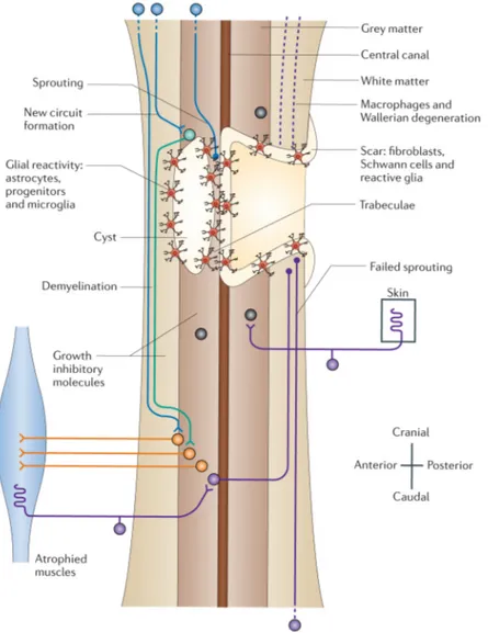
In spite of such a variety of causes and typologies, SCI pathophysiology seems to fol-low the same paradigm after the first insult (primary damage). In general, this process can be divided in an acute phase characterized by intertwined cellular death, hemorrhage and begin-ning of inflammatory response, and a chronic phase, in which the consequences of secondary damage started during the acute phase finally establish the formation of a glial scar at the in-jury site and, sometimes, of a cystic cavity full of liquid (Figure 3).
Figure 3. Diagram representing the spinal cord at the chronic stage of injury (adapted from Thuret et al., 2006).
The time at which a lesion reaches the “chronic stage” varies between species. Clinical data suggest that, for humans, an injury can be considered chronic 12 months after the first insult (Fawcett et al., 2006). The mechanisms of secondary injury have been deeply dissected in an-imal models of SCI (see below); nevertheless, recent studies indicate that similar mechanisms occur in humans as well (see Hagg and Oudega, 2006 for a review; Table 1).
!
Table 1. Similarities and differences between rodent and human response after SCI (adapted from Hagg and
Oudega, 2006).
!
After initial trauma, cells near the site of injury immediately die. Disruption of blood flow due to death of endothelial cells brings to hemorrhage and intravascular thrombosis, which in combination with loss of autoregulation (Senter and Venes, 1979) and vasospasm of intact vessels (Attar et al., 2001), causes the anoxia and hypoperfusion of spinal tissue surrounding lesion and the appearance of edema (Tator and Fehlings, 1991). Vascular damage is one of the principal causes of secondary injury (Mautes et al., 2000): the high metabolic demand of grey matter cannot be satisfied because of incipient ischemia, moreover loss of autoregulation of microvascular tone leads to higher susceptibility of neurons to changes in systemic arterial
Secondary response Rodent Human
Vascular response Hemorrage, angiogenesis Hemorrage, angiogenesis
Inflammation Extensive Much less pronounced, despite similar
cytokine expression
Demyelination Yes Yes, but perhaps less pronounced
Axonal degeneration Some die-back and Wallerian degeneration Wallerian degeneration much more protrac-ted
Glial scar Extensive, with astroglial CSPG Not extensive, CSPFs mostly in blood ves-sels
Cyst formation Rat yes; mouse no Yes
Schwann cell response Some invasion Extensive Schwannosis CSPG: chondroitin sulphate proteoglycans
worsen by formation of free radicals (such as reactive oxygen species and peroxynitrite), compounds that induce peroxidation of lipid components of cellular membrane producing the disaggregation of membrane itself and the formation of new reactive species that continue the cascade (Cayli et al., 2004); besides, free radicals can damage mitochondrial respiratory chain enzymes, inducing the apoptotic death of the cell (Cuzzocrea et al., 2001; Tanaka et al., 2005). The breakdown of blood-spinal cord barrier allows the entrance of inflammatory cells in the site of injury (Mautes et al., 2000; Whetstone et al., 2003; Habgood et al., 2007). The inflammatory response is a complex mechanism: it is initiated by the recruitment of neu-trophils starting few hours after the SCI by chemoattractant molecules (chemokines, cy-tokines, adhesion molecules) that are increased by lysis of necrotic cells (Campbell et al., 2002; Kwon et al., 2004; Fleming et al., 2006; Donnelly and Popovich, 2008); neutrophils are then followed by macrophages derived from resident microglia (Popovich et al., 2002) and blood-born macrophages (Carlson et al., 1998). Infiltration of inflammatory cells allows the phagocytosis of tissue debris; moreover, these cells start to release pro-inflammatory cy-tokines (such as tumor necrosis factor alpha (TNFα) and interleukins 1 and 6), cytotoxic mol-ecules (such as reactive oxygen species, nitric oxide) and proteolytic enzymes (such as metal-loproteinases) that supply inflammatory responses and induce secondary damage (Schnell et al., 1999; Xu et al., 2001; Noble et al., 2002; Donnelly and Popovich, 2008).
An other important process related with the secondary damage of spinal cord is excito-toxicity due to change in ionic conductance and release of neurotransmitters. Cell lysis, de-myelination due to apoptosis of oligodendrocytes and depolarization of neuronal membrane leads to increased intracellular concentration of sodium and calcium and extracellular concen-tration of potassium (Liu et al., 2011). This creates a loop in which increased intracellular cal-cium concentration allows the exocytosis of glutamate from synaptic vesicles, that in turn ac-tivates NMDA (N-methyl-D-aspartate) or AMPA
(alpha-amino-3-hydroxy-5-methyl-4-isoxa-zole- propionic acid)/kainate receptors. The hyperactivation of these ionotropic receptors re-sults in increased intracellular sodium levels in neurons and astrocytes, that can cause the death of the cell due to water influx and lysis through depolarization (Matyja et al., 2005). Moreover, calcium influx trough NMDA and AMPA receptors provokes axonal injury and eventual apoptosis or necrosis via an increase in the activation of cellular enzymes, mitochon-drial damage, acidosis, and production of free radicals (Schanne et al., 1979; Mody and Mac-Donald, 1995; Das et al., 2005).
As mentioned above, oligodendrocytes principally die by apoptosis after injury (Emery et al., 1998); oligodendrocyte death can be caused by microglial activation (Li et al., 2005), axonal degeneration (that causes the loss of trophic support to the oligodendrocytes; Barres et al., 1993) or interaction between NGF, which is increased by lesion, and pro-apop-totic receptor p75, which is expressed by oligodendrocytes (Casha et al., 2001). This process provokes demyelination of spared axons, with a consequent impairment of signal conductance and general physiology of the neuron, that eventually loses its function and degenerates (Waxman, 1989). Besides, a great loss of ascending and descending fibers is observed in the white matter one month after SCI (Bao and Liu, 2003; Wingrave et al., 2003; Araki et al., 2004; Iwata et al., 2004), with denervation and retraction of proximal axons.
The pathophysiology of the acute phase is completed by reactive gliosis. In response to cytokines and other molecules produced by inflammatory cells and degenerating axons, astrocytes start to overexpress the glial fibrillary acidic protein (GFAP) and to proliferate (Giulian and Lachman, 1985; Nishino et al., 1993; Chiang et al., 1994; Kahn et al., 1995; Hama et al., 1997; Rostworowski et al., 1997). These cells become hypertrophic and their processes start to overlap together with infiltrating fibroblasts, Schwann cells, macrophages and glial progenitors such as NG2 positive cells (Jones et al., 2002; Guest et al., 2005; Goritz
tempt to preserve the fragile neural tissue from further damage (Fitch et al., 1999; Myer et al., 2006). Unfortunately, the glial scar represents also a barrier for axonal regeneration, a condi-tion that is reinforced by the produccondi-tion of transmembrane and secreted inhibitory molecules such as proteoglycans that form the extracellular matrix (ECM) (Jones et al., 2003), ephrin-B2 (Bundesen et al., 2003), semaphorin 3A (Pasterkamp et al., 2001), and tenascin (Apostolova et al., 2006) by the same astrocytes.
!
Experimental models of SCI
In order to study the mechanisms leading to secondary damage and to create tools to investi-gate the efficacy of possible therapies, researchers have developed several animal models mimicking different aspects of human SCI since Galen (2nd century A.D., Siegel, 1973). In-jury models can be distinguished on the basis of the specimen, the level of inIn-jury and the kind of injury itself. Rodents and feline are the most used because of economic and practical ad-vantages, although non-human primate models are emerging as the best choice for their simi-larities with humans (see Courtine et al., 2007 for a review).
!
Contusion and compression models
The models that best resemble the characteristics of human disease are those based on contu-sion or comprescontu-sion of spinal cord tissue, as the majority of patient lecontu-sions derive from blunt trauma and displacements of vertebral bone. These methods induce a mild to complete neuro-logical lesion with the appearance of scar surrounding a fluid-filled cyst, then they are partic-ularly suitable to study neuroprotective agents, tissue replacement and injury pathophysiology in general.
The first experimental contusion injury model was proposed by Allen in 1911: a weight-drop insult was applied on the exposed dorsal spinal cord of a dog, causing the
contu-sion and the displacement of neural tissue in the vertebral canal (Allen, 1911). This approach was then adapted to the rat (Gruner, 1992) and led to the development of the NYU/MASCIS (New York University Multicenter Animal Spinal Cord Injury Study) Impactor, a device that allows to induce a reproducible contusive damage to the rat spinal cord in controlled condi-tions (spinal level, drop weight, impact strength; Basso et al., 1996). Other tools created to induce contusion injuries in rodents are the OSU (Ohio State University) Impactor (Bresnahan et al., 1987; Stokes, 1992), which employs an electromechanical impact probe controlled by computer feedback, and the IH (Infinite Horizon) Impactor (Scheff et al., 2003), a device that applies a force-defined impact to the cord with a stainless steel-tipped impounder. All these instruments share similar advantages (reliability of performance and results, possibility to set and check all the parameters during and after the experiment, low maintenance costs), as well as similar limitations: while the force that induces lesion in humans is usually applied on the ventral surface of the cord or in a centripetal way (e.g. by torsion of the cord), the impactors described above act on the dorsal, surgically exposed surface of the neural tissue.
An alternative strategy to induce a lesion that might be more similar to human blunt SCI is represented by the clip compression model: after exposure by dorsal laminectomy, the spinal cord is compressed along the dorso-ventral axis between the arms of a modified an-eurysmal clip (Rivlin and Tator, 1978; Joshi and Fehlings, 2002a; 2002b). The force and dura-tion of compression are directly related with the funcdura-tional outcome, so the severity of the le-sion can be easily modulated; on the other hand, the principal disadvantage of this technique is represented by the difficulty of surgical manipulation of the cord. Compression injury can also be elicited by means of an inflated microscopic balloon inserted in the epidural space for a period that can vary from minutes (leading to a reversible functional lesion, Nesathurai et al., 2006) to hours (leading to irreversible damage because of secondary consequences
de-rived form initial compression, Lim et al., 2007). This method allows to operate on different species, from rodents to primate, and kindly resembles human injury.
!
Transection models
Transection models are based on the surgical cut of the spinal cord and can be divided in complete and partial transection models.
In complete transections, rostral and caudal segments of the spinal cord are fully dis-sociated; these models are used to mimic human complete injury, though in patients a plete anatomical lesion is very rare also in case of total functional loss. Nevertheless, a com-plete transection allows to evaluate the efficiency of treatments aimed at inducing axonal re-generation without the risk of misleading results due to the contribution of spared connections (Alilain et al., 2012; Lu et al., 2012). A complication related to complete transection models is the high post-operative care request to maintain critical physiological functions (as feeding and bowel/bladder function) and to control complications as infections, pain and dehydration of the animal.
Partial transections models are often criticized as they are less likely to represent hu-man lesions than contusion models. On the other hand, incomplete transection is still a widely used model because it represent a valuable tool to study the contribution of particular tracts to motor and sensory function (e.g. CSTs, Bradbury et al., 2002; García-Alías et al., 2009) and to assess the effectiveness of pro-regenerative treatments; moreover, in case of dorsal or lateral hemisection, it allows to compare the injured circuit response with that of the healthy, unin-jured counterpart (Muir and Whishaw, 2000; Anderson et al., 2007).
!
Ischemia models
Organotypic spinal cord slices and specific cellular line cultures can be maintained under hypoxic conditions, enabling researchers to analyze the molecular cascades that lead to neuronal cell death (An et al., 2011; Lian Jin et al., 2011). Compared to in vivo models, the advantage in the use of slice and cell cultures is that they are far less expensive and do not require postoperative cares, while a limit is represented by the possibility to dissect just an aspect a time of a complex pathology as spinal cord injury.
Hypoxia and ischemic damage can be induced also in vivo by means of vascular com-pression; the most characterized model is based on the insertion of a Fogarty balloon in the rat thoracic descending aorta; blood flow is blocked at the level of the subclavian artery by bal-loon inflation, then the spinal cord is reperfused. In rats, this kind of insult induces paraplegia accompanied by rigidity and spasticity (Lu et al., 2004a; Cizkova et al., 2007).
!
!
Chapter 2: Plasticity
Neural plasticity can be defined as the ability of the nervous system to functionally and struc-turally reorganize itself in response to experience: “The whole plasticity of the brain sums it-self up in two words when we call it an organ in which currents pouring in from the sense-or-gans make with extreme facility paths which do not easily disappear” (James, 1890).
In 1960s and 1970s, Hubel and Wiesel examined the plasticity of cat and monkey vis-ual systems and identified a ‘critical period’ during which deprivation of normal visvis-ual expe-rience through the closure of an eyelid by surgical suture (monocular deprivation, MD) irre-versibly altered neuronal connections and functions in the visual cortex (Wiesel and Hubel, 1963; Hubel and Wiesel, 1970; Hubel et al., 1977). Since then, the existence of critical peri-ods in early postnatal life during which neural circuits display a heightened plasticity in re-sponse to external stimuli has been established for various brain regions subserving major be-havioral functions, as for instance the organization of auditory maps for sound localization in barn owls (Knudsen and Knudsen, 1990), birdsong in zebrafinches (Brainard and Doupe, 2002) and language learning in humans (Newport et al., 2001. See Berardi et al., 2000 and Hensch, 2004 for a comprehensive review). Concerning the motor system, an example of crit-ical period is represented by the sensitivity to tail suspension, a manipulation that allows hindlimb unloading: in young rats (postnatal day 8-13), tail suspension leads to permanent motor impairment in the spinal cord, while it is innocuous in older animals (Walton et al., 1992).
During critical periods, neural circuits reach maturity: starting from an initial pool of less precise connections, experience models neural circuits stabilizing some connections and removing others (Holtmaat and Svoboda, 2009; Fu and Zuo, 2011). Because of this “en-hanced-plasticity status”, the recovery from injury is much more pronounced in the early
postnatal phase (Nicholls and Saunders, 1996; Payne and Lomber, 2001; Chen et al., 2002; Dumas et al., 2002; Mladinic et al., 2009; Choksi et al., 2010). When the development is completed, plasticity wanes in favor of the formation of stable, reliable networks with precise circuitries and connections. In the adult brain, even if decreased, synaptic plasticity continues through modifications of synaptic strength, as well as through formation and elimination of synapses (Holtmaat and Svoboda, 2009; Chen and Nedivi, 2010).
In the past years, a big effort was made to investigate the mechanism underlying the enhanced plasticity of immature circuits and the possibility to manipulate the capability of neural networks to respond to experience and insults after the closure of critical periods. Sev-eral factors have been identified using the visual system as a paradigmatic model (see Berardi et al. 2003; Tropea et al., 2009; Levelt and Hubener, 2012). Among them, the excitatory/in-hibitory balance emerges as a critical regulator for neural plasticity. Inexcitatory/in-hibitory circuits sculpt the pattern and timing of neuronal electrical activity; for instance, the increase of GABAergic transmission in the visual cortex defines the opening and the closure of the critical period for ocular dominance plasticity (Hensch, 2005): when GABAergic input is reduced during devel-opment, as in mice knockout for GAD65 (the synaptic biosynthetic enzyme of GABA), the visual cortex does not respond to experience modification such as MD (Hensch et al., 1998). A similar reduction of MD sensitivity can be achieved increasing the efficacy of glutamatergic transmission during the critical period (Fagiolini et al., 2003). In physiological conditions, GABAergic transmission matures later than glutamatergic circuits (Jiang et al., 2005). If GABAergic tone is precociously increased during development through infusion of benzodi-azepines, overexpression of BDNF, exposure to environmental enrichment (EE) or removal of PSA-NCAM (polysialic acid presented by the neural cell adhesion molecule), the onset and closure of the critical period are accelerated (Hanover et al., 1999; Huang et al., 1999; Iwai et
al., 2003; Cancedda et al., 2004; Di Cristo et al., 2007. See Sale et al., 2010 for a comprehen-sive review).
In the adult visual system, inhibitory circuitry stabilizes synaptic connections, thus re-ducing neural plasticity: pharmacological reduction of GABAergic transmission reactivates OD plasticity in response to MD in adult rats (Harauzov et al., 2010), and several lines of evi-dence demonstrate that inhibitory tone is reduced by manipulations that increase visual corti-cal plasticity, such as EE (Sale et al., 2007; Baroncelli et al., 2010), dark rearing (He et al., 2006) and fluoxetine treatment (Maya Vetencourt et al., 2008). Furthermore, GABA has been suggested to be crucial in masking existing horizontal connections and consequently in the rapid reorganization of cortical maps after, for instance, loss of afferents due to peripheral nerve transection or limb amputation (Jacobs and Donoghue, 1991; Chen et al., 1998; Wu and Kaas, 1999).
!
Plasticity in the spinal cord
As in other areas of the CNS, neuronal plasticity occurs in the spinal cord in response to expe-rience through a variety of mechanisms. During development spinal cord plasticity is in-volved in standard behaviors like locomotion and rapid withdrawal from pain (Waldenstrom et al., 2003), while in the adulthood plasticity contributes to the acquisition and maintenance of new motor skills (see Wolpaw, 2007 for an extensive review). Moreover, spinal cord plas-ticity has a role in the mechanisms that lead to functional recovery (adaptive plasplas-ticity) or secondary adverse consequences (maladaptive plasticity) after an injury (Goldberger, 1977; Sanes and Donoghue, 2000; Blesch and Tuszynski, 2002; Raineteau et al., 2002; Edgerton et al., 2004). It has been estimated that if as little as 10–15% of the descending spinal tracts are spared, some locomotor function can recover (Basso, 2000; Metz et al., 2000). Based mostly on the results of studies using animal models, reorganization of the CNS, including synaptic
plasticity, axonal sprouting, and cellular proliferation, has long been known to spontaneously occur following spinal cord lesions. This reorganization occurs in the spinal cord circuitry and in supraspinal structures.
Early after SCI, in a time window varying between species, a certain degree of sponta-neous recovery can occur (see Onifer et al., 2011 for a review). In this phase, the contribution of plasticity to functional return and recovery is difficult to discriminate from other possible processes including the recovery from spinal shock, a condition probably due to the break-down of membrane potentials, excessive neurotransmitters levels, and the loss of neuromodu-lators regulating the excitability in the spinal cord (as the 5HT mediated system) that leads to the loss of muscle tone and segmental spinal reflexes (Smith and Jeffery, 2005). The onset for return of spontaneous function in both humans and animals after SCI could be due to the restoration of motoneuron excitability by constitutive expression of 5HT2C receptors (Fouad et al., 2010; Murray et al., 2010), adaptations in polysynaptic flexor reflexes involved in lo-comotor circuits (Lavrov et al., 2006; Dietz et al., 2009) and synaptic rearrangements (Rossignol, 2006).
As would also be expected, SCIs (both complete and incomplete) produce consider-able reorganization in the spinal cord circuitry, with sprouting of collaterals from damaged fibers (regenerative sprouting) and sprouting of lesion-spared descending axons (compensato-ry sprouting, Tuszynski and Steward, 2012). Several experiments showed the existence of re-generative and compensatory sprouting by corticospinal tract axons (CSTs); for example, after cervical SCI in rats, spontaneous sprouting from the ventral CSTs occurred onto medial mo-toneuron pools in the cervical spinal cord, leading to spontaneous recovery (Weidner et al., 2001). Furthermore, Bareyre et al. reported that after incomplete spinal cord injury in rats, transected hindlimb CSTs sprouted into the cervical gray matter to contact short and long
pro-were maintained, whereas contacts with short PSNs that did not bridge the lesion pro-were lost. In turn, long PSNs arborize on lumbar motor neurons, creating a new intraspinal circuit relaying cortical input to its original spinal targets (Bareyre et al., 2004). Similarly, in rhesus monkeys a C7 spinal cord hemisection led to marked spontaneous plasticity of CSTs, with reconstitu-tion of 60% of pre-lesion axon density arising from sprouting of spinal cord midline-crossing axons, and this extensive anatomical recovery was associated with improvement in coordinat-ed muscle recruitment, hand function and locomotion (Rosenzweig et al., 2010). Other de-scending pathways, such as the neuromodulatory efferents, undergo anatomical plasticity after SCI. For example, compensatory plasticity of the rubrospinal tract was found to mediate the recovery of forepaw function following CST lesion in cats (Alstermark et al., 1987) and mon-keys (Belhaj-Saif and Cheney, 2000). Similarly, rat cervical SCI led to increased sprouting of reticulospinal fibers rostral to the injury (Weishaupt et al., 2013), and several experiments showed that serotoninergic neurons sprout after spinal cord trauma (Sharma et al., 1990; In-man and Steward, 2003; CaIn-mand et al., 2004); intriguingly, in a cortical model of lesion, sero-toninergic neurons displayed lack of axonal dieback and enhanced sprouting within the in-hibitory environment of the glial scar, probably thanks to significantly higher amounts of growth-associated protein-43 and/or β1 integrin than cortical neurons (Hawthorne et al., 2011).
Some recovery of motor control after cord hemisection in cats has been attributed to collateral sprouting of primary afferent axons (Goldberger et al., 1993; Helgren and Gold-berger, 1993), and early studies demonstrating enlarged excitatory postsynaptic potentials in chronic spinal cats attributed part of the increase to sprouting of primary afferent fibers (Nel-son and Mendell, 1979).
In response to SCI, synaptic plasticity as well as anatomical reorganization can also occur at cortical and subcortical regions. Several studies have reported that cortical territories
controlling intact body parts tend to enlarge and invade cortical regions that have lost their peripheral target (McKinley et al., 1987; Jain et al., 1997; Fouad et al., 2001; Ghosh et al., 2010). The underlying mechanisms are hypothesized to be similar to those mediating reorga-nization after cortical injury, including disinhibition of latent cortical connections and axonal sprouting at multiple levels of the neuraxis (reviewed in Raineteau and Schwab, 2001; Fouad and Tse, 2008; Kaas et al., 2008). Another mechanism may be injury-induced structural plas-ticity in the dendritic spines of cortical motoneurons, as suggested by the observation that in rodents, after cervical SCI, dendritic spine density and morphology in neurons of the motor cortex is modified over 3 days to 2 weeks (Kim et al., 2006). TMS studies in human SCI pa-tients revealed motor reorganization, as muscles immediately rostral to the lesion could be activated through bigger regions of the cortex (Levy et al., 1990; Topka et al., 1991). On the other hand, PET, EEG and fMRI studies showed that appropriate (e.g. foot, leg) motor areas can be activated by imagined movements, even in long-term paraplegic or tetraplegic patients (Cohen et al., 1991; Corbetta et al., 2002; Curt et al., 2002; see Endo et al., 2009 for a review).
Besides underlying spontaneous recovery, neuroplasticity can also lead to detrimental consequences (Brown and Weaver, 2012). Increase in neuronal excitability due to products of microglial activation and changes in sodium channel and glutamate receptor expression (Deumens et al., 2008), collateral sprouting of calcitonin gene-related peptide (CGRP)-con-taining primary afferent fibers (Christensen and Hulsebosch, 1997a; 1997b) and aberrant growth of descending serotoninergic axons in the spinal cord dorsal horn (Inman and Steward, 2003) have been indicated as the main causes of neuropathic pain, a condition that leads to abnormal sensations (dysesthesia) or to the insurgence of nociception after a non-painful stimulus (allodynia). In addition, SCI is often associated with autonomic dysreflexia, an
ab-pounding headache and slow heart rate (see Weaver et al., 2006 and Mathias, 2006 for re-view): the entering of sensory input in the spinal cord below the level of the lesion leads to exaggerated sympathetic (autonomic) responses that can be associated with an increased CGRP-containing primary afferent arbor in the dorsal horn (Krenz and Weaver, 1998; Krenz et al., 1999), or in the presence of a normal arbor, with loss of descending inhibitory influ-ences on spinal sympathetic reflexes (Gris et al., 2005). Moreover, modification in neuronal excitability after SCI can lead to the insurgence of spasticity, an involuntarily elevated muscu-lar activity that can be triggered by a variety of sensory inputs, such as cutaneous stimulation (Norton et al., 2008). Notably, up-regulation of 5HT2 receptors and lowered expression of the potassium-chloride co-transporter KCC2 (which keeps Cl- intraneuronal concentration low) in
motoneuron membranes have been shown to be involved in the generation of this phe-nomenon (Lee et al., 2007; Boulenguez et al., 2010).
!
!
Chapter 3: Experimental therapies for spinal cord injury
In general, the spinal cord tries to repair itself after the initial insult: spontaneous recovery can be observed in animal models (Muir and Whishaw, 1999; Weidner et al., 2001; Fenrich and Rose, 2009; Rosenzweig et al., 2010) as well as in patients (Fawcett et al., 2006; Scivoletto et al., 2007). After injury, immediately early genes are overexpressed, recapitulating a situation typical of the developing nervous system (Maier and Schwab, 2006); signs of reorganization of neural circuits, for instance the appearance of regenerative or compensatory sprouting, have also been observed (Ramon y Cajal, 1928; Bradbury and McMahon, 2006). Some stud-ies indicate that macrophages infiltrating the lesion site in the acute phase could contribute to neuroprotection (removal of cellular debris, release of protective cytokines) and to the reorga-nization of damaged tissue in particular temporal contexts and in species/strain specific man-ner, besides their well characterized pro-degenerative role (Schwartz, 2003; Donnelly and Popovich, 2008).
Acquired knowledge on processes that are occurring in acute, sub-acute and late phase of injury allowed researchers to develop and test various kinds of experimental therapies in the attempt to achieve axonal regeneration, remyelination of damaged axons and, in general, recovery of function. Some of these approaches have been demonstrated to be efficient in an-imals models and reached the clinical trial phase.
Cell transplantation therapies
The most evident anatomical consequence of spinal cord injury is the interruption of connec-tions between the proximal and the distal segment of the spinal cord itself due to axon section and cell death; then, cell transplantation represent a promising strategy to replace damaged tissue, bridge the lesion cavity, counteract axonal demyelination and, moreover, can be used as a tool to create a neuroprotective and pro-regenerative environment for severed axons.
!
Peripheral nerve grafts
The first experiment of regeneration induced by cellular transplantation was carried out in 1911 by Francisco Tello, who transplanted portions of peripheral nerves in rabbit cerebral cor-tex and found out that cortical axons could enter in and elongate on the peripheral tissue (Tel-lo, 1911). His pioneering work gave the basis for experiments with autologous peripheral nerve graft in injured spinal cord, which demonstrated that this treatment can induce axonal regrowth into the graft and functional improvement in rats with a complete spinal cord tran-section, alone (Richardson et al., 1980; Cheng et al., 1996) or in combination with other ex-perimental strategies (Lee et al., 2002; Levi et al., 2002; Tom and Houlé, 2008), with one case of partial functional recovery in a patient with a chronic thoracic injury (Cheng et al., 2004).
Today, cellular transplants are preferred to nerve bridges: single cells can be purified and expanded in vitro to reach the high amount of material required to fill the cystic cavity and, most importantly, cells can be manipulated to produce and release factors at the lesion site (Ramer et al., 2005).
!
Schwann cells
Several studies demonstrated that the part of the peripheral nerve that actually works on axon-al regeneration is represented by Schwann cells (SCs): this cells, indeed, create a more
per-missive environment after peripheral nerve injury by increasing the production of pro-prolif-erative factors (as neurotrophins and cell adhesion molecules) and decreasing the expression of inhibitory myelin proteins (see Oudega and Xu, 2006 for a review); in addition, SCs can recreate the myelin sheath around severed and intact central axons (Gilmore, 1971). Schwann cells have been successfully implanted in the rat spinal cord and regrowth of axons into the bridge graft was observed; a recovery of function has also been reported in some studies (Xu et al., 1997; Xu et al., 1999; Bunge, 2002; Takami et al., 2002). Unfortunately, regenerating axons were not able to cross the graft tissue and to reinnervate the host, and SCs seem to be ineffective on corticospinal axon regeneration (Fehlings and Vawda, 2011). For these reasons, combinatorial strategies with treatments aimed to reduce environment inhibition (as glial scar digestion, Fouad et al., 2005) and to increase pro-regenerative factors (as neurotrophins and cell adhesion molecules, Xu et al., 1995; Lavdas et al., 2010) have been evaluated. Schwann cells were tested in a study on patients with a chronic mid-thoracic lesion (Saberi et al., 2008); the study demonstrated that SCs could be safe for humans, as no negative consequences of the implant were recorded, but the beneficial outcome of this treatment was not clear (only one patient had a better performance after the therapy, but it was not possible to assess if it was caused by transplanted cells or not).
!
Olfactory ensheathing glia
Olfactory ensheathing glia (OEG) are cells derived from embryonic and adult olfactory bulb important for maturation and migration of olfactory neurons; these cells share some character-istics with SCs and astrocytes (Xu and Onifer, 2009), then they are considered a good tool to make a bridge between peripheral and central nervous systems. Olfactory glial cells have been tested in several injury models, giving encouraging results on regeneration of corticospinal
et al., 2000; Keyvan-Fouladi et al., 2003; Plant et al., 2003; López-Vales et al., 2007) or in combination with other treatments (Ruitenberg et al., 2003, 2005; Cao et al., 2004; Kubasak et al., 2008b); nevertheless, there have been cases in which a recovery was not observed (Takami et al., 2002; Ramer et al., 2004; Deumens et al., 2006; Lu et al., 2006; Bretzner et al., 2008), underlining the necessity to establish the optimal conditions for OEGs transplantation to work, as cell source, age of cells and graft strategy (Thuret et al., 2006). To date, OEGs have been used in some clinical trials, with beneficial effects observed in chronic patients (Lima et al., 2006; 2010).
!
Bone marrow stromal cells
Bone marrow stromal cells (BMSCs) have been used in transplantation therapy for many dif-ferent diseases; they can be easily isolated from bone marrow, constituting a minimally inva-sive source for autologous cell transplantation; moreover, BMSCs have anti-inflammatory and immunomodulatory effects, mesodermal differentiation potential and secrete several neu-rotrophic factors (Fehlings and Vawda, 2011). Even though there is no conclusive evidence about their capability to differentiate in neural or glial cells (Lu et al., 2004b), BMSCs have been used in experimental SCI therapy in rats (Urdzíková et al., 2006; Lu et al., 2007; Novikova et al., 2011) and non-human primates (Deng et al., 2006) with beneficial effects on locomotion and axonal regrowth. Anyway, there is great variability in results depending on cell source (age, donor, culture conditions; Neuhuber et al., 2005; Ruff et al., 2012).
Despite the lack of consistency from preclinical studies, there are ongoing clinical studies with autologous BM-derived cells on small patient cohorts. The few studies carried out so far demonstrated that BMSCs are safe for human application, but no clear amelioration above spontaneous recovery was observed in these patients (Callera and do Nascimento, 2006; Chernykh et al., 2007; Yoon et al., 2007; Saito et al., 2008).
Neural progenitors
Neural progenitor cells (NPCs) represent a source for reconstituting damaged circuits, re-myelinating axons and increasing plasticity and/or axonal regeneration. They can be obtained from embryonic stem cells (eNPCs) or from adult stem cells (aNPCs).
Embryonic stem cells (ESCs) are obtained from the inner cell mass of the early blasto-cyst; they can potentially differentiate in every cellular type of an organism and, moreover, can indefinitely proliferate in vitro maintaining their pluripotency (Richards et al., 2002; Ko et al., 2007). Embryonic stem cells can be expanded in vitro as neurospheres containing precur-sors of neurons, astrocytes and oligodendrocytes, besides still pluripotent stem cells (Svend-sen et al., 1999); studies on SCI models demonstrated that eNPCs transplantation resulted in migration of cells from injection site, differentiation in neurons, astrocytes and oligodendro-cytes and improvement in function (McDonald et al., 1999; Ogawa et al., 2002; Meng et al., 2008). Unfortunately, the use of ESCs is subjected to some major caveats, such as terato-genicity in vivo (Fong et al., 2010), need to subject the host to immunosuppression before im-plantation (ESC are allogenic) and ethical questions around the use of human embryos for clinical research.
Adult stem cells (ASCs) can be found in mammals in the periventricular sub-ependy-mal layer and in the subgranular zone of the dentate gyrus of the hippocampus in the brain and in the ependymal regions lining the central canal of the spinal cord (Hawryluk and Fehlings, 2008; Barnabé-Heider et al., 2010). They represent an autologous source of stem cells, then reducing ethical concerns about cellular transplantation and risk of rejection. As eNPCs, aNPCs can be expanded in vitro as neurospheres (Weiss et al., 1996; Shihabuddin et al., 1997), but differently from eNPCs, adult progenitors tend to remain undifferentiated or to differentiate principally in glia when transplanted in the adult nervous system (Cao et al.,
aNPCs transplantation induced functional recovery in experimental models of SCI (Karimi-Abdolrezaee et al., 2006; Bottai et al., 2008; Parr et al., 2008; Moreno-Manzano et al., 2009). Oligodendrocyte restricted progenitors (OPCs) constitute a very promising cellular type for SCI therapy (Blakemore et al., 2000). They can be obtained from NPCs or from en-dogenous stem cells with the administration of particular transcription factors (as sonic hedgehog) and several growth factors expressed by the neural plate during development (Bambakidis et al., 2008). When transplanted in animal models of SCI, OPCs differentiated in mature oligodendrocytes, increased remyelination and promoted recovery of function (alone or in combination with viral vectors expressing neurotrophins, Cao et al., 2005; Keirstead et al., 2005). Human ESC derived OPCs were proposed for a phase I clinical study (by Geron Corporation, Menlo Park, CA) that is still underway.
In order to overcome the ethical questions regarding the use of ESCs and the limited availability of ASCs, scientists developed techniques to dedifferentiate somatic cells into in-duced pluripotent stem cells (iPSC) (Nakagawa et al., 2008; Okita et al., 2008; Yamanaka, 2010). The great advantage of these cells is that they are patient-specific, thus reducing reject risks; moreover, powerful reprogramming technologies have been developed to generate iPSC and differentiate them directly from skin or peripheral blood cells, avoiding the use of genetic and viral manipulations (Vierbuchen et al., 2010; Yamanaka, 2010).
!
Autologous macrophages
Since the observation that the inflammatory response differs in peripheral and central nervous systems after injury, a compromised recruitment of macrophages has been indicated as one of the causes of lack of regeneration in lesioned spinal cord (Hirschberg and Schwartz, 1995). To obviate to this defective response, an autologous transplant of activated macrophages was at-tempted in a rat model of SCI: the transplant induced a partial recovery of function (assessed
both behaviorally and electrophysiologically) and the regeneration of axons beyond the lesion site (Rapalino et al., 1998). These positive results, together with the manageability of cellular preparations for transplant, led to the first clinical trial for cellular transplantation therapy (PROCORD, Proneuron Biotechnologies Inc.). The study enrolled 16 patients with clinically complete SCI; after the treatment, five patients showed amelioration toward an incomplete injury status (three patients became ASIA C, two patients became ASIA B). Moreover, no ad-verse effect due to the transplantation was observed (Knoller et al., 2005). A phase II random-ized multicenter trial was started but suddenly suspended for financial problems.
Conversely, the attempt to induce endogenous macrophages to be activated with mi-croinjections of zymosan, a glucan prepared from yeast cell wall, induced detrimental effects on hindlimb function and tissue damage (Popovich et al., 2002).
!
Gene therapy
After injury, neurons display the expression of several growth associated genes, as the imme-diate early genes L1, c-Jun and c-Fos and GAP-43 (43 kDa growth-associated protein, Jenk-ins et al., 1993; Chaisuksunt et al., 2000), in a similar fashion to that appearing during devel-opment. Unfortunately, after the initial intrinsic growth response, axons fail to regenerate (Maier and Schwab, 2006). One possible cause for this phenomenon could be the lack of trophic factors, whose levels decrease dramatically in the adult (Maisonpierre et al., 1990). For this reason the administration of growth factors, such as neurotrophins (NGF, BDNF, NT-3, NT-4/5), cytokines (LIF, IL-6, CNTF), GDNF family ligands and insulin-like growth factor (IGF), represents a promising strategy to increase regeneration and plasticity after SCI.
The delivery of these molecules can be addressed directly by local injection at the site of injury (Schnell et al., 1994) or by intrathecal infusion with a catheter (Jakeman et al., 1998)
to transduce cells - as fibroblasts (Jin et al., 2002), SCs (Weidner et al., 1999), BMSCs (Lu et al., 2005), OEGs (Cao et al., 2004), NPCs (Cao et al., 2005) - with viral vectors, such as ade-no- and adeade-no-associated virus (AAV, Grieger and Samulski, 2012) and lentivirus (LV, Lund-berg et al., 2008), containing genes encoding for growth factors and then graft these cells in the lesion site (Shumsky et al., 2003; Tobias et al., 2003; Tuszynski et al., 2003; Blesch et al., 2004; Mitsui, 2005; Sasaki et al., 2009). These vectors can also be used to directly infect the CNS in vivo: they can be transported retrogradely in the neurons and integrate in the host genome, producing a long lasting and stable over-expression of the desired gene in the infect-ed cells (Liu et al., 1997; Romero and Smith, 1998; Blits and Bunge, 2006; Taylor et al., 2006a; Kwon et al., 2007). The regulation of delivered genes in the injured spinal cord is an important issue: the constitutive expression of trophic factors within the lesion site could, for instance, enhance the axonal growth into the graft while preventing their elongation beyond the graft itself. Moreover, the availability of growth promoting molecules for an indefinite time might cause the massive infiltration of Schwann cells in the lesion site, leading to in-creasing graft size (Blesch and Tuszynski, 2003; Blesch et al., 2004).
Concerning the molecules evaluated so far, neurotrophins appear to have a differential effect on different circuits in the spinal cord, consistent with the distribution of their respec-tive receptors: NGF (nerve growth factor) promotes the sprouting and regeneration of cholin-ergic local motor, primary nociceptive sensory and coerulospinal axons (Tuszynski et al., 1994; Grill et al., 1997b; Romero and Smith, 1998). Adenoviral gene transfer of NGF to the dorsal spinal cord can also induce directed sensory axon regrowth across the dorsal root entry zone into the spinal cord (Romero et al., 2001). One of the major caveats linked with the use of NGF is the induction of severe hyperalgesia due to the effect on nociceptive fibers (Romero et al., 2000). In order to restrict sensory axon regeneration to their appropriate tar-gets, NGF gene transfer to the superficial layers of the spinal cord was combined with a
re-pulsive signal for axon growth (semaphorin 3A) into deeper layers (Tang et al., 2004a, 2007): this strategy allowed to limit the expression of NGF in the ventral cord, thus limiting the pen-etration of nociceptive fibers in this region and reducing pain.
BDNF acts on the regeneration of raphespinal, coerulospinal, rubrospinal, reticu-lospinal, vestibureticu-lospinal, local motor and proprioceptive sensory axons (Kobayashi et al., 1997; Liu et al., 1999; Jin et al., 2002; Kwon et al., 2002; Ruitenberg et al., 2004; Lu et al., 2005). The regrowth has been associated with the increase of the expression of regeneration-associated genes such as GAP-43 and βIII-tubulin (Kobayashi et al., 1997; Ruitenberg et al., 2004; Kwon et al., 2007). Concerning CST fibers, instead, BDNF administration fails to in-duce sprouting or regeneration of lesioned axons (Lu et al., 2001; Brock et al., 2010), also if it enhances the survival of corticospinal neurons when delivered both in the cortex (near the cell soma ) or in the spinal cord (Giehl and Tetzlaff, 1996; Lu et al., 2001; Brock et al., 2010).
Similar to BDNF, NT-3 expression enhances the regenerative response of raphespinal and proprioceptive sensory axons (Bradbury et al., 1999; Taylor et al., 2006b; Alto et al., 2009). Moreover, sprouting and regeneration of CST axons has been observed after NT-3 de-livery (Schnell et al., 1994; Grill et al., 1997a; Blits et al., 2000; Tuszynski et al., 2003; Fortun et al., 2009), also if it is ineffective to protect CST neurons from lesion-induced atrophy (Brock et al., 2010). This differential effect of BDNF and NT-3 on CST somata and axons can be explained with the differential signaling and retrograde transport of their respective recep-tors (trkB and trkC) from the axonal compartment to the soma (Franz et al., 2012).
NT-4 acts on raphespinal, rubrospinal, reticulospinal, coerulospinal and propriospinal sensory axons (Bregman et al., 1997; Kobayashi et al., 1997; Blesch et al., 2004); this neu-rotrophin increases branching into cell grafts, also if growth beyond transplanted tissue has not been observed.
GDNF induces growth of motor and dorsal column sensory axons after partial and complete spinal cord transections and induces remyelination; moreover, it ameliorates neu-ronal atrophy of CST motor neurons after injury (Bennett et al., 1998; Blesch and Tuszynski, 2003; Tang et al., 2004b).
IGF-I gene transfer supports CST neuron survival but, despite its essential role during CST development (Ozdinler and Macklis, 2006), fails to promote axonal regeneration in the adult injured spinal cord (Hollis et al., 2009b).
Until now, no clinical trial for gene therapy in human SCI has been attempted. A study carried out to treat Alzheimer’s disease showed that the intracerebroventricular infusion of NGF induced pain and weight loss in patients during the course of treatment, leading to the stop of the trial (Eriksdotter Jönhagen et al., 1998). A more detailed knowledge about safety of transgene expression and regulation is still needed before translating the pre-clinical, en-couraging results to humans.
!
Pharmacological therapies
Pharmacological therapies are meant to act on different aspects of SCI pathophysiology: avoid secondary damage (acute neuroprotective treatments), reestablish signal transduction, overcome the anti-regenerative barriers and activate or increase the intrinsic regenerative re-sponse.
!
Neuroprotective treatments
The most studied molecule in acute SCI therapy is methylprednisolone sodium succinate (MPSS), a drug belonging to the glucocorticoid class. Studies in animal models of acute SCI demonstrated that MPSS was neuroprotective when it was administered in high doses (Braughler and Hall, 1984; Braughler et al., 1987; Oudega et al., 1999). The neuroprotective
effect could be explained by MPSS antioxidant action through inhibition of lipid peroxidation and ΤΝFα (that leads to modulation of the inflammatory/immune cells response), improved vascular perfusion and prevention of calcium influx and accumulation (Hall, 1992). The effi-cacy of MPSS was evaluated in three large scale acute clinical trials, named NASCIS (Na-tional Acute Spinal Cord Injury Studies) I, II and III (Bracken et al., 1985; 1990; 1992; 1997; 1998). The results emerged form these studies highlighted that MPSS exerted some neurolog-ical benefit when administered in high doses within the very acute phase of SCI (8 hours since the first insult), leading to the use of corticosteroids as a standard care for acute SCI patients. Further analysis of the clinical trial results showed that functional improvements were modest and present only in a small cohort of patients; moreover, increased rates of gastrointestinal hemorrhage, sepsis, pneumonia, delayed wound healing, and death due to respiratory compli-cations were observed (Hurlbert, 2000; Bydon et al., 2013). Nevertheless, due to lack of alter-native therapies, MPSS is still considered for use to treat acute SCI (Hawryluk et al., 2008).
Minocycline, a second-generation tetracycline antibiotic commonly used in dermatol-ogy, has been demonstrated to be beneficial in neurodegenerative conditions in which in-flammation plays a major role, such as stroke, Parkinson’s disease, Huntington’s disease, amyotrophic lateral sclerosis and multiple sclerosis (Yong et al., 2004). In experimental mod-els of SCI the systemic administration of minocycline inhibited microglial activation, reduced delayed oligodendrocyte death and attenuated axonal dieback (Stirling et al., 2004; Festoff et al., 2006; Yune et al., 2007), promoting functional recovery in mice and rats (Lee et al., 2003; Wells et al., 2003; Stirling et al., 2004; Teng et al., 2004). The promising data obtained from preclinical analysis led to a clinical trial from the University of Calgary. The study reached phase II and demonstrated that intravenous injections of minocycline were feasible and safe for patients with an acute, complete SCI; also if the results showed a tendency towards
im-provement in treated patients vs placebo, however, no clear functional recovery was observed (Casha et al., 2012).
In vivo and in vitro studies demonstrated that gangliosides, glycosphingolipids
con-taining sialic acid that are abundantly expressed in the CNS, could mimic or at least increase the action of neurotrophic factors, thus enhancing neurite outgrowth and plasticity and reduc-ing excitotoxicity and apoptotic cell death (Ferrari and Greene, 1998). A particular kind of ganglioside, the monosialotetrahexosylganglioside (GM-1 or Sygen, Fidia Pharmaceutical Corporation, Washington, DC) has been proven to induce neuroprotection and functional re-covery when administered in an experimental model of SCI (Bose et al., 1986). The first clin-ical trial for GM-1 in acute SCI (the prospective randomized Maryland GM-1 study, enrolling 37 patients) showed that the GM-1 administration promoted a significant amelioration in the ASIA motor score (Geisler et al., 1991). The results led to the Sygen Multi-Center Acute Spinal Cord Injury Study, enrolling more than 750 patients. In this study GM-1 was adminis-tered after MPSS (along the NASCIS II protocol) because of ethical reasons (at that time clin-icians believed that MPSS was safe and efficient, so all patients received it). Unfortunately, the functional outcome measured at the end of the trial evaluation (26 weeks after SCI) did not differ between GM-1 and placebo groups, also if a positive trend and a faster amelioration of sensitive and motor functionality was observed in GM-1 patients, in particular in those with incomplete SCI (Geisler et al., 2001).
Thyrotropin releasing hormone (TRH) is a tripeptide involved in the regulation of an-terior pituitary gland function. The application of TRH induced neurological recovery in ex-perimental SCI models in a dose-dependent fashion (Faden et al., 1984; Takami et al., 1991), indeed TRH antagonizes secondary injury effectors such as platelet activating factor, excito-toxic amino acids, endogenous opioids and peptidoleukotrienes (Dumont et al., 2001). The only clinical trial completed so far showed that the TRH induced a significant recovery of
function in case of incomplete SCI, whereas it was ineffective in complete lesions (Pitts et al., 1995).
Since the observation that endogenous opioids rise after SCI and participate to the acute phases of pathophysiology (Holaday and Faden, 1980; Krumins and Faden, 1986; Long et al., 1987; McIntosh et al., 1987), the use of opioid receptor antagonists has been investigat-ed in experimental SCI models. These studies demonstratinvestigat-ed that naloxone, a non specific opi-oid receptor antagonist, could increase sensorimotor recovery and prevent post-traumatic is-chemia in several models of SCI (Young et al., 1981; Faden et al., 1984; Winkler et al., 1994). Regardless some discrepancies in preclinical data (Wallace and Tator, 1986; Haghighi and Chehrazi, 1987), naloxone was assessed for human therapy in a phase I clinical trial (Flamm et al., 1985) and as one of the treatment evaluated in NASCIS II (Bracken et al., 1990, 1992). The authors concluded that the use of naloxone could be beneficial for the treatment of in-complete SCI, but further analysis should be addressed to clarify drug dosage and administra-tion timing (Bracken and Holford, 1993).