Abstract. Background: Activating mutations in the K-ras oncogene mainly occurr in codons 12 and 13 and may be predictive of response to drugs directly linked to the K-ras signaling pathway, such as panitumumab and cetuximab. Materials and Methods: K-ras analysis was carried out on DNA extracted from paraffin-embedded tumor samples after microdissection. Exons 1 and 2 were amplified by PCR and then sequenced. Results: A never-reported K-ras mutation (CAG>TAG) determining a premature stop signal at codon 22 (Gln22Stop) was found in a patient with metastatic colorecta cancer. BRAF and p53 were not found to be modified and microsatellite instability was not present. The patient, however, was found to be unresponsive to an anti-EGFR MAb treatment. Conclusion: This study is the first report of a novel K-ras truncating mutation in a patient with metastatic colorectal cancer and is also suggestive for the evaluation of alternative pathways to better identify individuals who are likely to benefit from targeted therapies.
K-ras proto-oncogene is frequently mutated in colorectal cancer and has been associated with colorectal cancer initiation and progression (1-2). Approximately 90% of the activating mutations, determining single amino acid substitution in the GTPase pocket that leads to a block of the GTP hydrolytic activity of K-ras-p21 protein, are found in codons 12 (GGT) and 13 (GGC) of exon 1 and nearly 5% in codon 61 (CAA) located in exon 2 (3-4). The most frequently observed types of mutations are G>A transitions
(5) and G>T transversions (6) and these alterations were found to be associated with gender and sub-localization of the tumor (7).
To date, frequencies and specific types of activating K-ras point mutations in colorectal cancer have been investigated in several studies including a selected although limited population of patients (1, 2, 8, 9). At the same time, other studies have analysed the frequency of the type of K-ras point mutation in colorectal cancer in a large series of unselected, incident colorectal cancer patients from various countries, i.e., UK (10-11), former Yugoslavia (6), Czech Republic (12), Norway (7), Switzerland (13), Mexico (5), USA (14) and the Netherlands (15-18). All studies, except that performed in former Yugoslavia (6), have identified the G>A transition in codon 12 as the most frequently found type of K-ras mutation. Moreover, the majority, including the most recently published studies, reported only the presence of classical somatic mutations in codons 12 and 13 (19-20), while few other authors reported cases presenting mutations in other codons, even without mutations in codons 12 and 13. As an example, in the large Netherlands Cohort Study, 37% mutations were found in codons 12 and 13, but 6.6% were found in codons 8, 9, 10, 15, 16, 19, 20 or 25 (18). Moreover, Rouleau et al. reported a mutation in codon 19 in two patients, although no polymorphism has been described in this codon. In these two cases, the intensity of the mutated allele in the sequence electropherogram was less then 30% of the normal allele. It should be emphasized, however, that the intensity of mutated and normal allele in heterozygous polymorphism should have been similar (21).
A recent study by Akagi et al. identified a novel G to T transversion K-ras mutation at the third position of codon 19 in colon adenocarcinoma, determining a substitution of phenylalanine (TTT) for leucine (TTG) and, moreover an oncogenic K-ras activity (22). This mutation occurred at an early stage of tumor development, like mutations at codons 12, 13 and 61 (1).
Correspondence to: Raffaele Palmirotta, MD, Ph.D., Department of Laboratory Medicine and Advanced Biotechnologies, IRCCS San Raffaele, Via della Pisana 235, 00163, Rome, Italy. Tel: +39 06 66130425, Fax: +39 0666130407, e-mail: raffaele.palmirotta@ sanraffaele.it
Key Words: Colorectal cancer, K-ras, panitumumab.
A Novel K-ras Mutation in Colorectal Cancer.
A Case Report and Literature Review
RAFFAELE PALMIROTTA1, ANNALISA SAVONAROLA1, VINCENZO FORMICA2, GIORGIA LUDOVICI1, GIROLAMO DEL MONTE3, MARIO ROSELLI2and FIORELLA GUADAGNI1
1Department of Laboratory Medicine and Advanced Biotechnologies, IRCCS San Raffaele Pisana, 00163 Rome; 2Medical Oncology, Tor Vergata Clinical Center, University of Rome Tor Vergata, Rome;
Mutations located at codons 15, 16, 18 and 31 were detected in few cases of adrenocortical tumors while they were not found in other tissues, strongly suggesting that the presence of these hot spots was highly related to adrenocortical tumors (23).
A study performed on 126 primary colorectal cancer tissues and 40 colorectal cancer cell lines identified a G to A transition, determining 146 missense substitutions (Ala146Thr) as a new type of recurrent somatic mutation involved in approximately 4% of colorectal cancer cases (24).
A more recent study by Wójcik et al., reported a 15-bp insertion in exon 2 resulting in tandem duplication of codons 62-66 with a presumably functional addition of 5 amino acids (25). The evaluation of K-ras mutations at codon 22 has been also performed (27, 28). C to A transversion substituting lysine (AAG) for normal glutamine (CAG) at codon 22 of K-ras was found in colon cancer (26-27), while Miyakura et al. presented, for the first time, point mutations of the K-ras oncogene at codons 22 (arginine for glutamine) and codon 12 (a substitution of serine for glycine) occurring concurrently in the same allele (28).
The majority of K-ras mutations and polymorphisms reported in the literature are available on the websites www.sanger.ac.uk/genetics/CGP/cosmic/ and dbSNP http:// www.ncbi.nlm.nih.gov/SNP, respectively. The relatively low frequency of mutations outside of classical hot spots may be due to the fact that most clinical studies, however, limited the test to K-ras codons 12 and 13. Indeed to date, only two K-ras mutation test kits are commercially available and have met the essential requirements of the relevant European Directives (CEMark) for diagnostic use in the European Union. In both cases, only the 7 key mutations in the K-ras gene, at codons 12 and 13, are detectable (29).
K-ras mutational status has recently been proven to be highly predictive of the activity of two monoclonal antibodies directed against epidermal growth factor receptor (EGFR) (cetuximab and panitumumab) so that the exclusion of the use such drugs in the presence of mutated K-ras is becoming a standard in clinical practice (29). According to the emerging needs to tailor the treatment on the basis of mutational analysis, additional K-ras-activating mutations should be considered for predicting treatment response. Here we report, for the first time, a point mutation of codon 22 determining a premature stop signal and the synthesis of a truncating form of K-ras-p21 protein in a subpopulation of cells from a human adenocarcinoma of the ascending colon. Case Report
A 43-year-old female was diagnosed with an adenocarcinoma of the ascending colon following investigations for increasing abdominal pain in the right lower quadrant associated with diarrhea. Baseline computed tomography (CT) scan revealed no
distant cancer disseminations and the patient underwent radical right hemicolectomy two weeks after diagnosis. The histological evaluation showed a pT3 pN0 (0/23) moderately differentiated mucinous adenocarcinoma and, after recovery from surgery, the patient received 6-month adjuvant chemotherapy, with an oxaliplatin/leucovorin/fluoruracil-based regimen. After 5 years from completion of adjuvant chemotherapy disease relapsed with multiple cancer dissemination to main lymph node regions (inguinal bilateral, right axillar and supraclavicular, retroperitoneal and mesenteric lymphonodal stations). A first-line bevacizumab/ irinotecan/fluorouracil-based chemotherapy was started with a RECIST-defined stable disease at the repeated CT scan after three months of therapy and a progression of disease after six months. Because of the poor activity of the bevacizumab/ irinotecan/fluorouracil combination and the lymphoid-specific cancer dissemination sparing the usual metastatic sites of liver and lungs, a possible differential diagnosis with lymphoma was raised and an inguinal lymph node biopsy was performed to address the histological nature of the metastatic disease. Histological examination revealed lymph node tissue infiltration from adenocarcinoma cells in keeping with metastatic cancer of colorectal origin.
In order to use an anti-EGFR monoclonal antibody (panitumumab)-containing second-line regimen, it was decided to assess EGFR immunohistochemical expression and K-ras gene mutational status.
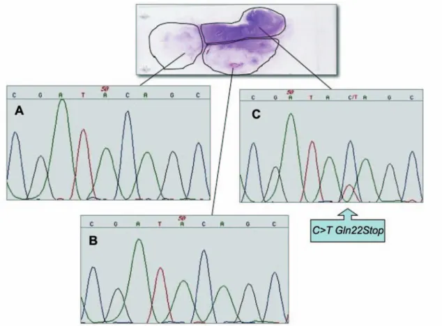
Paraffin-embedded sections were collected on microscope slides. Tumor and tumor-free areas were identified within 15 μm-thick deparaffinized sections lightly counterstained with hematoxylin and microdissected by gentle scraping with sterile scalpels into 1.5 ml polypropylene vials, using a hematoxylin and eosin-stained step section from the same block as a guide. Areas representative of colorectal tumor were identified and the sample was microdissected into three parts (Figure 1).
DNA extraction from each microdissected area was performed as reported elsewhere (30).
Exons 1 and 2 of K-ras were individually amplified using a seminested amplification protocol (31). DNA extractions and set-up of PCR reactions were performed in a laboratory distinct from that in which amplified DNAs are manipulated. Sequencing reactions were performed using Big Dye Terminator (Applied Biosystem, Foster City, CA, USA), and run on an ABI 3130 Genetic Analyzer (Applied Biosystem). All sequencing analyses were carried out on both strands in order to exclude pre-analytical and analytical errors and were repeated on PCR products obtained from new nucleic acid extractions.
Furthermore, DNA samples were subjected to mutational analysis of p53 (exons 5, 6, 7, 8 and 9) and BRAF (exon 15) performed by PCR and direct sequencing, as described above. Primers and PCR conditions are available upon request. To characterize genomic instability (MSI, microsatellite instability)
DNA from the blood of the same patient and from microdissected tumoral areas was analyzed using microsatellite markers mononucleotide repeats, BAT26 and BAT40, known to be the most sensitive markers of MSI status and widely regarded as sufficient for the identification of colorectal cancer with mismatch repair (MMR) defect. Primers and PCR conditions used for amplifications were as described (32).
All analyses were confirmed in duplicate experiments, using independently extracted DNA samples.
A further genetic characterization of other molecular markers was not feasible due to insufficiency of extracted nucleic acids and the paraffin tissue.
Results
EGFR immunohistochemical expression in the lymph node biopsy material was mild (score: 1+ in 10% of tumor cells). The mutational analysis for K-ras exons 1 and 2 was performed by PCR and direct sequencing using somatic DNA extracted from paraffin-embedded tumoral tissue. Figure 1 shows sequencing results, and the arrow indicates the point
mutation at the first letter of K-ras codon 22, which is a C to T transitions that determines a premature stop signal at codon 22 (Gln22Stop) in one of the three microdessected areas. Regarding the detected K-ras variant, the intensity of this mutated band was about 35% of that of the normal band (Figure 1). Tumor morphological examination showed a heterogeneous spreading of cancer cells within the same tumor. Therefore it was hypothesized that this heterozygous mutation could not be present in all cancerous cells. For this reason, after a morphological reassessment by an independent pathologist, the microdissected area containing the mutation was further microdissected into three microareas using a new histological slide in order to identify Gln22Stop. Several experiments, however, did not reveal the presence of the sequence variant.
The original embedded samples were then subjected to mutational analysis of exons 5-9 of the p53 gene, which encodes the DNA-binding region where cancer-associated mutations most frequently occur (31), and exon 15 of the BRAF gene, containing the polymorphisms Val600Glu, recently identified as genetic determinants of primary resistance to EGFR-targeted therapies in colorectal cancer (33).
Figure 1. Sequence analysis of the K-ras exon 1 performed on DNA extracted from each microdissected area of paraffin-embedded tumor sections. Wild-type sequence (panels A and B) and C to T transitions (panel C) that determine a premature stop signal at codon 22 (Gln22Stop) in one of the three microdessected areas. The mutation was named according to recommendations of the Nomenclature System for Human Gene Mutations. The GenBank mRNA sequence (M54968) of K-ras was used as a reference.
No mutations were found in either gene, and MSI status assessed by the analysis of the microsatellite loci BAT 26 and BAT 40, did not show constitutional alterations in the allele pattern of tumor samples compared with that of peripheral venous blood.
Discussion
We report a case of a patient with an adenocarcinoma of the ascending colon who, after 5 years from completion of adjuvant chemotherapy disease, relapsed with multiple cancer dissemination to main lymph node regions. In view of the use of an anti-EGFR monoclonal antibody (panitumumab)-containing second-line regimen (34), EGFR immunohistochemical expression in the lymph node biopsy material and K-ras gene mutational status in the primary colorectal tumor were performed (19). EGFR expression was mild in 10% of tumor cells (1+) whilst K-ras gene sequencing obtained different results according to the region of the tumor tissue analysed. In fact, the tumor sample was microdissected into three parts and two of them revealed wild-type K-ras gene, while in the third microdissected area, a point substitution in the codon 22 region (CAG>TAG; Gln22Stop) was found, determining an early STOP signal and encoding a truncated form of p21 protein. The patient was then put on a panitumumab/capecitabine-containing regimen but a rapid clinical worsening was observed and the patient eventually died after 4 months from the beginning of second-line therapy.
Several preclinical (35) and clinical (36) studies have previously shown that the presence of mutated K-ras alleles is an independent predictive marker of anti-EGFR mAb resistance. On the basis of these results, the European Union drug regulatory body, the European Medicines Agency, has approved the use of panitumumab only for patients with metastatic colorectal cancer whose tumors display wild-type K-ras (29): cetuximab and panitumumab have shown efficacy in approximately 10% to 20% of patients with metastatic colorectal cancer. In particular, the role of oncogenic K-ras has been extensively analyzed, leading to the conclusion that the occurrence of K-ras mutations represents a predictive parameter of anti-EGFR mAb resistance (36).
The most frequent K-ras gene alterations are detected in codons 12 and 13 and minor proportion in codons 61 and 146. Some studies, although limited in number, described mutations identified at codons other than those generally described (33). This novel type of K-ras mutation might be more frequent than we presently know, probably because current conventional analytical techniques, such as mutant-allele specific amplification, have focused exclusively on the identification of conventional mutations (29). Due to the low frequency of these rare or sporadic mutations, their clinical relevance in colorectal cancer is still unclear.
Similarly, mutations in codon 22 are described in a limited
number of cases. Previous studies have identified a C to A transversion substituting lysine (AAG) for normal glutamine (CAG) at the 22nd codon of the K-ras gene in a primary colon cancer. The biological activity of the mutation, tested by in vivo studies, suggested that this variant could be advantageous for in vivo tumor growth. Moreover, it is of particular interest that the 22nd amino acid residue of RASp21 is located between the GTPase domain and the effector domain (26, 27). Another study performed on a differentiated adenocarcinoma reported point mutations which caused substitutions of serine for glycine (GGT to AGT) at codon 12 and arginine for glutamine (CAG to CGG) at codon 22 concurrently in the same allele (28).
Our patient presented a never before reported premature stop signal at K-ras codon 22 determining the synthesis of a completely inactive truncated K-ras protein. To our knowledge, there are no reports that describe nonsense mutations of K-ras gene resulting in a premature stop signal. Only two studies described a G deletion mutation from the contiguous G base pairs located between codons 68 and 69 (AGG GAC) (37) and a single nucleotide deletion of one G in codon 12 of K-ras (38) both in somatic nucleic acid recovered from lung carcinomas.These mutations cause frameshift resulting in a stop codon several bases downstream; the resulting short truncated peptides probably have no apparently transforming activity because most of the carboxy part of the protein cannot be synthesized.
In our sample, the mutation detected only in a microdissected paraffin-embedded tissue area was not found in other tumoral areas of the slide. The mutated allele showed a lower intensity as compared to wild-type, suggesting the presence of the sequence variant in a small subclone of tumor cells. However, the mutational analysis conducted on more microareas obtained by a further microdissection of the tumoral area in question, did not reveal the presence of mutations, suggesting the presence of a cellular clone which was not identified during the review of the slide.
This observation corroborates the heterogeneity of a cancer population and also confirms previously published data demonstrating that intraneoplastic diversity could determine the genesis of potential metastatic clones and explain the presence of drug-resistant tumors (39, 40).
Heterogeneous multiple K-ras gene alterations in the same tumor have been documented, suggesting that different cellular clones arising from the same original transformed cell may harbour different genetic disruption of the same gene (28, 41). How these mutational clonality of K-ras may overall influence cancer growth and resistance to anti-EGFR drugs is unknown. To date, K-ras mutational tests commonly provide an on/off result (i.e. wild-type or mutated), without considering the relative percentage of tumor cells in the entire tumor mass harbouring wild-type, mutated, or other type of K-ras gene disruptions.
K-ras mutations, however, only account for 30% to 40% of nonresponsive patients. Therefore, the identification of additional genetic determinants of primary resistance to EGFR-targeted therapies in colorectal cancer would be of primary importance. Although the mechanism of action is still under study (35), genetic and biochemical evidence indicates that BRAF is the principal downstream effector of K-ras (42, 43). Because BRAF mutations are linked to MSI, a condition generally associated with better prognosis and resistance to standard chemotherapy (44), we also investigated MSI in our patient. Neither alteration for the BRAF gene V600E polymorphism nor genomic instability tested with mononucleotidic markers BAT26 and BAT40 were evident.
The presence of an inactivating K-ras mutation in a subclone of the cancer population does not seem to be the cause of the unresponsive EGFR-related treatment performed in this case. Other related K-ras pathways evaluated did not show any alteration. Therefore, other mechanisms should be considered for this specific case. One hypothesis which might explain the poor response to panitumumab is that tumor cells with a minimal inactivation of K-ras may develop alternative pathways to trigger downstream signals, thus escaping anti-EGFR tumor control in a similar fashion to cells that express constitutively activated K-ras.
In conclusion, although K-ras mutational analysis still remains the most important predictive parameter for EGRF-related therapies, further studies, considering other genes and pathways, should be performed in order to improve the predictivity of targeted therapies.
Acknowledgements
With the greatest appreciation we kindly thank Dr. Renato Covello for helping in histological examination of the tumor material. The Authors wish to thank Barbara Leone, Marco Ciancia and Raffaella Liberatore for their excellent technical assistance.
This study was partially supported by the Italian Ministry of Health Research Grant, Finalized Project RFPS20067342220 -Biotechnology strategies to improve rehabilitation outcome of fragile cancer patientsand by Grant LILT – Lega Italiana per la Lotta contro i Tumori.
References
1 Fearon ER and Vogelstein B: A genetic model for colorectal tumorigenesis. Cell 61: 759-767, 1990.
2 Vogelstein B, Fearon ER, Hamilton SR, Kern SE, Preisinger AC, Leppert M, Nakamura Y, White R, Smits AM and Bos JL: Genetic alterations during colorectal-tumor development. N Engl J Med 319: 525-532, 1988.
3 Andreyev HJ, Norman AR, Cunningham D, Oates JR and Clarke PA: Kirsten ras mutations in patients with colorectal cancer: the multicenter ‘RASCAL’ study. J Natl Cancer Inst 90: 675-684, 1998. 4 Bos JL, Fearon ER, Hamilton SR, Verlaan-de Vries M, van Boom JH, van der Eb AJ and Vogelstein B: Prevalence of ras gene mutations in human colorectal cancers. Nature 327: 293-297, 1987.
5 Martinez-Garza SG, Nunez-Salazar A, Calderon-Garciduenas AL, Bosques-Padilla FJ, Niderhauser-Garcia A and Barrera-Saldana HA: Frequency and clinicopathology associations of K-ras mutations in colorectal cancer in a northeast Mexican population. Dig Dis 17: 225-229, 1999.
6 Urosevic N, Krtolica K, Skaro-Milic A, Knezevic-Usaj S and Dujic A: Prevalence of G-to-T transversions among K-ras oncogene mutations in human colorectal tumors in Yugoslavia. Int J Cancer 54: 249-254, 1993.
7 Breivik J, Meling GI, Spurkland A, Rognum TO and Gaudernack G: K-ras mutation in colorectal cancer: relations to patient age, sex and tumor location. Br J Cancer 69: 367-371, 1994. 8 Bos JL: Ras oncogenes in human cancer: a review. [published
erratum appears in Cancer Res. 1990; 50: 1352]. Cancer Res 49: 4682-4689, 1989.
9 Baisse B, Bouzourene H, Saraga EP, Bosman FT and Benhattar J: Intratumor genetic heterogeneity in advanced human colorectal adenocarcinoma. Int J Cancer 93: 346-352, 2001. 10 Hughes R, Cross AJ, Pollock JR and Bingham S:
Dose-dependent effect of dietary meat on endogenous colonic N-nitrosation. Carcinogenesis 22: 199-202, 2001.
11 Bingham SA, Pignatelli B, Pollock JR, Ellul A, Malaveille C, Gross G, Runswick S, Cummings JH and O’Neill IK: Does increased endogenous formation of N-nitroso compounds in the human colon explain the association between red meat and colon cancer? Carcinogenesis 17: 515-523, 1996.
12 Beranek M, Bures J, Palicka V, Jandik P, Langr F and Nejedla E: A relationship between K-ras gene mutations and some clinical and histological variables in patients with primary colorectal carcinoma. Clin Chem Lab Med 37: 723-727, 1999. 13 Cerottini JP, Caplin S, Saraga E, Givel JC and Benhattar J: The
type of K-ras mutation determines prognosis in colorectal cancer. Am J Surg 175: 198-202, 1998.
14 Samowitz WS, Curtin K, Schaffer D, Robertson M, Leppert M and Slattery ML: Relationship of Ki-ras mutations in colon cancers to tumor location, stage and survival: a population-based study. Cancer Epidemiol Biomarkers Prev 9: 1193-1197, 2000.
15 Moerkerk P, Arends JW, van Driel M, de Bruine A, de Goeij A and ten Kate J: Type and number of Ki-ras point mutations relate to stage of human colorectal cancer. Cancer Res 54: 3376-3378, 1994.
16 Span M, Moerkerk PT, De Goeij AF and Arends JW: A detailed analysis of K-ras point mutations in relation to tumor progression and survival in colorectal cancer patients. Int J Cancer 69: 241-245, 1996.
17 Kampman E, Voskuil DW, van Kraats AA, Balder HF, van Muijen GN, Goldbohm RA and van’t Veer P: Animal products and K-ras codon 12 and 13 mutations in colon carcinomas. Carcinogenesis 21: 307-309, 2000.
18 Brink M, de Goeij AFPM, Weijenberg MP, Roemen GMJM, Lentjes MHFM, Pachen MMM, Smits KM, de Bruïne AP, Goldbohm RA and van den Brandt PA: K-ras oncogene mutations in sporadic colorectal cancer in the Netherlands Cohort Study. Carcinogenesis 24(4): 703-710, 2003.
19 Amado RG, Wolf M, Peeters M, Van Cutsem E, Siena S, Freeman DJ, Juan T, Sikorski R, Suggs S, Radinsky R, Patterson SD and Chang DD: Wild-type K-RAS is required for panitumumab efficacy in patients with metastatic colorectal cancer. J Clin Oncol 26: 1626-1634, 2008.
20 Lievre A, Bachet JB, Boige V, Cayre A, Le Corre D, Buc E, Ychou M, Bouche O, Landi B, Louvet C, Andre T, Bibeau F, Diebold MD, Rougier P, Ducreux M, Tomasic G, Emile JF, Penault-Llorca F and Laurent-Puig P: K-RAS mutations as an independent prognostic factor in patients with advanced colorectal cancer treated with cetuximab. J Clin Oncol 26: 374-379, 2008.
21 Rouleau E, Spyratos F, Dieumegard B, Guinebretière JM, Lidereau R and Bièche I: K-RAS mutation status in colorectal cancer to predict response to EGFR targeted therapies: the need for a more precise definition. Br J Cancer 99(12): 2100, 2008. 22 Akagi K, Uchibori R, Yamaguchi K, Kurosawa K, Tanaka Y and
Kozu T: Characterization of a novel oncogenic K-ras mutation in colon cancer. Biochem Biophys Res Commun 352: 728-732, 2007. 23 Lin SR, Tsai JH, Yang YC and Lee SC: Mutations of K-ras
oncogene in human adrenal tumors in Taiwan. Br J Cancer 77: 1060-1065, 1998.
24 Edkins S, O’Meara S, Parker A, Stevens C, Reis M, Jones S, Greenman C, Davies H, Dalgliesh G, Forbes S, Hunter C, Smith R, Stephens P, Goldstraw P, Nicholson A, Chan TL, Velculescu VE, Yuen ST, Leung SY, Stratton MR and Futreal PA: Recurrent K-RAS codon 146 mutations in human colorectal cancer. Cancer Biol Ther 5(8): 928-932, 2006.
25 Wójcik P, Kulig J, Okoń K, Zazula M, Moździoch I, Niepsuj A and Stachura J: K-RAS mutation profile in colorectal carcinoma and novel mutation-internal tandem duplication in K-RAS. Pol J Pathol 59(2): 93-96, 2008.
26 Tsukuda K, Tanino M, Soga H, Shimizu N and Shimizu K: A novel activating mutation of the K-ras gene in human primary colon adenocarcinoma. Biochem Biophys Res Commun 278: 653-658, 2000.
27 Yuen ST, Davies H, Chan TL, Ho JW, Bignell GR, Cox C, Stephens P, Edkins S, Tsui WW, Chan AS, Futreal PA, Stratton MR, Wooster R and Leung SY: Similarity of the phenotypic patterns associated with BRAF and K-RAS mutations in colorectal neoplasia. Cancer Res 62(22): 6451-6455, 2002. 28 Miyakura Y, Sugano K, Fukayama N, Konishi F and Nagai H:
Concurrent mutations of K-ras oncogene at codons 12 and 22 in colon cancer. Jpn J Clin Oncol 32(6): 219-221, 2002.
29 van Krieken JH, Jung A, Kirchner T, Carneiro F, Seruca R, Bosman FT, Quirke P, Fléjou JF, Plato Hansen T, de Hertogh G, Jares P, Langner C, Hoefler G, Ligtenberg M, Tiniakos D, Tejpar S, Bevilacqua G and Ensari A: K-RAS mutation testing for predicting response to anti-EGFR therapy for colorectal carcinoma: proposal for an European quality assurance program. Virchows Arch 453(5): 417-423, 2008
30 Palmirotta R, Matera S, Curia MC, Aceto G, el Zhobi B, Verginelli F, Guadagni F, Casale V, Stigliano V, Messerini L, Mariani-Costantini R, Battista P and Cama A: Correlations between phenotype and microsatellite instability in HNPCC: implications for genetic testing. Fam Cancer 3(2): 117-121, 2004. 31 Catalano T, Curia MC, Aceto G, Verginelli F, Cascinu S, Cama A, Mariani-Costantini R, Teti D and Battista P: Mutations in the p53 and Ki-ras genes, microsatellite instability and site of tumor origin in colorectal cancer. Oncol Rep 14: 625-631, 2005. 32 Ottini L, Falchetti M, D’Amico C, Amorosi A, Saieva C, Masala
G, Frati L, Cama A, Palli D and Mariani-Costantini R: Mutations at coding mononucleotide repeats in gastric cancer with the microsatellite mutator phenotype. Oncogene 16(21): 2767-2772, 1998.
33 Di Nicolantonio F, Martini M, Molinari F, Sartore-Bianchi A, Arena S, Saletti P, De Dosso S, Mazzucchelli L, Frattini M, Siena S and Bardelli A: Wild-type BRAF is required for response to panitumumab or cetuximab in metastatic colorectal cancer. J Clin Oncol 26(35): 5705-5712, 2008.
34 Van Cutsem E, Peeters M, Siena S, Humblet Y, Hendlisz A, Neyns B, Canon JL, Van Laethem JL, Maurel J, Richardson G, Wolf M and Amado RG: Open-label phase III trial of panitumumab plus best supportive care compared with best supportive care alone in patients with chemotherapy-refractory metastatic colorectal cancer. J Clin Oncol 25(13): 1658-1664, 2007.
35 Benvenuti S, Sartore-Bianchi A, Di Nicolantonio F Zanon C, Moroni M, Veronese S, Siena S and Bardelli A: Oncogenic activation of the RAS/RAF signaling pathway impairs the response of metastatic colorectal cancers to anti-epidermal growth factor receptor antibody therapies. Cancer Res 67: 2643-2648, 2007.
36 Lièvre A, Bachet JB, Le Corre D, Boige V, Landi B, Emile JF, Côté JF, Tomasic G, Penna C, Ducreux M, Rougier P, Penault-Llorca F and Laurent-Puig P: K-RAS mutation status is predictive of response to cetuximab therapy in colorectal cancer. Cancer Res 66: 3992-3995, 2006.
37 Wang YC, Lee HS, Chen SK, Yang SC and Chen CY: Analysis of K-ras gene mutations in lung carcinomas: correlation with gender, histological subtypes, and clinical outcome. Cancer Res Clin Oncol 124: 517-522, 1998.
38 Vachtenheim J, Horáková I, Novotná H, Opáalka P and Roubková H: Mutations of K-ras oncogene and absence of H-ras mutations in squamous cell carcinomas of the lung. Clin Cancer Res 1: 359-365, 1995.
39 Dexter DL and Leith JT: Tumor heterogeneity and drug resistance. J Clin Oncol 4(2): 244-257, 1986.
40 Sweeney C, Boucher KM, Samowitz WS, Wolff RK, Albertsen H, Curtin K, Caan BJ and Slattery ML: Oncogenetic tree model of somatic mutations and DNA methylation in colon tumors. Genes Chromosomes Cancer 48(1): 1-9, 2009.
41 Kimura K, Nagasaka T, Hoshizima N, Sasamoto H, Notohara K, Takeda M, Kominami K, Iishii T, Tanaka N and Matsubara N: No duplicate K-RAS mutation is identified on the same allele in gastric or colorectal cancer cells with multiple K-RAS mutations. J Int Med Res 35(4): 450-457, 2007.
42 Yan J, Roy S, Apolloni A, Lane A and Hancock JF: Ras isoforms vary in their ability to activate Raf-1 and phosphoinositide 3-kinase. J Biol Chem 273: 24052-24056, 1998.
43 Zhang BH, Guan KL: Activation of B-Raf kinase requires phosphorylation of the conserved residues Thr598 and Ser601. EMBO J 19: 5429-5439, 2000.
44 Locker GY, Hamilton S, Harris J, Jessup JM, Kemeny N, Macdonald JS, Somerfield MR, Hayes DF and Bast RC Jr; ASCO: ASCO 2006 update of recommendations for the use of tumor markers in gastrointestinal cancer. J Clin Oncol 24: 5313-5327, 2006.
Received April 9, 2009 Revised June 10, 2009 Accepted June 18, 2009