BIOS - Research Doctorate School in BIOmolecular
Sciences
Ph.D. in BIOMATERIALS - XX Cycle (2005-2007)
P
OLYMERIC
N
ANOSTRUCTURED
F
ORMULATIONS
FOR
T
ARGETED
A
DMINISTRATION OF
B
IOACTIVE
A
GENTS
Dr. Cesare Errico
S
UPERVISOR
: P
ROF
.
E
MO
C
HIELLINI
Tutor:
Dr. Federica Chiellini
Laboratory of Bioactive Polymeric Materials for Biomedical and Environmental Applications (BIOlab)
Department of Chemistry & Industrial Chemistry, University of Pisa (Italy)
Department of Biomaterials, Institute of Polymers Science and Technology CSIC, Madrid (Spain)
Chemotherapy is an effective treatment for cancer and other serious diseases such as cardiovascular restenosis and AIDS. It is a complicated procedure in which many factors are involved in determining its success or failure. Furthermore it carries a high risk due to drug toxicity, with the more effective drugs tending to be more toxic. Patients have to tolerate severe side effects and sacrifice their quality of life. The inefficiency and side effects of chemotherapy are caused mainly by the formulation, pharmacokinetics and toxicity of chemotherapeutic agents and the drug resistance. Hence, the development of effective carriers for both existing and newly developed drugs may be as important as the discovery of new anticancer drugs. All these concerns lead to the concept of sustained, controlled and targeted release of drugs.
The ideal goal of a drug delivery system is to deliver high efficacy drug(s) at the right time to the desired location, at high concentration but safe enough, over a sufficiently long period.
Effective drug delivery has also important implications from a market viewpoint. More effective drug delivery can provide drug companies with a means of expanding market share or of revitalizing therapeutics with previously unrealized potential because of poor pharmacokinetic profiles. Nanomedicine is not only important from the social and welfare aspects, but also for its economic potential.
In the framework of a long–standing research activity ongoing in the Bioactive Polymeric Materials Group at the Department of Chemistry and Industrial Chemistry of the University of Pisa, the present work was aimed at the development of polymeric nanoparticles for the site specific and controlled delivery of drugs for cancer treatment and tissue engineering application. The primary objective of the present PhD was the advancement of knowledge and the theoretical understanding of the relations among variables that play important roles in the development and optimization of procedures for the preparation of polymeric nanoparticle suspensions. The work was conducted keeping in mind the practical aspects of preparing nanodelivery structures for bioactive agents administration in cancer therapy and tissue engineering applications.
A variety of polymers including PLGA, PHB, VAM41, multi block copolymers of PCL-PLU, and Chitosan, were investigated. Retinoic Acid and Chondroitin Sulphate, were
loaded into nanoparticles as hydrophobic and hydrophilic model drugs respectively. Proteins have been also co-encapsulated as stabilizing agent or as model proteic drugs. For the preparation of RA loaded nanoparticles several methods were investigated and set up: dialysis, nanoprecipitation, co-precipitation and colloidal-coating. Colloidal-coating is an original solvent-free method developed in the framework of the research activity carried out in the present PhD thesis. The first two methods were employed for the preparation of formulations based on PLGA, PHB and the multi block copolymer PCL-co-PLU, while the last two for formulations based on VAM41. The resulting nanoparticle suspensions were analyzed macroscopically in terms of homogeneity and formation of precipitates, and characterized from a dimensional and morphological point of view by means of light scattering (LS) and scanning electron microscopy (SEM). Particles were purified and storage modalities were set up.
Drug release studies were carried out by an original method that might better reflect bioavailability data. Since HSA is one of the most abundant plasma protein it was added to the releasing medium overcoming problems related to the poor solubility of RA in water. Solubility problems often hamper the evaluation of drugs release kinetics of hydrophobic drugs in phosphate buffer solution (PBS) at 37°C, which is commonly used in in vitro environment and that should match the in vivo condition. The obtained results showed that HSA affected the stability and life of nanoparticle formulations.
The activity of RA encapsulated by colloidal-coating method was evaluated on SK-N-SH human neuroblastoma cell line that is known to undergo inhibition of proliferation and neuronal differentiation upon treatment with RA. The data obtained suggested that the anticancer activity of RA was not impaired by incorporation and purification processes.
A careful in vitro investigation of PHB and the relative nanoparticles cytotoxicity was carried out using two type of assays aimed at the evaluation of the interactions of the materials with cell metabolism (WST-1 tetrazolium salts) and cell endocytosis functions (Neutral Red Uptake). In vitro cytotoxicity of PHB resulted fairly low as highlighted by the high IC50 values obtained both with Tetrazolium Salts and Neutral Red assays. PHB
based nanoparticles prepared by dialysis method exhibited as well a very good cytocompatibility and can be considered fully biocompatible
It was also investigated the preparation of suitable vehicles for intra-articular injection in regenerated cartilages tissue, in form of nanoparticles, to allow Chondroitin Sulphate sustained release. Synthetic bioerodible (VAM41), biodegradable (PLGA), natural
develop the nanostructured formulations. The limit of nanoprecipitation method mainly concerns the possibility to achieve an efficiently loading of hydrophilic drug substances inside the nanoparticles. Drug is easily lost in water during the preparation and incompatibilities between the polymer and the drug could worse the situation. Due to the wide differences among the utilized polymers the nanoprecipitation and co-precipitation methods were adjusted and modified in relation to the physical-chemical characteristics of the utilized material.
PLGA, VAM 41 and Chitosan nanoparticles were prepared. No detectable amounts of CS were loaded onto the PLGA nanoparticles indicating the not suitability of the method for the encapsulation of this drug or a strong incompatibility between the hydrophobic polymer and hydrophilic drug. CS was detected into VAM41 and Chitosan NPs. A quantitative determination of the drug encapsulation was not possible due to several occurrences. Spectrophotometric UV assays have been based on changes in the absorption spectrum of the dye 1,9-dimethylmethylene blue (DMMB) when bound to Glycosaminoglycans (GAGs). CS interacted somehow with VAM41 and the characteristic UV band of absorbance of DMMB was shifted to higher wavelengths. Another attempt to evaluate encapsulation was done by IR spectrophotometry. The ratio between the intensity of the peak characteristic for CS and the intensity of the peak characteristic for VAM41 was evaluated and related to the percentage of CS in the samples. Although the developed method was effective for evaluating the encapsulation of CS it was not possible to verify whether the observed interaction affected these results or not.
Chitosan nanoparticles were insoluble in all the tested solvents. Hence, an approximate evaluation of CS content was carried out by XPS analysis. The presence of sulphur in the sample qualitatively confirmed the encapsulation of CS into the NPs.
The reported results and relevant discussions, which give exhaustive theoretical understanding of the phenomena occurring during colloidal formations, contribute to both basic and applied research in biomedical science and pharmaceutical technology.
TABLE OF CONTENTS
1 INTRODUCTION 1
1.1 BIOMATERIALS 1
1.1.1. Definitions and Classifications 1
1.1.2. Polymeric Biomaterials 3
1.1.2.1. Poly-3-Hydroxybutirate (PHB) 7
1.1.2.2. Copolymers of maleic anhydride (MAn) and alkyl vinyl ethers (RVE) (VAM) 7
1.1.3. Biodegradation and Bioerosion 9
1.2 BIOLOGICAL EVALUATION OF BIOMATERIALS 11
1.2.1. Biocompatibility and Hemocompatibility 11
1.2.2. Cytotoxicity 13
1.2.3. Genotoxicity 15
1.2.4. Carcinogenicity and Teratogenicity 16
1.2.5. Hemocompatibility 16
1.3 DRUG DELIVERY TECHNOLOGY 16
1.3.1. Polymer–Based Drug Delivery Systems 20
1.3.1.1 Diffusion controlled delivery systems 21
1.3.1.1.1 Membrane systems 21
1.3.1.1.2 Matrix systems 22
1.3.1.2 Degradable delivery systems 23
1.3.2. Colloidal Drug Delivery Systems 26
1.3.2.1. Liposomes and niosomes 27
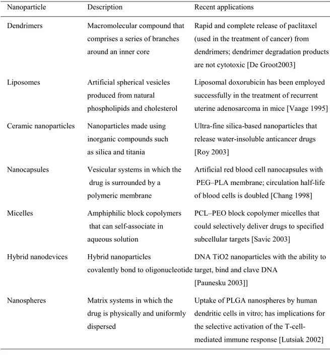
1.3.2.2. Nanoparticles 29
1.4. NANOPARTICLES IN CANCER THERAPY 31
1.4.1. Routes of Administration 32
1.4.1.1. Systemic administration 32
1.4.1.2. Local administration 35
1.4.1.3. Oral administration 35
1.4.1.4. Gene therapy and gene silencing therapy 36
1.4.2. Tumour targeting 39
1.4.2.1. Epitope-Paratope interaction 39
1.4.2.2. Targeted Delivery Through Enhanced Permeability and Retention (EPR) 41
1.4.2.3. Targeting Through Angiogenesis 42
1.4.2.4. Targeting Through Magnetic Devices 43
1.5. DRUG DELIVERY IN TISSUE ENGINEERING 44
1.5.1. Growth Factors role and Delivery 46
1.5.2. Cell-Based Gene Delivery 48
1.5.3. Direct Gene Delivery 48
1.6. TECHNIQUES OF NANO- AND MICROPARTICLE PREPARATION 49
1.6.1. Emulsion-Solvent Evaporation/Extraction Methods 50
1.6.1.1. Single Emulsion Method 50
1.6.1.2. Double Emulsion Method 51
1.6.2. Phase Separation 51 1.6.3. Solvent displacement 53 1.6.3.1. Nanoprecipitation Method 53 1.6.3.2. Co-Precipitation Method 54 1.6.3.3. Dialysis Method 55 1.6.4. Self assembling 56
1.6.5. Rapid Expansion of Supercritical Fluid Solution 57
1.6.6. Spray drying 58
1.7. DRUGS & STABILIZING AGENTS 59
AIM OF THE WORK 69
2. GLOSSARY 71
2.1. POLYMERS 71
2.2. DRUGS 73
2.3. REAGENTS 74
3.3. MATERIALS 75 3.1.1. Solvents 75 3.3.1.1. Chloroform 75 3.3.1.2. Acetic Acid 75 3.3.1.3. Acetone 75 3.3.1.4. Trifluoroethanol 99% (TFE) 75
3.3.1.5. Dimethy sulfoxide (DMSO) 75
3.3.1.6. Ethanol 75 3.3.1.7. Tetrahydrofuran (THF) 75 3.3.1.8. N,N- Dimethylformamide 99.9% (DMF) 75 3.1.2. Reagents 76 3.1.2.1. Hydrochloric Acid 37-38 % 76 3.1.2.2. Polyvinylpyrrolidone (PVP) 76
3.1.2.3. Fibrinogen from human plasma 76
3.1.2.4. Dialysis Membranes 76 3.1.2.5. Minisart filters 76 3.1.2.6. Metallic needles 76 3.1.2.7. Deionised water 76 3.1.2.8. 10 N NaOH solution 76 3.1.2.9. 1 N NaOH solution 77 3.1.2.10. 10 N HCl solution 77 3.1.2.11. 1 N HCl solution 77
3.1.2.12. Dulbecco’s Phosphate Buffered saline 77
3.1.2.13. 3% mannitol solution 77
3.1.2.14. Human Serum Albumin (HSA) 77
3.1.2.15. Poly (DL-lactide-co-glycolide) (PLGA) 77
3.1.2.16. VAM41 (2–methoxyethanol hemiester of poly(maleic anhydride–alt–butyl vinyl ether) 77
3.1.2.17. Poly (3-hydroxybutyric acid) natural origin (PHB) 77
3.1.2.18. Retinoic Acid (RA) minimum 98% 77
3.1.2.19. Pluronic F-127 78 3.1.2.20. Chitosan 78 3.1.2.21. Chondroitin Sulphate (CS) 78 3.1.2.22. Natriumtripolyphospate (TPP) 78 3.1.2.23. Buffers pH 3, 8 Titrisol 78 3.1.2.24. 1-9 Dimethylmethylene blue (DMMB) 78 3.1.2.25. Phosphotungstic acid 78
3.1.3. Cell Culture Materials 78
3.1.3.1. Cell line 78
3.1.3.2. Antibiotics 78
3.1.3.3. Fetal Bovine Serum (FBS) 78
3.1.3.4. Glutamine 79
3.1.3.5. Dulbecco's Modified Eagles Medium (DMEM) 79
3.1.3.6. Complete Dulbecco's Modified Eagles Medium (Complete DMEM) 79
3.1.3.7. Phosphate Buffer Saline without Ca2+ and Mg2+ (PBS4) 79
3.1.3.8. Cell proliferation reagent (WST–1) 79
3.1.3.9. Neutral Red kit 79
3.1.3.10. Disposable sterile plastic ware 79
3.1.4. Instrumental Characterizations (Instruments) 79
3.1.4.1. Ultrasonic processor 79
3.1.4.2. Absorbance measurements on microplates 80
3.1.4.3. CO2 incubator 80
3.1.4.4. Freeze-drying 80
3.1.4.5. Laminar flow cabinet 80
3.1.4.6. Optical Microscopy (OM) 80
3.1.4.7. Particle size analysis 80
3.1.4.8. pH measurements 80
3.1.4.9. Refrigerated centrifuge 80
3.1.4.10. Scanning Electron Microscopy (SEM) 81
3.1.4.11. Environmental Scanning Electron Microscopy (ESEM) 81
3.1.4.13. Sputter coater 81
3.1.4.14. UV–Vis spectroscopy 81
3.1.4.15. AT-IR spectroscopy 81
3.4. FORMULATION OF RETINOIC ACID LOADED NANOPARTICLES 82
3.2.1. PLGA Based Nanoparticles Prepared by Dialysis Method 82
3.2.1.1. Preparation of Unloaded Nanoparticles 82
3.2.1.2. Preparation of RA Loaded Nanoparticles 82
3.2.2. PHB Based Nanoparticles Prepared by Dialysis Method 83
3.2.2.1. Preparation of Unloaded Nanoparticles 84
3.2.2.2. Preparation of RA Loaded Nanoparticles 84
3.2.3. PCL-PLU Based Nanoparticles Prepared by Dialysis Method 84
3.2.4. PLGA Based Nanoparticles Prepared by Nanoprecipitation Method 85
3.2.4.1. Preparation of Unloaded Nanoparticles 85
3.2.4.2. Preparation of RA Loaded Nanoparticles 85
3.2.4.3. Preparation of RA and HSA Loaded Nanoparticles 86
3.2.5. PHB Based Nanoparticles Prepared by Nanoprecipitation Method 86
3.2.5.1. Preparation of RA Loaded Nanoparticles 86
3.2.6. Pcl-Plu Based Nanoparticles Prepared By Nanoprecipitation 87
3.2.6.1. Preparation of RA Loaded Nanoparticles 87
3.2.7. VAM41 Based Nanoparticles Prepared by Co-precipitation Method 87
3.2.7.1. Preparation of RA Loaded Nanoparticles 87
3.2.8. VAM41 Based Nanoparticles Prepared by Colloidal-Coating Method 88
3.2.8.1. Preparation of Unloaded Nanoparticles 88
3.2.8.2. Preparation of RA Loaded Nanoparticles 88
3.2.9. Nanoparticles Purification 89
3.2.10. Cryoprotection 89
3.3. NANOPARTCLES CHARACTERIZATION 89
3.3.1. Granulometry in Suspension 89
3.3.2. Morphological Analysis 90
3.3.2.1. Scanning Electron Microscopy (SEM) 90
3.3.2.2. Transmission Electron Microscopy (TEM) 90
3.3.3. Evaluation of Retinoic Acid Incorporation 90
3.3.4. Evaluation of HSA Incorporation 92
3.3.5. In Vitro Evaluation of the Activity of the Encapsulated Retinoic Acid 92
3.4. IN VITRO ASSESSMENT OF CYTOTOXICITY 92
3.4.1. Cell Propagation 93
3.4.1.1. Cryopreservation and Cell Thawing 93
3.4.1.2. Cell counting by Dye–Exclusion Hemocytometry 93
3.4.1.3. Subculturing of Continuous Cell Lines Grown in Monolayers 94
3.4.2. Sample Preparations 94
3.4.2.1. Preparation of Polymer Solutions 94
3.4.2.2. Preparation of Nanoparticle Samples 94
3.4.3. Quantitative Evaluation of Cytotoxicity 95
3.4.3.1. WST–1 cell proliferation assay 95
3.4.3.2. Neutral Red 96
3.5. IN -VITRO DRUG RELEASE STUDIES 96
3.5.1. Releasing Medium: PBS 96
3.5.2. Releasing Medium: PBS containing HSA 96
3.6. FORMULATION OF CHONDROITIN SULPHATE LOADED NPS 97
3.6.1. PLGA Based Nanoparticles Prepared by Nanoprecipitation-Solvent Evaporation Method 97
3.6.2. PLGA Based Nanoparticles Prepared by Double Nanoprecipitation 98
3.6.3. VAM41 Based Nanoparticles Prepared by Modified Co-Precipitation Method 98 3.6.4. Chitosan Based Nanoparticles Prepared by Modified Co-Precipitation Method 99
3.7. NANOPARTICLES CHARACTERIZATION 100
3.7.1. Evaluation of CS Encapsulation in PLGA and VAM41 nanoparticles 100
3.7.1.1. Determination of CS by UV Spectrophotometry 100
3.7.2. Chondroitin Sulphate encapsulation in nanoparticles evaluated by IR spectrophotometry 101
4. RESULTS & DISCUSSION 103
4.1. NANOPARTICLES: SIGNIFICANCE & FORMULATIONS DEVELOPMENT 103
4.2.1. Dialysis Method 105
4.2.1.1. PLGA Based Nanoparticles Prepared By Dialysis Method 105
4.2.1.2. PHB Based Nanoparticles Prepared by Dialysis Method 112
4.2.1.3. PCL-co-PLU Based Nanoparticles Prepared by Dialysis Method 115
4.2.2. Nanoprecipitation Method 115
4.2.2.1. PLGA Based Nanoparticles Prepared by Nanoprecipitation Method 116
4.2.2.2. PLGA Based Nanoparticles Loaded with RA and HSA Prepared by Nanoprecipitation 118
4.2.2.3. PHB Based Nanoparticles Prepared by Nanoprecipitation 120
4.2.2.4. PCL-co-PLU Based Nanoparticles Prepared by Nanoprecipitation 122
4.2.3. Co-Precipitation Method 125
4.2.4. Colloidal–Coating Method 128
4.3. NANOPARTICLES PURIFICATION 131
4.4. NANOPARTICLE FORMULATIONS CHARACTERIZATION 133
4.4.1. Release Studies 133
4.4.2. Determination of RA / HSA interactions 139
4.4.3. Evaluation of the Activity of the Encapsulated Retinoic Acid 140
4.4.4. In-Vitro Citotoxicity Studies 143
4.5. METHODS FOR ENCAPSULATING CHONDROITIN SULPHATE 144
4.5.1. Nanoprecipitation-Solvent Evaporation Method 144
4.5.1.1. PLGA Based Nanoparticles Prepared by Nanoprecipitation-Solvent Evaporation Method 144
4.5.2. Double Nanoprecipitation Method 146
4.5.2.1. PLGA Based Nanoparticles Prepared by Double Nanoprecipitation 146
4.5.3. Modified Co-Precipitation Method 148
4.5.3.1. VAM41 Based Nanoparticles Prepared by Modified Co-Precipitation Method 148 4.5.3.2. Chitosan Based Nanoparticles Prepared by Modified Co-Precipitation Method 150
4.6. NANOPARTICLES CHARACTERIZATION 152
4.6.1. Evaluation of CS Encapsulation in PLGA NPs 152
4.6.1.1. Determination by UV Spectrophotometry 152
4.6.1.2. Determination by IR Spectrophotometry 154
4.6.2. Evaluation of CS Encapsulation in VAM41 NPs 154
4.6.2.1. Determination of CS Encapsulation in VAM41 NPs by UV Spectrophotometry 154 4.6.2.2. Determination of CS Encapsulation in VAM41 NPs by IR Spectrophotometry 156
4.6.3. Evaluation of CS encapsulation in Chitosan NPs 158
4.6.3.1. Determination of CS encapsulation in Chitosan NPs by microanalysis 158
5. CONCLUDING REMARKS 161
REFERENCES 165
ACKNOWLEDGMENTS 185
1 INTRODUCTION
1.1 BIOMATERIALS
In the last decades the development of biomaterials science has been of enormous impact in the scientific world, especially in the field of regenerative medicine. The peculiarity of biomaterials science is its multidisciplinary nature, which involves connections between scientific, medical, and engineering disciplines. The continuous flow of knowledge in materials science, molecular and cell biology, human physiology and pathology and the constant demand for new technological solutions have driven the biomaterials science to be one of the most promising areas of scientific and social interest.
1.1.1. Definitions and Classifications
The first generation of materials to be used inside the human body was developed during the 1960s and 1970s. The objective of these early biomaterials was to “achieve a suitable combination of physical properties to match those of the replaced tissue with a minimal toxic response in the host” [Hench 1980]. The general required feature of these biomaterials stood in their biological “inertness”, as the reduction to a minimum of the immune response to the foreign body. The advanced approaches concerned the production of bioactive materials that could either elicit a controlled action and reaction in the physiological environment or the development of bioresorbable biomaterials that exhibited controlled chemical breakdown and elimination [Hench 1980, Wilson 1984]. Finally, the third generation of biomaterials involves the unification of the former concepts. These materials are thus designed to stimulate specific cellular responses at the molecular level (bioactivity) and simultaneously possess bioresorbable features [Hence 2002]. At present, the two most widely accepted definitions of biomaterials are:
o “A biomaterial is a nonviable material used in a medical device, intended to interact with biological systems” [Williams 1987].
o “A biomaterial is a material intended to interface with biological systems to evaluate, treat, augment or replace any tissue, organ or function of the body” [European
Indeed, the design and manufacture of biomaterials address several scientific aspects and need to assure specific requirements. In particular, a deep investigation of the chemical, physical and biological characteristics of the environment in which the material is expected to perform its action is of outmost importance. The acquired knowledge may lead to a better understanding of the factors that determine the final performances of the materials, and therefore allowing for the optimization of their design. The biofunctionality of the device under investigation should hence be associated to proper mechanical and chemical characteristics. Nevertheless, these products must retain their functionality and safety over the desired time, as ensured by their biocompatibility.
A general classification based on the chemical nature of biomaterials comprises [Barbucci 1994]:
o Biologically derived materials include natural proteins (collagen, elastin, gelatin), polysaccharides (hyaluronic acid, chitosan, dextran, cellulose), and proteins produced with biotechnological techniques (genetic engineering), as well as entire cells and natural tissues. They are often used in combination with other synthetic biomaterials, especially in dermatology.
o Synthetic polymers can be employed in a wide range of applications, of either intracorporeal or extracorporeal type. The most common uses include short–term devices (sutures, adhesives, artificial tendons, contact lenses), long–term cardiac valves, complex systems aimed at substituting the function of body organs (such as artificial kidney, heart, pancreas), catheters, filters for hemodialysis, orthopaedic material, and drug delivery devices.
o Metals are mainly used in orthopaedic and dental implants, as well as in the production of pacemakers and heart valves. The most commonly used metals are stainless steel, cobalt–based alloys, titanium and its alloys.
o Composites can be manufactured by the combination of polymers, ceramics, and metals. Composites currently used in biomaterial applications include bone particle– or carbon fiber–reinforced methyl methacrylate bone cement and ultra–high– molecular–weight polyethylene, porous surface orthopaedic implants, and dental filling composites.
o Ceramics are widely employed in orthopaedic implants, as bone fillers, and in dental and ear–nose–throat implants. The most common bioceramics are aluminum oxides,
1.1.2. Polymeric Biomaterials
The interest for polymeric biomaterials and their application for medical purposes are growing very fast. Polymeric materials have found applications in diverse biomedical fields, such as ophthalmology, dentistry, orthopaedics, implantation of medical devices, bioartificial organs, drug delivery and many more [Jagur-Grodzinski 1999].
Compared with other types of biomaterials, such as metals and ceramics, polymers offer the advantage of being engineered and processed in different ways to meet specific end– use requirements. Besides the broad variety of commercially available polymers, the synergy between advanced organic chemistry and polymer synthesis has led to the development of precious tools for the design of polymer architecture and the grafting of bioactive molecules [Drotleff 2004, Hawker 2005]. Therefore, according to key “device” characteristics such as mechanical resistance, degradability, permeability, solubility, and transparency, polymers can be selected and specifically tailored for appropriate surface or bulk properties.
Whereas the original uses of polymers in clinical medicine focused primarily on the replacements of connective tissues, new applications are emerging. Nowadays, two important application fields of polymeric biomaterials are tissue engineering and controlled drug delivery.
Tissue engineering is aimed at restoring, maintaining, or enhancing tissues and organs functions by assuring synergistic interactions of living cells and synthetic materials. Although transplantation and reconstructive surgery are the most used therapies to address the loss and failure of organs and tissues, they remain imperfect solutions. Tissue engineering could actually afford a valid alternative [Griffith 2002].
Drug delivery main issues are the control of the pharmacokinetics and biodistribution of drugs in order to restrict their action to the treated tissue and keep their concentration at therapeutically relevant levels for the desired time.
Polymers are currently used as physical carriers for drugs, components of prodrugs, conjugates or in complexes with proteins or nucleic acids, as well as direct therapeutics in their own right [Twaites 2005]. The role of polymers in drug delivery covers multiple aspects, from the enhancement of the physical-chemical stability of the drug to the regulation of drug release and targeting. The goal of drug delivery and the involvement of polymeric biomaterials is hence to increase drug efficacy and reduce the toxicity of
traditional drug administration, create easier dosage regimes and achieve a better patient compliance. [Vasir 2005].
As a further step towards the development of third–generation “smart” materials for biomedical use, the possibilities of synergistically combine the approaches of both conventional tissue engineering and controlled drug delivery has been recently envisaged. The aim of such association is to afford systems that not only constitute a supportive scaffold for cell proliferation, but also guide cell development and organization by progressive and controlled release of cell growth and differentiating factors [Saltzmann 2002]. This concept, which could promote the formation of highly organized and complex tissues, is actually under investigation by the most eminent research groups involved in biomaterial science.
Generally, polymeric biomaterials are classified as synthetic polymers and polymers of natural origin. The field of applications of synthetic and natural polymeric biomaterials is indeed very wide, including surgical devices, implants and supporting materials (e.g. artificial organs, prostheses and sutures), drug delivery systems with different routes of administration and design, carriers of immobilized enzymes and cells, biosensors components of diagnostic assays, bioadhesives, ocular devices, and materials for orthopaedic applications [Angelova 1999]. Biopolymers and their applications are summarised in Table 1.1-1.2 [Park 1992, Jagur-Grodzinski 1999, Saltzmann 2002, Kashyap 2004, Twaites 2005].
Table 1 - Natural polymers and their application.
Polymer Main applications and comments
Proteins and protein-base polymers Resorbable. Biocompatible and not toxic. Elastic materials applied in implantology and tissue engineering.
Collagen Sutures, drug release.
Albumin Drug stabiliser and drug release.
Gelatin Used in the preparation of gels and in drug release
Polyaminoacids Not toxic, not antigenic and biocompatible. Used as carriers
for oligomeric drugs. Polysaccharides and vegetal derived polymers
Carboxymethylcellulose Drug release, dialysis membranes, cell immobilisation (ionotropic gelation and formation of polyelectrolyte complexes)
Starch Drug delivery
Cellulose sulphate In polyelectrolytic complexes for imunoisolation
Agarose Used in clinical analysis and as matrix
Alginate Biocompatible. Applied in gel preparation and to immobilise
cell matrix and enzymes. For the release of bioactive molecules, injectable microcapsules for neurodegenerative diseases and hormones deficiency.
Polysaccharides and human/animal derivatives
Hyaluronic acid Moisturizing agent, wound dressing, artificial tears in ophthalmology, various orthopaedic applications
Eparin and Eparin-like Trombolytic anticoagulant properties. Used in surgery for glycosaminoglycans ionotropic gelation and in capsules preparation.
Microbial Polysaccharides
Dextran and derivatives Excellent reological properties, plasma expander. Used in drug delivery.
Chitosan and derivatives Natural polycation. Biocompatible, not toxic. Used in gel and film preparation. Applied in drug delivery.
Table 2 - Synthetic polymers used for biomedical applications
Polymer Main applications and comments
Aliphatic polyesters
Poly(lactic acid), poly(glycolic acid) Used in sutures, drug delivery systems and tissue engineering. and their copolymers Biodegradable. Copolymerized to regulate degradation time. Poly(hydroxybutirate)s Biodegradable, used as matrix for drug
Poly(γ–caprolactone) and copolymers Delivery systems, cell microencapsulation. Poly(alkylene succinate), etc. Properties can be tuned by chemical modification,
copolymerization and blending.
Polyanhydrides Biodegradable, useful in tissue engineering and for the release of bioactive molecules.
Polyorthoesters Surface–eroding polymers. Application in sustained drug delivery, ophtalmology.
Polycyanoacrylates Biodegradable, depending on the length of the alkyl chain. Used as surgical adhesives, glues, and in drug delivery.
Polyacrylonitrile Dialysis membranes
Polyphosphazenes Can be tailored with versatile side–chain functionality. Applications in drug delivery.
Polyamides Used for sutures, hemofiltration membranes. Inhibitors of DNA
transcription
Polyurethanes Used for prostheses, vascular grafts, catheters tissue adhesives, artificial cardiac structures, coatings
Polyethylene Used for orthopaedic materials and catheters.
Polypropylene Plasmapheresis membranes, sutures, external medical devices.
Poly(methyl methacrylate) This and its copolymers are used as dental implants, in bone
replacement and as intraocular lenses.
Poly(hydroxyethyl methacrylate) Used for contact lenses, ocular prostheses, skin coatings, catheters.
Polytetrafluoroethylene (Teflon®) Used for vascular grafts, clips and sutures facial prosthes, coatings.
Polydimethylsiloxanes Used for implants in plastic surgery, orthopaedics, blood bags,
pacemakers, drug delivery devices, membrane ossigenators. Poly(N-vinylpyrrolidone) in hydrogels for controlled release of drugs from metal stents,
or in blood substitutes.
Polysulfone Heart valves, penile prostheses
Poly(vinyl chloride) Used for tubing, blood bags.
Polystyrene Used as substrates for cell cultures.
Poly(ethylene oxide) Different polymer derivatives and copolymers used in a variety of biomedical applications.
Poly(ethylene glycol) Several applications as spacer, in bioconjugations, coatings, etc. Covalently bound in Pegvisomant for agromegaly;
and in Pegfilgrastim to increase neutrophilis production in bone marrow for neutropenia.
Polylisine Applied in gene delivery, formation of conjugate vectors
1.1.2.1. Poly-3-Hydroxybutirate (PHB)
A majority of drug delivery systems using natural polymers have been based on proteins and polysaccharides [Park 2005]. Poly (3-hydroxyalkanoates), synthesized by a wide variety of micro-organisms, have recently attracted a great deal of industrial attention. In particular, much interest focused on the use of poly (3-hydroxybutyrate) (PHB) and related copolymers as carriers or implants for drug delivery. When compared with chemically produced polymers such as polyglycolate, polylactate, and poly (lactide-co-glycolide) (PLGA), which are mostly well known as biologically degradable drug carriers with good retarding characteristics, PHB advantages are that it is easily processed, fully biologically degradable and compatible with animal tissue.
In addition, the controllable retarding properties of PHB can be manipulated by variations in processing and molecular weight of the polymer. Thus, it could be anticipated to be an ideal basic material for matrix composition [Kim 2000]. PHB is produced biosynthetically by bacteria from natural raw materials, and indeed can be readily broken down by micro-organisms under different conditions, such as those present in sewage. The novel method of manufacture has several implications which may be advantageous for biomedical applications. Biosynthesis avoids the use of chemical catalysts or initiators that could cause toxicological problems if they remain in a pharmaceutical product. The involvement of microbial enzymes results in polymers which are regular from a stereochemical point of view, which is likely to result in highly reproducible physical properties. Hydrolytic degradation of PHB in vitro proceeds to the monomer, D-(-)-3-hydroxy butyric acid. This acid is a normal constituent of blood and, in common with acetoacetate and acetone, is one of the three ketone bodies that are produced endogenously by the process known as ketogenesis. It is therefore thought that PHB can be well tolerated in vivo [Pouton 1996].
1.1.2.2. Copolymers of maleic anhydride (MAn) and alkyl vinyl ethers (RVE) (VAM) In order to have suitable matrices for the preparation of colloidal drug delivery systems, amphiphilic polymeric materials were developed by hemiesterification of alternating copolymers of maleic anhydride (MAn) and alkyl vinyl ethers (RVE) by the research group involved in the present work. These synthetic polymers possess favourable
systems of high and low molecular weight active agents. Each repeating unit carries has a hydrophilic ionisable acid group, a hydrophobic but potentially degradable ester residue, and an alkyl ether moiety of variable structure. By accurate choice of these groups, the polymeric material may result insoluble under physiological conditions. However, because of the progressive ester hydrolysis, the number of ionisable groups along the chain increases (Figure 1.1), affording a partially degraded structure, which becomes water soluble, thus allowing to classify the system as bioerodible.
O O O OH O R' Physiological conditions Degradation (Hydrolysis) O O O R' NaO ONa
Water insoluble Water soluble
R
Figure 1.1 - Bioerosion of poly(maleic anhydride–alt–alkyl vinyl ether) hemiesters.
The fraction of ionized carboxyl groups necessary for matrix solubilization and the pH at which ionization occurs depend on the hydrophilic/hydrophobic balance of both ether and ester residues [Chiellini 1992a, Heller 1978, Woodruff 1972]. Moreover, the presence of hydrophobic chains in either position tends to slow down the swelling and the hydrolysis process. Therefore, the release pattern from systems based on these polymers can be tuned easily by an appropriate choice of the ester and the ether chains.
Quite a large number of examples are reported in the literature in which hemiesters of copolymers of maleic anhydride and alkyl vinyl ethers have been utilized for the formulation of release systems. For instance, the n–butyl hemiester of maleic anhydride– methyl vinyl ether alternating copolymer was used in ophthalmology for the preparation of monolithic systems or as pH sensitive covers of microcapsules [Chiellini 1992a]. Similarly, alkyl hemiesters of maleic anhydride–oligo(oxyethylene) vinyl ether copolymers were formulated as monolithic inserts for the controlled release of pilocarpine [Chiellini 1988, Saettone 1988] and α–interferon [Chiellini 1992a, 1992b].
These polymers attracted particular interest as controlled delivery systems of protein drugs. Indeed, the presence of carboxyl groups should favour the incorporation of
matrices, such as PLA and PLGA copolymers. Moreover, the presence of poly(ethylene glycol) segments in the side chains can promote the establishment of non–covalent interactions among the polymeric matrix and hydrophilic multifunctional drugs, such as peptides and proteins [Chiellini 2001, 1992b]. Materials belonging to this class were successfully formulated as bioerodible nanoparticles loaded with α–interferon [Chiellini 2001, Cowdall 1999a, 1999b]. Additionally, it is well known that the presence of PEG segments on the surface of release systems may develop antiopsonizing effects, resulting in lower toxicity and longer circulation time [Kim 1998, Gref 1995].
1.1.3. Biodegradation and Bioerosion
Biopolymers used for medical application must often be biodegradable and polymers degradation may occur under active or passive processes. Generally, the human body is described as an aqueous environment with a pH of 7.4 and a temperature of 37°C. It is important to observe that actually, the physiological environment of particular organs or tissues may be different and it is also subjected to drastic changes in case of disease. The saline solution of the human body is an excellent electrolyte, which induces hydrolysis and corrosion processes on the implanted foreign body or to the polymeric drug carrier. Catalysts produced by cells and tissues may accelerate these processes as well as isolate, attack and destroy the implanted device. The majority of the polymers employed for the production of medical devices are subjected to water diffusion and consequently to hydrolysis reactions. The hydrophilicity of the polymer is a fundamental parameter to be considered in the selection of the material and it is strictly related to the final application of the device; i.e. it is preferable to choose bioresorbable materials for the production of sutures. The degradation of polymers in vivo (biodegradation) is not only related to water absorption but also to a complexity of factors that highly accelerate the degradation process. Numerous cells, e.g. the cells involved in inflammation, produce enzymes which catalyses some of the reactions responsible for significant alteration of the molecular structure of the implanted devices. In particular, phagocytes migrate to the areas of inflammation where the implant is located. Through specific recognition mechanisms (mediated by antibodies) or non-specific absorption of proteins, phagocytes adsorb on the surface of the foreign body. The consequence of the described behaviours is the enhancement of the metabolic activity and of the immune response of the body, which lead to the activation of the degradation processes on the implanted system.
Biomaterial degradation is affected by both material physical-chemical properties and general conditions of the host (age, health, therapeutic treatments) as well as by the specific location of the implant and the type of the surrounding tissues. Scaffolds made of biostable polymers require the removal of the device once the therapeutic activity is exhausted and may induce a chronic inflammation. Nonetheless, when biodegradable polymers are employed, they should not generate harmful or immunogenic products as a result of their degradation.
Generally, hydrophobic biomaterials displaying a bioerodible surface are used for devices that require a long half-life in vivo. Typically polyanhydrides and polyorthoesters are mostly applied [Angelova, 1999].
A polymer is considered “bioerodible” if its solubility in water changes from not soluble to soluble, under physiological conditions. As formerly explained, the solubilization of the polymer may not necessarily involve the chemical breakdown of polymer backbone but can simply be associated to pH changes and ionic transfers. Therefore, bioerosion includes both physical processes (dissolution) and chemical processes (hydrolytic cleavage of chemical bonds, also mediated by enzymes). The suffix “bio” imply an erosion process that takes place under physiological conditions and distinguishes bioerosion from other degradative processes due to temperature, radiations or to the presence of either strong acids or bases. Biodegradation includes also the concept of bioresorption. Bioresorption (also called bioassorption) concerns the removal of the polymer and of its degradation products from the biological environment, mostly performed by cells by means of phagocytosis [Kohn, 1996]. The removal of solubilised polymer chains and degradation products may also ensue from renal excretion [Griffith, 2002b]
The bioerosion of a solid polymeric implant correspond to macroscopic changes of the device aspect, alterations of the physical-chemical properties, swelling, weight loss, shape changes and loss of functionality. Two different bioerosion modalities have been described in literature, namely bulk and surface erosions. In bulk erosion, water diffusion inside the polymer matrix is faster than the rate at which the polymer is converted into a water-soluble material. Therefore, water uptake is followed by an erosion process that occur inside the entire volume of the solid device. Typically, bulk erosion of polymeric materials induces the formation of pores that drive the physical decomposition of the device.
Conversely, in surface erosion, water penetration inside the polymeric device is slower than the dissolution process on the surface. Differently from bulk erosion, the gradual dissolution of polymer surface has a minor but progressive influence on the shape and mechanical characteristics of the device.
1.2 BIOLOGICAL EVALUATION OF BIOMATERIALS
1.2.1. Biocompatibility and Hemocompatibility
The fundamental characteristic of biomaterials is their capability of interacting with organic tissues and biological fluids without inducing secondary alterations or damaging processes in the host living organism. Summarising, when biomaterials are in contact with body tissues, they should not cause toxic, inflammatory, or carcinogenic responses (biocompatibility) and their interactions with blood should not elicit coagulative or cell– disruptive activities (hemocompatibility). Until the 1970s, the term “biocompatibility” was related to the biological inertness of the material. At present, the concept of inertia is questionable because there is no material that does not elicit a response when in contact with body compartments. Moreover, with the improvements in material technology and instrumentation, new materials have appeared on the scientific scenario. The characteristic of such materials is to degrade in the biological tissues and release degradation products that can interact “positively” with the living cells and their metabolism, thus bringing about favourable biological reactions for the implant life and functionality [Gatti 2002]. Because of this insight and development, biocompatibility was defined as “the ability of a material to perform with an appropriate host response in a specific application” [Williams 1987].
In general, any foreign material in contact with body tissues or fluids generates a specific reaction. The type and level of this reaction will depend on the particular body organ where this reaction originates and will consist of a complex and integrated defensive system. In particular, a reaction of inflammatory type can occur, involving the progressive intervention of macrophages and leukocytes and subsequent secretion of mucopolysaccharides and procollagen, which will finally give rise to a fibrous tissue coating the foreign body. A further reaction can be of immune type, due to the antigenic activity exerted by the material or by its release products. Other adverse reactions can be
represented by thrombogenesis, hemolysis, carcinogenesis or mutagenesis phenomena [Marconi 2002].
More in detail, a biocompatible material must satisfy the following requirements:
o the material must not release soluble components, unless such release is intentional (e.g. controlled drug delivery systems);
o the host organism must not degrade the implant, unless this degradation is programmed and desired (e.g. resorbable sutures);
o the mechanical, physical, and chemical properties of the material must be modeled upon the required function and must be constant over the desired period of time; o the material must not be toxic, carcinogenic or theratogenic and must not negatively
interact with the host immune system;
o the implant must be able to undergo sterilization processes and preserve its properties.
Polymeric biomaterials may be considered as constituted of mixtures of chemicals. Some of these chemicals are bound to the polymer backbone or held in the material matrix, while others are able to migrate and disperse in the biological environment, where they perform their functional role. Polymer matrix can also be contaminated by residuals of the solvents employed in both synthesis and processing of the material, as well as being constituted of polymers with a high polydispersity index and present chains with different molecular weights. The type and amount of these chemicals may determine the biocompatibility of the material.
The evaluation of the biocompatibility is hence necessary to ensure the safety of the product and, in order to be declared suitable for clinical application a new material must undergo a series of in vitro and in vivo biocompatibility tests.
Evaluation under in vitro conditions can provide rapid and inexpensive data on biological interactions, minimizing the use of animals in research. However, this must be tempered with an appreciation that the obtained results may not be relevant to the implant situation. Up to now, no in vitro protocol exists that is universally considered as substitute of in vivo tests.
After the in vitro assessment of tissue compatibility, materials must undergo the in vivo evaluation by means of animal models. Several standard procedures for the biological identification and quantification of the risks associated with the biomedical materials have been defined by the International Organisation of Standardisation [ISO 10993, ISO 1992]
for Testing and Materials (ASTM) in the United States. The most important assays (sometimes performed both in vitro and in vivo) include those aimed at the evaluation of hemocompatibility, in terms of hemolysis and thrombogenicity, irritation and sensitization effects, local effect after implantation, systemic toxicity, genotoxicity, carcinogenicity, and reproductive toxicity [Gatti 2002].
1.2.2. Cytotoxicity
The term “cytotoxicity” is referred to toxic effects (death, alterations in cellular membrane permeability, enzymatic inhibition, etc.) at the cellular level [Ratner 1996]. Nowadays, in vitro systems range from simple subcellular fraction, primary cultures and cell line, to three-dimensional organotypic cultures.
In vitro cell culture represents certainly a powerful tool for the preliminary screening of biomaterials cytotoxicity. A wide variety of cell types ranging from stem cells, undifferentiated fibroblast like or epithelial like cells as well as highly differentiated and tissue specific cells can be isolated from many tissues and species, cultured over extended periods of time and/or cryopreserved for future use. Moreover, since most of the chemicals in charge for the development of diseases and death in animal and human are ultimately excreted at the cellular level, cultures of mammalian cells are particularly indicated for cytotoxicity evaluation. Cell-based assays can provide essential information on potential effects of chemicals on specific cell properties and provide relevant basis for further molecular studies [Chiellini F. 2006a].
The main cell culture assays for testing cytotoxicity are: o Direct contact with the biomaterial surface
o Exposure of the cells to solutions of the biomaterial or to extracts of solutions in which the biomaterial is placed (elution), depending on the solubility of the material in the culture medium
o Indirect contact via, for example a diffusion layer such as agar.
The choice of the method varies with the characteristics of the investigated material, the rational for doing the test, and the application of the data for evaluating biocompatibility. Commonly, the interpretation of the assays is based on morphologic effects on cell cultures, which are classified as light, moderate or strong grade.
When the cell culture is exposed to specific amounts of material, it is possible to evaluate the concentration of material that induces the 50% of cell death in respect to the control cell culture (IC50 value). The calculated values are useful for the establishment of the
initial concentration of material to be employed in in vivo experiments.
In the selection of the experimental method is important to weight the benefits and drawbacks of each assay, which are strictly related to the modality of the diffusion of toxic compounds from the tested material to the cells.
In the direct contact and agar-diffusion methods, the diffusion of toxic products generates a concentration gradient that is expected arranged in concentric areas. The major difference between the two is that in direct contact method, the physical movement of the device may determine a trauma on the surrounding cells. Their morphology and vitality will result altered by the physical presence of the material and by the elevated concentration of toxic agents, if released. Despite the presence of cells in direct contact with the device, if the released toxic agents have good solubility in water they can diffuse in the culture medium and hence influence the entire cell population.
In the agar-diffusion method the diffusion of the toxic compounds is still occurring but the physical trauma is prevented.
In the elution tests, the interpretation of the assay considers the entire cell population. In this case, the toxic compounds are uniformly distributed in the plate and capable of affecting the entire cell population. Generally, a deeper experience in morphology evaluation is required when the elution tests are performed.
The assay that involves the direct contact of the cells with the surface of the material mimics the clinical application of a device in a biological fluid, e.g. placed in the blood compartment. Aimed to a better reproduction of the physiological conditions, the material is directly located in a serum containing cell medium and kept at 37°C. In vivo, the proteins of the serum are responsible for the transport of insoluble compound in the blood. Similarly, when involved in the in vitro assays they are thought to enhance the solubilization of the substances extracted or released from the material. The direct contact assay is useful for the evaluation of specific surface geometries of a manufactured device as well as for the study of the raw material.
In all the methods described, the quantitative evaluation of cytotoxicity can be performed by means of chromogenic assays. Cell response to the presence of the exgoneous material is usually measured in terms of cell vitality, proliferation and metabolic activity
(Tetrazolium salts), cell membrane damage (LDH release) and endocytosis capability (Neutral Red).
A significant role is also played by cell adhesion on the biomaterial surface. Depending on the final application of the device, cell adhesion may be desirable or not. Chemical and physical factors, such as material surface charge, wettability, topography exposure of specific moieties, can affect cellular adhesion. Recently, new techniques for the evaluation of cytocompatibility in terms of cell adhesion have been reviewed. Immunochemistry, spectrophotometric analysis and image analysis have been applied [Manso 2002, Owen 2005].
The arising of positive or negative false results in cytotoxicity assays can be obviated by an accurate morphological investigation, performed by means of different techniques, such as optical microscopy, confocal laser microscopy, epifluorescence, scanning electron microscopy and transmission electron microscopy.
When the cytotoxicity profile of a material has been determined, application–specific tests are then performed to assess the biocompatibility. Current experience indicates that a material that is judged non-toxic in vitro will be non-toxic by in vivo assays. This does not necessarily mean that materials that are toxic in vitro could not be used in a given clinical application. The clinical acceptability of a material depends on many different factors, of which target cell toxicity is but one.
1.2.3. Genotoxicity
A material is defined mutagenic when it determines an increase of the number of mutations in specific genes and/or in entire chromosomes. Presently, in vitro methods for the quantitative evaluation of genotoxicity are not well developed. The most applied test aimed at the evaluation of genotoxicity is Ames test. This test verifies the mutagenic potential of a material on cell culture by comparison to a well know genotoxic material, applied as positive control, and a negative control [Gatti 2002].
The advances in genomic and proteomic will lead to deeper investigation of gene expression profiles at either the transcriptional or translational level, with expression profiles specifically related to the tested material or extracts [Butte 2004,Carpenter 2004].
1.2.4. Carcinogenicity and Teratogenicity
At present, the carcinogenicity of the materials is assayed in vivo. Polyethylene is commonly used as reference material, because it has been assessed that it does not induce neoplastic tissue formation. The main drawbacks of the assay are the long experimental time and the lack of absolute certainty that, once implanted, the material would not induce a carcinogenic response.
Recently, in vitro tests have been set for the evaluation of the teratogenicy of a material during the cellular reproduction cycle. Future development of these assays concern the exploitation in vitro of cells containing manipulated DNA, similar to human DNA [Gatti 2002].
1.2.5. Hemocompatibility
Blood is a complex tissue with particular chemical, biological and biomechanical features. Blood is composed of a multitude of cell types that participate in a vast array of functions, including tissue repair and immune responses as well as oxygen transport. Moreover, blood also contains several soluble multicomponent protein systems that systematically interact in various ways to perform key functions. Because of the range and criticality of these functions, any source of cytotoxicity to blood cells or significant interaction with serum proteins can cause considerable harm.
The sensitiveness of blood to the presence of a foreign body is appreciable also when it is withdrawn. Its properties and functionality may change with the extraction modality and affected by the chemical surface of the container. The reliability and reproducibility of in vitro hemocompatibility tests is therefore inadequate and do not predict the real hemocompatibility of the material [Gatti 2002].
1.3 DRUG DELIVERY TECHNOLOGY
The efforts in the identification of new lead compounds are often followed by failings in the late clinical trials or in the postmarket follow-up (phase IV). The main reason of this failure is that the 95% of all new potential therapeutics have poor pharmacokinetics and pharmaceutical properties [Orive 2004, Willis 2004]. Therefore, the development of
required time and concentration is essential. The aim of drug delivery technology is hence the maximization of the therapeutic efficacy and the minimization of the adverse effects of the drugs of interest. Indeed, drug delivery technology offers the possibility of mitigating the costs associated with drug failure, makes feasible the administration of labile drugs with good therapeutic potential but unsuitable physical-chemical properties, as well as provide solutions to many problems related to classical therapeutics currently available on the market.
Controlled drug delivery technology is a multidisciplinary field that embraces chemistry, pharmaceutical technology and medicine, all contributing to human health care. Furthermore, the controlled release of active agents finds interesting applications also in tissue engineering and, more in general, in regenerative medicine. In tissue engineering, a new approach for driving cell orientation, proliferation and differentiation consist in the loading of required active molecules in the scaffold and design their release in agreement with tissue demand. Indeed, the synergic application of drug delivery concepts and the availability of advanced technology for the processing of the materials will lead to the production of multifunctional devices for regenerative medicine applications.
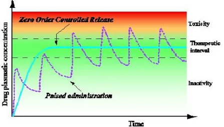
Conventional drug administration usually leads to a “pulsed pattern” of the drug concentration in the blood, characterised by a typical peak and valley trend. During the therapy, the plasmatic concentration of the bioactive agent may fall outside the therapeutic range and the administration of the drug must be repeated after precisely time intervals in order to maintain the therapeutic action. Moreover, the active agent can be sensitive to changes in tissue pHs as well as been attacked by enzymatic degradation, loose some activity and have a reduced half-life. To avoid the rapid breakdown or clearance in vivo, the drug is commonly administered in high concentration, resulting sometimes in strong toxic effects or induced inflammatory processes. On the other side, when the plasmatic concentration drops below the therapeutic level there is a significant decrease of the therapeutic benefits (Figure 1.2). The possibility of optimizing the pharmaceutical form of bioactive agents by means of drug delivery technology gives a great chance to the new biotechnological drugs such as peptides, proteins, and oligonucleotides that usually are very active and often have very narrow therapeutic window.
Figure 1.2. - Variation of drug plasmatic concentration within time following conventional administration and by using controlled release devices.
The drug delivery systems offer several advantages as compared to conventional dosage forms. The system is usually a reservoir of the therapeutic agent, with a specific time release of the drug, thus leading to a partial control of the pharmacokinetic that become complete if the system is also designed for the targeted release of the drug and influences its biodistribution. In addiction, the delivery system safeguards the drug from the attack of enzymatic degradation, it can enhance the penetration of the active agent in the diseased tissues improving its efficiency and reducing the toxicity. A better compliance and convenience of the patient are also achieved [Veronese 2002].
Compared to tradition drug formulation and administration, the advantages of a controlled delivery system can be summarised as follows:
− continuous maintenance of drug levels within the desired therapeutic range; − reduction of harmful side effects;
− potentially decreased amount of drug;
− lower number of administrations and possibly less invasive, leading to improved patient compliance;
− administration of pharmaceuticals with short in vivo half–life (for example peptides and proteins).
In the development of drug delivery systems, the rational should be modified according to the specific biological substance and/or the particular therapeutic situation. Although clinical introduction of the first controlled release systems occurred 30 years ago,
nowadays these systems have an incredible impact on nearly every branch of modern medicine, including cardiology, ophthalmology, endocrinology, oncology, pneumology, immunology, and pain management. In the United States alone, the market size for drug delivery systems in 1997 was over 13 billions US$, in 2002 it raised to about $47 billions and is projected to grow to about $67 billion in 2006. As matter of fact, every year a considerable number of improved devices are presented and adopted for health care. In addition, many of highly profitable blockbuster drugs will reach patent expiry by 2006 and lose about $37 billion in market value to generic competition. Optimizing products through drug delivery might be a successful strategy to re-launch competitive pharmaceutics [Langer 1998, Fuji-Keizai 2003].
Common types of controlled drug release systems are reported by following: o Oral systems
o Formulates for parenteral administration o Therapeutic transdermal delivery systems o Ocular systems
o Intrauterin and intravaginal systems
Recently, the interest of pharmaceutical scientists is mainly focus on the formulation of nanosized drug delivery devices for the administration of protein drugs [Pawar 2004, Bilati 2005] oligonucleotides, DNA and RNA for gene regulation [Lambert 2001, Ravi Kumar 2004, Zhu 2004]. Next generation of drug delivery systems will be influenced by the “intelligent material design” in terms of developing systems sensitive to drug concentration itself and to certain physiological stimuli. Recognitive molecular systems with biosensing properties will be integrated in the delivery device so that the release of the therapeutic will occur only under specific conditions [Kayser 2005].
Among the general characteristics that drug delivery systems should present, it is possible to mention the ability to incorporate the drug without damaging it, tuneable release kinetics, long in vivo stability, biocompatibility, in terms of lack of toxicity and immunogenicity, potential of targeting specific organs and tissues. All of these features are related to the nature of the materials that constitute the delivery system. In particular, the pursuit of an adequate compromise of both bulk and surface property represents an important issue to be addressed. Organic macromolecules have highly tuneable physical–
or functionalized. Accordingly, they very likely represent the best–suited class of materials for drug delivery technology [Kashyap 2004].
1.3.1. Polymer–Based Drug Delivery Systems
Polymeric systems have had an enormous impact on pharmacological therapy. In the past, many efforts were focused on using polymers that have a history of medical use and then adapting their microstructures to provide the desired delivery rate. Intentional design of materials is a more modern approach to solve specific issues of drug delivery [Kashyap 2004]. First generation drug delivery devices were based on drug encapsulation in non– degradable macromolecular matrices from which the drug could diffuse. Therefore, the release kinetics was completely dependent on the diffusive behaviour of the drug through the polymer matrix [Langer 1998]. Today, a second generation of drug delivery devices are under investigation. The polymer matrix is engineered in order to play an active role and permit the selective release of the incorporated drug to a specific site or against a selected tissue (drug targeting). Moreover, due to the in vivo degradability, the release kinetic profile can be precisely modulated [Uhrich 1999]. The first category of release systems consisted of simpler devices that could not be easily applied to particular cases, for example to protein delivery. On the other hand, the second–generation systems are characterized by high selectivity and efficiency, although they need careful control of the macromolecular structure of the polymer matrix. The particular requirements for controlled release applications dictate on each occasion the choice of polymer. Normally, the demand is less severe if the device is designed for external use or for oral administration; it is much more stringent if the system must be given intravenously, subcutaneously or for application into internal body compartments. For the external use, the polymer may be non–biodegradable, such as those having carbon–carbon or silicon– oxygen backbones, whereas biodegradable materials are in great demand for internal applications.
Typical examples of biodegradable and non–biodegradable polymers used in controlled release technology are listed in Table 1.3.
Table 3 - Non–biodegradable and biodegradable polymers commonly used in controlled release applications.
Non–degradable polymers Degradable polymers
Polysiloxanes Polylactides Polyglycolides
Polyacrylates Polycyanoacrylates Polysaccharides
Polyurethanes Polyorthoesters Polycarbonates
Polyethylene Polyphosphazenes Poly(ε–caprolactone)
Polymethacrylates Polyanhydrides
Drug delivery systems can be roughly classified in two main categories, namely systems in which the biological substance release is controlled respectively by the drug diffusion and by the degradation of the device. A historical excursus of the most applied delivery systems is reported briefly by following.
1.3.1.1 Diffusion controlled delivery systems
Diffusion controlled release systems are generally based on non–degradable polymeric materials [Uhrich 1999, Langer 1998], such as:
o Ethylene–vinyl acetate copolymers (EVAc) o Ethylene vinyl alcohol copolymers (EVA)
o Polymers and copolymers of 2–hydroxyethyl methacrylate (HEMA) and more in general poly(methacrylate)s
o Polysiloxanes (silicones)
In these systems, the drug diffuses through the polymer matrix or membrane because of the thermodynamic driving force arising from the different concentration of the drug inside and outside the device. Two different classes of release devices can be identified, namely membrane (reservoir) devices and matrix (monolithic) systems.
1.3.1.1.1 Membrane systems
In membrane systems, the drug is surrounded by a polymeric film (Figure 1.2) whose porosity is not homogeneous, and which determines the drug release rate [Theeuwess
1991]. These devices are more complex than matrix systems, but they offer a higher control of release profiles.
Drug time = 0 Polymer membrane Released drug Polymer membrane time = t
Figure 1.2 - Schematic representation of membrane release systems.
The drug can be a dry powder dispersed in a liquid or entrapped in a solid polymer matrix. The membrane can be a microporous or macroporous polymer film. Its composition changes from one component to a mixture of polymers, or to a heterogeneous matrix in which hydrophilic polymer particles are dispersed in a hydrophobic polymer matrix. The membrane and the active core can be assembled using different technologies: drug and solid membrane lamination into films; drug coating with a volatile solution of the polymer; drug microencapsulation; tubular membrane loading with dissolved or dispersed drug; drug loading in membrane capsules.
This latter approach led to the development of Norplant, small silicone capsules containing contraceptives that are slowly released by diffusion through the polymer for 5 years. The main limit of these systems is that the sustained delivery of ionic species and of molecules with molecular mass over about 400 cannot be accomplished.
1.3.1.1.2 Matrix systems
To address the issues of matrix systems, drugs can be physically embedded in polymers at large enough concentrations to create a series of interconnecting pores through which the drug can slowly diffuse.
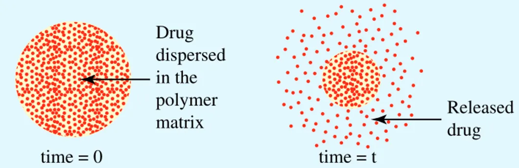
In these systems, the matrix may consist of hydrophobic or viscous hydrophilic polymers in which the solid drug is dispersed (Figure 1.3). Generally, the drug is sparingly soluble in the polymer matrix. These release systems are cheap and readily available, since they
is obtained later by extrusion. The release mechanism is based on the diffusion of the drug molecules to the device surface where they are delivered. This process takes place as long as the higher concentration of the drug in the system core affords a constant flow of drug molecules through the matrix.
In this dissolution–diffusion process, the interface between the drug reservoir and the release moiety progressively moves towards the core of the device.
Drug dispersed in the matrix Drug released from the matrix time = 0 time = t
Figure 13 - Schematic representation of matrix release systems.
1.3.1.2 Degradable delivery systems
In degradable delivery systems, the drug is loaded in a bioerodible and/or biodegradable polymer matrix. The release takes place because of a combination of processes, such as matrix degradation and drug diffusion. The use of these materials was considered in order to avoid the problems related to the physiological excretion or mechanical removal of the not degradable drug delivery devices after their function is completed. Of course, these devices are the preferred ones for internal application. It is worth noting that the combination of diffusion through pores and polymer degradation process provides a further control of the drug release rate.
In ideal systems, the degradation occurs only at the surface of the device, affording a progressive delivery of the drug (Figure 1.4).