Regulation of Mouse Neuropeptide Y Y
1
Receptor Gene
Transcription: A Potential Role for Nuclear Factor-
kB/Rel
Proteins
RITA MUSSO, MARIAGRAZIA GRILLI, ALESSANDRA OBERTO, SILVANA RICCI GAMALERO, and CAROLA EVA Institute of Pharmacology and Experimental Therapeutics, Medical School, University of Torino, 10125 Torino, Italy (R.M., A.O., S.R.G., C.E.), and Division of Pharmacology, Department of Biomedical Sciences and Biotechnology, University of Brescia, 25124 Brescia, Italy (M.G.) Received May 30, 1996; Accepted October 14, 1996
SUMMARY
We previously isolated a 1.3-kb genomic fragment in the 5 9-flanking region of the murine neuropeptide Y (NPY) Y1receptor gene, which is able to drive the expression of LacZ reporter gene in neuronal cells. We determined the ability of deletion mutants of this region to modulate transcription of the heterol-ogous luciferase gene in the Y1receptor-expressing neuroblas-toma/glioma NG108-15 cells and the Y1receptor-deficient 293 cells. Results suggest the presence of a cell type-specific core promoter (2399 to 2218 from the initiator ATG) and, upstream, of two positive and two negative regulatory elements. Se-quence analysis of the Y1 receptor promoter identified two decameric sequences corresponding to consensus binding
sites for nuclear factor-kB/Rel proteins. Gel shift analysis indi-cated that a 29-bp oligonucleotide comprising the two putative kB sites, which we refer to as Y1-kB sequence, specifically bindskB-related complexes in nuclear extracts from rat brain areas, NG108-15 cells, and the murine T cell clone A.E7. In nuclear extracts from A.E7 and NG108-15 cells, the Y1-kB sequence specifically binds an additional complex whose mo-lecular nature remains to be elucidated. Through transient transfection studies, we also demonstrated that the Y1-kB se-quence acts as an enhancer element, inferring its potential role in regulation of the Y1receptor gene expression.
NPY is the most abundant and widely distributed neu-ropeptide within the central nervous system, where it par-ticipates in the control of a large number of physiological functions, including effects on food intake, psychomotor ac-tivity, and central endocrine secretion and potent vasoactive effects on the cardiovascular system (1, 2). Two major sub-types of NPY receptors, Y1and Y2, have been defined on the basis of pharmacological criteria; the Y1receptor is consid-ered to be a postjunctional receptor, and the Y2receptor is considered to be a prejunctional receptor (3). We and others previously reported the molecular cloning of the Y1receptor cDNA from rat (4, 5), mouse (6), and human (7, 8) tissues; its primary structure shows that it belongs to the superfamily of G protein-coupled receptors. In peripheral tissues, the Y1 receptor is found predominantly at the sympathetic postjunc-tional sites in blood vessels, where it mediates the contractile response to NPY of vascular smooth muscle, both directly and indirectly by potentiating the action of other pressure agents, such as norepinephrine (3). In the central nervous system, the Y1receptor has been linked with different phys-iological processes, including stimulation of feeding behavior
(9), stimulation of luteinizing hormone-releasing hormone release (9), a sedative anxiolytic effect (10, 11), and modula-tion of inflammamodula-tion and nocicepmodula-tion (12, 13).
Recent studies have shown that a marked plasticity in the expression of the Y1receptor and its mRNA can be induced under different circumstances. For example, peripheral tis-sue inflammation evokes up-regulation of Y1receptor mRNA in dorsal root ganglia; in the same tissue, peripheral axotomy changes expression of the Y1receptor mRNA level (12, 13). The molecular mechanisms responsible for regulation of Y1 receptor expression are unknown; however, like the mecha-nisms forb2receptors and other G protein-coupled receptor genes (14 –16), they may result from alteration of the tran-scriptional regulatory pathway.
We recently cloned the murine gene of the Y1receptor, and we isolated a 1.3-kb genomic fragment of the 59 flanking region that is able to drive the expression of the lacZ reporter gene in the mouse neuroblastoma/rat glioma NG108-15 cell line and in rat corticostriatal neuron primary cultures but not in the Y1receptor-deficient rat glial and human embry-onic kidney 293 cells (6). Sequence analysis of this region ABBREVIATIONS: NPY, neuropeptide Y; Y1R-LUC, Y1receptor/luciferase fusion gene;kB-Y1-LUC, Y1-kB/Y1receptor/luciferase fusion gene;
Y1-kB, wild-type kB site(s) from the Y1receptor gene; mY1-kB, mutated kB site(s) from the Y1receptor gene; DTT, dithiothreitol, HEPES,
4-(2-hydroxyethyl)-1-piperazineethanesulfonic acid; IL, interleukin; Ig-kB, kB sequence from the immunoglobulin k light chain enhancer region;
AP-1, activator protein-1.
Copyright © by The American Society for Pharmacology and Experimental Therapeutics All rights of reproduction in any form reserved.
MOLECULAR PHARMACOLOGY, 51:27–35 (1997).
27
at ASPET Journals on November 21, 2017
molpharm.aspetjournals.org
revealed the presence of several potential recognition se-quences for known transcription factors, including two decameric sequences corresponding to consensus sites for members of the NF-kB/Rel family of transcription factors, three AP-1 sites, three half-palindromic estrogen-responsive elements, and one cAMP-responsive element (Fig. 1A).
In the current study, we further characterized the up-stream promoter region of the murine Y1 receptor gene through the use of transient transfection assays with NG108-15 and 293 cells. Analysis of Y1-R/LUC constructs containing deletions of the Y1 receptor regulatory region suggested the presence of a 181-bp cell type-specific core promoter spanning nucleotides2399 through 2218 from the initiator ATG and, upstream of this region, of two positive and two negative regulatory elements. Furthermore, we present evidence that members of the NF-kB/Rel family of transcription factors may participate in regulation of the Y1 receptor gene expression. The functional role of this family of inducible, ubiquitous transcription factors has been widely characterized in the periphery, where these proteins respond to a variety of signals and control expression of several genes mainly implicated in inflammatory and immune reactions (for reviews, see Refs. 17–19). More recently, several groups have shown that NF-kB is also abundant in brain, where it was found as both an inducible and a constitutively activated form (20 –27). However, currently, very little is known about the role of NF-kB-related factors in regulation of the
expres-sion of genes whose products play a functional role in the central nervous system.
Materials and Methods
Cell culture. The mouse neuroblastoma/rat glioma NG108-15
cells were plated onto Falcon Petri dishes coated with 10 mg/ml
poly-L-lysine (Mr70–1503 10
3) and were cultured in the minimum
essential medium containing 10% fetal bovine serum and 13 HAT
supplement (all from GIBCO, Grand Island, NY). The human em-bryonic kidney 293 cells were grown in minimum essential medium and 10% fetal bovine serum. Mouse fibroblast NIH 3T3 cells were cultured in Dulbecco’s modified Eagle’s medium containing 10% calf
serum (GIBCO). All culture media contained 2 mM glutamine, 100
units/ml penicillin, and 100mg/ml streptomycin.
Plasmid construction. A SalI/BglII fragment from the original
murine Y1receptor genomic clone (from nucleotides21523 to 2218
relative to the initiator ATG) was first subcloned into the polylinker
of pBluescript SK2 (Stratagene, La Jolla, CA) (6) (Fig. 1B). The
SalI/BglII fragment was isolated by gel electrophoresis; then, the BglII-digested end (all restriction enzymes were from Boehringer-Mannheim Biochemicals, Indianapolis, IN) was filled in with the Klenow fragment of DNA polymerase I (Boehringer-Mannheim) and excess of dNTPs (0.4 mM). The resulting SalI-blunt fragment was
ligated into the SalI/ClaI sites of pBluescript SK2after the ClaI site
of the plasmid was filled to obtain a blunt end. This construct
(pBS-Y1PR) contains 1305 bp of the 59 flanking region of the murine
Y1 receptor gene and includes the first three sites of initiation of
transcription (6). The Y1R-LUC expression plasmids were
con-Fig. 1. Murine Y1R-LUC
expres-sion plasmids. A, Putative cis-act-ing elements residcis-act-ing in the
up-stream region of the murine Y1
receptor gene. Above each box, relative position of the proximal nucleotide in each motif in rela-tion to the initiator ATG. Below each box, nucleotide sequences.
ERE, estrogen-responsive
ele-ment; CRE, cAMP-responsive el-ement. Underlined, sequences in
the Y1-kB motif corresponding to
thekB site. B, Luciferase fusion
constructs containing deletion
fragments of the murine Y1
recep-tor 59 flanking region. Top,
restric-tion fragments that were used for deletion mutants.
at ASPET Journals on November 21, 2017
molpharm.aspetjournals.org
structed through subcloning into the polylinker of pGL2-basic (Pro-mega, Madison, WI) with the following restriction fragments from
pBS-Y1PR: a 1305-bp SalI/HindIII fragment (p1305-LUC), a 985-bp
HpaI/HindIII fragment (p985-LUC), a 686-bp SmaI/HindIII frag-ment (p686-LUC), a 618-bp NsiI/HindIII fragfrag-ment (p618-LUC), a 490-bp SspI/HindIII fragment (p490-LUC), a 306-bp PstI/HindIII fragment (p306-LUC), and a 181-bp EcoRI/HindIII (p181-LUC) frag-ment (see Figs. 1B and 2B). Constructs containing an upstream
sequence between nucleotides21113 and 2960 (p895-LUC,
p876-LUC, p808-p876-LUC, and p742-LUC; see Figs. 1B and 2B) were obtained
through digestion of pBS-Y1PR with HpaI and subsequent treatment
with Bal31 enzyme (Boehringer-Mannheim). The plasmid DNA was then rendered blunt with the Klenow enzyme, digested a second time with HindIII, and subcloned into the SmaI/HindIII sites of pGL2-basic. The junctions between the insert DNAs and luciferase gene of the fusion constructs were confirmed through sequence analysis (28).
Oligonucleotides containing the Y1-kB and mY1-kB (see below)
were synthesized with BglII overhangs, annealed, and inserted into
the BamHI site of pBluescript SK2, resulting inkB-pBS SK2and
mkB-pBS SK2.
To construct pkB-985-LUC, pkB-686-LUC, pmkB-686-LUC, and
pkB-181-LUC plasmids, the deletion fragments of the Y1receptor 59
flanking sequence were obtained from pBS-Y1PR using the unique
HindIII site from the pBluescript SK2polylinker and the
appropri-ate restriction sites in the upstream region of Y1receptor gene and
were ligated immediately downstream of the wild-type and mutated
kB sites of kB-pBS SK2and mkB-pBS SK2. The resulting plasmids
were digested by XbaI, rendered blunt by Klenow enzyme, digested a second time with HindIII, and subcloned into the SmaI/HindIII
sites of the pGL2-basic vector (see Fig. 2B). To obtain the p
kB-893-LUC, pkB-873-LUC, pkB-811-LUC, and pkB-741-LUC plasmids, the
pkB-686-LUC plasmid was digested by SmaI/HindIII to remove the
696-bp insert, resulting in kB-pGL2. The plasmid pBS-Y1PR was
then digested with HpaI and subsequently treated with Bal31 en-zyme. The plasmid DNA was rendered blunt by Klenow enzyme, digested a second time with HindIII, and ligated into the SmaI/ HindIII sites ofkB-pGL2 (see Fig. 2B). The sequences of the
result-ingkB/Y1R-LUC expression plasmids were confirmed through
re-striction analysis and nucleotide sequence determination.
Sequencing of the Y1R-LUC expression plasmids revealed seven
nucleotide errors compared with the previously published 59 flanking
region of the Y1receptor gene.1
Transient transfection experiments. Transfection of reporter
plasmids into NG108-15, 293 and NIH 3T3 cells was performed according to the calcium phosphate coprecipitation method (29). In
all experiments, pSV-b-galactosidase control vector (Promega),
con-taining theb-galactosidase gene linked to the simian virus 40 early
1The corrected DNA sequence is in the EMBL/GenBank database
(acces-sion No. Z18281).
Fig. 2. kB-Y1R-LUC expression
plasmids. A, Putative regulatory
elements residing in the 59
flank-ing region of the Y1 receptor
gene. B, kB-Y1R-LUC fusion
genes containing the Y1-kB motif
ligated immediately upstream of different deletion fragments of the
murine Y1 receptor promoter.
Above each kB-Y1R-LUC
plas-mid, corresponding luciferase fu-sion constructs containing the deletion fragments of the murine
Y1 receptor 59 flanking region.
Mutant constructs were obtained as described in Materials and Methods and were confirmed by sequence analysis and restriction mapping. Restriction fragments that were used for deletion mu-tants are shown in Fig. 1.
1
at ASPET Journals on November 21, 2017
molpharm.aspetjournals.org
promoter/enhancer, was included as an internal control for the dif-ferent transfection efficiencies between experiments. When cells
reached;50% confluence, each 35-mm Petri dish received equimolar
amounts (2.5mg) of test plasmid and pSV-b-galactosidase. Cells were
harvested 48 hr after transfection, and the activities of luciferase and b-galactosidase were assayed as previously described (30). As con-trols, the plasmid pGL2-basic, containing the promoterless luciferase gene, and the plasmid pGL2-promoter vector (Promega), containing the luciferase gene driven by the simian virus 40 promoter, were transfected into parallel cultures of each cell line. In all of the cell lines tested, the pGL2-basic was inactive, whereas the pGL2-pro-moter vector showed high levels of luciferase activity.
Pharmacological treatments of transiently transfected NG108-15 cells were performed 8 and 20 hr before processing of cells.
Nuclear extracts and electrophoretic mobility shift assays.
Nuclear extracts from rat brain areas were prepared essentially as described by Kang et al. (31). Nuclear extracts from cell lines and from A.E7 cells were prepared according to a small-scale protocol
(25) with minor modifications. Briefly, 5–103 106cells were scraped
into cold phosphate-buffered saline, washed once in phosphate-buff-ered saline, and pelleted for 10 sec in an Eppendorff centrifuge. Cells
were then resuspended in 400ml of cold buffer A (10 mM
HEPES-KOH, pH 7.9, 1.5 mM MgCl2, 10 mM KCl, 0.5 mM DTT, 0.2 mM
phenylmethylsulfonyl fluoride), allowed to swell on ice for 10 min, and vortexed for 10 sec. Samples were centrifuged for 10 sec, and the
pellets were resuspended in 50ml of cold buffer C (20 mM
HEPES-KOH pH 7.9, 25% glycerol, 420 mMNaCl, 1.5 mMMgCl2, 0.2 mM
EDTA, 0.5 mM DTT, 0.2 mM phenylmethylsulfonyl fluoride) and
incubated on ice for 20 min for high salt extraction. Cellular debris were removed by centrifugation for 2 min at 4°, and the supernatant
was stored at270°. Protein concentration was assessed by BioRad
(Hercules, CA) Bradford assay according to the manufacturer’s in-structions. DNA binding reaction were initiated by the combination
of 2mg of nuclear extracts with 20,000 cpm (0.1 ng) of g-32P-labeled
oligonucleotide probes in 13 lipage buffer (10 mMTriszHCl, pH 7.5,
50 mMNaCl, 1 mMDTT, 1 mMEDTA, 10% glycerol) containing 0.5mg
of poly(dI/dC) in a total volume of 10ml. In competition experiments,
indicated amounts of unlabeled competitor oligonucleotides were
added with32P-labeled probes. Reactions were carried out for 20 min
at room temperature, and protein/DNA complexes were resolved on
nondenaturing 4% polyacrylamide gels in 13 Tris/glycine/EDTA
buffer (13 5 50 mMTris, 380 mMglycine, and 2.7 mMEDTA). Gels
were then dried and subjected to autoradiography at room temper-ature.
Synthetic DNA oligonucleotides. Oligonucleotide sequences
and their respective complementary strands were synthesized with a DNA synthesizer (Applied Biosystems, Norwalk, CT) and purified through denaturing gel electrophoresis. Oligonucleotides were an-nealed to complementary strands by heating to 68° and cooling slowly to room temperature. For gel shift analysis, double-stranded
oligonucleotides were end-labeled with [g-32P]ATP (. 7000 Ci/mmol;
ICN Pharmaceuticals, Costa Mesa) and T4 polynucleotide kinase
(Boehringer-Mannheim) to obtain a specific activity of.108cpm/mg.
The sequences of the oligonucleotides were as follows: Y1-kB,
59-GATCATGGGATTTCATTGGGATTTCACTT-39 (sense); mY1-kB,
59-GATCCATctcATTTCATTctcATTTCACTT-39 (sense); Ig-kB,
59-CA-GAGGGGACTTTCCGAGAGGC-39; and octamer (octamer binding
site from the IL-2 gene enhancer region), 5
9-TATGTGTAATATGTA-AAACATTTTGACACC-39. Sequences corresponding to thekB site
are underlined.
Statistical analysis. Statistical analysis was performed by using
the Mann-Whitney U test.
Results
Analysis of the effect of progressive deletion within the Y1receptor promoter on heterologous gene
expres-sion. The ability of several deletion mutants of the 59
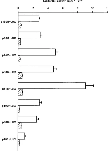
flank-ing region of the murine Y1receptor gene to drive the expres-sion of the luciferase reporter gene was analyzed in transient transfection experiments. As shown in Fig. 3, the 1.3-kb genomic fragment, spanning nucleotides 21523 through 2218 relative to the initiator ATG (p1305-LUC), drives the luciferase activity in NG108-15 cells, whereas a very low enzyme activity was determined in 293 cells. Sequential de-letion from nucleotides 21523 through 21026 (p985-LUC, p895-LUC, p876-LUC, and p808-LUC) had no significant effect on luciferase activity (Figs. 3 and 4). Further deletion to nucleotide 2960 (p742-LUC) resulted in an ;2-fold in-crease in luciferase activity in NG108-15 cells but not in 293 cells, suggesting the presence of a negative regulatory ele-ment between nucleotides21026 and 2960 that is operative in NG108-15 cells. The promoter activity remained un-changed by removal of the region between2960 and 2904 (p686-LUC), whereas further reduction of the upstream se-quence to nucleotide 2836 (p618-LUC) increased the lucif-erase activity by ;2-fold in NG108-15 cells but not in 293
Fig. 3. Deletion analysis of the murine Y1receptor gene promoter. The
promoter activity of the Y1R-LUC expression plasmids was determined
in (open bars) NG108-15 and (hatched bars) 293 cells. Left, tested
reporter plasmids. The luciferase activity was normalized to
b-galacto-sidase activity obtained by cotransfecting cells with the control plasmid
pSV-b-galactosidase (in cpm). Values are mean 6 standard error from
eight or more transfection experiments, each performed in triplicate, with plasmid DNAs from at least two different preparations. Cultures transfected with the promoterless plasmid pGL2-basic had a mean
luciferase activity of 0.166 0.018 and 0.04 6 0.003 cpm 3 1026in
NG108-15 and 293 cells, respectively.
at ASPET Journals on November 21, 2017
molpharm.aspetjournals.org
cells, suggesting that the sequence between nucleotides2904 and2836 contains a negative regulatory element that con-tributes to lower Y1receptor gene expression in this cell type (Fig. 3). Further deletion to nucleotide 2708 (p490-LUC) decreased the luciferase activity to the level driven by the undeleted Y1 receptor promoter (p1305-LUC) in NG108-15 cells, suggesting the presence of a positive cis-acting element between nucleotides2836 and 2708. Extension of 59 deletion to nucleotide2524 (p306-LUC) did not affect the luciferase activity significantly, whereas further deletion to nucleotide 2399 (p181-LUC) reduced the reporter gene expression by 2.8-fold in NG108-15 cells, suggesting the presence of a pos-itive cis-acting element between nucleotides2524 and 2399. The remaining 181 bp-genomic fragment, spanning nucleo-tides 2399 and 2218 of the Y1 receptor gene, drives the luciferase activity in NG108-15 cells at a level significantly above the pGL2-basic (see legend to Fig. 3), indicating that it represents the minimal promoter region still capable of di-recting expression of the Y1receptor gene in the neuroblas-toma/glioma cell line. It should also be pointed out that the same sequence is unable to drive luciferase activity in 293 cells. Furthermore, all of the Y1receptor deletion mutants/ fusion constructs drove negligible luciferase activity when transiently transfected into the mouse fibroblast NIH 3T3 cell line (data not shown).
Binding of Y1-kB sequences to members of the family
of NF-kB transcription factors. Detailed sequence analy-sis of the Y1receptor promoter region reveals the presence of putative binding sites for known transactivating factors that may play a role in the regulation of the tissue-specific
expres-sion of this gene (Fig. 1A). Particularly, we focused our in-terest on the sequence located in position 21302 through 21282 in the Y1receptor gene regulatory region because it contains two decameric sequences corresponding to consen-sus sites for members of the NF-kB/Rel family of transcrip-tion factors (6). We performed experiments to verify whether the aforementioned sequences are indeed binding sites for transcriptional control proteins belonging to this family. A well-studied model of gene regulation mediated by kB/Rel proteins is represented by A.E7 cells, a CD41murine T cell clone in whichkB-mediated gene expression has been exten-sively analyzed. Kang et al. (31) demonstrated that these untransformed cells constitutively express at least two nu-clear complexes belonging to the NF-kB/Rel family: the p50-p65 (relA) heterodimer and the p50 homodimer. In this well-characterized model, we initially studied binding properties of the putativekB sequences from the Y1receptor gene.
An oligonucleotide comprising the twokB sequences from the Y1receptor gene (Y1-kB oligonucleotide) was synthesized, radioactively labeled at the 59 end with T4 kinase, and incu-bated with nuclear extracts prepared from A.E7 cells.
Sub-Fig. 4. Enhancer activity of the Y1-kB sequence in NG108-15 cells.
Histograms show the mean6 standard error of at least eight
transfec-tion experiments, each performed in triplicate. Each reporter plasmid tested (left) was independently prepared at least two times. The
lucif-erase activity was normalized tob-galactosidase activity from a
co-transfected internal control plasmid pSV-b-galactosidase (in cpm). The
luciferase activity of pkB-741-LUC, pkB-686-LUC, and pkB-181-LUC
was significantly more than that of p742-LUC, p686-LUC, and
p181-LUC, respectively (p, p , 0.05).
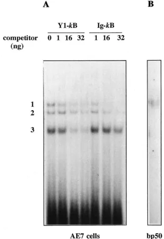
Fig. 5. Analysis of the binding properties of the Y1-kB sequence on
nuclear extracts from a T cell clone endowed with constitutive NF-kB/
Rel activities and on the p50 subunit. A, Oligonucleotide sequence
comprising the twokB sequences from the Y1receptor gene (Y1-kB)
binds three specific nuclear complexes in extracts from a murine T cell clone, A.E7. Electrophoretic mobility shift assay obtained through
in-cubation of 2mg of A.E7 nuclear extracts with32P-labeled Y
1-kB probe
in the absence (0 ng) or in the presence of 1, 16, or 32 ng of unlabeled
Y1-kB or Ig-kB as competitors. B, Affinity purified, bacterially produced
p50 subunit of the NF-kB/Rel family can bind the Y1-kB oligonucleotide
probe. bp50, bacterially purified p50.
1
at ASPET Journals on November 21, 2017
molpharm.aspetjournals.org
sequently, binding reactions were analyzed in a gel shift assay; results are shown in Fig. 5A. The Y1-kB probe could detect three nuclear complexes with different migration properties. All complexes proved to result from specific inter-action with the DNA sequence because they were displaced, in a dose-dependent manner, by the unlabeled oligonucleo-tide Y1-kB. To further analyze the relationship between these nuclear complexes and NF-kB/Rel proteins, an oligonucleo-tide sequence containing a classickB site (Ig-kB) was tested for the ability to compete for binding to the Y1-kB oligonu-cleotide. As shown in Fig. 5A, only the top two migrating complexes were competed, whereas the binding of the third complex was unaffected. These results suggested that the two higher-molecular-weight complexes able to bind the Y1-kB sequence were indeed kB-related proteins, whereas the third complex, which was present in nuclear extracts from A.E7 cells, was also able to specifically bind the exam-ined sequence, but as shown by the results of the competition analysis with the Ig-kB oligonucleotide, we could exclude that it was akB-related protein or even a degradation prod-uct of the higher-molecular-weightkB complexes. To further prove the ability of the Y1-kB sequence to bind kB-related complexes, we confirmed that bacterially expressed, affinity purified p50 protein, one of the members of the NF-kB/Rel family, was indeed able to bind the Y1-kB probe in a gel shift assay (Fig. 5B).
Nuclear extracts were also prepared from NG108-15 cells and tested in a gel shift assay for binding to the32P-labeled Y1-kB sequence. In analogy with what we observed in A.E7
extracts, three retarded complexes were detected (Fig. 6A,
lane 1). When the same extracts were incubated with the
Ig-kB oligonucleotide probe, two complexes were detected, comigrating with the top two complexes bound to the Y1-kB probe (Fig. 6A, lane 2). A detailed competition analysis of the Y1-kB bound complexes was performed (Fig. 6B). Increasing amounts (1–8 ng) of unlabeled Y1-kB oligonucleotide (lanes
2–4) and Ig-kB (lanes 5–7) were used. The two
higher-molec-ular-weight complexes were displaced by both competitors, whereas binding of the third complex seemed to be affected by the Y1-kB but not by the Ig-kB oligonucleotide. None of the complexes were competed by an unrelated oligonucleotide containing the octamer protein binding site (lanes 9–12), even when a higher concentration (16 ng) of competitor was used (lane 12).
Interaction between the Y1-kB sequences and DNA-bind-ing factors was further investigated in nuclear extracts from several rat brain regions, including cortex, hippocampus, striatum, cerebellum, and olfactory bulb. Results of a repre-sentative gel shift assay are shown in Fig. 7. Surprisingly, in this situation, a single DNA binding activity was detected, which seemed to be specific because it could be competed by unlabeled Ig-kB sequence [Fig. 7, lane 6, shows competition on nuclear extracts from rat cortex, but the same results were obtained with extracts from the other rat brain regions (not shown)].
The Y1-kB oligonucleotide sequence from the Y1
re-ceptor gene acts as enhancer element in NG108-15 cells. Although the deletion from nucleotides 21523 to 21203 (p985-LUC) in the Y1receptor sequence did not affect luciferase activity in our in vitro model (Fig. 4), based on the essential role of the NF-kB/Rel family of transcription factors for the expression of several genes (for reviews, see Refs. 17–19), we surmised that the Y1-kB sequence may partici-pate in the regulation of the transcriptional activity of this gene. To test this possibility, we constructed a series of ex-pression plasmids (pkB-Y1R-LUC) in which an oligonucleo-tide corresponding to the Y1-kB sequence was placed imme-diately upstream of deletion fragments of the Y1receptor 59 flanking region that did not contain the endogenous sites (Fig. 2B). Results indicated that the Y1-kB sequence, when placed upstream of nucleotides2959, 2904, and 2399 from the initiator ATG (pkB-741-LUC, 686-LUC, and pkB-181-LUC, respectively), enhances by .2-fold the luciferase activity in NG108-15 cells (Fig. 4). Specificity of the effect was demonstrated by the fact that mutation of selected nu-cleotides within the motifs, which abolished binding activity in gel shift assay (not shown), completely abolished enhancer activity (Fig. 8). Interestingly, the Y1-kB sequence seemed to be functional in NG108-15 cells but not in 293 cells (Fig. 8). Furthermore, the Y1-kB motif failed to increase luciferase activity when placed upstream of nucleotides 21203 (pkB-985-LUC), 21111 (pkB-893-LUC), 21091 (pkB-873-LUC), and 21029 (pkB-811-LUC) suggesting that the 70-bp se-quence spanning nucleotides 21029 and 2959 of the Y1 receptor promoter contains a negative regulatory element that inhibits Y1-kB enhancer activity in NG108-15 cells (Fig. 4). ThekB-related factors were found as both inducible and constitutively activated complexes in the central nervous system (20–27). ThekB nuclear activity interacting with the Y1receptor gene is constitutive. We verified whether specific extracellular signals might further activate the enhancer Fig. 6. Gel shift analysis of nuclear complexes specifically interacting
with the Y1-kB probe in NG108-15 cell extracts. A, Comparison of the
migration properties of complexes intercepted by (lane 1) Y1-kB and
(lane 2) Ig-kB oligonucleotide probes in NG108-15 cell extracts. B,
Competition analysis of the complexes bound by the Y1-kB
oligonu-cleotide probe. Competition was performed by adding the indicated
amounts (in ng) of the unlabeled oligonucleotide sequences Y1-kB
(lanes 2– 4) or Ig-kB (lanes 5–7) or the unrelated oligonucleotide
se-quence for octamer binding proteins (lanes 9 –12).2, No competitor
(lanes 1 and 8). Apparent discrepancies in the migration properties of complexes in A and B are due to differences in gel running length in the two different sets of experiments.
at ASPET Journals on November 21, 2017
molpharm.aspetjournals.org
activity of the Y1-kB sequence. To investigate this possibility, we treated NG108-15 cells transfected with pkB-686-LUC or pkB-181-LUC plasmids with various agents that are known to modulate activity of these transcriptional regulators in either peripheral or central nervous system-derived cells (17, 22, 25–27). In particular, we tested cytokines, such as IL-1 (30 units/ml), IL-2 (2 nM), and tumor necrosis factor-a (100
ng/ml); lipopolysaccharide (25 mg/ml); concanavalin A (25 mg/ml); 12-O-tetradecanoylphorbol-13-acetate (0.1 mM);
hy-drogen peroxide (50 and 100 mM), KCl (30 mM); and gluta-mate (100mM). However, none of these agents were able to
further augment the transcriptional activity of pkB-686-LUC or pkB-181-LUC expression plasmids (data not shown).
Discussion
The Y1receptor subtype plays important roles in mediat-ing NPY-induced control of several functions, includmediat-ing
car-diovascular system activity, neuroendocrine secretion, food intake, and nociception (1, 2). In situ hybridization studies have shown that Y1 receptor mRNA is indeed highly ex-pressed in several regions of the rat forebrain, in the hypo-thalamus, and in dorsal root ganglia (4, 13, 32, 33). We previously isolated the murine gene encoding the Y1receptor and demonstrated that the 59 flanking region of this gene contains a functional promoter that is active in neuronal cells (primary cultured neurons and NG108-15 cells) but not in glial or 293 cells (6).
In the current study, we investigated which regions of the Y1receptor promoter contain potential negative or positive
cis-acting elements participating in the regulation of gene
expression. For this purpose, luciferase constructs compris-ing the 1.3-kb 59 flanking regions of the Y1receptor or vari-ous 59 deletions of it were transfected into a cell line that expresses the Y1receptor endogenously, NG108-15 cells, and in the Y1receptor-deficient 293 cell line. Deletional analysis has shown the presence of several potential areas of tran-scriptional regulation. Our data suggest the presence of at least two positive acting regulatory elements lying between nucleotides 2836 and 2708 and between nucleotides 2524 and2399. Although the sequence of the fragment contained between2836 and 2708 has no clear homology to any known regulatory element, it is noteworthy that an AP-1 binding site resides between nucleotides 2473 and 2479. Further mutational and deletional analysis will be required to define whether this site functions as a cis-acting element. In pre-liminary experiments, we were able to show that the Y1 receptor promoter/luciferase reporter gene can be positively modulated by treatment with phorbol esters but that deletion of the sequence containing the AP-1 site (2473 to 2479) fails to suppress this type of responsiveness (data not shown). Fig. 7. The Y1-kB oligonucleotide sequence binds a single kB-related
complex in nuclear extracts from several rat brain regions. Protein extracts were from hippocampus (lane 1, hipp.), striatum (lane 2), cerebellum (lane 3, cereb.), cortex (lanes 4 and 6), and olfactory bulb (lane 5, olf. bulb). Lane 6, binding competition was performed with 16
ng of the unlabeled oligonucleotide Ig-kB and nuclear extracts from
cortex.
Fig. 8. Mutational analysis of the Y1-kB sequence. A three-nucleotide
mutation was made in eachkB motif from the Y1-kB oligonucleotide
sequence as described in Materials and Methods. The pmkB-686-LUC
fusion plasmid was prepared by ligating the mutated Y1-kB motif
(mY1-kB) immediately upstream of the 686-bp (SmaI/HindIII) deletion
fragment of the Y1receptor promoter. Promoter activity of the
p1305-LUC, p686-p1305-LUC, pkB-686-LUC, and pmkB-686-LUC was determined
in (open bars) NG108-15 and (hatched bars) 293 cells. The enhancer
activity of the mutated Y1-kB motif was depicted in NG108-15 cells. p,
p, 0.05 versus p1305-LUC. pp, p , 0.05 versus p686-LUC.
1
at ASPET Journals on November 21, 2017
molpharm.aspetjournals.org
A second type of regulation that may exist for the Y1 receptor gene expression involves silencer domains. Our work suggests the presence of at least two negative regula-tory elements contained between nucleotides 21026 and 2960 and between nucleotides 2904 and 2836. To assess the cell type specificity of these sequences, 293 and NIH 3T3 cells were transfected with the corresponding luciferase con-structs. All deletion mutants were ineffective in modulating transcription of the heterologous luciferase gene in these cell lines, indicating that the negative regulatory elements of the Y1receptor gene are not responsible for repression in these cell lines.
Reporter gene assay also suggested that the core promoter (2399 to 2218) of the Y1receptor gene exhibited substantial cell type specificity. Ball et al. (34) recently reported that the human Y1receptor gene is under the control of three promot-ers that are activated in a tissue-specific manner. It is note-worthy that the core promoter of the murine Y1receptor gene displays a high sequence homology with the corresponding region of the human promoter directing the expression of the most abundant Y1receptor transcript.
Detailed analysis of the sequence of the murine Y1receptor 59 flanking region reveals the presence of many putative binding sites for known transcription factors (6) (Fig. 1A). We focused our attention on the murine Y1 receptor promoter region that contains two decameric sequences located in tan-dem in position 21302 to 21282 bp, relative to the ATG. These sequences correspond to consensus sites for members of the kB-Rel family of transcription factors (17). In the current study, we showed that this sequence can indeed bind kB-related nuclear complexes in a specific manner and acts as an enhancer element in transiently transfected NG108-15 cells.
NF-kB/Rel proteins are constitutive and inducible tran-scription factors that are present in most cell types. EachkB complex corresponds to homodimers and heterodimers whose subunits belong to a superfamily that comprises at least five DNA binding proteins: p50, p52 (p50B), p65 (RelA), c-rel, and RelB (17–19). The inducible form of NF-kB contains an ad-ditional inhibitory subunit called IkB and can be activated in response to stimuli that mostly represent pathogenic condi-tions, including viruses, bacterial lipopolysaccharide, inflam-matory cytokines, and oxidants. It was previously suggested that only a limited number of lymphoid cells contain consti-tutively active NF-kB-related factors (35, 36). More recent evidence, however, indicates that in the central nervous sys-tem, members of thekB family of transcription factors are constitutively active and are present in the nucleus of cul-tured neurons as well as in neurons in vivo (21, 24, 25).
Our data demonstrate that the Y1-kB sequence binds with high affinity members of thekB/Rel family of transcription factors in nuclear extracts from rat brain areas, from the NG108-15 neuronal cell line, and from the murine T cell clone A.E7. Interestingly, different binding properties were observed in nuclear extracts from different sources. In nu-clear extracts from rat brain regions, a single kB-related complex was detected. In nuclear extracts prepared from the cell line NG108-15, as well as in extracts from the murine T cell clone A.E7, three complexes with different migration properties interacted specifically with the Y1-kB sequence, but only two of them seemed to bekB-related nuclear activ-ities in competition experiments. The molecular nature of the
third complex, specifically interacting with the Y1-kB se-quence, remains to be elucidated. Furthermore, detailed mu-tation studies are necessary to better define the binding requirements of each complex to the Y1-kB sequence contain-ing the twokB sites. Also, it will be interesting to clarify the significance of the differences in binding activities in rat brain extracts compared with extracts from NG108-15 cells and from the murine T cell clone A.E7. It is noteworthy that the single complex identified by the Y1-kB oligonucleotide probe in rat brain extract is reminiscent of the binding spec-ificity of akB-binding site recently identified and character-ized in the regulatory region of the amyloid precursor pro-tein, which in rat brain extracts recognizes specific complexes that are either identical or very similar to p50 homodimers (25).
In transient transfection assays, we also demonstrated that the Y1-kB sequence behaves as an enhancer element when placed upstream of deletion fragments of the Y1 recep-tor regularecep-tory region. Surprisingly, the Y1-kB site does not enhances the transcriptional activity of the fusion gene con-structs when placed$70 bases upstream of nucleotide 2959 relative to the initiator ATG. These results suggest that the 70-bp region lying between21029 and 2959 bases of the 59 flanking region of the Y1receptor gene might contain a neg-ative regulatory element that is able to suppress the en-hancer activity of Y1-kB sequence in NG108-15 cells.
The NF-kB activity interacting with the Y1receptor gene promoter seems to be a constitutive activity, but it is well known that these transcriptional activators can be present as both activated and inducible forms in neurons (20, 22, 25– 27). A wide variety of stimuli can modulate NF-kB/Rel activ-ities, depending on the cell type (17–19). However, the treat-ment of NG108-15 cells with several agents, including inflammatory cytokines, oxidants, bacterial lipopolysaccha-ride, and neurotransmitters, failed to stimulate the tran-scriptional activity of the Y1-kB sequence. It is possible that in NG108-15 cells, the intracellular signals that activate NF-kB proteins are coupled to specific membrane receptors that we were unable to identify, or that in this tumoral cell line, the Y1-kB binding activity is maximally up-regulated. To answer these questions, we are analyzing the modulation of the Y1-kB binding activity in primary cultures of neuronal cells.
The results that we report are, to our knowledge, the first demonstration that akB-related motif contained in the reg-ulatory region of a neuropeptide receptor gene binds kB-related nuclear proteins and might be activated by this fam-ily of transcription factors. The functional significance of these data remains to be elucidated. Kaltschmidt et al. (22) suggested that in neurons, thekB-related factors might par-ticipate in the normal physiology and development of the nervous system. Sequence analysis revealed the presence of putative kB-related sequences in the regulatory region of other neuropeptide receptor genes, such as the vasoactive intestinal peptide receptor (37) and thed-opioid receptor (38). It is possible that this family of transcription factors partic-ipates in the control of neurotransmission by transcription-ally regulating the expression of neuropeptide and neuro-transmitter receptor genes.
In most cell types, the kB-related proteins mediate an immediate-early response to stimuli that represent stress conditions. Vascular responsiveness to NPY was shown to be
at ASPET Journals on November 21, 2017
molpharm.aspetjournals.org
increased in conditions that were occurring physiologically during prolonged stress or in disease states, such as hyper-tension (39). In addition, NPY seems to play a critical role in the transmission of stress-related information to the hypo-thalamic/hypophysial system and in the activation of neu-roendocrine responses essential for the survival of the organ-ism (40). An interesting possibility is that the Y1receptor for NPY may represent one of thekB site-containing genes that is modulated in the mammalian nervous system by kB-re-lated factors in response to stimuli that require an immedi-ate defensive response.
Acknowledgments
We thank Prof. F. Altruda (Department of Biology, Genetics and Medical Chemistry, University of Torino, Torino, Italy), Dr. D. For-nasari (CNR Center of Cellular and Molecular Pharmacology, Mi-lano, Italy), and Dr. L. Varesio (Laboratory of Molecular Biology, Istituto G. Gaslini, Genova, Italy) for critical reading of the manu-script.
References
1. Heilig, M., and E. Widerlo¨v. Neuropeptide Y: an overview of centrally distribution, functional aspects, and possible involvement in neuropsychi-atric illness. Acta Psychiatr. Scand. 82:95–114 (1990).
2. Allen, J. M., and J. I. Koenig. Central and peripheral significance of neuropeptide Y and its related peptides. Ann. N. Y. Acad. Sci. 611: (1992). 3. Wahlestedt, C., and D. J. Reis. Neuropeptide Y-related peptides and their receptors: are the receptors potential therapeutic drug targets? Annu. Rev.
Pharmacol. Toxicol. 32:309–352 (1993).
4. Eva, C., K. Keinanen, H. Monyer, P. Seeburg, and R. Sprengel. Molecular cloning of a novel G protein-coupled receptor that may belong to the neuropeptide receptor family. FEBS Lett. 271:81–84 (1990).
5. Krause, J., C. Eva, P. H. Seeburg, and R. Sprengel. Neuropeptide Y1
subtype pharmacology of a recombinantly expressed neuropeptide recep-tor. Mol. Pharmacol. 41:817–821 (1992).
6. Eva, C., A. Oberto, R. Sprengel, and E. Genazzani. The murine NPY-1 receptor gene: structure and delineation of tissue specific expression.
FEBS Lett. 314:285–288 (1992).
7. Herzog, H., Y. J. Hort, H. J. Ball, G. Hayes, J. Shine, and L. A. Selbie. Cloned human neuropeptide Y receptor couples to two different second messenger systems. Proc. Natl. Acad. Sci. USA 89:5794–5798 (1992). 8. Larhammar, D., A. G. Blomqvist, F. Yee, E. Jazin, H. Yoo, and C.
Wahl-estedt. Cloning and functional expression of a human neuropeptide Y/pep-tide YY receptor of the Y1 type. J. Biol. Chem. 267:10935–10938 (1992). 9. Kalra, S. P., A. Sahu, P. S. Kalra, and W. R. Crowley. Hypothalamic
neuropeptide Y: a circuit in the regulation of gonadotropin and feeding behavior. Ann. N. Y. Acad. Sci. 611:273–283 (1990).
10. Heilig, M., B. So¨derpalm, J. A. Engel, and E. Widerlo¨v. Centrally admin-istered neuropeptide Y (NPY) produces anxiolytic-like effects in animal anxiety models. Psychopharmacology 98:524–529 (1989).
11. Wahlestedt, C., E. Merlo Pich, G. F. Koob, F. Yee, and M. Heilig. Modu-lation of anxiety and neuropeptide Y-Y1 receptors by antisense oligonu-cleotides. Science (Washington D. C.) 259:528–531 (1993).
12. Ru-Rong, J., X. Zhang, Z. Wiesenfeld-Hallin, and T. Ho¨kfelt. Expression of neuropeptide Y and neuropeptide Y (Y1) receptor mRNA in rat spinal cord and dorsal root ganglia following peripheral tissue inflammation. J.
Neu-rosci. 14:6423–6434 (1994).
13. Zhang, X., Z. Wiesenfeld-Hallin, and T. Ho¨kfelt. Effect of peripheral axo-tomy on expression of neuropeptide Y1 receptor mRNA in rat lumbar dorsal root ganglia. Eur. J. Neurosci. 6:43–57 (1994).
14. Collins, S., J. Altschmied, C. Herbsman, M. G. Caron, D. L. Mellon, and R. J. Lefkowitz. A cAMP response element in theb2-adrenergic receptor gene confers transcriptional autoregulation by cAMP. J. Biol. Chem. 265: 19330–19335 (1990).
15. Hershey, A. D., P. E. Dykema, and J. E. Krause. Organization, structure, and expression of the gene encoding the rat substance P receptor. J. Biol.
Chem. 266:4366–4374 (1991).
16. Zhou, Q.-Y., C. Li, and O. Civelli. Characterization of gene organization and promoter region of the rat dopamine D1 receptor gene. J. Neurochem. 59:1875–1883 (1992).
17. Grilli, M., J. J.-S. Chiu, and M. J. Lenardo. NF-kB and Rel participants in
a multiform transcriptional regulatory system. Int. Rev. Cytol. 143:1–62 (1993).
18. Liou, H. C., and D. Baltimore. Regulation of the NF-kB/rel transcription factor and IkB inhibitor system. Curr. Opin. Cell Biol. 5:477–487 (1993). 19. Bauerle, P. A., and T. Henkel. Function and activation of NF-kB in the
immune system. Annu. Rev. Immunol. 12:141–179 (1994).
20. Kaltschmidt, C., B. Kaltschmidt, and P. A. Bauerle. Brain synapses con-tain inducible forms of the transcription factor NF-kB. Mech. Dev. 43:135– 147 (1993).
21. Kaltschmidt, C., B. Kaltschmidt, H. Neumann, H. Wekerle, and P. A. Bauerle. Constitutive NF-kB activity in neurons. Mol. Cell Biol. 14:3981– 3991 (1994).
22. Kaltschmidt, C., B. Kaltschmidt, and P. A. Bauerle. Stimulation of iono-tropic glutamate receptors activates transcription factor NF-kB in primary neurons. Proc. Natl. Acad. Sci. USA 92:9618–9622 (1995).
23. Bakalkin, G. Y., T. Yakovleva, and L. Terenius. NF-kappa B-like factors in the murine brain: developmentally-regulated and tissue-specific expres-sion. Mol. Brain Res. 20: 137–146 (1993).
24. Rattner, A., M. Korner, M. D. Walker, and Y. Citri. NF-kB activates the HIV promoter in neurons. EMBO J. 12:4261–4267 (1993).
25. Grilli, M., M. Ribola, A. Alberici, A. Valerio, M. Memo, and P. F. Spano. Identification and characterization of a NF-kB/Rel binding site in the regulatory region of the amyloid precursor protein gene. J. Biol. Chem. 270:26774–26777 (1995).
26. Grilli, M., F. Goffi, M. Memo, and P. F. Spano. Interleukin-1b and gluta-mate activate the NF-kB/Rel binding site from the regulatory region of the amyloid precursor protein gene in primary neuronal cultures. J. Biol.
Chem. 271:15002–15007 (1996).
27. Guerrini, L., F. Blasi, and S. Denis-Donini. Synaptic activation of NF-kB by glutamate in cerebellar granule neurons in vitro. Proc. Natl. Acad. Sci.
USA 92:9077–9081 (1995).
28. Sanger, F., S. Nicklen, and A. R. Coulson. DNA sequencing with chain-termination inhibitors. Proc. Natl. Acad. Sci. USA 74:5463–5467 (1977). 29. Chen, C., and H. Okayama. High-efficiency transformation of mammalian
cells by plasmid DNA. Mol. Cell. Biol. 7(8):2745–2752 (1987).
30. Glembotski, C. C., C. E. Ironns, K. A. Krown, S. F. Murray, A. B. Sprenkle, and C. A. Sei. Myocardiala-thrombin receptor activation induces hyper-trophy and increases atrial natriuretic factor gene expression. J. Biol.
Chem. 268:20646–20652 (1993).
31. Kang, S.-M., A.-C. Tran, M. Grilli, and M. J. Lenardo. NF-kB subunit regulation in nontransformed CD41T lymphocytes. Science (Washington
D. C.) 256:1452–1456 (1992).
32. Mikkelsen, J. D., and P. J. Larsen. A high concentration of NPY (Y1)-receptor mRNA-expressing cells in the hypothalamic arcuate nucleus.
Neurosci. Lett. 148:195–198 (1992).
33. Larsen, P. J., S. P. Sheikh, and J. D. Mikkelsen. Neuropeptide Y Y1 receptors in the rat forebrain: autoradiographic demonstration of [125I][Leu31,Pro34]-NPY binding sites and neurons expressing Y1 receptor
mRNA. J. Recept. Signal Transd. Res. 15:457–472 (1995).
34. Ball, H. J., J. Shine, and H. Herzog. Multiple promoters regulate tissue-specific expression of the human NPY-Y1 receptor gene. J. Biol. Chem. 270:27272–27276 (1995).
35. Griffin, G. E., K. Leung, T. M. Folks, S. Kunkel, and G. J. Nabel. Activation of HIV gene expression during monocyte differentiation by induction of NF-kB. Nature (Lond.) 339:70–73 (1989).
36. Schreck, P., P. Rieber, and P. A. Bauerle. Reactive oxygen intermediates as apparently widely used messengers in the activation of NF-kB transcrip-tion factor and HIV-1. EMBO J. 10:2247–2258 (1991).
37. Sreedharan, S. P., J.-X. Huang, M.-C. Cheung, and E. J. Goetzl. Structure, expression, and chromosomal localization of the type I human vasoactive intestinal peptide receptor gene. Proc. Natl. Acad. Sci. USA 92:2939–2943 (1995).
38. Augustin, L. B., R. F. Felsheim, B. H. Min, S. M. Fuchs, J. A. Fuchs, and H. H. Loh. Genomic structure of the moused opioid receptor gene.
Bio-chem. Biophys. Res. Commun. 207:111–119 (1995).
39. Zukowska-Grojec, Z., and C. Wahlestedt. Origin and actions of neuropep-tide Y in the cardiovascular system, in The Biology of Neuropepneuropep-tide Y and
Related Peptides (M. F. Colmers and C. Wahlestedt, eds.). Humana Press,
Totowa, 315–388 (1993).
40. McDonald, J. K., and J. I. Koenig. Neuropeptide Y actions on reproductive and endocrine functions, in The Biology of Neuropeptide Y and Related
Peptides (W. F. Colmers and C. Wahlestedt, eds.). Humana Press, Totowa,
419–456 (1993).
Send reprint requests to: Carola Eva, Ph.D., Instituto di Farmacologia e Terapia Sperimentale, Via Pietro Giuria, 13, 10125 Torino, Italy. E-mail: [email protected]
1
at ASPET Journals on November 21, 2017
molpharm.aspetjournals.org