Article
CYTOTOXIC EFFECT OF TRABECTEDIN IN
HUMAN ADRENOCORTICAL CARCINOMA CELL
LINES AND PRIMARY CELLS
Andrea Abate1, Elisa Rossini1, Sara A. Bonini1, Martina Fragni1, Deborah Cosentini2, Guido A.M.
Tiberio3, Diego Benetti4; Constanze Hantel5,6, Marta Laganà2, Salvatore Grisanti2, Massimo Terzolo7; Maurizio Memo1, Alfredo Berruti2, Sandra Sigala1,*
1
Section of Pharmacology, Department of Molecular and Translational Medicine, University of Brescia, Brescia, Italy;
2 Oncology Unit, Department of Medical and Surgical Specialties, Radiological Sciences, and Public Health,
University of Brescia at ASST Spedali Civili di Brescia, Brescia, Italy;
3
Surgical Clinic, Department of Clinical and Experimental Sciences, University of Brescia at ASST Spedali Civili di Brescia, Brescia, Italy;
4 Thoracic Surgery Unit, ASST Spedali Civili of Brescia, Brescia, Italy; 5
Klinik für Endokrinologie, Diabetologie und Klinische Ernährung, Universitätsspital Zürich, Zurich, Switzerland;
6
Medizinische Klinik und Poliklinik III, University Hospital Carl Gustav Carus Dresden, Germany;
7 Department of Clinical and Biological Sciences, University of Turin, Internal Medicine 1, San Luigi Gonzaga
Hospital, Orbassano, Italy.
* Correspondence: [email protected]; Tel.: + 0039 +030 3717663
Received: date; Accepted: date; Published: date
Abstract: Mitotane is the only drug approved for the treatment of AdrenoCortical
Carcinoma (ACC). The regimen to be added to mitotane is a chemotherapy including etoposide, doxorubicin, and cisplatin. This pharmacological approach however has a limited efficacy and significant toxicity. Evidence indicates that ACC seems to be sensible to alkylating agents. Trabectedin is an anti-tumor drug that acts as an alkylating agent, with a complex mechanism of action. Here, we investigated whether trabectedin could exert a cytotoxic activity in in vitro cell models of ACC.
Cell viability was evaluated by MTT assay on ACC cell lines and primary cell cultures. The gene expression was evaluated by q-RT-PCR, while protein expression and localization was studied by western blot and immunocytochemistry. Combination experiments were performed to evaluate their interaction on ACC cell line viability.
1 2 3 4 5 6 7 8 9 10 11 12 13 14 15 16 17 18 19 20 21 22 23 24 25 26 27 28 29 30 31 32 33 34 35 36 37 38
Trabectedin demonstrated high cytotoxicity at sub-nanomolar concentrations in ACC cell lines and patient-derived primary cell cultures. The drug was able to reduce /β catenin nuclear localization, although it is unclear whether this effect is involved to the observed cytotoxicity. Trabectedin/mitotane combination exerted a synergic cytotoxic effect in NCI-H295R cells.
Trabectedin has antineoplastic activity in ACC cells. The synergistic cytotoxic activity of trabectedin with mitotane provides the rationale for testing this combination in a clinical study.
Keywords: trabectedin, adrenocortical carcinoma, cytotoxicity, in vitro, cell lines,
primary cell cultures
1. Introduction
Adrenocortical carcinoma (ACC) is a rare and aggressive tumor characterized by an estimated incidence of 0.7-2.0 cases/million people per year and an overall 5-year survival rate less than 15% in patients with metastatic disease [1]. To date, radical surgery at experienced centers still remains the only potential curative treatment for patients with early disease stage and those with locally advanced ACC responding to neoadjuvant treatment. However, 30-70% of radically operated patients recur within two years often with metastatic disease [1]. Clinical manifestations of ACC can be consequences of adrenal hormone excess (functional tumors) or growing abdominal masses. Standard systemic therapies for patients with advanced ACC are mitotane and chemotherapy [2]. The dichlorodiphenyl trichloroethane derivative mitotane represents the only drug approved for the treatment of ACC since many decades, although its mechanism of antineoplastic activity is not fully understood [3] Clinical evidence shows that the efficacy of mitotane is strictly dependent on the attaining and maintenance over time of circulating blood levels within the 14-20 mg/dL range [2]. Due to the pharmacokinetic characteristics of mitotane, this value is usually reached in about 2-3 months [4] and this long latency could impair at least in part the efficacy of the treatment. The standard chemotherapy regimen to be added to mitotane as first-line treatment for advanced ACC is the combination of EDP (Etoposide, Doxorubicin, Cisplatin) [5, 6]. The EDP-M regimen however has a limited efficacy and its administration is burdened by significant toxicity. Moreover, no efficacious second line therapies are available up to now [7, 8]. Therefore, new treatment strategies are needed. DNA alkylation appears to be a critical point for inducing cytotoxicity in the ACC, indeed, cisplatin is a fundamental component of the EDP-M scheme. Furthermore, results obtained both in vitro [9] and in vivo [10] support the antineoplastic activity of another alkylating agent such as temozolomide against ACC. Trabectedin (ET-743) is a powerful anti-tumor drug isolated from the Caribbean tunicate Ecteinascidia turbinata. The compound is approved for the treatment of soft tissue sarcoma (STS) and relapsed platinum-sensitive ovarian 39 40 41 42 43 44 45 46 47 48 49 50 51 52 53 54 55 56 57 58 59 60 61 62 63 64 65 66 67 68 69 70 71 72 73 74 75 76 77 78 79
cancer (OC). Although trabectedin acts as an alkylating agent, its mechanism of action is more complex than expected. Indeed, the drug interferes with the minor groove, binding the guanine to the exocyclic N2 amino group [11], modifying the recognition of GC-enriched sequences of transcriptional factors, often involved in the oncogene transcription. It has been observed that trabectedin interferes with FUS-CHOP and EWS-CHOP fusion proteins, inhibiting their ability to induce the transcription of different oncogenes [12, 13] Furthermore, in vitro studies demonstrate that trabectedin down regulates the Wnt/ β-catenin pathway in cell cultures of biliary tract adenocarcinoma [14] Finally, trabectedin was shown to modulate the activity of immune cells in the tumor microenvironment, reducing the production of pro-inflammatory mediators (for a review see [11]). These assumptions leaded us to hypothesize that trabectedin could exert a cytotoxic effect also in ACC. To explore this issue, we took advance of the experimental in vitro model of ACC cell lines as well as patient-derived ACC primary cell cultures.
2. Results.
2.1. Trabectedin induced cytotoxicity in ACC cell lines
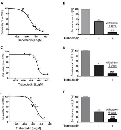
Exposure of NCI-H295R cells to increasing concentrations of trabectedin (0.0625 – 0.750 nM) for 4 days led to a concentration-dependent reduction of cell viability. Sigmoidal concentration-response function was applied to calculate the IC50 value of trabectedin, which was 0.15 nM (95% confidence interval [CI]: 0.13 - 0.17nM). The drug was highly active in inducing cytotoxicity, as its efficacy reached about 90% at the highest concentration tested (Fig. 1A).
80 81 82 83 84 85 86 87 88 89 90 91 92 93 94 95 96 97 98 99 100 101
Figure 1. Cytotoxic effect of trabectedin in ACC cell models. (A) Concentration-response curve of trabectedin-induced inhibition of cell viability of in NCI-H295R cells. Cells were treated with increasing concentration of trabectedin (0.0625 nM – 0.75 nM) for 4 days. (B) Cytotoxic effect lasted after trabectedin withdrawn. Cells were treated with the trabectedin IC50 (0.15 nM) for 4 days, then trabectedin was withdrawn from medium and cells were kept in culture for further 4 days. (C) MUC-1 cells were treated for 5 days with increasing concentrations (0.125 nM – 2.0 nM) of trabectedin. (D) MUC-1 cells were treated with 0.80 nM trabectedin for 5 days then trabectedin was withdrawn from medium and cells were kept in culture for further 5 days. (E) The sub-clone HAC-15 cells were treated for 6 days with increasing concentrations (0.125 nM – 1.5 nM) of trabectedin. (F) HAC-15 cells were treated with 0.50 nM trabectedin for 6 days, then trabectedin was withdrawn from medium and cells were kept in culture for further 6 days. Cell viability was analyzed by MTT assay. Results are expressed as percent of viable cells vs untreated cell ± SD; * p < 0.0001 vs control; # p < 0.001 vs control; ** p < 0.01 vs control; ## p < 0.0001 vs trabectedin-treated cells.
The cytotoxic effect of trabectedin induced DNA fragmentation (Supplemental Figure 1) and apoptotic cell death (Supplemental Figure 2). Cells were then plated and cultured in complete medium added with 0.15nM trabectedin, cell viability was assessed at 4 days of treatment, then the drug was withdrawn and cells were kept in drug-naive complete medium, to evaluate whether the trabectedin cytotoxic insult was a long-lasting effect. Results show that trabectedin treatment induced a cell damage that progressed also in the absence of the drug (Fig. 1B).
102 103 104 105 106 107 108 109 110 111 112 113 114 115 116 117 118 119 120 121
The cytotoxic effect of trabectedin was then studied in other ACC experimental cell line models. As shown in Fig. 1, trabectedin exerted a cytotoxic effect in other ACC cell line models as well, although with different sensitivity, accordingly with their different phenotype, indeed, as indicated in the Methods, HAC-15 is a subclone of NCI-H295R, while MUC-1 is a EDP-M resistant cell line recently established. Concentration-response curves of trabectedin in MUC-1 and HAC-15, are reported in Fig. 1C and 1E. Analysis of the curves allowed the evaluation of the respective IC50, which was 0.80 nM (95% CI: 0.77 - 0.83 nM) in MUC-1 cells, 0.50 nM (95% CI: 0.30 - 0. 82 nM) in HAC-15 cells. In line with results obtained in NCI-H295R cells, trabectedin induced a cell damage leading to cell death that continued in drug-withdrawn conditions (Fig. 1D, 1F). Supplemental Figure 3 reported results obtained with SW13 cells, which is of adrenal origin, but it has been suggested to be a small cell carcinoma. These cells are as well sensitive to the cytotoxic effect of trabectedin and the IC50 was 0.098 nM (95% CI: 0.0093 – 0.104 nM). When cells were exposed to the IC50 trabectedin for 3 days and then transferred in drug-free medium, the cytotoxic insult elicited by trabectedin induced cell death.
2.2. Trabectedin-induced cytotoxicity in ACC primary cell cultures
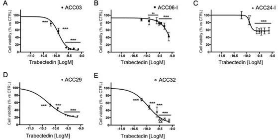
Primary cell cultures were prepared from tissue samples obtained from ACC patients who underwent surgery, as described in the Methods section. Trabectedin exerted a concentration-dependent reduction of human ACC primary cell viability (Fig. 2), however, as expected, due to the different patient tumor stage and tumor cell characteristics, ACC primary cells displayed a different drug sensitivity.
Figure 2. Cytotoxic effect of trabectedin in primary cell cultures derived from ACC patients. Cells were treated with increasing concentration of trabectedin (0.0625 nM – 0.75 nM) for four days. Cell viability was analyzed by MTT assay. Results are expressed as percent of viable cells vs untreated cells ± SD; ** p < 0.001; *** p < 0.0001.
Table 1 reports the in vitro efficacy of trabectedin in ACC primary cultures, measured as percentage of maximum cytotoxic effect, and the trabectedin IC50 for each cell culture. In particular, ACC03, ACC29 and ACC32 displayed the higher sensitivity, as the trabectedin-induced cytotoxicity was over 80% compared to untreated cells, with the IC50 that was within low nanomolar concentrations (range: 0.08 - 0.13 nM). 122 123 124 125 126 127 128 129 130 131 132 133 134 135 136 137 138 139 140 141 142 143 144 145 146 147 148 149 150 151 152 153
Table 1. Effects of trabectedin in ACC primary cultures.
Primary culture
identification IC50 (95% confidence interval) Maximum effect
ACC03 0.13 nM (0.12 nM to 0.14 nM) 92 ± 0.63%
ACC06-I ambiguous 57 ± 11.8%
ACC24-I 0.13 nM (0.10 nM to 0.17 nM) 41 ± 8.2%
ACC29 0.053 nM (0.048 nM to 0.58 nM) 77.2 ± 0.5% ACC32 0.11 nM (0.087 nM to 0.13 nM) 88.6 ± 4.9%
2.3. Trabectedin enhanced cytotoxicity induced by mitotane in NCI-H295R cell line
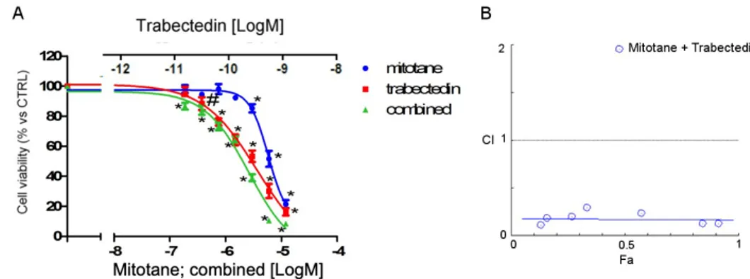
As mitotane is the reference drug for the treatment of ACC, to evaluate whether trabectedin could enhance the cytotoxicity of mitotane in NCI-H295R cells, a combined treatment was performed, applying the Chou-Talalay approach (Fig. 3).
Figure 3. Effect of the trabectedin/mitotane combination on NCI-H295R cell viability. Cells were treated with increasing concentrations of trabectedin and mitotane alone or in combination at fixed concentration molar ratio (trabectedin: mitotane; 1: 10000 molar ratio, for 4 days. Cell viability was measured by MTT. (A) Concentration-response curves. Cells were exposed to increasing concentrations of trabectedin and mitotane alone or in combination at fixed concentration trabectedin : mitotane = 1:10.000 molar ratio. Results are expressed as percent of viable cells vs untreated cell ± SD. (B) Combination Index plot. Dose and effect data obtained were converted to Fa values and analyzed with CompuSyn software. #p < 0.01; *** p < 0.0001.
The concentration-response of each drug and of the combined treatment is reported in Fig.3A. The trabectedin/mitotane combined treatment in NCI-H295R cells induced a synergistic cytotoxic effect compared to each single compound. 154 155 156 157 158 159 160 161 162 163 164 165 166 167 168 169 170 171 172
Results obtained were converted to Fa values and analyzed with CompuSyn software and the Combination Index plot, showing the synergistic effect was shown in Fig. 3B. The Combination Index value is <1 at each drug combination tested, thus suggesting a mainly synergic effect.
2.4. Trabectedin affects β-catenin localization in NCI-H295R cell line
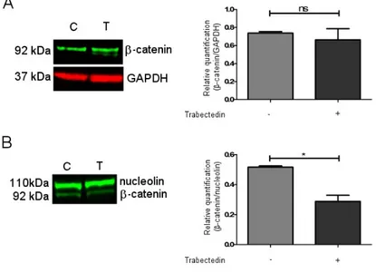
It has been shown in the cholangiocarcinoma model that the Wnt/β catenin pathway could be a target of trabectedin [14]. Based on these findings, we investigated whether trabectedin could influence β-catenin expression and function in NCI-H29R cells. Western blots (Supplemental Fig. 4) were performed on trabectedin-exposed NCI-H295R cells and results demonstrated that the total protein expression of β-catenin was not modified by drug exposure (Fig. 4A), while, when the nuclear/cytoplasm subcellular localization was investigated, we observed that after 3 days of trabectedin treatment at its IC50 value, the β-catenin nuclear concentration was reduced by 44 ± 7.3% in treated cells compared to vehicle-treated cells. (Fig. 4B).
Figure 4. Effect of trabectedin on the β -catenin expression by western blot (WB) technique. NCI-H295R cells were exposed to 0.15 nM of trabectedin for 3 days. (A) A representative WB on total lysate is shown. (B) A representative WB on nuclear protein fraction is shown. * p < 0.01.
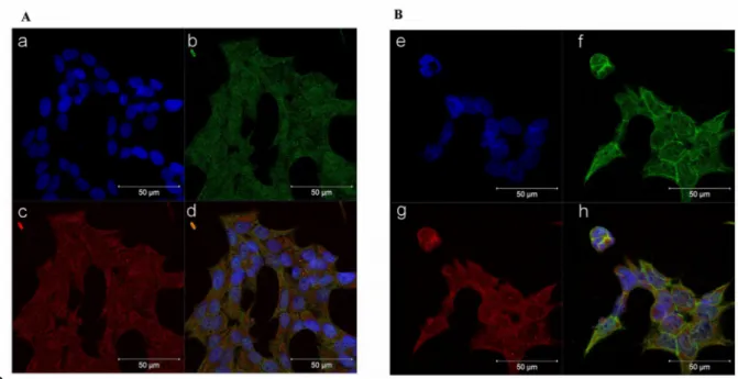
Representative figure of subfraction proteins is reported in Supplemental Fig. 5. Furthermore, immunocytochemistry experiments performed in the NCI-H295R cells and reported in Fig. 5 indicate that after 72 hours of trabectedin treatment, the co-localization of β-catenin with the constitutive proteasome subunit PSMB5 was increased, thus suggesting that the drug induced a translocation of β-catenin from the nucleus to the cytoplasm, in particular to the proteasome, leading to its 173 174 175 176 177 178 179 180 181 182 183 184 185 186 187 188 189 190 191 192 193 194 195 196 197 198 199
degradation. Indeed, β-catenin translocation from nucleus to cytoplasm is necessary to turning off its transcription-factor activity and to direct the protein to proteasomal degradation [15]. A time course is shown in Supplemental Figure 6.
Figure 5. Trabectedin exposure affects the subcellular localization of β-catenin in NCIH295R cells. Cells were treated with 0.15 nM trabectedin for 3 days. Untreated (A) and trabectedin (B) treated cells were analyzed for β-catenin localization following by incubation with Hoechst for nuclear staining. Panels a, e: Hoechst; panels b, f: β-catenin; panels c, g: constitutive proteasome subunit PSMB5; panels d, h: merge. The scale bar of 50 µm is automatically inserted by the software ZEN Black.
2.5. Trabectedin affects the expression of β-catenin target genes
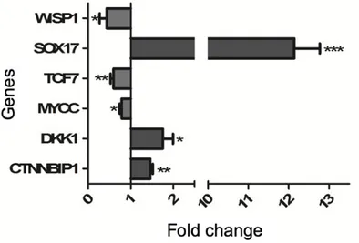
Whether the reduction of β-catenin nuclear localization can affect the gene expression regulator was investigated taking advance of the specific Wnt\ β-catenin pathway signaling RT2 profiler array, as described in Methods. Representative genes modified by trabectedin treatment were then validated by qRT-PCR analysis in vehicle-treated and trabectedin-treated NCI-H295R cells. In particular, we selected target genes among those which are known to be critical in cancer development, namely TCF7, DKK1, WISP1, MYCC and SOX17. Trabectedin exposure significantly increased the expression of genes that encode proteins involved in the reduction of Wnt activity, i.e. CTNNBIP1, DKK1, SOX17, while the protoncogene MYCC and genes activating the Wnt pathway, namely TCF7 and WISP1 are significantly reduced in NCI-H295R cells after exposure to trabectedin (Fig. 6). 200 201 202 203 204 205 206 207 208 209 210 211 212 213 214 215 216 217 218 219 220 221 222
Figure 6. Validation of selected Wnt/ β-catenin pathway by qRT-PCR in trabectedin-treated NCI-H295R cells. RNA was extracted and retro-transcribed as described in Materials and methods. Genes were measured by q-RT-PCR, using SYBR Green as fluorochrome. Results are presented as fold change ± SEM; * p < 0.01; ** p < 0.05; *** p < 0.001.
3. Discussion
Trabectedin displays a manifold mechanism of action, which impacts on tumor cell biology and tumor microenvironment. The alkylating activity together with the inhibition of transcription, due to the interaction with the RNA polymerase II and DNA repair proteins, appear to be the hallmarks of the antiproliferative activity of this drug [16].
The present study was undertaken to investigate the trabectedin antitumor activity in in vitro experimental cell models of ACC. The results revealed that trabectedin is highly cytotoxic at sub-nanomolar concentrations both in ACC cell lines and primary cell cultures derived from patients who underwent surgery for ACC. Trabectedin showed high potency and efficacy, reaching a reduction of cell viability of about 90% compared to untreated cells. These results are in line with those obtained in soft tumor sarcoma (STS) cells [17] and in ovary carcinoma cell lines [18]. Also the IC50 obtained in ACC cells were in the same range of the above mentioned cell lines. Noteworthy, the cytotoxic damage induced by trabectedin showed an increasing trend when ACC cell lines were withdrawn from the drug and maintained in the normal growth medium. These data suggest that the insult induced by trabectedin are not repaired upon withdrawal and this is consistent with the trabectedin capability to inhibit the transcription-dependent nucleotide excision repair pathways, leading to p53-independent apoptosis [19].
Noteworthy, the sensitivity of tumor cells to trabectedin antineoplastic activity varied consistently among the different cell lines evaluated, being maximum against NCI-H295R and less evident against MUC-1, that are derived from an ACC patient whose tumor was resistant to the standard EDP-M scheme. This would 223 224 225 226 227 228 229 230 231 232 233 234 235 236 237 238 239 240 241 242 243 244 245 246 247 248 249 250 251 252
imply that trabectedin could be efficacious when administered upfront, while its efficacy could decrease if the drug is employed as second line approach in EDP-M treated patients. This hypothesis is strengthened by results obtained with the primary cell cultures. Indeed ACC06-I and ACC24-I cells, established from metastases of EDP-M pretreated patients, displayed a limited sensitivity to trabectedin, with a cytotoxic efficacy around 50%; while ACC03 and ACC32, belonging to untreated local relapse and primary ACC, respectively, were highly susceptible to the trabectedin cytotoxic effect, with almost no alive cells left after 4 days trabectedin exposure. It should be noted, however, that a degree of variability to trabectedin does exist among patients, as ACC29 cells derive from a patient that underwent 4 EDP-M cycles before surgery, but cells still maintained a good response, reaching about 80% of cytotoxicity when they are exposed to the highest trabectedin concentration used. Finally, the SW13 cell line, probably belonging from a small cell carcinoma of adrenal, was highly sensitive to trabectedin cytotoxic effect.
The observation of the in vitro synergic cytotoxic effect when NCI-H295R cells were treated with the trabectedin and mitotane combination has an interesting potential clinical implication, since the therapeutic potential of the combination can be obtained at low doses of each drug, thus leading to more efficacious therapeutic approach and ameliorating the toxicity profile. The pharmacometabolic interactions between the two drugs could be a limit of their association in clinics, as mitotane is known to be a CYP3A4/5 inducer and trabectedin is extensively metabolized by the CYP3A4 (www.micromedexsolutions.com, accessed October 10 2019). It should be underlined, however, that a limit of the mitotane therapy is its latency of action, due to the long half-life that allowed the achievement of the initial response after about three months [4] This period could be critical, especially for patient with advanced disease. It could be thus speculate that a sequential/combined trabectedin plus mitotane treatment could be of clinical interest, with the advantage of a prompt cytotoxic effect, which could be improved when the two drugs are overlapping. Periodical evaluation of trabectedin plasma concentration could be indicated, to highlight the possible pharmacometabolic induction of trabectedin by the mitotane.
The Wnt/ β-catenin pathway activation in ACC is notoriously a major tumor driver in the pathogenesis of ACC [20] and a mechanism of ACC resistance to modern immunotherapy [21, 22]. NCI-H295R cells harbor an activating point mutation in the β-catenin gene CTNNB1 that modifies the Ser45 of exon 3, leading to enhanced Wnt/ β-catenin transcriptional activity and increased nuclear localization [23, 24]. Despite the constitutive activation of CTNNB1 gene, however, the Wnt/β-catenin pathway in the NCI-H295R cell model retains, at least partially, the capability to respond to exogenous regulatory stimuli. This was shown to be induced by progesterone in previous studies of our group [25, 26], as well as in a study testing the Wnt/ β-catenin inhibitor PNU-7654 [24]
253 254 255 256 257 258 259 260 261 262 263 264 265 266 267 268 269 270 271 272 273 274 275 276 277 278 279 280 281 282 283 284 285 286 287 288 289 290 291 292 293 294
Noteworthy, Peraldo-Neia [14] recently showed in preclinical cell models of cholangiocarcinoma that trabectedin is able to inhibit the Wnt/β-catenin pathway, leading to downregulation of MYCC expression, proliferation inhibition and apoptosis enhancement. Here, we provided preliminary evidence that trabectedin treatment reduced the nuclear localization of β-catenin in NCI-H295R cells and increased the co-localization of β-catenin with cytoplasmatic proteasomal protein markers, thus suggesting a proteosomal degradation. The reduction of β-catenin nuclear localization found a functional effect in the modification of the expression of some target genes involved in the activity and in the regulation of this pathway. We provided indications that trabectedin lead to the increased expression of CTNNBIP1 (β-Catenin-Interacting-Protein-1) and DKK1 (Dickkopf-related protein) that inhibits [27] and antagonizes [28, 29] the Wnt signaling pathway, respectively. The apparent discrepancy of the DKK1 mRNA increase found its rationale in the evidence that alkylating drugs directly stimulate DKK1 gene expression ([30]). Thus, result we observed may be the balance between the reduction of DKK1 mRNA expression induced by a decrease of nuclear β-catenin ([31]) and the mRNA increase in response to the alkylating action of trabectedin. Moreover, we observed a strong increase of the gene encoding the trascription factor SOX17, a Wnt signaling antagonist, [32-34] and reduction of TCF7 gene expression, that is involved in the trabectedin-induced reduction of cell viability [24]. The Wnt downstream regulator WISP1 (WNT1-inducible signaling pathway protein-1) (for a review see: [35] and MYCC gene expressions (reviewed in [36]), were also reduced. We are aware that gene expression does not linearly correlate with the protein translation, and that these data need confirmation with the protein expression. Nevertheless our results begin shedding light on the possible involvement of this crucial pathway in the trabectedin mechanism of action, strongly indicating that it is even more complex than hitherto demonstrated.
4. Materials and Methods
4.1. Cell lines
The NCI-H295R, HAC-15 and SW13 cell lines were purchased by ATCC (American Type Culture Collection, U.S.A.) and maintained as suggested. MUC-1 cells were kindly given by Dr. Hantel, and were maintained according to [37]. Media and supplements were supplied by Sigma Aldrich Italia, (Milano, Italy). NCI-H295R cells are the worldwide known in vitro ACC model [38]. HAC-15 cells derived from NCI-H295R cell line and retain the ACTH-sensitivity [39]. The SW13 cell line has been established from a small cell carcinoma in the adrenal cortex, but its histopathologic characteristics are still under investigation [40]. Finally, MUC-1 cells derive from a neck metastasis of an EDP-M treated patient [37].
295 296 297 298 299 300 301 302 303 304 305 306 307 308 309 310 311 312 313 314 315 316 317 318 319 320 321 322 323 324 325 326 327 328 329 330 331 332 333
4.2. Primary cultures
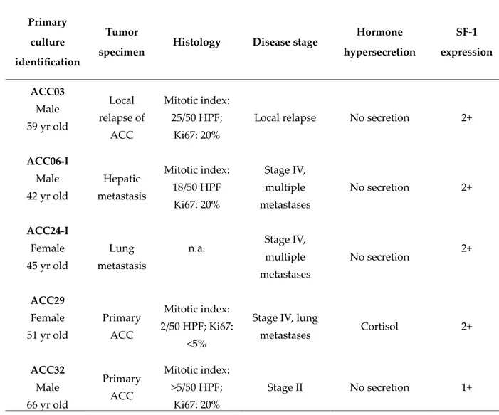
The ACC primary cultures belonged to five patients underwent surgical resection of primary or metastatic ACC. One of them (ACC29) presented a cortisol-secreting tumor, while others were non-secreting tumors. The clinical characteristics of patients are summarized in Table 2.
Table 2. Clinical and immunohistochemical characteristics of ACC patients.
Primary culture identification
Tumor
specimen Histology Disease stage
Hormone hypersecretion SF-1 expression ACC03 Male 59 yr old Local relapse of ACC Mitotic index: 25/50 HPF; Ki67: 20%
Local relapse No secretion 2+
ACC06-I Male 42 yr old Hepatic metastasis Mitotic index: 18/50 HPF Ki67: 20% Stage IV, multiple metastases No secretion 2+ ACC24-I Female 45 yr old Lung metastasis
n.a. Stage IV,
multiple metastases No secretion 2+ ACC29 Female 51 yr old Primary ACC Mitotic index: 2/50 HPF; Ki67: <5%
Stage IV, lung
metastases Cortisol 2+ ACC32 Male 66 yr old Primary ACC Mitotic index: >5/50 HPF; Ki67: 20% Stage II No secretion 1+
The project was approved by the local Ethics Committee and informed consent was signed by each patient enrolled in the study. Primary ACC cultures were obtained according to the previously published procedure [41]. The adrenal origin of primary cells was confirmed by the positivity to the Steroidogenic Factor 1 gene expression [37, 42]. 334 335 336 337 338 339 340 341 342 343 344 345 346 347 348
4.3. Cell viability assay
Cell viability was evaluated by 3-(4,5-Dimethyl-2-thiazol)-2,5-diphenyl-2H-tetrazolium bromide (MTT) dye reduction assay according to the manufacturer protocol (Sigma Aldrich). Drug- or vehicle-treated cells were incubated with MTT dye (at final concentration of 0.5mg/mL) and solubilized by DMSO. Absorbance was measured by a spectrophotometer at 570 nm.
4.4. Cell treatments
Cells were plated in 96 wells-plate at the concentration of 6x10³ cells/well for NCI-H295R and HAC-15 and 5x10³ cells/well for SW13, MUC-1 and ACC primary cultures. For the concentration-response curves, cells were exposed to increasing concentrations of trabectedin for different time, according to the respective doubling time. NCI-H295R cells line and ACC primary cultures were exposed to trabectedin (0.0625 – 0.75 nM) for 4 days, while SW13 cell treatment lasted for 3 days. HAC-15 cells were treated with increasing concentration of trabectedin (0.125 – 1.5 nM) for 6 days. Finally, MUC-1 were exposed to trabectedin (0.125 – 2.0 nM) for 5 days.
4.5. Drug combination experiments
Trabectedin and mitotane combination experiments were performed to evaluate their interaction on NCI-H295R cell viability, according to Chou and Talaly method [43]. NCI-H295R were treated for 4 days with trabectedin (0.018 – 1.2 nM) and mitotane (0.18 – 12 µM) alone or in combination with a fixed ratio (trabectedin / mitotane = 1 / 10000), as recommended for the most efficient data analysis [44]. Cells were analyzed for cell viability using MTT. Data were then converted to Fraction affected (Fa, range from 0 to 1 where Fa = 0 indicating 100% of cell viability and Fa = 1 indicating 0% of cell viability) and analyzed using the CompuSyn software (ComboSyn inc. Paramus, NJ, USA) to calculate the Combination Index (CI), being the CI value < 0.9 an indication of synergism, a CI = 09-1.1 an indication of additive effect and CI > 1.1 and indication of antagonism.
4.6. Quantitative RT-PCR (qRT-PCR).
Total RNA was extracted from cells by RNAeasy kit (Qiagen, Milano, MI, Italy) and 1µg was transcribed into cDNA, using murine leukemia virus reverse transcriptase (Promega Italia, Milano, MI, Italy). Gene expression was evaluated by qRT-PCR (ViiA7, Applied Biosystems, Milano, Italy) using SYBRGreen as 349 350 351 352 353 354 355 356 357 358 359 360 361 362 363 364 365 366 367 368 369 370 371 372 373 374 375 376 377 378 379 380 381 382 383 384
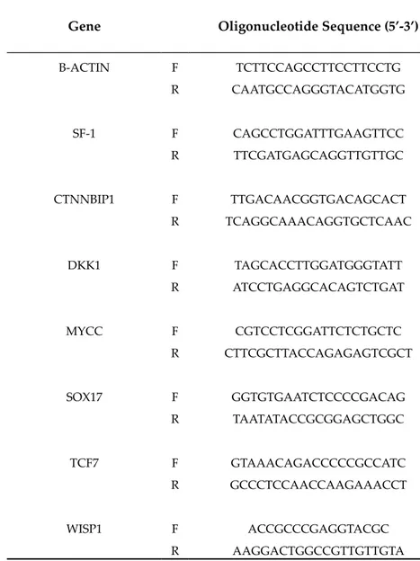
fluorochrome, as already described [45]. Sequences of oligonucleotide primers used were reported in Table 3.
Table 3. Sequences of oligonucleotide primers for qRT-PCR.
Gene Oligonucleotide Sequence (5’-3’)
Β-ACTIN F R TCTTCCAGCCTTCCTTCCTG CAATGCCAGGGTACATGGTG SF-1 F R CAGCCTGGATTTGAAGTTCC TTCGATGAGCAGGTTGTTGC CTNNBIP1 F R TTGACAACGGTGACAGCACT TCAGGCAAACAGGTGCTCAAC DKK1 F R TAGCACCTTGGATGGGTATT ATCCTGAGGCACAGTCTGAT MYCC F R CGTCCTCGGATTCTCTGCTC CTTCGCTTACCAGAGAGTCGCT SOX17 F R GGTGTGAATCTCCCCGACAG TAATATACCGCGGAGCTGGC TCF7 F R GTAAACAGACCCCCGCCATC GCCCTCCAACCAAGAAACCT WISP1 F R ACCGCCCGAGGTACGC AAGGACTGGCCGTTGTTGTA
Reactions were performed under the following conditions: 1 cycle at 95°C for 10 min, 40 cycles at 95°C for 15 s, 62°C for 1 min. Differences of the threshold cycle (Ct) values between the β-actin housekeeping gene and the gene of interest (ΔCt) were then calculated, as an indicator of difference in the amount of mRNA expressed [46].
4.7. WNT/ β catenin pathway gene profile
Total RNA was extracted from vehicle-treated NCI-H295R cells and from cells treated with the IC50 of trabectedin for 4 days, using RNAeasy kit (Qiagen). 385 386 387 388 389 390 391 392 393 394 395 396 397
cDNA was synthesized from 0.5µg RNA using RT2 First Strand Kit (Qiagen) and the mixture was added into a 96-wells WNT pathway signaling RT2 profiler array, according to the manufacturer’s instructions (Qiagen). Thermal cycling was performed using ViiA7, with an initial denaturation at 95 °C for 10 min, 40 cycles at 95 °C for 15 s, and 60 °C for 1 min. Qiagen’s online Web analysis tool was used to calculate the fold change in trabectedin-treated vs untreated cells, by determining the ratio of mRNA levels to control values using the Δ threshold cycle (Ct) method (2−ΔΔCt). All data were normalized to an average of five housekeeping genes. Validation of selected genes was performed by q-RT-PCR as previous described.
4.8. Western Blot
NCI-H295R cells were treated with IC50 value of trabectedin for 4 days. Whole cell lysates were prepared in ice-cold buffer with protease and phosphatase inhibitor cocktails (Roche, Milano, Italy) as previously described [25]. Fractionated cell lysates were prepared according to [47]. Equal amounts of protein were separated by electrophoresis on a 4-12% NuPAGE Bis-Tris Gel System (Life Technologies, Milano, Italy) and electroblotted to a nitrocellulose membrane. Primary and secondary antibodies are listed in Table 4. Signal was detected and quantified with the Odyssey® CLx Infrared Imaging System (LI-COR Biosciences, Lincoln, U.S.A.).
Table 4. Primary and secondary antibodies
Target Characteristic Company Final
concentration 398 399 400 401 402 403 404 405 406 407 408 409 410 411 412 413 414 415 416 417 418 419 420 421 422 423 424 425 426 427
β-CATENIN Rabbit mAb Cell Signaling Tecnology
6 ng/mL (WB) 15 ng/mL (IF)
GAPDH Mouse mAb Santa Cruz Biotechnology 1 µg/mL
C23 (nucleolin) Rabbit pAb Santa Cruz Biotechnology 0,2 µg/mL
PSMB5 Mouse mAb Abcam 10 µg/mL
Secondary anti-mouse IRDye 680CW conjugated LI-COR 0,67 µg/mL Secondary anti-rabbit IRDye 800CW conjugated LI-COR 0,67 µg/mL
Secondary anti-mouse Alexa Fuor 555
Conjugated Immunological Sciences 5 µg/mL
Secondary anti-rabbit Alexa Fuor 488
Conjugated Immunological Sciences 5 µg/mL
4.9. Immunofluorescence
Cells were grown onto 12 mm poly-L-lysine coated coverslips and treated with IC50 value of trabectedin for 3 days. Cells were then fixed in ice-cold methanol for 20 min and permeabilized with 0.2% Triton X-100 in PBS for 1 hr. Nonspecific binding was blocked by incubation in PBS containing 0,2% Triton X-100 and 1% of BSA for 5 minutes. Cells were then incubated overnight at 4°C with rabbit monoclonal antibody against human β-catenin primary antibody and with mouse monoclonal antibody against human PSMB5. After extensive washes, the Alexa Fluor488 anti-rabbit secondary antibody (Life Technologies) was applied for 1 hour at room temperature, followed by counterstaining with Hoechst (Sigma Aldrich) for 5 min. After rinsing in PBS, coverslips were mounted using FluorPreserveTM Reagent and cell staining was detected using a Zeiss LSM 510 META confocal laser-scanning microscope (Carl Zeiss AG, Oberkochen, Germany). Zen software was used for image analysis and processing.
4.10. Chemicals
Trabectedin was kindly given by Pharma Mar S.A. (Madrid, Spain), dissolved in DMSO and stored at -20°C in 10mM aliquots. Mitotane was supplied by 428 429 430 431 432 433 434 435 436 437 438 439 440 441 442 443 444 445 446
Selleckchem Chemicals (DBA Italia, Milano, Italy), dissolved in DMSO and stored at -80°C in 200mM aliquots.
4.10. Statistical analysis
Statistical analysis was carried out using GraphPad Prism software (version 5.02, GraphPad Software, La Jolla, CA, USA). One-way ANOVA with Bonferroni׳s correction was used for multiple comparisons. Unless otherwise specified, data are expressed as mean ± SD or SEM of at least three experiments run in triplicate. P values < 0.05 were considered statistically significant.
5. Conclusions
Our results indicated that trabectedin, at concentrations superimposable to those employed in in vitro experimental models of STS and ovary carcinoma, exert a cytotoxic effect in different ACC cell models and effect persists upon drug withdrawal. These findings, together with the high lipophilicity of trabectedin and the lyophilic milieu of these tumors, suggest that the drug may accumulate in ACC cells and could be efficacious in treating ACC patients. This point however is speculative and needs to be extensively studied in a dedicated prospective clinical trial.
Finally, our data on the inhibitory effect of trabectedin on Wnt/β-catenin are interesting due to the major role of this pathway in proliferation and resistance to antineoplastic agents of ACC cells [21, 22, 48].
Supplementary Materials: The following are available online at www.mdpi.com/xxx/s1, Figure S1 Effect of trabectedin on DNA integrity, Figure S2 Trabectedin promoted apoptotic cell death in NCI-H295R cells, Figure S3 Cytotoxic effect of trabectedin on SW13 cell line, Figure S4 Representative figure of subfractions proteins of NCI-H295R. EN = Nuclear Extract, EC = Cytosolic Extract.
Author Contributions: Conceptualization, S.S., A.B., A.A.; Methodology, S.S., A.A., S.A.B., G.A.M.T., D.B.; Formal Analysis, A.A., E.R., M.F., S.A.B.; Data Curation: A.A., S.S., D.C., M.L.; Writing – Original Draft Preparation, A.A.; Writing – Review & Editing, M.M., C.H., S.G., M.T., S.S., A.B.; Supervision, S.S., A.B.; Funding Acquisition, S.S., A.B., C.H.
Funding: This work was supported by Pharma Mar S.A. (Madrid, Spain). C.H. received funding by the Uniscientia Foundation (keyword: tumor model)
Conflicts of Interest: The Authors declare no conflict of interest.
References 447 448 449 450 451 452 453 454 455 456 457 458 459 460 461 462 463 464 465 466 467 468 469 470 471 472 473 474 475 476 477 478 479 480 481 482 483 484 485 486
1. Terzolo M, Daffara F, Ardito A, Zaggia B, Basile V, Ferrari L , Berruti A. Management of adrenal cancer: a 2013 update. J Endocrinol Invest. 2014, 37(3), 207-217.
2. Fassnacht M, Dekkers O, Else T, Baudin E, BerrutiA, de Krijger RR, Haak HR, Mihai R, Assie G, Terzolo M. European Society of Endocrinology Clinical Practice Guidelines on the Management of Adrenocortical Carcinoma in Adults, in collaboration with the European Network for the Study of Adrenal Tumors. Eur
J Endocrinol. 2018, 179(4), G1-G46. https://doi.org/10.1530/EJE-18-0608.
3. Waszut U, Szyszka P, Dworakowska D.Understanding mitotane mode of action. Journal of J Physiol
Pharmacol. 2017, 68(1), 13-26.
4. Micromedex® (electronic version). IBM Watson Health, Greenwood Village, Colorado, USA. Available at: https://www.micromedexsolutions.com/ (Accessed on 31/10/2019).
5. Berruti A, Terzolo M, Sperone P, Pia A, Della Casa S, Gross DJ, Carnaghi C, Casali P, Porpiglia F, Mantero F et al. Etoposide, doxorubicin and cisplatin plus mitotane in the treatment of advanced adrenocortical carcinoma: a large prospective phase II trial. Endocr Relat Cancer. 2005, 12(3), 657–666. 6. Fassnacht M, Terzolo, M, Allolio B, Baudin E, Haak H, Berruti A, Welin S, Schade-Brittinger C, Lacroix A,
Jarzab B et al. Combination chemotherapy in advanced adrenocortical carcinoma. N Engl J Med. 2012, 366(23), 2189-2197. https://doi.org/10.1056/NEJMoa1200966
7. Sperone P, Ferrero A, Daffara F, Priola A, Zaggia B, Volante M, Santini D, Vincenzi B, Badalamenti G, Intrivici C et al. Gemcitabine plus metronomic 5-fluorouracil or capecitabine as a second-/third-line chemotherapy in advanced adrenocortical carcinoma: a multicenter phase II study. Endocr Relat Cancer 2010, 17(2), 445–453. https://doi.org/10.1677/ERC-09-0281
8. Grisanti S, Cosentini D, Laganà M, Abate A, Rossini E, Sigala S, Berruti A. Are we failing in treatment of adrenocortical carcinoma? Lights and shadows of molecular signatures. Curr Opinion Endocr Metab Res. 2019, 8, 80-87. https://doi.org/10.1016/j.coemr.2019.07.007
9. Creemers SG, van Koetsveld PM, van den Dungen ES, Korpershoek E, van Kemenade FJ, Franssen GJ, de Herder WW, Feelders RA, Hofland LJ Inhibition of Human Adrenocortical Cancer Cell Growth by Temozolomide in Vitro and the Role of the MGMT Gene. J Clin Endocrinol Metab. 2016, 101(12), 4574-4584. 10. Cosentini D, Badalamenti G, Grisanti S, Basile V, Rapa I, Cerri S, Spallanzani A, Perotti P, Musso E,
Laganà M, Ferrari V, Luppi G, Dalla Vola A, Incorvaia L, Sigala S, Volante M, Terzolo M, Berruti A. Activity and safety of temozolomide in advanced adrenocortical carcinoma patients. Eur J Endocrinol. 2019, 181(6), 681-689. https://doi.org/doi: 10.1530/EJE-19-0570.
11. D'Incalci M, Galmarini CM. A Review of Trabectedin (ET-743): A Unique Mechanism of Action. Mol
Cancer Ther. 2010, 9(8), 2157-63. https://doi.org/10.1158/1535-7163
12. Pérez-Losada J, Pintado B, Gutiérrez-Adán A, Flores T, Bañares-González B, del Campo JC, Martín-Martín JF, Battaner E, Sánchez-García I. The chimeric FUS/TLS-CHOP fusion protein specifically induces liposarcomas in transgenic mice. Oncogene. 2000, 19(20), 2413-2422.
13. Owen L, Kowaleski A, Lessnick S. EWS/FLI mediates transcriptional repression via NKX2.2 during oncogenic transformation in Ewing's sarcoma. PLoS One. 2008, 3(4), e1965. https://doi.org/ 10.1371/journal.pone.0001965.
14. Peraldo-Neia C, Cavalloni G, Soster M, Gammaitoni L, Marchiò S, Sassi F, Trusolino L, Bertotti A, Medico E, Capussotti L et al Anti-cancer effect and gene modulation of ET-743 in human biliary tract carcinoma preclinical models. BMC Cancer. 2014, 14, 918. https://doi.org/ 10.1186/1471-2407-14-918.
15. MacDonald BT, Tamai K, He X. Wnt/beta-catenin signaling: components, mechanisms, and diseases. Dev
Cell. 2009, 17(1), 9-26. https://doi.org/10.1016/j.devcel.2009.06.016.
16. Lea VH, Inaib M, Williams RM, Kanb T. Ecteinascidins. A Review of the Chemistry, Biology and Clinical Utility of Potent Tetrahydroisoquinoline Antitumor Antibiotics. Nat Prod Rep. 2015, 32(2), 328–347. https://doi.org/10.1039/c4np00051j
17. Li WW, Takahashi N, Jhanwar S, Cordon-Cardo C, Elisseyeff Y, Jimeno J, Faircloth G, Bertino JR. Sensitivity of soft tissue sarcoma cell lines to chemotherapeutic agents: identification of ecteinascidin-743 as a potent cytotoxic agent. Clin Cancer Res. 2001, 7(9), 2908-2911.
18. Mabuchi S, Hisamatsu T, Kawase C, Hayashi M, Sawada K, Mimura K, Takahashi K, Takahashi T, Kurachi H, Kimura T. The activity of trabectedin as a single agent or in combination with everolimus for clear cell carcinoma of the ovary. Clin Cancer Res. 2011, 17(13), 4462–4473.
19. Cvetkovic RS, Figgitt DP, Plosker GL. ET-743. Drugs. 2002, 62(8), 1185-1192
20. Gaujoux S, Grabar S, Fassnacht M, Ragazzon B, Launay P, Libé R, Chokri I, Audebourg A, Royer B, Sbiera S et al. β-catenin activation is associated with specific clinical and pathologic characteristics and a poor
487 488 489 490 491 492 493 494 495 496 497 498 499 500 501 502 503 504 505 506 507 508 509 510 511 512 513 514 515 516 517 518 519 520 521 522 523 524 525 526 527 528 529 530 531 532 533 534 535 536 537 538 539 540 541
outcome in adrenocortical carcinoma. Clin Cancer Res. 2011, 17(2), 328-336. https://doi.org/10.1158/1078-0432.CCR-10-2006
21. Cosentini D, Grisanti S, Dalla Volta A, Laganà M, Fiorentini C, Perotti P, Sigala S, Berruti A. Immunotherapy failure in adrenocortical cancer: where next? Endocr Connect. 2018, 7(12), E5–E8. https://doi.org/10.1530/EC-18-0398
22. Fiorentini C, Grisanti S, Cosentini D, Abate A, Rossini E, Berruti A, Sigala. Molecular Drivers of Potential Immunotherapy Failure in Adrenocortical Carcinoma. J Oncol. 2019, 2019, 6072863. https://doi.org/10.1155/2019/6072863
23. Tissier F, Cavard C, Groussin L, Perlemoine K, Fumey G, Hagneré AM, René-Corail F, Jullian E, Gicquel C, Bertagna X, et al. Mutations of beta-catenin in adrenocortical tumors: activation of the Wnt signaling pathway is a frequent event in both benign and malignant adrenocortical tumors. Cancer Res. 2005, 65(17), 7622-7.
24. Leal LF, Bueno AC, Gomes DC, Abduch R, de Castro M, Antonini SR. Inhibition of the Tcf/beta-catenin complex increases apoptosis and impairs adrenocortical tumor cell proliferation and adrenal steroidogenesis. Oncotarget 2015, 6(40), 43016-32. https://doi.org/10.18632/oncotarget.5513
25. Fiorentini C, Fragni M, Perego P, Vezzol S, Bonini SA, Tortoreto M, Galli D, Claps M, Tiberio GA et al. Antisecretive and Antitumor Activity of Abiraterone Acetate in Human Adrenocortical Cancer: A Preclinical Study. J Clin Endocrinol Metab. 2016, 101(12), 4594-4602.
26. Fragni M, Fiorentini C, Rossini E, Fisogni S, Vezzoli S, Bonini SA, Dalmiglio C, Grisanti S, Tiberio GAM, Claps M et al. In vitro antitumor activity of progesterone in human adrenocortical carcinoma. Endocrine 2019, 63(3), 592–601. https://doi.org/10.1007/s12020-018-1795-x
27. Graham TA, Clements WK, Kimelman D, Xu W. The crystal structure of the beta-catenin/ICAT complex reveals the inhibitory mechanism of ICAT. Mol Cell. 2002, 10(3), 563-71.
28. Lee AY, He B, You L, Xu Z, Mazieres J, Reguart N, Mikami I, Batra S, Jablons DM. Dickkopf-1 antagonizes Wnt signaling independent of beta-catenin in human mesothelioma, Biochem Biophys Res Commun. 2004, 323(4), 1246–1250.
29. Huang Y, Liu L & Liu A. Dickkopf-1: Current knowledge and related diseases. Life Sci. 2018, 209, 249-254.
https://doi.org/10.1016/j.lfs.2018.08.019
30. Shou J, Ali-Osman F, Multani AS, Pathak S, Fedi P, Srivenugopal KS. Human Dkk-1, a gene encoding a Wnt antagonist, responds to DNA damage and its overexpression sensitizes brain tumor cells to apoptosis following alkylation damage of DNA. Oncogene. 2002, 21(6), 878-889.
31. González-Sancho JM, Aguilera O, García JM, Pendás-Franco N, Peña C, Cal S, García de Herreros A, Bonilla F, Muñoz A. The Wnt antagonist DICKKOPF-1 gene is a downstream target of beta-catenin/TCF and is downregulated in human colon cancer. Oncogene. 2005, 24(6), 1098-1103. 32. Fu DY, Wang ZM, Li-Chen, Wang BL, Shen ZZ, Huang W, Shao ZM. Sox17, the canonical Wnt antagonist,
is epigenetically inactivated by promoter methylation in human breast cancer. Breast Cancer Res Treat. 2010, 119(3), 601-12. https://doi.org/10.1007/s10549-009-0339-8
33. Zhang W, Glöckner SC, Guo M, Machida EO, Wang DH, Easwaran H, Van Neste L, Herman JG, Schuebel KE, Watkins DN et al. Epigenetic inactivation of the canonical Wnt antagonist SRY-box containing gene 17 in colorectal cancer. Cancer Res. 2008, 68(8), 2764-72 https://doi.org/10.1158/0008-5472.CAN-07-6349 34. Tan DS, Holzner M, Weng M, Srivastava Y, Jauch R. SOX17 in cellular reprogramming and cancer. Semin
Cancer Biol. 2019, S1044-579X(19), 30030-6 https://doi.org/10.1016/j.semcancer.2019.08.008
35. Gurbuz I Chiquet-Ehrismann R. CCN4/WISP1 (WNT1 inducible signaling pathway protein 1): a focus on its role in cancer. Int J Biochem Cell Biol. 2015, 62, 142-6. https://doi.org/10.1016/j.biocel.2015.03.007
36. Katoh M. Multilayered prevention and treatment of chronic inflammation, organ fibrosis and cancer associated with canonical WNT/βcatenin signaling activation (Review). Int J Mol Med. 2018, 42(2), 713-725. https://doi.org/10.3892/ijmm.2018.3689
37. Hantel C, Shapiro I, Poli G, Chiapponi C, Bidlingmaier M, Reincke M, Luconi M, Jung S, Beuschlein F. Targeting heterogeneity of adrenocortical carcinoma: Evaluation and extension of preclinical tumor models to improve clinical translation. Oncotarget. 2016, 7(48), 79292-79304. https://doi.org/10.18632/oncotarget.12685
38. Rainey WE, Saner K, Schimmer BP. Adrenocortical cell lines. Mol Cell Endocrinol. 2004, 228(1-2), 23-38. 39. Parmar J, Key RE, Rainey WE. Development of an adrenocorticotropin-responsive human adrenocortical
carcinoma cell line. J Clin Endocrinol Metab. 2008, 93(11), 4542-6. https://doi.org/10.1210/jc.2008-0903.
542 543 544 545 546 547 548 549 550 551 552 553 554 555 556 557 558 559 560 561 562 563 564 565 566 567 568 569 570 571 572 573 574 575 576 577 578 579 580 581 582 583 584 585 586 587 588 589 590 591 592 593 594 595
40. Wang T, Rainey WE. Human adrenocortical carcinoma cell lines. Mol Cell Endocrinol 2012, 351(1), 58-65. https://doi.org/10.1016/j.mce.2011.08.041
41. Fragni M, Palma Lopez LP, Rossini E, Abate A, Cosentini D, Salvi V, Vezzoli S, Poliani PL, Bosisio D, Hantel C et al. In vitro cytotoxicity of cabazitaxel in adrenocortical carcinoma cell lines and human adrenocortical carcinoma primary cell cultures. Mol Cell Endocrinol. 2019, 498, 110585. https://doi.org/10.1016/j.mce.2019.110585
42. Sbiera S, Schmull S, Assie G, Voelker HU, Kraus L, Beyer M, Ragazzon B, Beuschlein F, Willenberg HS, Hahner S, Saeger W, Bertherat J, Allolio B, Fassnacht M. High diagnostic and prognostic value of steroidogenic factor-1 expression in adrenal tumors. J Clin Endocrinol Metab. 2010, 95, E161-171. https://doi.org/10.1210/jc.2010-0653.
43. Chou TC, Talalay P. Quantitative analysis of dose-effect relationships: the combined effects of multiple drugs or enzyme inhibitors. Adv Enzyme Regul. 1984, 22, 27–55.
44. Chou TC.) Theoretical basis, experimental design, and computerized simulation of synergism and antagonism in drug combination studies. Pharmacol Rev. 2006, 58(3), 621-681.
45. Sigala S, Bodei S, Missale C, Zani D, Simeone C, Cunico SC, Spano PF. Gene expression profile of prostate cancer cell lines: effect of nerve growth factor treatment. Mol Cell Endocrinoy. 2008, 284(1-2), 11-20. https://doi.org/10.1016/j.mce.2007.12.015
46. Livak KJ, Schmittgen TD. Analysis of relative gene expression data using real-time quantitative PCR and the 2(-Delta Delta C(T)) Method. Methods. 2001, 25(4), 402-408.
47. Porrini V, Sarnico I, Benarese M, Branca C, Mota M, Lanzillotta A, Bellucci A, Parrella E, Faggi L, Spano et al. Neuroprotective and Anti-Apoptotic Effects of CSP-1103 in Primary Cortical Neurons Exposed to Oxygen and Glucose Deprivation. Int J Mol Sci. 2017, 18 E184. https://doi.org/10.3390/ijms18010184. 48. Mohan DR, Lerario AM, Hammer GD. Therapeutic Targets for Adrenocortical Carcinoma in the
Genomics Era. JEndocr Soc. 2018, 2(11), 1259-1274. https://doi.org/10.1210/js.2018-00197
49. Fragni, M., Fiorentini, C., Rossini, E., Fisogni, S., Vezzoli, S., Bonini, S.A., Dalmiglio, C., Grisanti, S., Tiberio, G.A.M., Claps, M., Cosentini, D., Salvi, V., Bosisio, D., Terzolo, M., Missale, C., Facchetti, F., Memo, M., Berruti, A., Sigala, S. In vitro antitumor activity of progesterone in human adrenocortical carcinoma. Endocrine. 2019 63, 592–601. https://doi.org/10.1007/s12020-018-1795-x.
© 2020 by the authors. Submitted for possible open access publication under the terms and conditions of the Creative Commons Attribution (CC BY) license (http://creativecommons.org/licenses/by/4.0/). 596 597 598 599 600 601 602 603 604 605 606 607 608 609 610 611 612 613 614 615 616 617 618 619 620 621 622 623 624 625