UNIVERSITÀ DEGLI STUDI DELLA TUSCIA DI VITERBO DIPARTIMENTO DI SCIENZE ECOLOGICHE E BIOLOGICHE
Corso di Dottorato di Ricerca in
Genetica e Biologia Cellulare – XXVII Ciclo
IDENTIFICATION OF NOVEL MUTANTP 53 FUNCTIONS INVOLVED IN THE MODULATION OF THE TUMOUR MICROENVIRONMENT
s.s.d. BIO/11
Tesi di dottorato di:
Dott. Valentina Ubertini
Coordinatore del corso Tutore
Prof. Giorgio Prantera Dott. Gianluca Bossi
Table of contents
Abstract
1Chapter 1: Introduction
2Wild-Type and Mutant p53 2
p53 structure 2
p53 functions 3
p53 Mutations 5
Gain-of-function of mutant p53 5 Mutant p53 GOF in genomic instability 7 Mutant p53 GOF in cell death and drug resistance 8 Mutant p53 GOF in Cell Migration and Invasion 8 Mutant p53 GOF in cell proliferation 8
The tumour microenvironment 10
Cancer cells 10
Stromal cells 11
Cancer associated fibroblasts 11
Cells of the immune system 11
Tumour associated macrophages 11
Tumour associated neuthrophils 12
T lymphocytes 12
B lymphocytes 13
Natural Killer cells (NK) 13
Myeloid-derived suppressor cells (MDSCs) 13
Dendritic cells (DCs) 13
Adipocytes 14
Cancer-releated inflammation in tumour promotion 14
Chapter 2: Aims of the study
17Chapter 3: Materials and methods
18Cell culture 18
Cytokine arrays 18
Semi-quantitative and quantitative reverse transcriptase–PCR 18
Western blotting 19
Transfection and luciferase reporter assay 20
ChIP assays 20
Co-IP 21
MAFF RNAi 21
IL-8 gene expression 22
Endothelial cell injury 22
Bromodeoxyuridine incorporation assays 22
Xenograft in vivo assay 22
Chemical induction of mammary tumours 23
DNA extraction from tails biopsies 23
Genomic DNA genotyping 23
Generation of the mutant p53 murine breast cancer cell line 24
Generation of the orthotopic model of breast cancer 24
Immunohistochemestry 24
Statistical analysis 25
Chapter 4: Results-Part 1
26
Mutant but not wild-type (wt) p53 suppresses sIL-1Ra gene expression in tumour cells 26
Mutant but not wtp53 reduces sIL-1Ra promoter activity 28
Mutant but not wtp53 is physically recruited to the sIL-1Ra promoter 30
MafF works as a common player in the regulation of sIL-1Ra gene expression by mutant and
wtp53 proteins 33
Upon mutant p53 depletion, de-repressed sIL-1Ra blocks IL-1β response 37
Mutant p53 sustains IL-1β-driven tumour malignancy through sIL-1Ra suppression 39
Chapter 4: Results-Part 2
43
Generation of mutant p53 mice sensible to chemical induction of mammary tumours 43
Mutant p53 induces less differentiated tumours with respect to the wild type 44
Mutant p53 might play relevant roles in TAMs recruitment 45
Production of a mutant p53 cancer cell line used to generate an immunocompetent model of breast cancer 47
Chapter 5: Discussion
49
1
Abstract
The tumour suppressor TP53 is one of the most frequently mutated genes in human cancers. The majority of these mutations, located into the DNA binding domain, results in the production of mutated proteins that lose the wild-type functionality but can acquire new oncogenic functions (gain-of-function, GOF). We previously showed that p53R273H mutation promotes tumour growth, stromal invasion and angiogenesis in xenografted HT-29, suggesting a role of mutant p53 in the modulation of the tumour microenvironment (TME). In the first part of the study it has been explored whether p53 mutants might have role in modulating cytokines/secretory factors production. Cytokines profile analyses on conditioned media derived from a panel of human cancer cell lines showed that endogenous mutant p53 depletion increases significantly the secreted form of IL-1Ra (sIL-1Ra). The sIL-1Ra is a naturally occurring anti-inflammatory cytokine that acts as a specific antagonist of IL-1β. Confirmatory analyses showed that mutant p53 represses 1Ra at transcriptional level, identifying by ChIP assays specific sIL-1Ra regulatory regions required for mutant p53 physical recruitment. Moreover, further studies identified the transcriptional co-factor MafF as a required player to recruit by protein-protein interaction mutant p53 on the sIL-1Ra promoter. Indeed, we showed that MafF endogenous depletion (RNAi) impairs mutant p53 recruitment on the sIL-1Ra promoter restoring its activity. Functional studies showed that mutant p53 contributes to maintain a prone inflammatory response by sIL-1Ra down-regulation. Indeed, either mutant p53 depletion or recombinant sIL-1Ra (Kineret) delivery impairs IL-1b response in vitro and in vivo. Taken together, these results reveal a novel oncogenic mutant p53 GOF activity, exerted by the repression of sIL-1Ra that contributes to generate a pro-inflammatory tumour microenvironment promoting tumour malignancy. The second part of the study has been dedicated to the identification and characterization of a more physiological experimental model, as immunocompetent mice, to better study mutant p53/TME crosstalk. The analysis of chemically induced mammary tumours in p53+/R172H, p53R172H/R172H and p53+/+ genetically engineered mutant p53R172H knock-in mice, showed that mutant p53 occurrence strongly impacts on tumour onset, tumour differentiation and increases the recruitment of macrophages into the tumour milieu. Finally, in order to identify a feasible, less expensive and more reliable system than the genetically engineered mouse, we focused on the generation and initial characterization of a syngeneic orthotopic model of breast cancer in immunocompetent mice, by using a breast cancer cell line, named 44-1, derived from a p53R172H/R172H chemically induced mammary tumour. This model will be useful to confirm the whole data obtained and to examine in depth the fine regulation exerted by mutant p53 on the tumour microenvironment.
2
Chapter 1
Introduction
Wild-Type and Mutant p53
p53 structure
The human TP53 gene resides on chromosome 17p13.11-3 and encodes one of the most intensively studied oncosuppressors, the p53 protein. The genomic organization of TP53 exhibits a high degree of similarity among different species. Eleven exons are interrupted by 10 introns. Exon 1 of TP53 is a non-coding exon, whereas exons 2–11 code for the 393-amino-acid protein. Exons 5–8 are regions highly conserved through evolution.4
p53 protein is composed of several structural and functional domains (Figure1): a N-terminus
region containing a transactivation domain (residues 1-42) and a proline-rich region(residues 61-94), a central core containing the DNA binding domain (residues 102-292), and a C-terminal
region (residues 301-393) containing an oligomerization domain (residues 324-355), a strongly
basic regulatory domain (residues 363-393), a nuclear localization signal (NLS) and a nuclear export signal (NES).5
3
The N-terminal domain of p53 is required to activate transcription and to regulate p53 function and stability by interacting respectively with transcription factors (such as p300 and CBP), and Mdm2, an E3 ubiquitin ligase that targets p53 for degradation in the proteasome.6-9 The DNA-binding domain is required for sequence-specific DNA DNA-binding (the consensus sequence contains two copies of the 10-bp motif 5’-PuPuPuC(A/T)-(T/A)GPyPyPy-3’, separated by 0-13 bp).10,11 The oligomerization domain (residues 324–355) allows p53 dimerization and tetramerization.12-14 The basic C-terminus of p53 (residues 361–393) is a negative regulatory domain that is believed to modulate sequence-specific DNA binding.15,16 According to the allosteric/conformational model, the C-terminus controls whether p53 exists in a “latent” or “active” conformation for high-affinity DNA binding. The basic C-terminus could force p53 into a non-DNA-binding “latent” conformation by interacting with the core or other domains of the protein.17 Post-translational modification (phosphorylation/acetylation) of the C-terminus following cellular stress could convert p53 into an “active” conformation, inducing an enhanced transcriptional activity.17,18 The C-terminus has also been implicated in induction of cell death.19
p53 functions
Under non-stress condition, p53 is a short-lived nuclear protein with a half-life of ∼5–20 min in most cell types The Mouse double minute 2 homolog (MDM2), an E3 ubiquitin-protein ligase ,is the most critical negative regulator for p53.20,21
p53 can be activated by a wide variety of stress signals, including DNA damage, hypoxia, and oncogene activation.15 Following activation, p53 normally functions as a sequence-specific transcription factor binding to p53 responsive elements on target genes regulatory regionsas a homotetramer,22 and plays important roles in cell cycle control, senescence, apoptotosis,15,23,24 and metabolism25 in order to suppress cancer. A large number of genes have been shown to be transcriptional targets of p53. The p53 pathway utilizes G1/S and G2/M checkpoint mechanisms to arrest cell-cycle progression, thus preventing propagation of DNA damage while cells attempt to repair it. It is well-established that p53 can induce G1 arrest through transcriptional induction of p21, a cyclin-dependent kinase inhibitor.26,27 p53 was also reported to transcriptionally activate GADD45 (growth arrest and DNA-Damage inducible 45) and 14-3-3s (tyrosine 3-monooxygenase/tryptophan 5-monooxygenase activation protein, sigma polypeptide), which in turn leads to G2 arrest.28,29 However, if the damage is too severe,
4
activation of the p53 pathway results in apoptotic cell death to avoid the division of cells with unrepaired DNA and possible malignant transformation.19,30 To this aim, p53 induces expression of a set of target genes involved in apoptosis, PUMA (p53 up-regulated modulator of apoptosis), Bax (BCL2-associated X protein), Noxa (PMAIP1), PIG3 (tumour protein p53 inducible protein 3), Killer/DR5 (tumour necrosis factor receptor superfamily, member 10b), Fas (Fas cell surface death receptor), Perp (p53 apoptosis effector related to PMP-22), and p53AIP1 (tumour protein p53 regulated apoptosis inducing protein 1).24Recent studies have shown that p53 can also regulate apoptosis through a transcription-independent pathway. In response to stress, a fraction of the p53 protein translocates to mitochondria, where p53 interacts with anti-apoptotic Bcl-xL and Bcl-2 to inhibit their functions, resulting in the release of cytochrome c from the mitochondria and thereby induces apoptosis.31,32
Another important function of p53 is the induction of senescence. Many DNA-damaging agents used in chemotherapy can activate p53 and induce senescence. Many senescence signals activate p53, which in turn transactivates p21 and induces p53-dependent senescence.33-36 However, the mechanism by which p53 induces senescence is not as clear as the mechanisms for apoptosis and cell cycle arrest.
An important feature of p53 is its involvement in the autophagy regulation.37 p53 has been reported to promote autophagy through different mechanisms, which may contribute to the role of p53 in tumour prevention. p53 promotes autophagy through inhibition of the mTOR (mammalian target of rapamycin) pathway, which is a critical negative regulator of autophagy.38 p53 also induces the expression of several genes, including DRAM (DNA-damage regulated autophagy modulator 1), PUMA, ISG20L1 (interferon-stimulated exonuclease gene 20 kDa-like 1), and Ei24 (etoposideinduced 2.4), to promote autophagy.39-41
In addition to transcriptional regulation of protein-coding genes, recent studies have shown that p53 can transcriptionally regulate the expression of miRNAs as a new mechanism of p53 tumour suppressive functions.42,43 p53 regulates the expression of miR34-a/b/c through direct binding to the p53-responsive elements in their promoters. miR-34 family members repress the expression of several targets genes involved in the regulation of cell cycle, cell proliferation, and survival, including cyclin E2, CDK4/6, and BCL2. Ectopic expression of miR-34 family members promotes p53-mediated apoptosis, cell cycle arrest, and senescence.44-46 Since then, a group of miRNAs has been reported to be directly induced by p53 oncosuppressive functions (miR-145, miR-107, miR-192/194/215, miR-15a/16-1, miR-215, and let-7).42,43 In addition to the
5
transcriptional regulation p53 promotes the post-transcriptional maturation of specific miRNAs (miR-16-1, miR-143, miR-145),47 and affects the miRNA target selection by regulating RNA-binding proteins, such as RBM38 (RNA-RNA-binding motif protein 38), which competes with miRNAs for binding to 30-UTRs of mRNAs of target genes.48
p53 Mutations
More than 30,000 somatic mutation data of p53 appear in the international agency for research on cancer (IARC) TP53 database version R17 (http://www-p53.iarc.fr/).49
The frequencies of reported TP53 mutations vary considerably between cancer types, ranging from ~10% in haematopoietic malignancies to 50–70% in ovarian, colorectal and head and neck cancers.50 Alterations have been found in virtually every region of the protein.51 The majority of this p53 alterations result in missense point mutations, leading to the substitution of a single amino acid residue. These substitutions occur throughout the p53 protein, but most commonly cluster within the DNA binding domain, with six ‘‘hotspot’’ amino acids that are most frequently substituted (R175, G245, R248, R249, R273 and R282).52 The mutations in the structured core of p53 can have significant consequences to p53 protein folding. Indeed the mutations have been divided into two categories: structural mutants (such as R175H) that can cause unfolding of the p53 protein, and DNA-contact mutants (such as R273H) that change amino acids critical for DNA binding.53 These mutations abrogate the TP53 tumour suppressor functions, reducing the ability of the cell to induce a proper p53 response; if both alleles are mutated, or if the remaining allele is lost (loss of heterozygosity), the cells will be totally deprived of anticancer protection by p53. Furthermore, many mutant (mut) p53 isoforms can exert dominant–negative effects over co-expressed wtp53, by forming mixed tetramers that are incapable of DNA binding and transactivation. Hence, through such mechanism , the cell may be rendered totally deficient of wtp53 function, in particular if the mutant protein is expressed at higher level.54
It is becoming clear that at least some of these mutp53 proteins give rise to a more aggressive tumour profile, indicating that they have acquired novel functions, different from the wild type (wt) oncosuppressive functions.
Gain-of-function of mutant p53
The concept of mutp53 gain-of-function (GOF) was first suggested 20 years ago, when the introduction of mutp53 into p53-null cells was shown to increase their ability to form colonies
6
in soft agar in vitro and tumours in mice.55 Since then, numerous publications have demonstrated many mutp53 GOFs in a large number of cell lines with a variety of p53 mutations.56 The GOF hypothesis has recently been reinforced by in vivo experiments showing that mice expressing mutp53 have a more aggressive and metastatic tumour profile than p53-null or p53 wild-type mice.57-60 During these years, different models for mutp53 GOF have been proposed.61
Model 1: Mutant p53 binds directly to target genes DNA to alter their expression.
Since mutp53 proteins retain the N-terminal transcriptional transactivation domain, much of their activity have been related to a direct ability to regulate target gene expression. The amino acid substitutions within the DNA-binding domain may change sequence-specific DNA binding raising the possibility that some mutp53 proteins may recognize a specific mutp53 response element, allowing them to act as an oncogenic transcription factor. However, a mutp53-specific consensus sequence has not been identified yet. It has been demonstrated that mutp53 directly interacts with other parts of the DNA, including sequences matrix attachment regions62 or G/C-rich DNA around transcription start sites (TSS) of target genes63 and providing other mechanisms to directly regulate gene expression.
Model 2 : Mutant p53 binds transcription factors to enhance or prevent their function.
The best-described transcriptional function of mutp53 is its ability to interact with other transcription factors and modulate the expression of their target genes. In some cases, mutp53 increases the activity of the transcription factor partner, whereas in other cases inhibits its activity. Following exposure to low levels of DNA damage, the DNA topoisomerase 2-binding protein 1 (TopBP1) recruits mutp53 and the transcriptional cofactor p300 to target gene pro-moters. PLK2 kinase can phosphorylate mutp53 and stimulate the binding of mutp53 to p300. The phosphorylated mutp53–p300 complex subsequently interacts with NF-Y to induce transcription of a number of genes controlling different phases of the cell cycle.64-66 Mutp53 complexes with the KLF17 transcription factor and decreases the metastatic suppressor function of KLF17 in breast cancer cell lines by reducing its recruitment on EMT target gene promoters.67 In response to TGF-β treatment, SMAD2 promotes the mutp53--p63 complex, leading to the inhibition of p63-driven gene expression.68 It has been recently demonstrated
7
that certain mutp53 proteins are able to form prion-like aggregates,69 which may contribute to the binding and inhibition of p53 family members.70
Model 3 : Mutant p53 interacts with proteins changing their function directly.
Mutp53 is also known to bind and modulate the function of proteins that are not directly involved in the transcription process. Recent data showed mutp53 binds and inhibits the tumour suppressor DAB2IP in the cytoplasm of cancer cell lines, promoting tumour progression. Indeed, interfering mutp53-DAB2IP interaction reduces aggressiveness of xenografted cancer cells.71 By interacting with MRE11, a DNA nuclease involved in DNA repair, mutp53 prevents the MRN (MRE11–RAD50–NSB1) complex to phosphorylate and activate Ataxia Telangiectasia Mutated protein (ATM), the primary double strand break sensor, inhibiting the DNA double-strand breaks processing.72,73 The structural mutp53 proteins have been demonstrated to induce H-Ras activity by binding and suppressing the activity of the cell cycle regulator BTG2, and preventing it from repressing H-Ras.74
Mutant p53 GOF in genomic instability
Genomic instability is defined as an increased rate of DNA alterations. One of the first link between mutp53 and genomic instability was shown in 1998 by Gualberto et al, who clearly demonstrated that human mutp53 can disrupt normal spindle checkpoint control, leading to accumulation of cells with polyploidy genomes.75 Ectopic expression of murine p53 R172H mutation (the equivalent of human R175H mutation) in p53-null primary mouse mammary epithelial cells results in aberrant centrosome amplification, multipolar mitoses, and consequently increased numbers of chromosomes76. In vitro data also suggest that mutp53 can facilitate structural chromosomal abnormalities by interacting with and inhibiting proteins involved in DNA repair. MRE11 sequestered by mutp53 proteins limit ATM phosphorylation and activation resulting in bypassing the G2/M DNA damage checkpoint.77 Recently, it has been demonstrated that mutp53 forms a transcriptional repressive complex with E2F4 protein onto the regulatory regions of BRCA1 and RAD17 genes inhibiting their expression. BRCA1 and RAD17play a pivotal role in DNA damage repair.78
8
Mutant p53 GOF in cell death and drug resistance
One distinctive characteristic of many p53 GOF mutants is the ability to confer an elevated resistance to a variety of pro-apoptotic signals, such as serum starvation,79 γ-irradiation and chemotherapy treatment (e.g. doxorubicin and cisplatin),80 12-O-tetradecanoylphorbol 13-acetate,81 TNFα82 and vitamins.83 Recently it has been suggested that mutp53P151S displays Anoikis resistance,84 which is essential for survival of metastatic cells.85 Several mechanisms can be attributed to mutp53-dependent death resistance. For instance mutp53 was found to transcriptionally induce the expression of the multidrug resistance gene MDR1 by stimulating its promoter. MDR1 is an adenosine triphosphate-dependent efflux pump that transports foreign substances out of cells and clears drug accumulation in cells.86 Furthermore, mutp53 was found to modulate the expression of genes directly involved in cell death regulation by inducing the expression of the antiapoptotic Bcl-xL,87 EGR1,88 and repressing the proapoptotic gene Fas.89
Mutant p53 GOF in Cell Migration and Invasion
Advanced stages in tumour progression are characterized by acquisition of cancer cells ability to invade adjacent tissue, migrate toward distant sites, and seed metastases. Recently, it has been demonstrated that mutp53 enhances EMT in prostate tumour cells by elevating the expression of Twist1, a key regulator of EMT.90 This is further supported by another recent study suggesting that mutp53 enhances EMT by modulating the miR-130b–Zeb1 (zinc finger E-box binding homeoE-box 1) axis in endometrial cancer.91 An important role of mutp53 in the regulation of EMT has been demonstrated also in invasive breast carcinoma. Mutp53 is able to induce cell-motility by suppressing KLF17, a transcription factor which reduces cell migration and invasion by decreasing CD44, PAI-1 and Cyclin-D1 expressions.67 Furthermore, recent data show that mutp53 upregulate the expression of myosin-X, a protein that transports integrins to the filopodia tips and thus regulates filopodia stability.Myosin-X is frequently highly expressed in breast cancers and mediates adhesion, migration, invasion, and metastasis of breast cancer cells in vitro and in vivo.92
Mutant p53 GOF in cell proliferation
When p53 is mutated the wild type functions leading to cell cycle arrest or cell death following various types of stress are compromised, leading to enhanced cell proliferation, one of the
9
typical hallmarks of cancer cells. Moreover, mutp53 has been demonstrated to upregulate or downregulate target genes involved in the regulation of cell cycle. Mutp53 interacts with the transcription factor NF-Y, increasing the expression of NF-Y target genes involved in cell cycle control (Cyclin A, Cyclin B2, cdk1, cdc25C).64 Another GOF activity of mutp53 increases the expression of hsMAD1, whose levels are correlated with the proliferative status of the cell.93 It has been recently demonstrated a novel GOF activity of mutp53 by which it upregulates REGγexpression. REGγ is a subunit of the 11S activator, which binds and activates the 20S proteasome to degrade specific proteins, such as cell-cycle inhibitors, promoting cell proliferation.94 Mutp53 binds to the promoter of ID2, a member of the inhibitor of differentiation or DNA binding (Id) family, and downregulates its expression, leading to enhanced cell proliferation.95 Mutp53 has also been demonstrated to induce proliferation by binding MAP2K3 promoter and activating its transcription.164
10
The tumour microenvironment.
The tumour microenvironment (TME) is a concept in evolution that defines the behavior of cancer not only by the genetics of the tumour cells, but also by the surrounding milieu needed for survival, growth, proliferation, and metastasis.96The TME is a dynamic network composed by cancer cells, stromal tissue (immune cells, fibroblasts, adipocytes, cytokines, and vascular tissue), as well as the extracellular matrix that surrounds it all.97
Cancer cells
The environment surrounding cancer cells is deficient in oxygen, low in nutrients and glucose, and is usually characterized by a low pH.97,98 Cancer cells have adapted for survival in these conditions and in some cases use these harsh conditions to their benefit.99,100 For example, cancer cells are able to convert from aerobic to anaerobic metabolism in order to survive to the hypoxic environment and to fluctuating oxygen tension.101 Cancer cells generate bicarbonic and lactic acids (lactate is the principal end product of glycolysis). Such acidic microenvironment favor tumour invasion102 and suppress anticancer immune effectors.103
HIF-1 is a transcription factor that is activated under hypoxia. It has been shown to promote the transcription of genes that enable the switch from oxidative phosphorylation to fermentation for energy production. Many of these target genes are enzymes of the glycolytic pathway.104,105 A large number of genes target of HIF-1 promotes tumour growth in hypoxic conditions.100 For example, vascular endothelial growth factor (VEGF), erythropoietin, and nitric oxide synthase (NOS) are all activated by HIF-1. Thus, HIF-1 is also implicated in angiogenesis.
Cancer cells modulate the TME by the production of secretory factors such as Matrix Metalloproteinases (MMPs). Recently it has been demonstrated that breast cancer cells can produce MMP-9, which promotes tumour vascularization and invasion.106
Cytokines are also differentially produced by tumour with respect to normal cells. For example, breast cancer cell lines produce a variety of proinflammatory cytokines with respect to human mammary epithelial cells. Among these, markedly up-regulated IL-6, RANTES and, MCP-1 cytokines are associated with tumour progression and macrophage activation.107
11 Stromal cells
The TME contains not only malignant cells but also fibroblasts, immune cells, the tumour vasculature and lymphatic vessels, as well as, pericytes and sometimes adipocytes.108
Cancer associated fibroblasts
Cancer-associated fibroblasts (CAFs) are the most prominent cell type within the tumour stroma of various cancers, most notably breast, prostate and pancreatic carcinoma.109,110CAFs differ from normal fibroblasts and may arise during disease progression under the effect of cytokines delivered/produced in the milieu, eliciting pro-tumourigenic functions.109 CAFs produce a variety of secretory factors that sustains tumour progression, e.g. Tumour growth factor β (TGF-β) that has been reported to induce EMT in malignant cells and contributes to the immune-suppressive microenvironment,111 and CXCL12 (SDF1-a), a chemokine that can induce angiogenesis and enhance cancer cells proliferation.112
Cells of the immune system
Tumour associated macrophages
Most solid tumours recruit macrophages through the production of specific cytokines and chemokines, such as colony-stimulating factor 1 (CSF1), vascular endothelial growth factor A (VEGFA), semaphorin 3A (SEMA3A), CC-chemokine ligand 2 (CCL2) and CXC-chemokine ligand 12 (CXCL12).113-115 Tumour associated macrophages (TAMs) present into the tumour milieu interact with a wide range of growth factors, cytokines and chemokines, which are thought to educate the TAMs to assume a specific phenotype and, consequently, a functional role as tumour promoter.116 Macrophages range from M1 to M2 polarization, with M1 producing pro-inflammatory cytokines with a tumouricidal role and M2 producing immunosuppressive cytokines. TAMs resemble M2 macrophages,115 they carry on their tumour-promoter role by influencing important aspects of tumour biology: they produce molecules (e.g. EGF) that affect cell growth directly, regulate inflammatory responses and adaptive immunity, enhance angiogenesis, and catalyze changes of the ECM structure.113,117,118 The M2 polarization factors are IL-4, IL-6, IL-10 and IL-13, M-CSF, glucocorticoids, TGFb and prostaglandin E2 (PGE2). These factors can be produced by neoplastic cells and fibroblasts (e.g. IL-10, TGF-b), and by Th2 lymphocytes (e.g. IL-4,IL-13).119
12 Tumour associated neuthrophils.
Neutrophils recruited into human tumours mileu occurs mainly in response to interleukin-8 (IL8), which is strongly induced by hypoxia.120 The contribution of tumour-associated neutrophils (TANs) to primary tumour growth and metastasis is controversial. Evidences indicate TANs promote tumour growth in mouse cancer models and have pro-tumourigenic effects by enhancing angiogenesis, immune suppression and increasing degradation of the extracellular matrix (ECM).121-124 By contrast, following immunological or cytokine activation, it has been observed TANsantitumoural functions . Under these conditions, neutrophils can actively eliminate disseminated tumour cells, as well as indirectly through inhibition of TGF-b.125-128
T lymphocytes
Many different T cell populations infiltrate the tumour milieu at the invasive tumour margin and in draining lymphoid organs.
CD8+ memory T cells: are antigen ‘experienced’ cells with capability of killing tumour cells. They are strongly associated with a good prognosis.129
CD4+ T helper 1 (TH1) cells: are characterized by the production of the cytokines interleukin-2 (IL-2) and interferon gamma (IFN-γ); elevated numbers of these cells in the TME correlate with a good prognosis.129
CD4+ T helper 2 (TH2) and T helper 17 (TH17) cells: TH2 cells producing IL-4, IL-5 and IL-13, which support B cell responses, or TH17 cells, producing IL-17A, IL-17F, IL-21 and IL-22 that favor antimicrobial tissue inflammation, are generally thought to promote tumour growth.129 CD4+ T regulatory cells (Tregs): exert an immune suppressive function by producing IL-10, transforming growth factor beta (TGF-b) and by cell-mediated contact through cytotoxic T-lymphocyte antigen 4 (CTLA4), inhibiting recognition and clearance of tumour cells by the immune system.130 High numbers of Tregs in the TME correlate with bad prognosis in many types of cancer.131,132
γδ T lymphocytes: have a strong cytotoxic activity against a wide range of malignant cells, including cancer stem cells.133,134 it is not yet certain whether the presence of γδ T cells in the TME reflects a good or bad prognosis.
13 B lymphocytes
B lymphocytes are important mediators of humoral immunity. They can promote cancer progression by secreting pro-tumourigenic cytokines and altering TH1-to-TH2 ratios. Their importance in supporting tumour growth is evident in B cell–deficient mice, which exhibit resistance to engraftment of certain syngeneic tumours.135-137
Natural Killer cells (NK)
The ability of NK cells to contribute to host control of hematologic malignancies has been well-documented but recent work has indicated that NK cells also can contribute to control solid tumours. The Interleukin 15 (IL-15) released by tumour cells and IL-15 receptor α (IL-15Rα) expressed on cancer cells are fundamental to induce NK cells to destroy tumour cells in mouse models.138 A substantial body of evidence suggest that the recognition by the NK receptor NKG2D of Rae-1 family ligands in the mouse or the MICA and MICB ligands in humans contribute to tumours cells recognition by the immune system.139-141 However, although they are present in the TME, NK cells might not be able to exert their tumour-killing function. Studies showed that NK cells in the tumour stroma have an anergic phenotype that is induced by malignant cell-derived TGF-β.129
Myeloid-derived suppressor cells (MDSCs)
MDSCs are immunosuppressive precursors of dendritic cells, macrophages and granulocytes. In cancer milieu they mainly function to disrupt tumour immunosurveillance by interfering with T cell activation, cytotoxic activity, antigen presentation and cell polarization.136,142
Dendritic cells (DCs)
DCs have important functions in antigen processing and presentation. The DCs found in the TME are thought to be defective because they cannot stimulate an immune response to tumour-associated antigens. The hypoxic and inflammatory TME further impairs DC function to activate immune function, and some DCs have been found to suppress T cell responses at the tumour site.136
14
Adipocytes
In some cancers (e.g. intra-abdominal tumours that metastasize to the omentum), adipocytes actively aid the recruitment of malignant cells through the secretion of adipokines and also promote the growth of malignant cells by providing fatty acids as fuel for the cancer cells.143 Adipose stromal cells can be recruited to growing tumours, where they can also differentiate into pericytes and incorporate into vessel walls.144
Cancer-releated inflammation in tumour promotion
It is well established that chronic-inflammation predisposes to different forms of cancer, but inflammatory components are also present in the microenvironment of most neoplastic tissues for which a firm causal relationship to inflammation has not been established, indicating that a chronic-inflammatory microenvironment can also promote tumour progression. The hallmarks of cancer-related inflammation include the presence of inflammatory cells and inflammatory mediators (e.g. chemokines, cytokines and prostaglandins) in tumour tissues with tissue remodeling and angiogenesis similar to that seen in chronic-inflammatory responses, and tissue repair.117
An important transcription factor involved in cancer inflammation is nuclear factor kappa-light-chain-enhancer of activated B cells (NF-κB). The NF-κB and inflammation have generally a double role in cancer. On one hand, NF-κB activation is part of the immune defense, which eliminates transformed cells, in particular during acute inflammatory processes, where the activation of NF-κB is accompanied by a high activity of cytotoxic immune cells against cancer cells.145 On the other hand, NF-κB is constitutively activated in many types of cancer and can exert a variety of pro-tumourigenic functions. These cancers seems to be characterized by a chronic-inflammatory condition with often only moderately elevated levels of NF-κB activity. NF-κB induces cytokines that regulate the immune response (such as TNFα, IL-1, IL-6 and IL-8), as well as adhesion molecules required for the recruitment of leukocytes to the inflammation sites. NF-κB signaling was shown to control a great variety of other cellular processes, including cell proliferation,146,147 apoptosis,148 EMT and metastasis,149 and angiogenesis (via upregulation of VEGF and its receptors).150,151Enhanced NF-κB activity can be directly induced by mutations of NF-κB genes and/or oncogenes that activate the NF-κB signaling pathway, or through increased cytokine release from the TME.152 In the canonical activation pathway, excitatory signaling can be mediated through Toll-like receptors (TLRs), Interleukin-1 receptor (IL-1R),
15
tumour necrosis factor receptor (TNFR) and antigen receptors. Typical stimulating signaling molecules are tumour necrosis factor α (TNFα) and interleukin-1 β (IL-1β).153,154
IL-1β is a pro-inflammatory cytokine that can be produced by malignant or microenvironmental cells. IL-1β is involved in all phases of the malignant process, such as tumourigenesis, tumour invasiveness, progression, as well as activation/suppression of anti-tumour immunity. In the malignant process, IL-1 activates pre-malignant, malignant cells, as well as surrounding cells of the microenvironment which induces production of inflammatory mediators, such as growth factors, MMPs, angiogenic factors (VEGF) and cytokines (IL-8, MCP-1), with which promote growth and invasiveness. In experimental tumour models and in cancer patients, increased local levels of IL-1 usually correlate with tumour invasiveness and a bad prognosis.155
The pro-inflammatory cytokine TNF-α has a critical role in chronic-inflammatory diseases. Although originally shown to be toxic to tumour cells at high doses, the tumour-promoting function of TNF-α has been clearly demonstrated in mice.156 TNF-α has been shown to contribute in tumour initiation and progression by stimulating the production of genotoxic reactive nitrogen/oxigen species (RNS/ROS). Tumour or inflammatory cells in the TME throught TNF-α production can promote cell survival, invasion, angiogenesis, impair the immune surveillance through T-cell suppression and inhibition of the cytotoxic activity of activated macrophages.157
Mutant p53 in the tumour microenvironment.
In recent years there has been increased attention on the potential role of mutp53 in the TME modulation . Indeed, expression of mutp53 in surrounding stromal fibroblasts enhances tumour growth and facilitates metastasis in prostate tumour.158
Malignant cells are able to induce angiogenesis in order to respond to the growing demands of nutrients and oxygen by the tumour mass. There is indeed a correlation between mutp53 and VEGF expression and tumour aggressiveness.159,160 To facilitate angiogenesis, mutp53, together with E2F1, were found to induce the expression of ID4, which in turn promotes the expression of pro-angiogenic factors such as IL-8 and GROα, leading to augmented angiogenesis of the cancerous tissue.161 Accordingly, mutp53 depletion reduces growth, stromal invasion and angiogenesis in xenograft tumours87.
Recently it has also been demonstrated a relation between mutp53 and cancer inflammation. Indeed, mutp53 augments and prolongs the response of epithelial cells to low amounts of
16
inflammatory cytokine, enforcing a chronic state of NF-kB activation, contributing to the expression of NF-kB pro-inflammatory target genes (Cooks et al 2013). By binding and inhibiting the tumour suppressor DAB2IP in the cytoplasm of cancer cell lines, mutp53 fuels NF-kB activation while it dampens activation of ASK1/JNK by TNF-α.71 GOF activities of mutp53 proteins were also found to upregulate CXC-chemokine expression contributing to multiple aspects of tumourigenesis.162
17
Chapter 2
Aims of the study
A previous work published by Bossi et al.,87 showed that conditional depletion of mutp53 in
vivo reduces tumour growth, angiogenesis and stromal invasion in HT29 xenograft tumours,
suggesting a potential role of mutp53 in supporting tumour growth and progression by modulating the TME .
To investigate the mutp53/TME crosstalk, the first part of my study aimed to explore whether mutp53 might have roles in the modulation of cancer cell secretome. Following secretory factors identification, studies in vitro and in xenograft models have been performed to understand the molecular mechanisms responsible for the mutp53 dependent regulation and the biological significance in mutp53 GOF .
To deeply understand the role of mutp53 in TME modulation, the identification of more physiological experimental models would be desirable. Indeed, the immune system is considered as a double edge sword that helps fight cancer but can also support its progression. For these reasons, the second part of the study has been performed by using immunocompetent mice. To this aim, subsequent studies have been addressed in genetically engineered mice carrying the p53R172H mutation corresponding to the human host-spot p53R175H. Since R175H is the second most common site of missense mutation in human breast cancer, the mutp53/TME crosstalk has been investigated in breast cancers chemically induced (MPA-DMBA) in wtp53 and mutp53R172H knock-in mice.
Given that suitable murine breast cancer lines to analyze mutp53/TME crosstalk are currently missing, the final part of my studies aimed at the generation and characterization of a syngeneic orthotopic model of breast cancer in immunocompetent mice. This model would allow to validate the relevance of cellular or molecular players that will be identified as significantly involved in mutp53 driven TME modulation.
18
Chapter 3
Materials and Methods
Cell cultureHuman lines HT29 (colon adenocarcinoma), MDA-MB468 (breast adenocarcinoma), SKBR3 and MDA-MB231 (breast adenocarcinoma), engineered with an lentiviral-based TET-OFF inducible RNAi system carrying sh-RNA sequences specific for p53 (sh-p53) or control scrambled (sh-scr), are previously described.87,164 HEPG2 (hepatocellular carcinoma) and MCF7 (breast adenocarcinoma) cells were purchased from ATCC (Manassas, VA, USA). All but MDA-MB231 and MCF7 cells, cultured in Dulbecco’s modified Eagle’s medium (DMEM)-F12 1:1, were cultured in DMEM supplemented with 10% fetal bovine serum (GIBCO-BRL, Grand Island, NY, USA), L-glutamine (2mM) and Penicillin/Streptomycin (100 U/ml) (Life Technologies Inc., Eggenstein, Germany). HUVEC cells (Lonza, Basel, Switzerland), cultured in endothelial cell basal medium (EBM-2) supplemented with endothelial cell Bullet Kit (Lonza), were used between passages 4 and 5 for experiments.165 All cells were grown at 37 °C in a humidified atmosphere with 5% CO2.
Cytokine arrays
To generate CMs, engineered sh-p53 and sh-scr HT29, MDA-MB468, SKBR3 or MDA-MB231 cells were cultured 72 h with plus/minus (±) tetracycline derived DOX (1.0 μg/ml) (D9891, Sigma-Aldrich, Milan, Italy) to induce sh-RNA expression. Then, cells were seeded (6.0 ×10⁵/60mm dish) and 24 h later washed (3×) in phosphate-buffered saline (PBS) before refed with fresh DMEM± DOX. Seventy-two hours later, CMs were collected and analyzed with non-magnetic BIOPLEX-HU-27-PLEX kit (171A11127, Bio-Rad, Hercules, CA, USA) and Bio-Plex200 instrument, equipped with the Bio-Plex Manager Software 4.1 following the manufacturer’s guidelines.
Semi-quantitative and quantitative reverse transcriptase–PCR
TRIzol (15596-026; Invitrogen, Monza, Italy) extracted RNAs from cells or tumors were retro-transcribed with Moloney-Murine-Leukemia virus reverse-transcriptase (M-MLV-RT, Invitrogen) following the manufacturer’s instruction.
19
For semiquantitative PCR, cDNAs were amplified by Hot-Master Taq (5PRIME) with: hGAPDH (human glyceraldehyde 3-phosphate dehydrogenase; FOR 5'-ATGACATCAAGAACGTGGTG-3', REV 5’-CATACCAGGAAATGAGCTTG-3′); hp53 (FOR GTCTGGGCTTCTTGCATTCT-3'), REV 5'-AATCAACCCACAGCTGCAC-3'); mMCP-1/CCL2 (FOR 5'-AAGCTGTAGTTTTTGTCACCAAGC-3', REV TGCTTGAGGTGGTTGTGGAA-3'); mGAPDH (FOR GCCTGGAGAAACCTGCCAA-3', REV 5'-TTATGGGGGTCTGGGATGGA-3'). For qPCR, cDNAs were amplified with SYBR reagent in a real-time PCR machine (ABI 7900; Applied Biosystems, Foster City, CA, USA) with 50 cycles of two-step amplification with the following primers: hsIL1RA (FOR TTCCTGTTCCATTCAGAGACGAT-3', REV CCAGATTCTGAAGGCTTGCAT-3'); hIL-8 (FOR CTCTGTCTGGACCCCAAGGA-TTCCTGTTCCATTCAGAGACGAT-3', REV TGAATTCTCAGCCCTCTTCAAAA-3'); hMAFF (FOR TGCCCAGGTCCCATTTCTC-3', REV GGCCCACGAAGGGAATGT-3'); and hβ-actin FOR GCTGCCCTGAGGCACTCTT-3', REV 5'-ATGATGGAGTTGAAGGTAGTTTCGT-3'). All reactions were performed in triplicate in a final volume of 20 μl. Dissociation curves were run to confirm that single products were amplified in each reaction. QPCR data were analyzed using the 2^ − ΔCt method: 2^ − ΔCt = 2^ − (Ct target – Ct reference) where Ct target and Ct reference (hBeta-actin) are mean threshold cycles of PCR done in triplicates on the same cDNA samples. The relative mRNA levels with respect to control samples (set to 1.0) were obtained by the ratio 2 ^− ΔCt sample/2^ − ΔCt control sample.
Western blotting
Cells, rinsed (2×) with ice-cold PBS, were lysed in 1 × RIPA buffer16 supplemented with protease/phosphatase inhibitors cocktail (Sigma-Aldrich). Lysates (30μg/lane) were resolved on 10% or 13% SDS–PAGE (sodium dodecyl sulfate–polyacrylamide gel electrophoresis), and filters were immuno-reacted with the following antibodies: mouse anti-p53 (DOI),6 rabbit anti-MAFF (M8194, Sigma-Aldrich), mouse actin (Ab-1, Calbiochem, Billerica, MA, USA), rabbit anti-hIL-1R1 (EP409Y, Epitomic, Burlingame, CA, USA); and rabbit anti-p21 (sc-397, Santa Cruz Biotechnology, Dallas, TX, USA). To detect sIL-1Ra production, CMs, concentrated with Centricon-10 (4205-Amicon) following the manufacturer’s instruction, were supplemented with loading dye (Tris/HCl pH6.8, Glycerol and Bromophenol- Blue) and resolved by 13% SDS–PAGE. Filters were immuno-reacted with rabbit anti-hsIL-1Ra (ab2573, Abcam, Cambridge, UK). Secondary HRP-conjugated anti-mouse (Santa Cruz Biotechnology) or anti-rabbit (Calbiochem) antibodies and ECL kit (Amersham Biosciences Glattbrugg, Switzerland) were used to detect immuno-reactions. Images were acquired with the EPSON Expression 10000 XL scanner (Epson,
20
Long Beach, CA, USA), and densitometry was performed with the ImageJ software (NIH, Bethesda, MD, USA).
Transfection and luciferase reporter assay
Early transduced sh-scr and sh-p53 HT29 and MDA-MB468 cells16 were seeded (5 × 10⁴cells/6 well plate) and 24 h later transfected with pRA-1680-Luc17 (0.8 μg/well) and pRSVβ-GAL16 (0.2 μg/well) vectors with lipofectamine/plus reagents (Invitrogen) following the manufacturer’s guidelines. Cells were processed 48 h later. MCF7 cells were seeded (2 × 10⁵/60mm dish) and transfected with pRa-1680-Luc (8 μg/dish) and pRSVβ-GAL (2 μg/dish) by calcium phosphate procedure. After 24 h, cells refed with fresh medium were either treated with Nut-3 (10 μM, 10004372 Cayman) or DMSO and collected 48 h later. HEPG2 cells were seeded (2 × 10⁵/60mm dish) and co-transfected with pRA-1680-Luc (7 μg/dish) and pRSVβ-GAL (2 μg/dish) along with either empty, wtp53, p53R273H or p53R175H encoding vectors (1 μg/dish). Twenty-four hours after medium replacement, cells were treated with LPS (30 ng/ml; Escherichia Coli 055: B5, Sigma-Aldrich) and processed 48 h posttransfection. Luciferase and β-galactosidase assays were performed as reported earlier.164
ChIP assays
Cells upon treatments were cross-linked as reported.164 Sonicated chromatins were incubated with the following antibodies: rabbit anti-p53 (6 μl/reaction; ab-7 no. PC35, Oncogene, Billerica, MA, USA); rabbit anti-MAFF (8 μl/reaction; M8194, Sigma-Aldrich); goat anti-GFI-1 (4 μg/reaction; sc-8558, Santa Cruz Biotechnology); goat anti-HRT1 (4 μg/ reaction; sc-16424, Santa Cruz Biotechnology); anti-PAN-H3ac (10 μl/ reaction; no. 06-599, Millipore, Billerica, MA, USA); anti-H3K9ac (4 μl/reaction; no. 07-352, Upstate, Billerica, MA, USA); anti-H3K14ac (4 μl/ reaction; no. 07-353, Upstate); and anti-H3K4me3 (8 μl/reaction; no. 07-473, Upstate). ChIP-enriched DNA was determined with semi-quantitative and qPCR. Semi-quantitative PCRs were performed with Hot-Master Taq (5PRIME) and the following primers: hsIL-1Ra-I (FOR 5'-CTGGGATTACAGGCACATGC-3'; and REV 5'-TGTCTCCTTGGCCCTCAAAG-3'). QPCR were performed with SYBR reagent in a real-time PCR machine (ABI 7900; Applied Biosystems) with 50 cycles of two-step amplification and the following primers: hsIL-1Ra-I (FOR CCAGCCCAGCCATCATTTT-3'; REV TTGGCCCTCAAAGGAAGACA-3'); hsIL-1Ra-II (FOR GGGTGGCACAAGGCAAGT-3'; REV AACTCAGCATTTGGACAGGAATG-3'); hsIL-1Ra-III (FOR
5'-21
CCAAGGCTGTCCATTTTTCAA-3'; REV 5'-GATAGGGCTCCCTGCACATG -3′); and hsIL-1Ra-IV (FOR 5'-GCTTGGGTGAGTGACTATTTCTTTATAA-3'; REV 5'-TCCATTCTGTGACTGCAGCAA-3'). All reactions were performed in triplicate in a final volume of 20 μl. Dissociation curves were run to confirm that single products were amplified in each reaction. QPCR data were analyzed using the 2 − ΔΔCt method (fold enrichment relative to the NoAb); this includes normalization for both background levels and input chromatin: 2^ − ΔΔCt = 2^− (ΔCtIP − ΔCtnoAb), where ΔCtIP = CtIP – Ctinput and ΔCtnoAb = CtnoAb-Ctinput. CtIP and CtnoAb are mean threshold cycles of PCR done in triplicates on DNA samples immunoprecipitated with specific antibody and control (noAb) defined as 1.0.164
Co-IP
Cells, washed (2 × ) with cold PBS, were collected with prechilled lysis buffer (PBS pH8.3; 10mM EDTA; 0.1% Tween20) supplemented with protease/phosphatase inhibitors and lysate on ice by passing cell suspension through a 26-G needle several times. Lysates (1 mg/sample) were clarified (16 000 g, 10 min at 4 °C), precleared with Pierce Protein-G Agarose (20399, Thermo-Scientific, Waltham, MA, USA) and incubated (overnight at +4 °C) with either rabbit anti-MAFF (4 μl/sample, M8194 Sigma-Aldrich) or rabbit IgG (2729, Cell Signaling, Danver, MA, USA) antibodies. The day after, washed Protein G slurry (50 μl/sample) was added, and samples were incubated for 45 min at 4 °C. After washes (2× ) in cold lysis buffer, samples were resolved in 10% SDS–PAGE, and filters were incubated with p53 (DO1) and MAFF-specific antibodies.
MAFF RNAi
Human MAFF shRNA-specific sequence (RNAi-codex-portal/database)164 (5'-cgcgtccCTATCCAGCAAAGCTCTAAttcaagaga-TTAGAGCTTTGCTGGATAGtttttggaaat-3'; 5'-cgatttccaaaaaCTATCCAGCAAAGCTCTA-AtctcttgaaTTAGAGCTTTGCTGGATAGa-3'), were annealed and cloned into MluI/ClaI (Roche Applied Science, Basilea, Switzerland) digested pLV-THM vector generating pLV-THsh/MAFF. Lentivirus was produced and titered as described.87 For constitutive RNAi, cells were transduced at required MOI with polybrene (8.0 μg/ml) (Sigma-Aldrich, H-9268). After 16 h, cells were washed and replenished with fresh medium and cultured until processing. Stealth RNAi si-RNA duplex oligo ribonucleotides (Invitrogen Life Technologies) MAFF-specific or negative control were transduced with INTERFERin (Polyplus transfection) following the manufacture’s guidelines. Cell were processed 48 h later.
22 IL-8 gene expression
Sh-scr and sh-p53 HT29 and MDA-MB468 cells, cultured 72 h with DOX (1 μg/ml), were seeded (2.5 × 10⁵ cells/six-well plate) and 24h later washed 3 × ) in PBS before refed with DMEM+DOX. Cells were cultured 72h for medium conditioning and then incubated (1 h) with either recombinant human IL-1β (rhIL-1β; 400-002; RELIATech GmbH) or vehicle solution (PBS+0.1% bovine serum albumin). For neutralization assays, before rhIL-1β delivery sh-scr cells were pretreated (2h) with recombinant nonglycosylated human sIL-1Ra (0.2 and 0.5 μg/ml; anakinra, Kineret, SOBI, Stockholm, Sweden). After incubations, IL-8 gene expression was verified by qPCR.
Endothelial cell injury
Sh-scr and sh-p53 HT29 and MDA-MB468 cells, cultured 72 h with DOX, were seeded (1.5 × 10⁶ cells/100mm dish) and 24h later washed (×3) in PBS before refed with DMEM+DOX. After 72h, all cells were incubated (6h) with either rhIL-1β or vehicle solution, along with Kineret pretreatment as described above. After incubations, CMs were collected and delivered (1:10 diluted in serum-free EBM-2) to 24h cultured HUVEC cells (1.5 × 10⁵ cells/60mm dish). Cell viability was assessed 24h later by trypan-blue.
Bromodeoxyuridine incorporation assays
Sh-scr and sh-p53 HT29 and MDA-MB468 cells, cultured 72h with DOX, were seeded (1 × 10⁵ cells/35mm dish) and 96h later incubated (6h) with either rhIL-1β or vehicle solution, along with Kineret pretreatments as described above. After BrdU (20 μM; Sigma-Aldrich) incubation (2h) cells were processed as described.192 The percentage of BrdU-positive nuclei were estimated by counting 500 cells per field, five fields for each experimental condition.
Xenograft in vivo assay
Exponentially growing sh-scr and sh-p53 HT29 (5 × 10⁵ cell/mouse) or MDA-MB468 (5 × 10⁶ cells/mouse) cells were either injected (8 mice/group) subcutaneously in 45-days-old (20–23 gr) female nude mice (CD1/Swiss, Charles River, Lecco, Italy). After tumor appearance, all mice received DOX (2.0 g/l) as reported.6 LPS (1 μg/mouse, intraperitoneal) was delivered in physiological water to all mice, whereas Kineret (0.2 μg/mouse in DMEM) was delivered in peri-tumoral region to a subgroup of sh-scr tumor-bearing mice 1 h after LPS administration.
23
Treatments were performed three times a week till the end of the experiment. Tumor growth was followed by caliper measurements twice a week and tumor volumes were (TV) estimated by the formula: TV = a × (b2)/2, where a and b are tumor length and width, respectively. All the procedures involving animals and their care were approved by the Ethical Committee of the Regina Elena Cancer Institute (CE/532/12) and were conformed to the relevant regulatory standards in accordance with the Italian legislation.
Chemical induction of mammary tumours
C57BL/6 +/R172H mice166 were backcrossed with FVB+/+ mice for 3 generations (N3) obtaining mice with a 87,5% FVB and 12,5% with C57BL/6 background. To obtain p53+/R172H, p53R172H/R172H and p53+/+ female mice, we crossed heterozygous FVB p53+/R172H N3 brothers and sisters. The F1 female mice were used to the chemical induction of mammary tumours. A MPA (medroxyprogesterone acetate ) pellet (35 mg) has been implanted into the interscapular area of p53+/R172H, p53R172H/R172H and p53+/+ 6-weeks old female mice. Mice were then treated with DMBA (Dimethylbenz(a)anthracene) (1mg/dose dissolved in seed oil, by oral gavage administration) at weeks 9,10,12,13.168 Mice were euthanized when the primary tumor reached a volume of 1,0 cm3. Tumours were exiced, and each sample was both snap frozen in liquid nitrogen and stored at -80°C (For RNA analysis), and fixed in Formalin, Buffered, 10% (for immunohistochemical analysis).
DNA extraction from tails biopsies
3 mm of mouse tails were digested with DNA digestion buffer (50 mM Tris-HCl pH 8.0, 100 mM EDTA pH 8.0, 100 mM NaCl, 1% SDS, 0.5 mg/ml proteinase K (Life technologies)) and incubated overnight at 53°C with gentle shaking. DNA was then extracted by phenol/chloroform/isoamyl alcohol (25:24:1), precipitated in 100% ethanol, washed in 70% ethanol and resuspended in H2O.
Genomic DNA genotyping
Genotyping was performed by PCR analysis using 50 ng of genomic DNA and the following primers: FOR 5'-ACCTGTAGCTCCAGCACTGG-3', REV 5'-ACAAGCCGAGTAACGATCAGG-3' . PCR reactions consisted of 35 cycles of 95 °C for 60 s, 60 °C for 60 s and 72 °C for 180 s and run on 2% agarose electrophoresis gel.
24 Generation of the mutant p53 murine breast cancer cell line (44-1 cell line)
A part of an exciced chemically induced tumour derived from a p53R172H/R172H mouse has been cut in small pieces, treated with trypsin-EDTA supplemented with Penicillin/Streptomycin (100 U/ml) for 1h at 37°C in a humidified atmosphere with 5% CO2, in gentle agitation. Cell
suspension has been then plated in RPMI-1640 supplemented with 20% fetal bovine serum (GIBCO-BRL, Grand Island, NY, USA), L-glutamine (2mM) and Penicillin/Streptomycin (100 U/ml) (Life Technologies Inc., Eggenstein, Germany) for 48h in a humidified atmosphere with 5% CO2.
Fibroblasts were then removed by mild trypsinization. The derived cancer cell line, named 44-1, has been maintained as a mixed population in RPMI-1640 supplemented with 20% fetal bovine serum (GIBCO-BRL, Grand Island, NY, USA), L-glutamine (2mM) and Penicillin/Streptomycin (100 U/ml) (Life Technologies Inc., Eggenstein, Germany) in a humidified atmosphere with 5% CO2.
Generation of the orthotopic model of breast cancer
Exponentially growing 44-1 cells (1 × 10⁶ cell/mouse) were injected into the fat pad of the mammary gland of anesthetized 6-weeks old female FVB +/+ mice. Tumor growth was followed as described above. At the end of the experiments, mice were euthanized and tumours exiced, and each sample was both snap frozen in liquid nitrogen and stored at -80°C (For RNA analysis), and fixed in Formalin, Buffered, 10% (for immunohistochemical analysis).
Immunohistochemestry
Animals were sacrificed and tumours collected and processed for histological analysis, using standard methods and after staining with Hematoxylin & Eosin (H&E) the sections were analyzed. For immunohistochemical analyses, 3-μm thick tumour sections were dewaxed, rehydrated then incubated in 0.3% H2O2 in methanol for 30 min to inhibit endogenous
peroxidase. After antigen unmasking, carried out according to the manufacturer’s instructions, sections were incubated overnight at + 4°C with the following primary antibodies: Rat monoclonal [CI:A3-1] to F4/80 (abcam), Rat monoclonal [RB6-8C5] to Ly6g (abcam), Anti-p53 (Ab-7) (Pantropic) Sheep pAb. Detection was carried out with the Vectastain Elite ABC Kit (Vector Laboratories, Inc., Burlingame, CA), by using anti-rat or anti-sheep biotinylated secondary antibodies respectively, for 1h at room temperature. After incubation with avidin-biotin immunoperoxidase, immunohistochemical staining was visualized with
3-amino-9-25
ethylcarbazole (Vector Novared kit; Vector Laboratories, Inc.).Slides were counterstained with Mayer’s haematoxylin and mounted in Glycergel (Dako Corporation, Carpinteria, CA, USA) for microscopy.
Statistical analysis
All experiments were performed in triplicate. Numerical data are reported as means ± s.d.s. Significances were assessed by the Student’s t-test analyses.
26
Chapter 4
Results-part 1
Mutant but not wild-type (wt) p53 suppresses sIL-1Ra gene expression in tumour cells
Previous studies showed that the inducible depletion of mutp53 in HT29 xenograft tumours reduces tumour growth, angiogenesis and stromal invasion.87 Based on this, we enquired whether mutp53 might exert novel, unidentified roles in the regulation of TME. To address this issue, we investigated the cytokines production in a panel of human cancer lines, harboring different p53 hot-spot mutations, earlier engineered with a lentiviral-based TET-OFF inducible RNAi system encoding p53 (shp53) or scrambled (sh-scr) shRNA-specific sequences.87,164 Conditioned media (CMs) were generated from sh-p53 and sh-scr cells treated/untreated with doxycycline (DOX), a tetracycline analog. Cytokines profile showed that mutp53 affects the production of specific cytokines. Indeed, a significantly higher production of secreted Interleukin-1 Receptor Antagonist (sIL-1Ra) was found in CMs of HT29 (colon adenocarcinoma) and MDA-MB468 (breast adenocarcinoma) cancer cells upon depletion of endogenous p53R273H (sh-p53+DOX) with respect to controls (sh-p53; sh-scr+DOX) (Fig. 2A). Interestingly, no significant effects were observed in MDA-MB231 (breast adenocarcinoma) and SKBR3 (breast adenocarcinoma) cells upon depletion of their R280K and R175H mutants, respectively (Fig. 2A). Further analyses confirmed the increased sIL-1Ra production seen in mutp53R273H depleted cells at mRNA level (Fig. 2B) and as secreted protein (Fig. 2C). The sIL-1Ra is encoded by the IL1RN gene along with the closely related intracellular (ic) IL-1Ra, whose expression is controlled by the activity of two separate 5’ regulatory regions.169,170 The sIL-1Ra along with the two agonistic proteins IL-1α and β constitutes the IL-1 family.171 The sIL-1Ra acts as a specific antagonist of the IL-1 pro-inflammatory cytokines: it binds to both type I and II IL-1 receptors, with approximately equal affinity as compared with IL-1α and IL-1β, without exerting any agonist activity,172 thus blocking or reducing the IL-1 pro-inflammatory signals.
27 A
B
C
Figure 2. Mutant p53R273H suppresses sIL-1Ra protein production in cancer cells. (A) Cytokine arrays performed on CMs generated
from either engineered sh-p53 and sh-scr HT29, MDA-MB468, SKBR3 or MDA-MB231 cancer lines. Relative sIL-1Ra production was quantified with respect to controls (sh-scr-DOX) set to 1.0. Upper panels, western blotting (WB) analyses were performed, on derived protein lysates, with the indicated antibodies.(B) Reverse transcriptase–PCR (RT–PCR) (upper panel) and qPCR (lower panel) performed on total RNAs from engineered sh-p53 and sh-scr HT29 and MDA-MB468 to assess occurred mutp53 depletion and to analyze effects on sIL-1Ra gene expression. Relative sIL-1Ra mRNA level were quantified with respect to controls (sh-scr) set to 1.0.(C) WBs performed with the indicated antibody on CMs generated from the indicated cancer cells along with recombinant human sIL-1Ra (Kineret) (positive control).
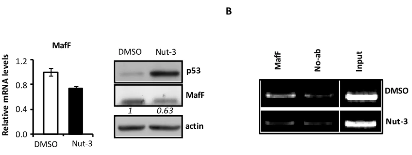
To explore whether the wtp53 protein might have roles in sIL-1Ra gene expression, we performed experiments with MCF7 cells upon delivery of Nutlin-3 (Nut-3), a well-known non-genotoxic wtp53 activator. Activated wtp53, as confirmed by induced p21 expression (Fig. 3A), increases significantly the sIL-1Ra mRNA (Fig. 3A, lower panel) and secreted protein (Fig. 3B) with respect to cells treated only with the Nut-3 solvent, dimethyl sulfoxide (DMSO). These results show that p53R273H suppresses, whereas wtp53 induces sIL-1Ra expression, suggesting a novel mutp53 GOF activity.
MDA-MB231 (p53R280K) 0 1 2 3 4 5 sh-scr sh-p53 DOX + -+ -+ -+ + p53 actin Fo ld s o f sI L-1 R a in d u ct io n in C M 0 1 2 3 4 5 SKBR3 (p53R175H) sh-scr sh-p53 DOX + -+ -+ -+ + p53 actin Fo ld s o f sI L-1 R a in d u ct io n in C M 0 1 2 3 4 5 MDA-MB468 (p53R273H) sh-scr sh-p53 DOX + -+ -+ -+ + p53 actin Fo ld s o f sI L-1 R a in d u ct io n in C M HT29 (p53R273H) p53 actin Fo ld s o f sI L-1 R a in d u ct ion in C M 0 1 2 3 4 5 sh-scr sh-p53 DOX + -+ -+ -+ +
B
sh-p53 sh-scr HT29 0 1 2 3 4 R e la ti ve m R N A le ve l MDA-MB468 sh-scr sh-p53 GAPDH p53 0 1 2 3 4 sIL-1Ra CM HT29 sIL-1Ra sh-p53 sh-scr Kineret -+ -+ -+ + -+ MDA-MB468 + -+ -+ -+ DOX28
A B
Figure 3. Wt p53 induces sIL-1Ra protein production in cancer cells.(A) MCF7
cells were treated with either Nut-3 (10 μM) or DMSO, and RNAs and proteins extracted 48 h later. (Upper panel) WB analyses performed with the indicated antibodies. (Lower panel) qPCR analyses. Relative sIL-1Ra mRNA level were quantified with respect to DMSO-treated cells set to 1.0. (B) WBs performed with the indicated antibody on CMs derived from MCF-7 cancer cells upon treatment with DMSO and Nut-3 as in (B).
Mutant but not wtp53 reduces sIL-1Ra promoter activity
To investigate whether the increased sIL-1Ra gene expression upon p53R273H depletion or wtp53 activation might occur by modulating the sIL-1Ra promoter activity, assays were performed with the sIL-1Ra regulatory region driving a luciferase reporter (pRA1680-Luc; − 1680 bp with respect to transcription start site), which exhibits patterns of expression and induction similar to that of the endogenous gene.169 Reporter assays were performed with sh-p53 and sh-scr HT29 and MDA-MB468 cells and with Nut-3- and DMSO-treated MCF7 cells. Significantly higher sIL-1Ra promoter activity was found in mutp53 depleted HT29 and MDA-MB468 cells and Nut-3-treated MCF7 cells with respect to their controls (sh-scr and DMSO) (Fig.
4).
Figure 4. Mutant p53 inhibits, whereas wtp53 increases the sIL-1Ra gene expression modulating its promoter activity. The activities
of sIL-1Ra regulatory region (−1680;+1 with respect to transcription start site) were analyzed in either: (i) early lentiviral-transduced sh-scr and sh-p53 HT29 and MDA-MB468 cells; and (ii) Nut-3 or DMSO-treated MCF7 cells. Outcomes were normalized to transfection efficiency (βGAL) and protein quantity. Relative Luc activity was quantified with respect to controls (sh-scr or DMSO) set to 1.0.
0 1 2 3 4 5 sh-scr sh-p53 0 1 2 3 4 5 sh-scr sh-p53 0 0,5 1 1,5 2 2,5 3 DMSO Nut-3 R e la ti ve L U C a ct iv it y CM sIL-1Ra DMSO MCF-7 Nut-3 DMSO Nut-3 p53 p21 Actin 0 1 2 3 4 sIL-1Ra R e la ti ve m R N A le ve l -1680LUC
29
To further confirm the achieved results, we analyzed the transcriptional activity of the endogenous sIL-1Ra promoter. As shown in Fig. 5, an increased occupancy of active chromatin markers was detected in sh-p53 HT29 cells with respect to controls (sh-scr). These results indicate that, under our experimental conditions, p53R273H suppresses sIL-1Ra gene expression by reducing its promoter activity, whereas wtp53 increases promoter activity.
Figure 5. Endogenous sIL-1Ra promoter activities. Chromatins were
isolated from sh-scr and sh-p53 HT29 cells, and ChIP assays performed to analyze the recruitments of histone H3 pan-acetylated (H3Ac pan); H3 acetylated at lysine 9 (H3K9ac), H3 acetylated at lysine 14 (H3K14ac) and H3 tri-methylated at lysine 4 (H3K4me3)
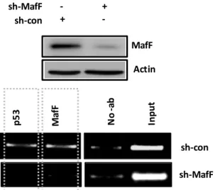
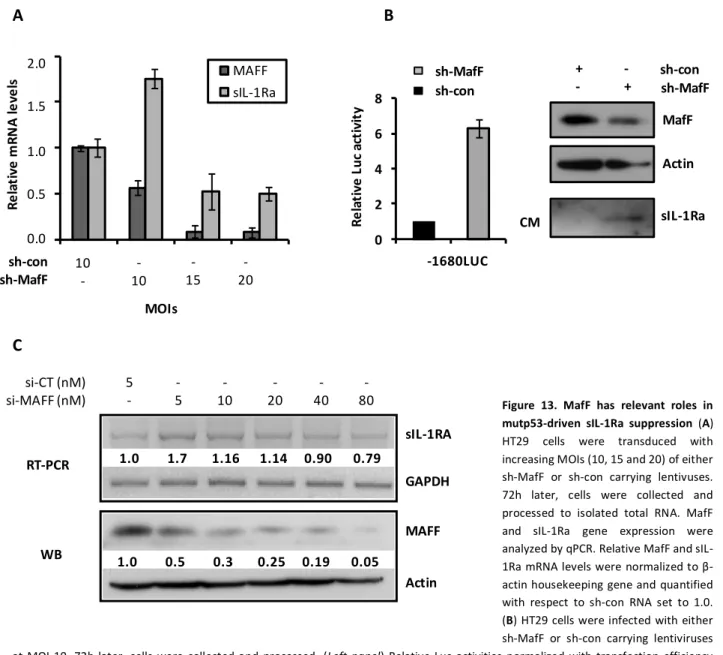
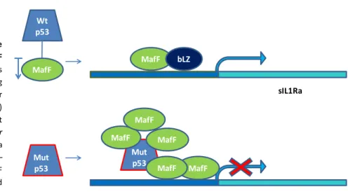
We next asked whether the sIL-1Ra suppression might occur in a p53R273H-specific fashion or whether other p53 mutants might similarly affect the sIL-1Ra gene expression. Thus, experiments have been performed in isogenic conditions with HEPG2 cells, a line widely used to study the production of acute-phase proteins (APPs), as sIL-1Ra was described as an APP.173 The sIL-1Ra promoter activity was evaluated upon transient ectopic expression of wtp53, p53R273H or p53R175H encoding cDNAs. The results showed that, with respect to control (empty vector), exogenous wtp53 significantly increases the sIL-1Ra promoter activity, whereas both ectopically expressed mutants inhibit its activity (Fig. 6A). Confirmatory experiments were similarly carried out in mutp53 depleted HT29 cells along with exogenous expression of either p53R175H or p53R273H mutants. As shown in Fig. 6B, the increased sIL-1Ra mRNA levels observed along with depletion of endogenous p53R273H, drops consistently upon p53R175H or p53R273H ectopic expression. These results show that mutp53-driven sIL-1Ra suppression might not occur in a mutation specific fashion, and the discrepancy with SKBR3 results might be related to the cell specificity of mutp53 GOFs.
H 3 K 1 4 A c H 3 K 4 m e 3 N o Ab In p u t H 3 A c p an H 3 K 9 A c sh-scr sh-p53 HT29