Biomolecules 2021, 11, 176. https://doi.org/10.3390/biom11020176 www.mdpi.com/journal/biomolecules Review
CAPE and Neuroprotection: A Review
Marwa Balaha 1,2, Barbara De Filippis 1, Amelia Cataldi 1,* and Viviana di Giacomo 1
1 Department of Pharmacy, University “G. d’Annunzio”, Chieti-Pescara, 66100 Chieti, Italy;
[email protected] (M.B.); [email protected] (B.D.F.); [email protected] (V.d.G.) 2 Department of Pharmaceutical Chemistry, Faculty of Pharmacy, Kafrelsheikh University,
Kafr El Sheikh 33516, Egypt
* Correspondence: [email protected]; Tel.: +3908713554467
Abstract: Propolis, a product of the honey bee, has been used in traditional medicine for many years.
A hydrophobic bioactive polyphenolic ester, caffeic acid phenethyl ester (CAPE), is one of the most extensively investigated active components of propolis. Several studies have indicated that CAPE has a broad spectrum of pharmacological activities as anti-oxidant, anti-inflammatory, anti-viral, anti-fungal, anti-proliferative, and anti-neoplastic properties. This review largely describes CAPE neuroprotective effects in many different conditions and summarizes its molecular mechanisms of action. CAPE was found to have a neuroprotective effect on different neurodegenerative disorders. At the basis of these effects, CAPE has the ability to protect neurons from several underlying causes of various human neurologic diseases, such as oxidative stress, apoptosis dysregulation, and brain inflammation. CAPE can also protect the nervous system from some diseases which negatively affect it, such as diabetes, septic shock, and hepatic encephalopathy, while numerous studies have demonstrated the neuroprotective effects of CAPE against adverse reactions induced by different neurotoxic substances. The potential role of CAPE in protecting the central nervous system (CNS) from secondary injury following various CNS ischemic conditions and CAPE anti-cancer activity in CNS is also reviewed. The structure–activity relationship of CAPE synthetic derivatives is discussed as well.
Keywords: caffeic acid phenethyl ester (CAPE); neuroprotection; neurodegenerative disease; propolis; CNS injury; CNS ischemia; CNS cancer; neuroinflammation; CAPE derivatives; neurotoxic substance(s)
1. Introduction
Propolis, a honeybee product, is a resinous product obtained from different plant parts mixed with beeswax and bee salivary enzymes and represents a multimillion dollar market [1,2]. It has been used in traditional medicine for many years due to its anti-microbial, anti-inflammatory, anti-oxidant, immunomodulator, anti-mutagenic, and carcinostatic effects [3]. Propolis consists of a large number of different compounds according to the ingredients the bees collect from the plants. Its main components are phenols and related compounds. Investigations have been conducted to purify and determine which substances are responsible for these effects. The results of these investigations have suggested that caffeic acid phenethyl ester (CAPE, phenethyl 3-(3-4 dihydroxyphenyl) acrylate) is the main component that mediates most of the beneficial effects ascribed to propolis [4]. As a consequence, CAPE has become one of the most studied natural product research topics in recent years. Grunberger and co-workers extracted and described CAPE in 1988 [5], and later Sigma-Aldrich produced commercial preparations for the market [6]. CAPE is a polyphenol with hydroxyl groups within the catechol ring which provide strong antioxidant properties to the molecule and can affect many biological activities. It has a lipophilic property because of its long carbon groups in an aromatic and aliphatic structure, which leads to adequate blood concentration after Citation: Balaha, M.; De Filippis,
B.; Cataldi, A.; di Giacomo, V. CAPE and Neuroprotection: A Review. 2021, 11, 176. https:// doi.org/10.3390/biom11020176 Academic Editor: John Mitrofanis
Received: 27 November 2020 Accepted: 25 January 2021 Published: 28 January 2021
Publisher’s Note: MDPI stays
neutral with regard to
jurisdictional claims in published maps and institutional affiliations.
Copyright: © 2021 by the
authors. Licensee MDPI, Basel, Switzerland. This article is an open access article distributed under the terms and conditions of the Creative Commons Attribution (CC BY) license (http://creativecommons.org/lice nses/by/4.0/).
intraperitoneal administration [7]. Celli et al. [8] investigated the stability of CAPE in rats and human plasma. It was found that CAPE undergoes hydrolysis to caffeic acid after 6 h in rat plasma in vitro and also in vivo, giving caffeic acid as the major metabolite. This type of hydrolysis does not occur in human plasma because it does not contain carboxylesterase, which is responsible for CAPE hydrolysis [9]. The broad spectrum of pharmacological activities shown by CAPE ranges from anti-oxidant/anti-inflammatory to anti-viral/anti-fungal properties, including anti-proliferative effects in various cancer models [10]. A recent in vivo study indicated that CAPE has the ability to cross the blood– brain barrier in rats [11,12]. This review focuses on the protective effects of CAPE in many diseases that can affect the central nervous system (CNS). CAPE was found to have a protective effect on different neurodegenerative disorders, either those occurring with age, such as Alzheimer’s disease (AD) and Parkinson’s disease (PD), or other neurodegenerative disorders, such as amyotrophic lateral sclerosis (ALS) and seizures. CAPE was proven to protect neurons from the main underlying causes of several human neurologic diseases—namely, oxidative stress, apoptosis dysregulation, and brain inflammation. In addition, CAPE was found to be able to protect the nervous system from the consequences of some diseases not directly affecting it, such as diabetes, septic shock, and hepatic encephalopathy (HE). A paragraph about the neuroprotective effects of CAPE against adverse reactions induced by different neurotoxic substances such as ethanol, methotrexate, acrolein, and others is also provided. The role of CAPE in the pathophysiology of CNS tumors and in neuronal injuries is carefully reviewed through the description of the modulatory activity of CAPE against biochemical and histopathological cascade mechanisms following ischemic conditions in the brain and spinal cord. Finally, a brief overview of CAPE synthetic derivatives follows. Five tables summarize the main findings on CAPE effects in different areas of neuroprotection. In the column named “Parameters measured”, the effect of CAPE is reported between parentheses where applicable. In some case, the effect of CAPE is not directly deducible, but depends on the experimental model or conditions. This review provides a useful insight into the role of CAPE in the management of numerous pathological conditions affecting the CNS. The description of the molecular mechanisms and the focus on CAPE derivatives can help in discovering new promising targets for neuroprotection and in finely tailoring structural modifications of CAPE derivatives.
2. Methodology
The online database PubMed was used for screening the studies for this review, with no chronological limits applied. Keywords used to search for articles included caffeic acid phenylethyl ester and neuroprotection, neurodegenerative disorders, CNS injury, CNS ischemia, CNS cancer, neuroinflammation, brain injury, spinal cord injury, and neuronal injury. The search retrieved more than 90 results, including three reviews with a section or hints about neuroprotective effect of CAPE or its protective effects against ischemia-reperfusion injury. None of these discussed the diverse signaling pathways involved. After having removed duplicates and narrowing the results according to the relevance to the core topic of the review, 77 papers were selected. A thorough analysis of the reference list of all the included studies identified additional relevant studies which, together with papers cited to introduce and clarify the various subjects, led to a total of 90 references.
3. CAPE Effects on Different Neurologic Disorders
With age, a lot of neurologic disorders become common, such as Alzheimer’s disease (AD), Parkinson’s disease (PD), and amyotrophic lateral sclerosis (ALS). Common causes for the development of these diseases are oxidative stress, inflammation, and apoptosis impairment. A main goal of anti-ageing strategies is the elimination of harmful agents— in other words, free radicals—from the environment to protect organs from aging and other oxidative stress-related pathologies. One study evaluated the effects of the long-term administration of CAPE on histological and biochemical alterations induced by
ageing in the brain and cerebellum of old rats. CAPE increased the superoxide dismutase (SOD), catalase (CAT), and glutathione peroxidase (GSH-Px) activities and glutathione (GSH) levels in both cerebral and cerebellar tissues to levels similar to those in young rats. Additionally, CAPE reduced the malondialdehyde (MDA, an important marker of lipid peroxidation) level and ageing-induced ultrastructural alterations, proving to be more effective than melatonin, which is used worldwide in order to prevent ageing-related pathologies [13]. Many studies addressing the protective effects of CAPE on different neurologic disorders are described below and summarized in Table 1. Other than the above-mentioned pathological conditions, the role of CAPE in psychosis, seizures, and the side-effects of other diseases are described.
3.1. Apoptosis
Apoptosis dysregulation has been suggested to be the underlying cause of several human neurologic diseases. Transient focal or global cerebral ischemia in rodents leads to neuronal apoptosis. Reactive oxygen species (ROS) production is thought to represent a relevant mechanism in the series of biochemical events ultimately leading to apoptosis. CAPE blocks neuronal death through inhibition of inflammation and of mitochondrial cytochrome c release, and through a reduction in free radical generation [14,15]. Primary cultures of cerebellar granule neurons (CGNs) are considered a suitable in vitro model to study the mechanisms of neuronal apoptosis, which can be induced by low K+ concentrations. CAPE exerts its anti-apoptotic effect on CGNs by blocking ROS formation and inhibiting the activity of both caspase-3 and caspase-9. Caspase-3 is the executioner caspase that is downstream of the intrinsic (mitochondrial) caspase-9 and extrinsic receptor activated (Fas) caspase-8 pathways. Interestingly, CAPE was found to completely block the activation of nuclear factor-κB (NF-κB) without interfering with the marked decrease in intracellular Ca2+ concentration induced by low K+ [14,16].
Another study investigated CAPE’s neuroprotective effect against apoptotic cell death in the developing rat brain after pentylenetetrazole (PTZ)-induced status epilepticus (SE). Prolonged seizures are associated with inadequate blood flow, increased excitatory amino acid release, and decreased glucose use and oxygen consumption. All these features are associated with impaired mitochondrial function and irreversible neuronal damage. CAPE showed diminished apoptosis and preservation of the number of neurons, as well as a reduced number of caspase-3-positive cells in the hippocampus and in the prefrontal cortex [17]. Another study [18] investigated the effect of CAPE on CGNs exposed to excitotoxicity by the overstimulation of glutamate receptors. Neuronal death caused by excitotoxicity plays an important role in a number of neurodegenerative disorders, including Alzheimer’s disease, Parkinson’s disease, and multiple sclerosis. CAPE inhibited necrosis mediated by p38 phosphorylation and apoptosis mediated by cytochrome c release and caspase-3 activation, while it appeared to have no effect on glutamate-induced neuronal death via the NF-κB pathway. Interestingly, CAPE proved effective in modulating neurotogenesis—i.e., the formation of new neurites—in PC12 cells challenged with the dopaminergic neurotoxin 1-methyl-4-phenylpyridinium (MPP+). CAPE induced the formation, elongation, and ramification of neurites and inhibited the shortage of neurites induced by the neurotoxin. These effects were associated with an increased expression of neuronal typical proteins responsible for axonal growth (GAP-43) and synaptogenesis (synaptophysin and synapsin I) [19].
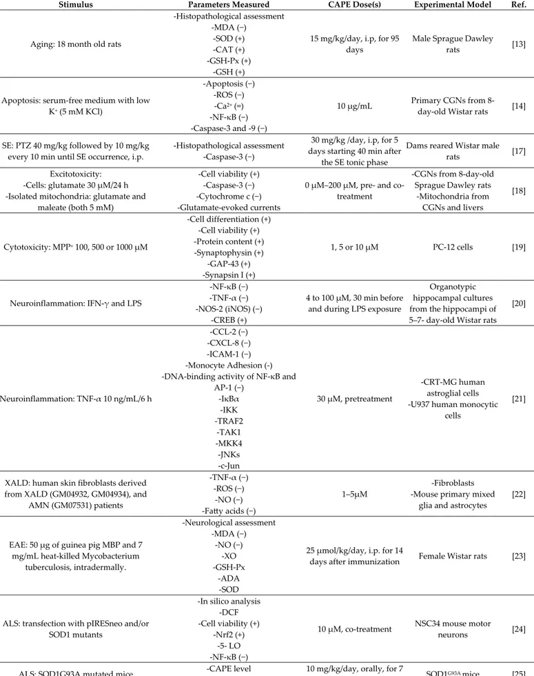
Table 1. CAPE protective effects in different neurological disorders. Where applicable, the effect of CAPE on the
parame-ters measured is reported between parentheses (+, − or =).
Stimulus Parameters Measured CAPE Dose(s) Experimental Model Ref.
Aging: 18 month old rats
-Histopathological assessment -MDA (−) -SOD (+) -CAT (+) -GSH-Px (+) -GSH (+)
15 mg/kg/day, i.p, for 95 days
Male Sprague Dawley rats [13]
Apoptosis: serum-free medium with low K+ (5 mM KCl) -Apoptosis (−) -ROS (−) -Ca2+ (=) -NF-κB (−) -Caspase-3 and -9 (−) 10 µg/mL Primary CGNs from 8-day-old Wistar rats [14]
SE: PTZ 40 mg/kg followed by 10 mg/kg every 10 min until SE occurrence, i.p.
-Histopathological assessment -Caspase-3 (−)
30 mg/kg /day, i.p, for 5 days starting 40 min after
the SE tonic phase
Dams reared Wistar male
rats [17]
Excitotoxicity: -Cells: glutamate 30 µM/24 h -Isolated mitochondria: glutamate and
maleate (both 5 mM)
-Cell viability (+) -Caspase-3 (−) -Cytochrome c (−) -Glutamate-evoked currents
0 µM–200 µM, pre- and co-treatment
-CGNs from 8-day-old Sprague Dawley rats
-Mitochondria from CGNs and livers [18] Cytotoxicity: MPP+ 100, 500 or 1000 µM -Cell differentiation (+) -Cell viability (+) -Protein content (+) -Synaptophysin (+) -GAP-43 (+) -Synapsin I (+) 1, 5 or 10 µM PC-12 cells [19]
Neuroinflammation: IFN-γ and LPS
-NF-κB (−) -TNF-α (−) -NOS-2 (iNOS) (−)
-CREB (+)
4 to 100 µM, 30 min before and during LPS exposure
Organotypic hippocampal cultures from the hippocampi of 5–7- day-old Wistar rats
[20] Neuroinflammation: TNF-α 10 ng/mL/6 h -CCL-2 (−) -CXCL-8 (−) -ICAM-1 (−) -Monocyte Adhesion (-) -DNA-binding activity of NF-κB and
AP-1 (−) -IκBα -IKK -TRAF2 -TAK1 -MKK4 -JNKs -c-Jun 30 µM, pretreatment -CRT-MG human astroglial cells -U937 human monocytic
cells
[21]
XALD: human skin fibroblasts derived from XALD (GM04932, GM04934), and
AMN (GM07531) patients -TNF-α (−) -ROS (−) -NO (−) -Fatty acids (−) 1–5µM -Fibroblasts -Mouse primary mixed
glia and astrocytes
[22]
EAE: 50 µg of guinea pig MBP and 7 mg/mL heat-killed Mycobacterium tuberculosis, intradermally. -Neurological assessment -MDA (−) -NO (−) -XO -GSH-Px -ADA -SOD
25 µmol/kg/day, i.p. for 14
days after immunization Female Wistar rats [23]
ALS: transfection with pIRESneo and/or SOD1 mutants
-In silico analysis -DCF -Cell viability (+)
-Nrf2 (+) -5-LO -NF-κB (−)
10 µM, co-treatment NSC34 mouse motor
neurons [24]
ALS: SOD1G93A mutated mice -CAPE level -Behavioural assessment
10 mg/kg/day, orally, for 7
- pp38 (−)
-Neuroinflammation, in vitro: 200 ng/mL LPS
-in vivo, a single intraperitoneal injection of 20 mg/kg LPS, i.p., 2 h after the last
CAPE injection -Cell viability -ERK2 -Akt -p38, -pERK1/2 -pp38 -pAKT -pJNK -EPO (+) -HO-1 (+) -iNOS (−) -COX-2 (−) -pAMPKα (+) -0.1 to 1.75 µM 30 min before LPS treatment, or
co-treatment -1 or 5 mg/kg once daily for
3 days -BV-2 murine microglial cell line -Eight-week-old male ICR mice [26]
PD: 6-OHDA 70 µM for 6 h, on day 8–10
-Cell viability (+) -Cytochrome c (−) -Caspase-3 (−) -Ca2+ 10 to 100 µM, pre-treatment for 4 h -Primary CGNs from 8-day-old Wistar rats -Rat liver mitochondria from 7-day-old Sprague–
Dawley rats
[27]
Dopaminergic neurodegeneration: 6-OHDA, 40 µM for RMN and 70 µM for
CGN -Free radicals (−) -Peroxynitrite (−) 10 µM, pre-treatment for 2 h -Rat RMN -Primary CGNs [28] PD: 6-OHDA 8 mg/mL, s.i.
-Fe, Cu, Zn and Mn (−) -ROS (−) -Protein content
-TH
-Mitochondrial functions: Ca2+-induced swelling, Ca2+ uptake and respiration
-In Vivo: 10 µmol/kg/day, i.p, 5 days -In Vitro: 0.5 or 10 µM Wistar rats [29] -Dopaminergic neurodegeneration, in vitro: LPS/72 h -PD, in vivo: LPS 3 µg/µL, intranigral, or 6-hydroxydopamine 2 µg/µL, intrastriatal,
30 min after first CAPE injection.
-NO (−) -ERK -p38 MAPK -HO-1 (+) -BDNF (+) -Nrf2 -In Vitro: 3–30 µM -In Vivo: 10 or 30 mg/kg/day, i.p, for 4 days
-In Vitro: rat organotypic midbrain slice cultures -In Vivo: mouse model of
dopaminergic neurodegeneration [30] PD: rotenone1 mg/kg, s.c., every 48 h, 9 injections -Behavioural assessment -Histopathological assessment -CD11b -COX-2 (−) -iNOS (−) -NF-κB (−) -Dopamine level (+) -TNF-α (−) -IL-1β 2.5, 5 or 10 mg/kg/day.
orally, every 48 h, 9 doses Male Swiss albino mice [31]
PD: CPF 80 mg/kg, s.c. -PON1 activity (+) -Lipid profile -TSA (+) -TAC (+) -TOC (−) -Histopathological assessment 10 µmol/kg/day, i.p, 21
days Male Swiss albino mice [32]
PD: MPTP–HCl 20 mg/kg, i.p, four in at 2 h intervals
-TH-positive neurons (+) -Cell viability (+) -CAPE and MPP+ levels
-DA (+) -MAO (−) -i- and nNOS (−)
-Caspase-1 (−) -Cytochrome c (−) -AIF (−) -Free radicals (−) -Peroxynitrite (−) 2, 5, or 10 mg/kg/day, 7days Eight-week-old male C57BL/6 mice [33]
Loss of memory (AD): STZ 3 mg/kg, bilaterally on day 1 and 3
-TBARS (−) -GSH (+) -SOD (+)
-CAT (+) -Nitrite (−) -AChE (−) -TNF-α (−) -eNOS (+) -NF-κB (−) -Behavioural assessment -Histopathological assessment Dementia (AD type): STZ; 3 mg/kg, on day
1 and 3, ICV
-MDA (−) -GSH (+) -TNF-α (−) -Behavioural tests
3, 6 mg/kg/day, i.p, 28 days Wistar rats [35]
Dementia (AD type): Aβ1-42O, unilateral stereotaxic, ICV -Behavioural assessment -ROS (−) -Nrf2 (+) -GSH -pGSK3α/β -Caspase-9
10 mg/kg/day, i.p, 1 h after
brain lesion, 10 days Male C57Bl/6 mice [36]
Seizures: 60 mg/kg PTZ, i.p, single dose
-Neurological assessment -MDA (−)
-NO (−) -XO -SOD (+)
100 µmol/kg, i.p., 2 days
prior to PTZ injection Female Swiss albino mice [15]
Psychosis: dizocilpine maleate (MK-801), 0.5 mg/kg/day for 5 days, i.p.
-Behavioural assessment -Histopathological assessment -MDA (−) -PC (−) -NO (−) -SOD -GSH-Px (−) -XO (−) -ADA (−) -CAT (=)
10 µmol/kg, 6 days, started
one day before MK-801, i.p. Wistar rats [37]
Diabetes: STZ 45 mg/kg, i.p, single dose
-NO (−) -SOD -GSH-Px (−) -GSH -XO (−) -CAT (−) -MDA (−) -iNOS (−) -TNF-α (−) -IFN-γ (−) -IL-10
25 µM/kg/day, two days after STZ treatment for 60
days
Male Wistar rats [38]
Endotoxic shock: LPS, 20 mg/kg, i.p,
-TNF-α (−) -IL-1α, -1β, -6 (−) -IL-4, -10 (+) -sICAM-1 (−) -Histopathological assessment 10 µmol/kg/day, 14 days before shock induction and
a single dose 30 min after induction
Male Wister rats [39]
Hepatic encephalopathy: thioacetamide:600 mg/kg, i.p., two doses
(0 and 24 h)
-Behavioural and motor assessment -Blood ammonia (=)
-ALT (−) -AST (−)
10 µmol/kg/day, i.p, starting 1 day before the first dose of thioacetamide
Male Wistar rats [40]
Optic nerve crushing, 10 s
-Apoptosis (−) -Astrocyte migration -Cell viability (+) -NF-κB (−) -IL-6 and -8 (−) -iNOS (−) -COX-2 (−) -TNF-α (−) -CCL-2 (−)
10 µmol/kg, i.p, 10 min after the surgery
Male Sprague Dawley
3.2. Neuro-Inflammation
Brain inflammatory events are observed in several neuropathologies or in response to infectious diseases. CAPE’s ability to control neuro-inflammation was compared with that of other drugs, such as acetyl-salicylate (anti-inflammatory), dexamethasone (gluco-corticoid), pyrrolidine dithiocarbamate (anti-oxidant), and SN 50 peptide (selective per-meant NF-κB inhibitor) in organotypic hippocampal slice cultures. Interestingly, CAPE presented a longer-lasting control (over 48 h after a single administration) of neuro-in-flammation compared to the other compounds. This effect can be ascribed to the interfer-ence CAPE exerts on several neuroinflammatory effectors, such as NF-κB, tumor necrosis factor (TNF)-α, and nitric oxide (NO). At the dose of maximal effect (100 µM), an increase in cAMP-responsive element binding protein (CREB) activity, a molecule with anti-in-flammatory activity, was also observed [20]. Another study examining the anti-inflamma-tory effect of CAPE on activated astroglial cells found that the modulation of NF-κB ac-tivity in astroglial cells can be a potential target for the treatment of inflammatory and degenerative CNS diseases. CAPE pre-treatment abrogated the TNF-α induced expres-sion of chemokine (C-C motif) ligand 2 (CCL-2) and intercellular adheexpres-sion molecule-1 (ICAM-1) via the inhibition of NF-κB activation in a cell-specific manner. CAPE inhibited the downstream pathways of inhibitor κB (IκB) degradation in monocytic cells while sup-pressing the activation of upstream IkB kinase in astroglial cells [21].
X-linked adrenoleukodystrophy (X-ALD) is a neuro-inflammatory disease associated with the demyelination of the cerebral white matter, characterized by the accumulation of very long chain fatty acids (VLCFA) caused by peroxisomal disorder due to mutations in the ABCD1 gene. CAPE treatment corrected both the metabolic VLCFA accumulation and the secondary inflammation. The effect of CAPE was mediated by the up-regulation of Abcd2 expression and of peroxisomal β-oxidation, leading to decreased VLCFA levels in ABCD1-deficient U87 cells. CAPE administration reduced the expression of inducible ni-tric oxide synthase (iNOS), inflammatory cytokines, and the activation of NF-κB in pri-mary astrocytes derived by ABCD1/ABCD2-silenced mice [22]. Inflammation is a major component in the pathogenesis of another disease, experimental autoimmune encephalo-myelitis (EAE), which is considered the animal model of multiple sclerosis (MS). In both EAE and MS, ROS can damage the myelin sheath and the blood–brain barrier (BBB). CAPE maintains cell membrane integrity and function, thus preventing protein leakage and accumulation by inhibiting the peroxidation of membrane lipids. CAPE may exert its anti-inflammatory effect and ameliorate clinical symptoms by inhibiting ROS production at the transcriptional level, through the suppression of NF-κB activation, and by directly inhibiting the iNOS catalytic activity [23].
ALS is a disease that causes the death of motor neurons controlling voluntary mus-cles. In one study in 2009 [24], CAPE was selected from a library of 2000 small anti-oxidant molecules for the mechanism of action and the ability to be in the CNS in a concentration sufficient to give a therapeutic effect. In two experimental models of ALS, represented by mouse motor neuron cells (NSC34) expressing mutant superoxide dismutase1 (SOD1) and motor neurons isolated from cases of familial SOD1-associated ALS, CAPE inhibited NF-κB-induced inflammation. CAPE also activated the nuclear factor erythroid 2-related factor 2 (Nrf2)–ARE pathway, which is usually down-regulated in ALS. Genes up-regu-lated by Nrf2 include GSH-Px, glutathione reductase, heme oxygenase 1 (HO-1); enzymes involved in GSH synthesis, and NADPH-regenerating enzymes. CAPE effects were tested also in an in vivo model of mice expressing a mutant superoxide dismutase (SOD1G93A) linked to human ALS. CAPE increased the post-onset survival and lifespan of the mice, showing a significantly greater number of surviving motor neurons and a decreased num-ber of activated microglia and astrocytes in the lumbar region of the spinal cord. In addi-tion, lower levels of phosphorylated p38, a mitogen-activated protein kinase that is in-volved in both inflammation and neuronal death, were observed in the spinal cords of SOD1G93A mice [25]. Microglial activation has been widely demonstrated to mediate in-flammatory processes that are crucial in several neurodegenerative disorders. CAPE was
proven to inhibit cyclooxygenase-2 (COX-2) and iNOS expression and the consequent NO production both in in vitro and in vivo models of microglia activation. Anti-neuroinflam-matory responses in microglial cells were mediated by 5′-adenosine monophosphate-ac-tivated protein kinase (AMPK)α, erythropoietin (EPO), and HO-1 [26].
3.3. Parkinson’s Disease
PD is a neurodegenerative disorder characterized by the progressive loss of dopa-minergic neurons of the substantia nigra pars compacta. 6-Hydroxydopamine (6-OHDA) is a neurotoxic synthetic organic compound used to selectively destroy dopaminergic neu-rons in the brain to induce PD in laboratory animals. The neurotoxicity and neuronal cell death induced by 6-OHDA are due to ROS production, cytochrome c release, and subse-quent caspase-3 activation. CAPE increased the viability of cerebellar granule neurons
dose dependently and markedly attenuated the 6-OHDA-induced toxicity. CAPE blocked Ca2+-induced cytochrome c release and caspase-3 activation, but no interaction of CAPE with caspase-3 cleavage was found [27]. These results were confirmed in the same cellular model and in rostral mesencephalic neurons (RMNs), where CAPE pretreatment in-creased neuronal viability from 39% to 91% in CGNs and from 33% to 60% in RMNs. CAPE is able to block 6-OHDA-induced dopaminergic neuronal death through the block-age of O2−· and peroxynitrite generation, suggesting its potential use as a neuroprotective drug for PD [28]. With regard to the in vivo experimental models, CAPE inhibited mito-chondrial permeability transition, a process that triggers cytochrome c release and caspase-3 activation, leading to neuronal death in rats treated with 6-OHDA to induce PD. The mechanism of protection involves the scavenging of free radicals and metal chelation without mitochondrial dysfunction [29].
CAPE can protect midbrain dopaminergic neurons in vitro and in vivo also by the induction of HO-1 and brain-derived neurotrophic factor (BDNF) expression [30]. Rote-none is commonly used to induce experimental PD because of its ability to block the com-plex I inhibition and to activate microglia, resulting in neuropathologic and phenotypic features of PD. In a rotenone Parkinsonian mice model, CAPE treatment raised the level of striatal dopamine and lessened the inflammatory burden by suppressing microglia cells and down-regulating the gene expression of COX-2, iNOS, and NF-κB. These effects led to an improvement in locomotor activity. CAPE exposure protected against rotenone-in-duced histopathological abnormalities and led to an increased number of surviving neu-rons as well [31]. In a model for PD induced by chlorpyrifos, CAPE showed positive ef-fects on the paraoxonase (PON1) activity, levels of lipid profile, total sialic acid (TSA), total anti-oxidant capacity (TAC), and total oxidant capacity (TOC) in plasma and brain tissue, preventing neurodegeneration [32]. In a murine model of PD induced by MPTP (1-methyl-4-phenyl-1,2,3,6-tetrahydropyridine), CAPE attenuated dopaminergic neuro-degeneration and dopamine loss. This effect was associated with a marked reduction in iNOS and caspase-1 expression in vivo. In Vitro, CAPE proved able to mitigate neurotox-icity by inhibiting the mitochondrial release of cytochrome c and apoptosis inducing fac-tor (AIF) induced by MPP+ [33].
3.4. Alzheimer’s Disease
Alzheimer’s disease (AD) is an age-associated neurodegenerative disease that can be induced in rodents by the intracerebroventricular (ICV) administration of a widely used diabetogenic drug, streptozotocin (STZ). Brain PI3-kinase activity and nitric oxide pro-duction mediated by the endothelial NOS (eNOS) are involved in the memory revival function of CAPE in STZ-ICV-administered rats. The basal NF-κB activity in the rat brain was found essential for memory functions as well. The anti-apoptotic and pro-survival functions of PI3-kinase activation regulate eNOS and NFκB activity, conferring neuropro-tection and improving memory [34]. A previous study by the same group showed that the CAPE down-regulation of oxidative stress and inflammation was accompanied by the amelioration of STZ-ICV-induced dementia. A great increase in the brain GSH levels and
a diminution of thiobarbituric acid reactive substances (TBARS) as well as of TNF-α con-tent were observed in the brains of rats treated with CAPE [35]. In another study, amyloid-beta oligomers were administered to mice to induce dementia and to study the AD onset and cognitive function impairment. CAPE treatment reversed cognitive deficits and im-proved learning and memory abilities. This action was accompanied by an induction of Nrf2 and heme oxygenase-1 via the modulation of glycogen synthase kinase 3β in the murine hippocampus [36].
3.5. Seizures and Psychosis
CAPE is useful as an adjunctive treatment of seizure disorders. Seizures induced by pentylenetetrazole (PTZ) are due to the activation of glutamate receptors and the inhibi-tion of GABA, an inhibitory neurotransmitter. Glutamate receptors’ activainhibi-tion enhances the ROS level, which in turn enhances glutaminergic activity, but the administration of CAPE increased the latency and decreased the duration of seizures in PTZ-treated mice. CAPE protected the brain tissue from oxidative damage because of its ability to scavenge ROS, to decrease MDA concentration, to increase the anti-oxidant SOD level, and to sig-nificantly attenuate NO generation [15].
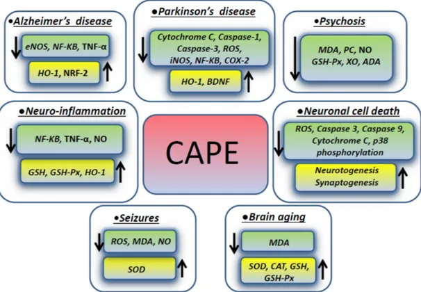
Psychosis has many different causes and can be experimentally induced by dizocili-pine maleate (MK-801), which causes neurotoxicity inducing the intracellular generation of free radical species through the N-methyl-D-aspartate (NMDA) receptor blockage. NMDA antagonists mediate cell death triggered by ROS, which are involved in mem-brane pathology in CNS and play a role in neuropsychiatric disorders, including schizo-phrenia. In one study focusing on the rat prefrontal cortex (PFC), which is the region mainly affected in schizophrenia, CAPE modulated the brain oxidant/anti-oxidant status, stabilizing the cellular membranous structures. A CAPE therapeutic effect was exerted by decreasing the levels of malondialdehyde, protein carbonyl (PC), and nitric oxide (NO). The activity of the enzymes glutathione peroxidase (GSH-Px), xanthine oxidase (XO), and adenosine deaminase (ADA) in prefrontal tissue were decreased as well, when compared to the MK-801 groups, whereas the catalase activity was not changed. In addition, CAPE treatment decreased the number of apoptotic cells in the PFC exposed to MK-801 [37]. Figure 1 summarizes the molecules involved in the effects exerted by CAPE in different neurological conditions described in the previous paragraphs.
Figure 1. CAPE effects on intracellular molecules and pathways in different neurologic disorders.
3.6. Other Diseases
Some pathological conditions, different from neurodegenerative disorders, such as diabetes, septic shock, and hepatic encephalopathy (HE), can affect the health of the nerv-ous system. Diabetes induces oxidative stress and inflammation in brain. In a murine model, CAPE significantly counteracted the effects of diabetes by decreasing the levels of nitric oxide and malondialdehyde and the activities of catalase, glutathione peroxidase, and xanthine oxidase. CAPE treatment also significantly suppressed the expression of in-flammatory cytokines such as TNFα and interferon (IFN)-γ and the iNOS activity, which were remarkably enhanced in the brain by diabetes [38].
Sepsis patients suffer from severe oxidative stress, with the overproduction of reac-tive oxygen species and reacreac-tive nitrogen species, resulting in direct cellular injury. NF-κB plays a central role in the induction of crosstalk between cytokines and inflammatory mediators, which leads to the pathophysiology of septic shock. The effect of CAPE against lipopolysaccharide (LPS) -induced endotoxemia, neuronal damage, and the associated systemic inflammatory response was investigated in male Wistar rats. CAPE prevented neuronal damage and preserved astrocyte morphology, with no sign of inflammatory cel-lular infiltration, edema, or cytoplasmic swelling. CAPE decreased the levels of inflam-matory cytokines (interleukin (IL)-1α, -1β, -6) and TNF-α in the plasma, increased the anti-inflammatory cytokine levels (IL-4, IL-10), and counteracted the imbalance leading to the inhibition of adhesion molecule expression (sICAM-1).This effect of CAPE on the inflam-matory cellular infiltration into the brain could be directly attributed to its inhibitory effect on NF-κB activation [39].
Hepatic encephalopathy (HE) is a major neurological complication secondary to se-vere liver failure, causing serious neurological problems. Thioacetamide-induced HE in rats was almost fully reversed by a combination of CAPE with the laxative lactulose. The survival rates were 37.5% in the HE group, 70% in the HE + lactulose group, 80% in the HE + CAPE group, and 100% in the HE + CAPE + lactulose group. The lack of death in the animals treated with both CAPE and lactulose can be ascribed to the direct
neuroprotec-tive effect of CAPE together with the prevention of ammonia production in the body. In-creased ammonia, high transaminase levels in blood, inIn-creased lipid peroxidation, and decreased antioxidant enzyme activities in most brain regions, along with impaired sen-sorymotor behavioral tests, were reversed to almost control values in the CAPE + lactu-lose-treated group [40].
Glaucoma is characterized by the death of retinal ganglion cells (RGCs) and visual field defects leading to irreversible blindness. CAPE prevented optic nerve crush-induced RGC apoptosis and neuroinflammation. These effects are mediated by the decreased ex-pression of the inflammatory cytokines IL-8 and IL-6, inducible nitric oxide synthase, cy-cloooxygenase-2, tumor necrosis factor-α, and chemokine C-C ligand-2. The hypertrophy of astrocytes and Müller cells (gliosis) was modulated by CAPE through the inhibition of NF-κB signaling [41].
4. CAPE Protective Effects against Different Neurotoxic Substances
Some substances can affect the nervous system functions by damaging brain cells or nerves. Different studies investigating the protective effect of CAPE against different neu-rotoxic substances are described in this paragraph and summarized in Table 2.
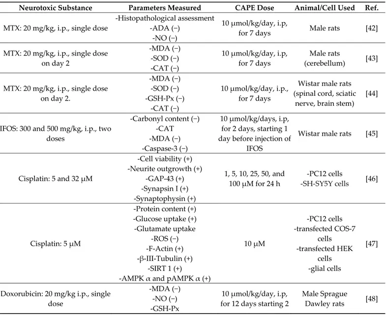
Table 2. CAPE protective effects against different neurotoxic substances. Where applicable, the effect of CAPE on the
parameters measured is reported between parentheses (+, − or =).
Neurotoxic Substance Parameters Measured CAPE Dose Animal/Cell Used Ref. MTX: 20 mg/kg, i.p., single dose
-Histopathological assessment -ADA (−)
-NO (−)
10 µmol/kg/day, i.p,
for 7 days Male rats [42]
MTX: 20 mg/kg, i.p., single dose on day 2 -MDA (−) -SOD (−) -CAT (−) 10 µmol/kg/day, i.p, for 7 days Male rats (cerebellum) [43]
MTX: 20 mg/kg, i.p., single dose on day 2. -MDA (−) -SOD (−) -GSH-Px (−) -CAT (−) 10 µmol/kg/day, i.p., for 7 days
Wistar male rats (spinal cord, sciatic
nerve, brain stem) [44]
IFOS: 300 and 500 mg/kg, i.p., two doses -Carbonyl content (−) -CAT -MDA (−) -Caspase-3 (−) 10 µmol/kg/days, i.p, for 2 days, starting 1 day before injection of
IFOS
Wistar male rats [45]
Cisplatin: 5 and 32 µM -Cell viability (+) -Neurite outgrowth (+) -GAP-43 (+) -Synapsin I (+) -Synaptophysin (+) 1, 5, 10, 25, 50, and 100 µM for 24 h -PC12 cells -SH-SY5Y cells [46] Cisplatin: 5 µM -Protein content (+) -Glucose uptake (+) -Glutamate uptake -ROS (−) -F-Actin (+) -β-III-Tubulin (+) -SIRT 1 (+)
-AMPK α and pAMPK α (+)
10 µM -PC12 cells -transfected COS-7 cells -transfected HEK cells -glial cells [47]
Doxorubicin: 20 mg/kg i.p., single dose
-MDA (−) -NO (−) -GSH-Px
10 µmol/kg/day, i.p, for 12 days starting 2
Male Sprague
-CAT (+) -SOD
days before doxorubicin
Cigarette smoke: 1 h daily for 4 weeks
-MDA (−) -SOD (+) -Apoptosis (−)
10 mmol/kg/day, i.p, for 4 weeks before the
exposure to cigarette smoke Rabbits [49] Ethanol: 3 mg/kg, oral -TOS (−) -TAS (=) -Histopathological assessment 10 µmol/kg, i.p., immediately after ehanol administration Rats [50] Acrolein: 1 M -Cell viability (+) -ROS (−) -GSH (+) -MAPKs -Akt/GSK3 -α/β-secretase 0-90 µM, pretreatment for 30 min HT22 mouse hippocampal cells [51]
K2CrO4: 2 mg/kg/day, i.p., for 30 days
-SOCS3, JAK2 and STAT3 -NO (−) -GSH (+) -SOD (+) -GSH-Px -AChE -TNF-α (−) -IL-6 (−) 20 mg/kg/day, orally,
for 30 days Wistar male rats [52]
CdCl2: 1.5 mg/kg
-Neurobehavioural assessment -Histopathological assessment
-AMPK and pAMPK -SIRT1 -Bcl-2 (+) -Bax (−) -Caspase-3 (−) -p-Tau -TLR4 -IL-6 (−) -IL1-β (−) -TNF-α (−)
10 µmol/kg/ day, for 4 weeks 7 weeks old Kunming mice [53] Chlorpyriphos: 10 mg/kg, oral -AChE (−) -TOS (−) -TAR -Histopathological assessment -Caspase-3 -Bcl-2 -Bax 10 µmol/kg, i.p., immediately after chlorpyriphos admnistration Wistar rats [54]
INH and ETM:
50 mg/kg/day, orally, for 30 days
-Histopathological assessment -MDA (−) -TOS (−) -TAC (+) -SOD (+) -PON-1 (+) -NO (−) 10 mol/kg/day, i.p., for 30 days Male Sprague-Dawley rats [55]
Sevoflurane: (2.9%) for 6 h at day 7 -Caspase-3, -8 and -9 (−) -Bax (−) -Bcl-2 (+) -Bcl-xL (+) -Bad (−) -MAPK (−) -JNK -ERK -PI3K (−) 10, 20 or 40 mg/kg, from postnatal day 1
to day15
Rat pups [56]
The methotrexate (MTX) is widely used as a chemotherapeutic agent to treat various neoplastic diseases. However, it has a pronounced neurotoxicity which represents a rele-vant clinical problem, being directed towards the brain, the spinal cord, or the nerve roots. Its neuronal toxicity is primarily caused by demyelination as a consequence of axonal loss. MTX inhibits the conversion of folic acid to tetrahydrofolate, a molecule needed for the synthesis of DNA during S-phase, and leads to improper DNA synthesis and subsequent cell death. ROS production was increased indirectly by an augmented activity of purine-catabolizing enzymes such as XO and ADA. In two studies conducted in 2006 from the same group, CAPE showed protective effects in Wistar rats treated with MTX. In the first study, CAPE reversed biochemical (ADA activity and NO levels) and histopathological parameters to the control levels in spinal cord tissue [42]. In the second study, CAPE sig-nificantly reducedthe MDA levels and SOD and CAT activities in rat cerebellum, leading to protection from oxidative damage caused by MTX treatment [43]. The decreased activ-ity of the two well-known anti-oxidant enzymes SOD and CAT following CAPE admin-istration could seem a contradiction. As a matter of fact, the latter could be explained by CAPE acting as non-enzymatic free radical scavengers, inhibiting oxidative stress with the consequent attenuation of anti-oxidant enzyme activity. The same protection from oxida-tive stress was found in rat spinal cord, brainstem, and sciatic nerve: CAPE treatment sig-nificantly decreased the MDA level and CAT and GSH-Px activities, whereas SOD activity was increased in comparison to the MTX group [44].
Ifosfamide (IFOS) is an alkylating chemotherapeutic agent used for a wide range of solid and hematologic malignancies. Commonly reported neurological complications of IFOS include acute alterations in consciousness, seizures, cerebral infarction, paralysis, neuropathies, leukoencephalopathy, and ototoxicity. A study in a rat model suggested that CAPE may help in counteracting IFOS toxicity and in reducing the development of changes in the brain in clinical practice. CAPE significantly decreased the MDA and PC levels, as well as caspase-3 activity, preventing neuronal apoptosis [45].
Cisplatin is another effective broad-spectrum chemotherapeutic drug. Peripheral sensory neuropathy is its major dose-limiting side effect. Recently, two studies by the same group demonstrated the protective activity of CAPE against neurotoxicity induced by cisplatin [46,47]. The first study used PC12 cells, a model of sympathetic neurons re-sponsive to nerve growth factor (NGF). The study showed that CAPE reduced the axonal damage induced by cisplatin by promoting neuroplasticity through the activation of NGF high-affinity receptors (trkA) and the up-regulation of axonal proteins related to neurite outgrowth and synaptic communication [46]. In addition, CAPE attenuated the down-regulation of cytoskeleton proteins (F-actin and β-III-tubulin) and energy-related markers (AMPK α, p-AMPK α, and SIRT1) as well. Moreover, the neuroprotective mechanism of CAPE also involves the activation of the neurotrophic signaling pathways MAPK/Erk and PI3k/Akt [47].
Doxorubicin, one of the most widely used anticancer agents, is known to be neuro-toxic and to induce oxidative injury and lipid peroxidation in the brain. One study con-cluded that doxorubicin-induced oxidant injury can be prevented in the rat brain by CAPE treatment, which decreased the level of MDA and increased the CAT activity [48].
Cigarettes contains various toxic substances that can be considered harmful to the nervous tissue. CAPE was found able to have an anti-apoptotic role in the hippocampal formation of rabbits exposed to cigarette smoking. CAPE-treated rabbits showed a de-creased MDA level and inde-creased SOD activity compared with untreated rabbits [49]. Sim-ilarly, brain injury in alcoholic patients can be caused by the oxidative degradation of the mitochondrial genome. The combination of CAPE and intralipid treatment has a syner-gistic protective effect against ethanol-induced neurotoxicity: the first decreased the total oxidant status (TOS), while the intralipid treatment increased the total antioxidant status (TAS). Histopathologic results confirmed the biochemical results [50].
Acrolein is a ubiquitous pollutant existing in alcoholic beverages, water, and foods such as cheese, donuts, and coffee, and is also formed during the combustion of organic materials, including engine exhaust, wood, tobacco, and over-heated cooking oils. Acro-lein-induced oxidative stress has been implicated in the pathophysiology of neurodegen-erative diseases, including Alzheimer’s disease. Acrolein induces the hyperphosphoryla-tion of microtubule-associated protein tau and promotes amyloid-β peptide (Aβ) aggre-gation in senile plaques. Pretreatment with CAPE significantly attenuated acrolein-in-duced neurotoxicity, ROS accumulation, and GSH depletion in HT22 mouse hippocampal cells. CAPE modulated the MAPK and Akt/GSK3β signaling pathways and restored the changes induced by acrolein in β-secretase (BACE-1) activity and/or in the activation of α-secretase (ADAM-10). These findings suggest that CAPE may provide a promising ap-proach for the treatment of acrolein-related neurodegenerative diseases [51].
Hexavalent chromium [Cr(VI)], a proven toxin and carcinogen, is commonly used in industry and its improper disposal leads to increasing levels of Cr(VI) in water, soil, and air, causing environmental pollution. Cr(VI) administration produced a significant in-crease in the pro-inflammatory cytokines, TNF-α and IL-6, in the rat cerebrum. Cr(VI) provoked oxidative stress and inflammation, leading to activation of the JAK/STAT sig-naling pathway, whose dysregulation represents a key factor in neurodegenerative dis-eases. The neuroprotective effects displayed by CAPE could be explained by its anti-in-flammatory potential. CAPE effectively decreased lipid peroxidation and NO production, and rejuvenated the altered GSH and anti-oxidant defense enzymes. In addition, it can exert a modulatory effect on the JAK2/STAT3 pathway via a SOCS3-independent mecha-nism. The study demonstrated that CAPE protects the brain against Cr(VI)-induced tox-icity through the attenuation of oxidative stress and inflammation [52]. Cadmium (Cd), a non-biodegradable heavy metal, is another environmental pollutant. Cd could cause dam-ages to the central and peripheral neuronal systems and can induce hippocampal damage and memory deficits. A recent study showed cognitive deficits and spatial memory im-pairments due to CdCl2-induced neurotoxicity in mice. CAPE significantly decreased CdCl2-induced neuronal apoptosis and the expression of Bax and cleaved caspase-3 while promoting Bcl-2 expression in mice hippocampus. CAPE also inhibited the CdCl2- initi-ated Aβ accumulation and activation of pro-inflammatory factors and microglia in the brain [53].
Neurotoxicity can be also caused by poisoning with organophosphate pesticides, which results in cholinergic syndrome. Seeking an alternative or supportive treatment for organophosphate insecticide poisoning, a study investigated the effect of CAPE on chlo-ropyriphos toxicity. Although CAPE had a significant effect on the protection of neuronal degeneration by decreasing the amount of oxidative stress (TOS), the results revealed that CAPE significantly inhibits the enzyme AChE. Consequently, it is not suitable for the management of organophosphate toxicity [54] where the acetilcholinesterase activity is already compromised.
In order to decrease the risk of drug resistance, a commonly used strategy is the com-bination of various drugs to treat a disease. Isoniazid (INH), a first-line drug for tu-bercolosis, has side effects on both the central and peripheral nervous system, while optic neuropathy has been reported with the administration of ethambutol (ETM). Tuberculosis treatment can be discontinuous because of such neurotoxic side effects. Interestingly, one
study demonstrated that CAPE can protect against INH- and ETM-induced neurotoxicity in the rat brain and sciatic nerve. CAPE normalized the levels of both MDA and TOS, increased by anti-tuberculosis medications, and prevented the down-regulation of SOD and PON-1 activities by scavenging the free radicals produced following INH and ETM administration. CAPE treatment reduced effectively the edema and vascular congestion caused by these medications in both the central and peripheral nervous systems [55].
Sevoflurane, a volatile anesthetic frequently used for pediatric anesthesia, was re-ported to promote neurodegeneration. A recent study demonstrated that CAPE effec-tively inhibited sevoflurane-induced neuroapoptosis in rat pups by modulating the ex-pression and phosphorylation of apoptotic proteins, MAPKs, and the PI3K/Akt pathway. CAPE significantly reduced sevoflurane-induced apoptosis, down-regulated the sion levels of caspases and pro-apoptotic proteins (Bax and Bad), and elevated the expres-sion levels of Bcl-2 and Bcl-xL when compared with sevoflurane treatment [56].
5. CAPE Protective Effects against Ischemia
Approximately 87% of all brain strokes are ischemic ones. Ischemic stroke is caused by a blockage in an artery that supplies blood to the brain. The blockage reduces the flow of blood and oxygen to the brain, leading to the damage or death of brain cells. If circula-tion is not restored quickly, the brain damage can be permanent. Ischemic brain damage is an evolving process which begins during the insult and extends into the recovery period after the reperfusion interval. The immediate area surrounding the infarct consists of neu-rons undergoing either necrosis or apoptosis.
Many studies have investigated the potential protective role of CAPE in various CNS ischemic conditions (Table 3). In one study, the effects of CAPE and alpha-tocopherol, another free radical scavenger, on ischemia–reperfusion cerebral injury were compared. CAPE proved to be better than alpha-tocopherol in reducing the cerebral level of MDA, a marker of oxidative stress [57]. CAPE was also found able to significantly reduce the total infarct volume following focal cerebral ischemia. Its effects were found to be related to its antioxidant activity and to the up-regulation of NO production [58], which has many ben-eficial properties in ischemia–reperfusion injury, including an increase in blood flow pro-duced by cerebral vasodilation. Interestingly, Khan et al. showed that the protective effect of CAPE after transient focal cerebral ischemia and following reperfusion is achieved through not only anti-oxidant and anti-inflammatory mechanisms, but also via hemody-namic effects in a dose-dependent manner in both short- and long-term ischemia/reper-fusion models. CAPE increased nitric oxide and glutathione levels, decreased lipid perox-idation and nitrotyrosine levels, and enhanced cerebral blood flow. A down-regulation of the inflammation by blocking of the NF-κB activity was also observed. The affected me-diators included adhesion molecules (ICAM-1 and E-selectin), cytokines (TNF-α and IL-1β), and inducible nitric oxide synthase. This action was further confirmed by a reduction in the expression of ED1, a marker of activated macrophage/microglia. CAPE also inhib-ited apoptotic cell death by down-regulating caspase-3 and up-regulating the anti-apop-totic protein Bcl-xL [59]. These results were supported by a study on rabbits [60], in which CAPE attenuated the elevation of the plasma level of MDA, CAT, and XO caused by cer-ebral ischemia injury and restored the plasma level of GSH and NO. In the same in vivo model, CAPE significantly attenuated the serum S-100B level, an index for brain damage, after middle cerebral artery occlusion [61]. As for the timing of CAPE administration, Cengiz et al. [62] showed that the pre-treatment reduced neuroglia activation and struc-tural changes (infarcted areas, pyknotic cells, vacuolization, and neuroglial cell infiltra-tion) in the rat cerebral cortex after middle artery cerebral ischemia/reperfusion. On the other hand, the post-treatment of ischemic brain injury with CAPE significantly reduced the infarct size, IL-1α production, and expression, in a dose-dependent manner, of TNF-α, hypoxia inducing factor (HIF)-1TNF-α, monocyte chemoattractant protein (MCP)-1, and in-doleamine 2,3-dioxegenase (IDO) in the cerebral cortex ipsilateral to the photothrombosis. In addition, CAPE elicited a significant increase in HO-1 expression and IL-10 production,
demonstrating that its anti-inflammatory properties are related to a remarkable neuropro-tective effect on ischemic brain injury [63]. Feng and collaborators reported in 2008 [64] that CAPE compensated the functional alterations in mitochondria isolated from mouse brain challenged by anoxia-reoxygenation. This effect was attributed to the inhibition of the decrease in the membranes’ fluidity and of lipoperoxidation and protein carbonyla-tion. A blockade of the enhanced release of cardiolipin and cytochrome c was also ob-served. Interestingly CAPE was used at concentrations ranging from pico- to micromolar, making CAPE more efficient in quenching free radicals than its derivatives, ferulic acid and ethyl ferulate.
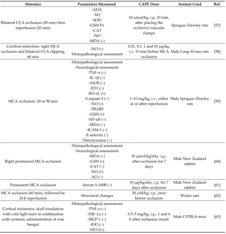
Table 3. CAPE protective effects against ischemia/reperfusion injury. Where applicable, the effect of CAPE on the
param-eters measured is reported between parentheses (+ or −).
Stimulus Parameters Measured CAPE Dose Animal Used Ref.
Bilateral CCA occlusion (20 min) then reperfusion (20 min) -ADA -XO -SOD -GSH-Px -CAT -NO -MDA (-)
10 µmol/kg, i.p. 10 min. after placing the occlusive vascular
clamps
Sprague–Dawley rats [57]
Cerebral infarction: right MCA occlusion and bilateral CCA clipping,
60 min
-NO (+)
-Histopathological assessment
0.01, 0.1, 1 and 10 µg/kg, i.v. 15 min before MCA
occlusion
Male Long–Evans rats [58]
MCA occlusion, 20 or 90 min
-Histopathological assessment -Neurological assessment -TNF-α (−) -IL-1β (−) -iNOS (−) -ED1 (−) -Bcl-xL (+) -Caspase-3 (−) -NO (+) -TBARS -GSH (+) -NF-κB (−) -MDA (−) -ICAM-1 (−) -E-selectin (−) -Nitrotyrosine (−) 1–10 mg/kg, i.v., either at or after reperfusion Male Sprague–Dawley rats [59]
Right permanent MCA occlusion
-Histopathological assessment -Neurological assessment -MDA (−) -GSH (+) -CAT (−) -NO (+) -XO (−) 10 µmol/kg/day, i.p., after occlusion for 7
days
Male New Zealand rabbits [60]
Permanent MCA occlusion -Serum S-100B (−) 10 µg/kg/day, i.p. for 7 days after occlusion
Male New Zealand rabbits [61] MCA occlusion (60 min), followed by
24 h reperfusion -Structural changes
50 µM/kg, i.p., once
before occlusion Wistar rats [62] Cortical ischaemia: skull irradiation
with cold light laser in combination with systemic administration of rose
bengal -Histopathological assessment -TNF-α (−) -HIF-1α (−) -MCP-1 (−) -IDO (−) -HO-1(+) 0.5–5 mg/kg, i.p., 1 and 6
-IL-1α (−) -IL-10 (+) Anoxia-reoxygenation -Mitochondrial oxygen consumption -Mitochondrial anisotropy -Mitochondrial TBARS -Mitochondrial protein concentrations -Protein carbonylation (−) -CL and Cytochrome c release (−)
10–10−5 µM before the
anoxia or just at reoxygenation
Male Kunming mice [64]
CCA ligation followed by exposure to hypoxia
-i- and nNOS (−) -Cytochrome c (−) -Caspase-1 and -3 (−)
40 mg/kg/day, 4 hrs before and/or after the
stimulus 7-day-old Sprague– Dawley rats [65] SAH -MDA (−) -GSH (+) -NO (+) -Histopathological assessment
10 µmol/kg, i.p. twice daily for 5 days after
SAH
15-week-old male
Wistar rats [66]
SAH -Histopathological assessment
10 mg/kg/day, twice a day, for 3 days starting 6
h after SAH.
Wistar rats [67]
Aortic occlusion, 21 min
-MDA (−) -SOD -CAT
-Histopathological assessment -Neurologic assessment
10 µmol/kg, i.p. 30 min
before the stimulus New Zealand rabbits [68]
The effect on isolated brain mitochondria was confirmed in another paper, where CAPE directly inhibits the Ca2+-induced cytochrome c release. In the same study, CAPE proved able to prevent neuronal death in neonatal rats exposed to hypoxia–ischemia. This condition is associated with clinical syndromes of neurological disability, such as seizures, intellectual impairment, and cerebral palsy. CAPE can effectively protect against neuronal and tissue loss in the cortex, hippocampus, and thalamus in vivo. In addition, CAPE hibited hypoxia-induced caspase-3 activation and the hypoxia-mediated expression of in-ducible nitric oxide synthase and caspase-1 in vivo. It also potently blocked nitric oxide-induced neurotoxicity in vitro [65].
In another model of stroke, CAPE was tested on subarachnoid hemorrhage (SAH) and was found able to significantly attenuate brain ischemia due to vasoconstriction after SAH; CAPE inhibited the marked narrowing in the lumens and the thickening in the walls of the basilar arteries as well. The tissue levels of MDA were also found to be decreased after CAPE administration following SAH because of ROS scavenging, lipid peroxidation prevention, and GSH and NO increases in the cerebral tissue [66]. In a similar model, CAPE was shown to reduce the hippocampal neuron loss induced by SAH, but no signif-icant effect of CAPE on the vasospasm was found [67].
Injury to the gray matter of the spinal cord, with consequent neuronal death, has generally been considered an important element in the pathology of spinal cord ischemic injury. The latter represents the main complication in the surgical repair of thoracic and thoraco-abdominal aneurisms and remains a persistent clinical problem. The most serious complication is paraplegia. Methylprednisolone (MP) proved effective in improving neu-rologic function after traumatic spinal cord injury. Ilhan et al. [68] compared the effects of CAPE and MP on histopathological changes, antioxidant status, lipid peroxidation, and neurologic recovery in temporary induced spinal cord ischemia in rabbits. CAPE reduced ischemic and reperfusion damage and provided a better neurologic outcome than MP. CAPE administration resulted in an improvement in microcirculatory environment dur-ing reperfusion, preventdur-ing endothelial cell lysis provoked by proteases released by acti-vated leukocytes.
6. CAPE Protective Effects against Injury
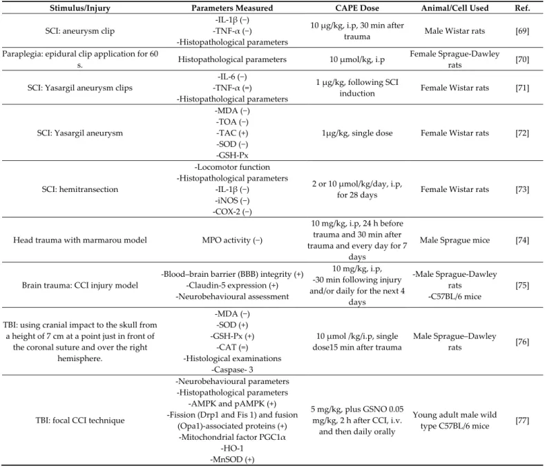
The secondary injury consists of the biochemical and histopathological cascade mechanisms in brain and spinal cord following a primary injury and leading to progres-sive neuronal death after trauma. Table 4 lists the studies investigating the protective ef-fect of CAPE against these mechanisms.
Table 4. Protective effects of CAPE against injury. Where applicable, the effect of CAPE on the parameters measured is
reported between parentheses (+, − or =).
Stimulus/Injury Parameters Measured CAPE Dose Animal/Cell Used Ref.
SCI: aneurysm clip
-IL-1β (−) -TNF-α (−)
-Histopathological parameters
10 µg/kg, i.p, 30 min after
trauma Male Wistar rats [69] Paraplegia: epidural clip application for 60
s. Histopathological parameters 10 µmol/kg, i.p
Female Sprague-Dawley
rats [70]
SCI: Yasargil aneurysm clips
-IL-6 (−) -TNF-α (=)
-Histopathological parameters
1 µg/kg, following SCI
induction Female Wistar rats [71]
SCI: Yasargil aneurysm
-MDA (−) -TOA (−) -TAC (+) -SOD (−) -GSH-Px
1µg/kg, single dose Female Wistar rats [72]
SCI: hemitransection -Locomotor function -Histopathological parameters -IL-1β (−) -iNOS (−) -COX-2 (−) 2 or 10 µmol/kg/day, i.p,
for 28 days Female Wistar rats [73]
Head trauma with marmarou model MPO activity (−)
10 mg/kg, i.p, 24 h before trauma and 30 min after trauma and every day for 7
days
Male Sprague mice [74]
Brain trauma: CCI injury model
-Blood–brain barrier (BBB) integrity (+) -Claudin-5 expression (+) -Neurobehavioural assessment
10 mg/kg, i.p, -30 min following injury and/or daily for the next 4
days
-Male Sprague-Dawley rats -C57BL/6 mice
[75]
TBI: using cranial impact to the skull from a height of 7 cm at a point just in front of
the coronal suture and over the right hemisphere. -MDA (−) -SOD (+) -GSH-Px (+) -CAT (=) -Histological examinations -Caspase- 3
10 µmol /kg/i.p, single dose15 min after trauma
Male Sprague–Dawley
rats [76]
TBI: focal CCI technique
-Neurobehavioural parameters -Histopathological parameters
-AMPK and pAMPK (+) -Fission (Drp1 and Fis 1) and fusion
(Opa1)-associated proteins (+) -Mitochondrial factor PGC1α
-HO-1 -MnSOD (+)
5 mg/kg, plus GSNO 0.05 mg/kg, 2 h after CCI, i.v. and then daily orally
Young adult male wild type C57BL/6 mice [77]
A clip compression model was used in rats to simulate the spinal cord injury (SCI). In the early phase of injury, CAPE suppressed the serum levels of pro-inflammatory cy-tokines—i.e., TNF-α and IL-1β. At the same time, CAPE decreased hemorrhage and ne-crosis occurrence both in the gray and white matter, as well as in the central canal, as demonstrated by the histopathological evaluation of the healing process [69]. Another study compared the effects of CAPE with those of methylprednisolone (MP) in the pre-vention of neurological deficits caused by secondary spinal cord injury. CAPE has been found to be superior to MP in preventing apoptosis without showing the latter side ef-fects—namely, gastrointestinal hemorrhage and infection [70]. The effects of intrathecal
CAPE administration following SCI were investigated as an alternative approach in CNS diseases because the systemic administration of pharmacological agents does not guaran-tee the attainment of the maximum effective dose in the damaged area. The intrathecal CAPE administration resulted in a decrease in the tissue and serum levels of IL-6, an in-flammatory cytokine that plays an essential role in secondary damage. Although no sig-nificant decrease was identified in the TNF-α level, CAPE led to a decrease in the edema and microhemorrhage that developed in association with a local inflammatory response in the damaged area. In addition, CAPE was more effective than MP in decreasing micro-hemorrhage [71]. Interestingly, the intratechal administration of CAPE and MP decreased also the MDA, TOA, and SOD levels, with a greater effect in the CAPE group [72]. Since CAPE and MP treatment substantially increased the TAC levels, it was concluded that the intrathecal injection of both compounds can inhibit lipid peroxidation and oxidant in-crease following SCI. In a model of SCI caused by hemi-transection, CAPE suppressed the expression of the mRNA of the pro-inflammatory cytokine IL-1β and of the two inflam-matory enzymes iNOS and COX-2. An enhanced recovery of locomotor function and a reduction in the lesion size were also found following CAPE treatment [73].
One of the early pathological events triggered by traumatic brain injury (TBI) is the breakdown of the blood brain barrier (BBB), which leads to the infiltration of fluid and circulating cells and molecules, resulting in an exacerbation of brain damage and tissue loss. Recently, myeloperoxidase (MPO) analysis was used to identify the level of poly-morphonuclear leukocyte (PMNL) infiltration after TBI. PMNLs worsen brain damage due to the release of cytotoxic mediators that interfere with BBB function. Hypochlorite acid is formed through the MPO-H2O2–Cl-system and is one of the oxidants released by activated leukocytes. CAPE administration to mice with TBI showed a significant decrease in MPO levels, thereby inhibiting the BBB inflammatory process [74]. Another study ex-amined the potential role of CAPE in reducing some of the pathological consequences of TBI in rodent models. The post-TBI administration of CAPE reduced the BBB permeability and cortical tissue loss both in rats and mice. The improvement was associated with the preservation of the levels of the tight junction protein claudin-5. On the other hand, it failed to offer significant improvements in either motor or memory tasks [75]. The admin-istration of a single dose of CAPE post-TBI can also have protective effects via decreasing the elevated MDA levels and restoring the activities of antioxidant enzymes (SOD and GSH-Px), with the exception of CAT. The morphology of neurons was well preserved, and the immunoreactivity of degenerating neurons after trauma was reduced in the CAPE-treated group [76].
Since TBI has a multi-mechanistic etiology, a recent study combined the endogenous nitric oxide (NO) metabolite S-nitrosoglutathione (GSNO) with CAPE to accelerate the rate and enhance the degree of recovery. GSNO is a natural component of the human body, present in the brain and other organs. It provides neuroprotection and improves neurobehavioral functions via anti-inflammatory and anti-neurodegenerative mecha-nisms. It acts via the S-nitrosylation of target proteins, including NF-κB, STAT3, COX-2, caspase-3, calpains, and i- and eNOS. The combination therapy improved cognitive and depressive-like behavior compared with GSNO or CAPE monotherapy. Separately, both GSNO and CAPE improved mitochondrial integrity/function and decreased oxidative damage; however, the combined therapy had greater effects on dynamin-1-like protein (Drp1) and manganese-dependent superoxide dismutase (MnSOD). Additionally, while CAPE alone activated 5′-adenosine monophosphate-activated protein kinase (AMPK), this activation was heightened in combination with GSNO. These results recommended that the combination therapy-based multi-mechanistic approach is worthy of investiga-tion in human TBI because no adverse effect for both GSNO and CAPE is known [77].
7. CAPE Anti-Tumoral Effects in CNS
In 2020, according to Cancer Stat Facts, an estimated 23,890 new patients in the USA will be diagnosed with primary cancerous tumors of the brain and spinal cord. The 5-year
survival rate for people with a CNS tumor is around 32.6% [78]. The most common types of brain tumors are gliomas and meningiomas. Gliomas are considered the most malig-nant form of brain tumors, and are ranked among the most aggressive human cancers. Choi et al. in 2007 [79] demonstrated that the anti-oxidant property of CAPE can exert a pro-apoptotic effect in Fas-mediated cell death in human malignant astrocytoma cells. Fas activation increased the intracellular ROS levels in a NADPH oxidase- and caspase-de-pendent manner. Choi’s results suggested that Finduced ROS production protects as-trocytoma CRT-MG cells from caspase-dependent apoptosis. The suppression of Fas-in-duced ROS levels augments Fas-mediated apoptosis by enhancing the enzymatic activity of caspase-3. CAPE, by inhibiting ROS production, dramatically sensitized astrocytoma cells to the Fas-induced loss of mitochondrial transmembrane potential and subsequent cell death. Another study demonstrated that CAPE suppressed the invasion and prolifer-ation of glioma cells by the down-regulprolifer-ation of phospholipase D1 (PLD1) expression at the transcriptional level. PLD is a class of enzymes functionally linked with oncogenic signals and tumorigenesis. CAPE exerted its effect via the inhibition of the binding of NF-κB to the PLD1 promoter. In addition, CAPE bound to a Cys837 residue of the protein PLD1 and inhibited its enzymatic activity. The activation of matrix metalloproteinases-2 induced by phosphatidic acid, a product of PLD activity, was also inhibited [80].
5-lipoxygenase (5-LO) is another enzyme involved in the pathophysiology of glioma. A strong 5-LO expression has been observed in glioma cells, and inhibiting this enzyme could impact glioma cell proliferation. Interestingly, CAPE (1 µM) was found to be effec-tive in reducing 5-LO activity more efficiently than the ferulic acid ester (FAPE) in two human glioma cell lines, Hs683 and LN319. CAPE administration led to cytotoxicity, cell growth impairment, and apoptosis occurrence [81].
In an in vivo model, Bio 30, a water-miscible CAPE-rich extract of New Zealand prop-olis, was analyzed for its anti-tumoral properties. Bio 30 suppressed completely the growth of both neurofibromatosis type 1 (NF1) and neurofibromatosis type 2 (NF2) tumor xenografts in mice, suggesting that CAPE-rich propolis might serve as an effective, inex-pensive, and safe NF therapeutic first approach. Seventy percent of human cancers and all NF tumors, but not normal cells, are highly dependent on PAK1 for their growth. PAK1 is a Rac/CDC42-dependent Ser/Thr kinase. Evidence was found that CAPE, by blocking the oncogenic PAK1 signaling pathways, is the anti-tumor ingredient of Bio 30 [82]. All the studies described above about the anti-tumor effect of CAPE in CNS are listed in Table 5.
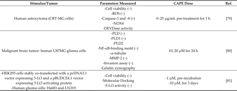
Table 5. CAPE anti-tumoral effects in CNS. Where applicable, the effect of CAPE on the parameters measured is reported
between parentheses (+ or −).
Stimulus/Tumor Parameters Measured CAPE Dose Ref.
Human astrocytoma (CRT-MG cells)
-Cell viability (−) -ROS (−) -Caspase-3 and -8 (+) -NOX4 -DEVDase activity 0–25 µg/mL pre-treatment for 1 h [79]
Malignant brain tumor: human U87MG glioma cells
-PLD (−) -PLD1 (−) -PLD2 -NF-κB-binding motif (−) -α-tubulin -MMP-2 (−) -Invasion assay (−) -Gelatin zymography 10, 20 µM for 24 h [80]
-HEK293 cells stably co-transfected with a pcDNA3.1 vector expressing 5-LO and a pBUDCE4.1 vector
expressing 5-LO activating protein -Human glioma cells: Hs683 and LN319.
-Cell viability (−) -Molecular Docking
-5-LO activity (−)
-1 µM, pre-incubation
Tumor xenografts in nu/nu mice: SC injection of NF1-deficient MPNST (S-462) cells or NF2-NF1-deficient
Schwannoma (HEI-193) cells
-Cell viability (−) -Tumor size (−)
Bio 30 (a CAPE-rich extract), 100–300 mg/kg, i.p., twice a week [82]
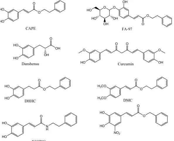
8. CAPE Derivatives
Due to the involvement of a variety of pathways in neurological disorders, the find-ing of molecules with multiple neuroprotective effects would be a great achievement for the slowing down of the course of these diseases. CAPE is supposed to be a promising neuroprotective agent with multiple targets. Many studies have aimed to explore the structure–activity relationship of CAPE in order to optimize its structure through a series of studies on CAPE derivatives. Figure 2 shows the chemical structures of CAPE and some of its analogues. Novel derivatives of CAPE were recently designed, synthesized, and evaluated in different in vitro models as potential neuroprotective agents. Phenolic hy-droxyl groups and double bonds in the structure of CAPE proved critical in conferring neuroprotective properties to the compound, whereas monophenol compounds showed a low anti-inflammatory activity. Compounds with a phenolic hydroxyl group on para-position exerted a higher anti-inflammatory activity than compounds with the same group on the ortho-position, indicating that conjugated double bond played an important role in anti-inflammatory activity. Only compounds with a retained catechol structure showed anti-oxidant properties. The amino group did not show the same anti-oxidant activity found for the bioisoteric and the hydroxyl group. Replacing the aromatic alkyl part of CAPE with different liposoluble groups improved the inflammatory and anti-oxidant activities and the BBB permeability [83], measured with a parallel artificial mem-brane permeability assay (PAMPA), a high-throughput technique developed to predict passive permeability through biological membranes. These findings were supported with a study on PC12 cells suggesting that the presence of the caffeic acid group is important to maintain the cytoprotective and neuritogenic activities of the compound [84]. Another group explored the neuroprotective effects of CAPE and another two caffeic acid deriva-tives, Danshensu and Curcumin. They are similar in structure, since they have an aromatic ring, but they differ in the numbers of hydroxyl groups attached to the aromatic ring and in the conjugated double bond. The structure–neuroprotective activity relationship against H2O2-induced oxidative damage in PC12 cells and the D-galactose (D-gal)-in-duced cognitive impairment was investigated in mice. The neuroprotection exerted by these compounds was mediated by the activation of the protein kinase A (PKA)-cyclic AMP response element-binding protein (CREB) pathway. It was found that 3, 4-dihydrox-yphenyl hydroxyl groups attached to the aromatic ring play a more important role than conjugated double bond in up-regulating the expression of the PKA/CREB pathway in the mouse hippocampus [85].
In two separate studies, CAPE proved active in suppressing the invasion and prolif-eration of glioma cells by the down-regulation of PLD1 and 5-LO expression. One study showed that CAPE analogues with a catechol moiety, dihydroxydihydrocinnamic acid phenethylester (DHHC) and dimethoxycinnamic acid phenethyl ester (DMC), inhibit PLD1 expression with a lower potency than CAPE due to differences in NF-κB transacti-vation. This was confirmed by structural analysis, which showed that both electrophiles in CAPE, Michael reaction acceptor and catechol moiety, are involved in the down-regu-lation of the PLD1 expression and in NF-κB transactivation [80]. The other study demon-strated that CAPE, but not FAPE, displayed substantial cytotoxicity against two glioma cell lines [81].
Even though CAPE stability in human plasma was demonstrated [9], its lipophilic properties, due to long carbon groups in an aromatic and aliphatic structure, can hamper CAPE absorption upon oral administration. Hence, FA-97 caffeic acid phenethyl ester 4-O-glucoside was synthesized to overcome the low water solubility and poor bioavailabil-ity of CAPE, which represent the main limits to its application in vivo. The in vivo and in vitro results showed that FA-97 is able to suppress oxidative stress-mediated neuronal cell