1
UNIVERSITÀ POLITECNICA DELLE MARCHE
SCUOLA DI DOTTORATO DI RICERCA DELLA FACOLTÀ DI
MEDICINA E CHIRURGIA
Curriculum: Scienze biomediche
XV ciclo
OVEREXPRESSION AND SILENCING OF
NICOTINAMIDE N-METHYLTRANSFERASE IN
HUMAN CANCER CELL LINES
Dottorando:
Relatore:
Dott. Riccardo Seta
Chiar.ma Prof.ssa
Monica Emanuelli
Referente:
Chiar.mo Prof. Gian Marco Giuseppetti
Triennio 2013-2016
2
TABLE OF CONTENTS
1 INTRODUCTION
4
1.1 LUNG CANCER 1.1.1 Epidemiology 5 1.1.2 Risk factors 6 1.1.3 Histopathological classification 8 1.1.4 Staging 10 1.1.5 Clinical manifestations 12 1.1.6 Diagnosis 13 1.1.7 Therapeutic strategies 14 1.2 ORAL CANCER 1.2.1 Epidemiology 17 1.2.2 Risk factors 18 1.2.3 Histopathological classification 19 1.2.4 Staging 201.2.5 Clinical manifestations and diagnosis 22 1.2.6 Therapeutic strategies 24 1.3 NICOTINAMIDE N-METHYLTRANSFERASE 1.3.1 Drug metabolism 26 1.3.2 Nicotinamide homeostasis 27 1.3.3 Characterization of human NNMT 28 1.3.4 NNMT polymorphisms 35
1.3.5 NNMT and non-neoplastic diseases 37 1.3.6 NNMT and Parkinson’s disease 41 1.3.7 NNMT and neoplastic diseases 43
1.4 AIM OF THE STUDY 51
2 MATERIAL AND METHODS
52
2.1 NNMT SILENCING IN A549 LUNG CANCER CELL LINE
2.1.1 Cell lines and reagents 53
2.1.2 A549 transfection 53
2.2 NNMT OVEREXPRESSION IN HSC-2 ORAL CANCER CELL LINE
3
2.2.2 Cloning 55
2.2.3 HSC-2 transfection 61
2.3 EFFICIENCY OF NNMT SILENCING AND OVEREXPRESSION
2.3.1 Total RNA extraction and cDNA synthesis 61
2.3.2 Real-Time PCR 62
2.3.3 Western blot analysis 63
2.3.4 Enzyme assay 64
2.3.5 Protein assay 65
2.4 EFFECT OF NNMT SILENCING AND OVEREXPRESSION ON CELL PHENOTYPE
2.4.1 MTT assay 65
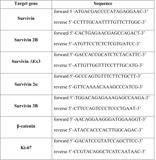
2.4.2 Soft agar colony formation assay 66 2.5 EXPRESSION LEVELS OF β-CATENIN, KI-67, AND SURVIVIN
ISOFORMS
2.5.1 Qualitative PCR 66
2.5.2 Real-Time PCR 67
2.6 STATISTICAL ANALYSIS 68
3 RESULTS
69
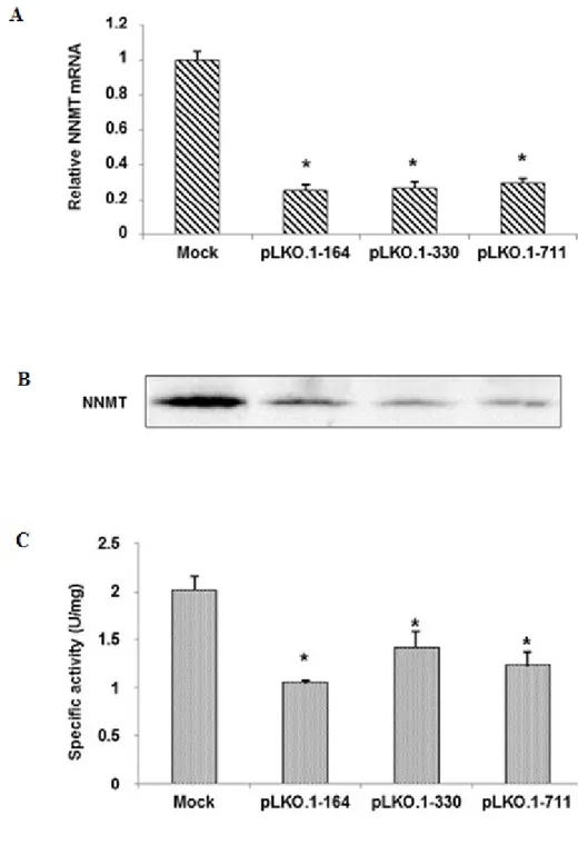
3.1 NNMT SILENCING IN A549 CELLS
3.1.1 Efficiency of NNMT silencing 70 3.1.2 Effect of NNMT silencing on cell proliferation 72 3.1.3 Effect of NNMT silencing on anchorage-independent cell growth 73 3.2 NNMT OVEREXPRESSION IN HSC-2 CELLS
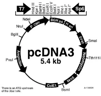
3.2.1 NNMT cloning into pcDNA3 vector 74 3.2.2 Efficiency of NNMT overexpression 76 3.2.3 Effect of NNMT overexpression on cell proliferation
indipendente
79 3.2.4 Effect of NNMT upregulation on β-catenin, survivin, and Ki-67
expression
80
4 DISCUSSION AND CONCLUSIONS
82
5 LIST OF ABBREVIATIONS
90
4
5
1.1 LUNG CANCER
1.1.1 Epidemiology
Lung cancer is the most common cancer in the world with 1.8 million of new cases in 2012, accounting for 12.9% of the total estimated tumors.
In men, lung cancer is the most common cancer worldwide (1.2 million, 16.7% of the total). It ranks second in more developed regions, whereas predominate in less developed countries with 751,000 cases, constituting 40% of the new cancer cases. The highest incidence rates are in Central and Eastern Europe (53.5 new cases per 100,000 person-years) and Eastern Asia (50.4 new cases per 100,000 person-years), whereas incidence rates are very low in Middle and Western Africa (2.0 and 1.7 new cases per 100,000 person-years, respectively). In women, the incidence rates are generally lower, with a geographical pattern that reflects in part differences in the uptake and consumption of tobacco. The highest lung cancer rates are in Northern America (33.8) and Northern Europe (23.7), but a relatively high incidence has been also estimated among women in Eastern Asia (19.2), despite tobacco smoking is generally rare in these populations. Nevertheless, the high incidence estimated could be related to other known risk factors for lung cancer including air pollution.
Lung cancer is the leading cause of tumor-related death worldwide with 1.59 million deaths (19.4% of the total) reported in 2012. It is the main cause of cancer death among men in both more and less developed regions, whereas in women has surpassed breast cancer as leading cause of cancer mortality in more developed regions [1].
In Western countries, such as United States and the United Kingdom, smoking prevention and cessation programs, as well as advanced medical technologies, have
6
decreased lung cancer mortality, especially in men. However, in developing countries, where the uptake and consumption of tobacco occurred later, mortality rates are increasing and more efforts to promote smoking cessation and avoid initiation are urgently needed. Nevertheless, at a global level, the mortality patterns of lung cancer closely follow those of incidence, due to the high fatality associated with the disease [2].
The 5-year survival rate is 52.2% for patients with localized disease. Survival declines to 24.3% and 3.6% for patients with regional and distant stage disease, respectively. As the majority of lung cancers (56%) are diagnosed at a distant stage, while only 15% of cases are diagnosed at a local stage, the development of new techniques as well as the identification of reliable biomarkers are required to detect lung cancer at an early stage [3].
1.1.2 Risk factors
Tobacco smoking is the major risk factor for lung cancer, accounting for 80% of the worldwide lung cancer burden in males and at least 50% in females. Tobacco smoke contains many kinds of carcinogenic factors responsible for tumor promoting effects as well as induction of mutations, which cause a 10-fold or greater increased risk of developing lung cancer in cigarette smokers compared to those who have never smoked [4]. Epidemiological studies have shown an approximately linear dose-response relationship between the number of cigarettes smoked per day and lung cancer mortality. A greater than linear increment in incidence or mortality is reported when considering the duration of the smoking habit, probably because of a very high dose of carcinogens that affect a number of steps in carcinogenesis. In former smokers, the risk of developing lung cancer is lower than in continuing smokers, with a correlation
7
between risk reduction and the length of time the person has quitted smoking. Nevertheless, generally even long-term former smokers have higher risks of lung cancer than those who never smoked [5].
Considering the high correlation between cigarette smoking and lung cancer, most of the worldwide burden of lung cancer could be avoided by promoting smoking cessation and preventing smoking initiation. Stopping tobacco use before middle age avoids more than 90% of the lung cancer risk attributable to tobacco. Smoking cessation can even be beneficial in individuals with an established diagnosis of lung cancer, decreasing side effects from therapy, as well as improving survival and quality of life. Moreover, smoking can alter the metabolism of many chemotherapeutic drugs, highlighting the importance of promoting smoking cessation even after the diagnosis of lung cancer is established.
Although tobacco smoking is considered the most well established risk factor for lung cancer, a number of non-smoking patients have developed this disease, accounting for up to 25% of all lung cancer cases. Deaths from lung cancer are increasing every year in non-smokers, where the disease occupies the seventh place worldwide in terms of mortality.
Environmental tobacco smoke (ETS), or second-hand smoke, is an established cause of lung cancer. The risk from ETS is less than from active smoking, with about a 20-30% increase in lung cancer observed among never smokers married for many years to smokers.
Several other factors are associated with an increased risk of developing lung cancer, including indoor air pollution, genetic susceptibility, and exposure to several occupational and environmental carcinogens, such as asbestos, arsenic, radon,
8
bis(chloromethyl) ether, hexavalent chromium, mustard gas, nickel, and polycyclic aromatic hydrocarbons.
Occupational observations can provide insights into possible mechanisms of lung cancer induction. The risk of lung cancer among asbestos-exposed workers is increased primarily among those with underlying asbestosis, raising the possibility that the inflammation and scarring produced by this fibrotic lung disease may be the trigger for asbestos-induced lung cancer [4].
The cellular effect of environmental carcinogens can be modulated by polymorphic variations in genes involved in carcinogens’ metabolism. Genetic polymorphisms that increase the activity of the cytochrome P450 family enzymes, specifically CYP1A1, or reduce the activity of the glutathione-S transferase family, are associated with the development of lung cancer.
Among first-degree relatives, the risk of lung cancer increases from 2- to 3-fold, suggesting that specific genes and/or genetic variants may contribute to susceptibility to lung cancer. In this regard, increased risk for lung cancer has been reported in individuals with inherited mutations in pRB and p53.
Genome-wide association studies have identified three separate loci associated with lung cancer (5p15, 6p21, and 15q25), that include genes involved in acetylcholine nicotinic receptors regulation and telomerase production. Furthermore, the 6q23-25 region was also identified as being linked to lung cancer susceptibility among light and never smokers [6].
1.1.3 Histopathological classification
According to the classification of World Health Organization (WHO), lung cancer is divided into two main histological groups:
9 - Small-cell lung cancer (SCLC) - Non-small cell lung cancer (NSCLC)
NSCLC is the most common type of lung cancer and includes squamous cell carcinoma (SCC), adenocarcinoma (ADC), and large cell carcinoma (LC).
The histological type of NSCLC correlates with the site of origin, reflecting the variation in respiratory tract epithelium of the bronchi to alveoli.
Squamous cell carcinoma accounts for approximately 30% of NSCLC and is the subtype most strongly associated with tobacco smoking, with a higher incidence in men than in women. Squamous cell carcinoma originate in the central part of the lungs, usually near a central bronchus, and may show keratinization and/or intercellular bridges that arise from bronchial epithelium.
Adenocarcinoma is the most common histologic type, accounting for about 40% of lung cancer. Adenocarcinomas exhibit glandular differentiation or mucin production, showing acinar, papillary, bronchioloalveolar, solid features or a mixture of these patterns. Although most cases of adenocarcinoma are associated with smoking, it develops more frequently than any other types of lung cancer in non-smokers, especially in women.
Large cell carcinoma is an undifferentiated non-small cell carcinoma that lacks the cytological and architectural features of small cell carcinoma and glandular or squamous differentiation. Large cell carcinoma accounts for approximately 10% of all lung cancers and predominate in smokers.
Small-cell lung cancer accounts for about 15% of all lung cancer cases, consisting of small cells with scant cytoplasm, ill-defined cell borders, finely granular nuclear chromatin, absent or inconspicuous nucleoli, and a high mitotic count.
10
SCLC is the most aggressive form of lung cancer, displaying a high incidence rate in men and is commonly associated with smoking [4].
Classification into histopathological subtypes as well as immunohistochemical and molecular characterization of lung cancer provide important information in the prognostic assessment and thus treatment decisions.
At molecular level, a number of gene mutations have been identified in lung cancer. Oncogenes of the Myc family are frequently activated in both SCLC and NSCLC. Mutations that inactivate the tumor suppressor gene TP53, encoding the p53protein, are detected in up to 50% of NSCLC and in over 70% of SCLC. Another common alteration is the inactivation of the suppressor gene RB1, encoding the Rb protein, occurring in over 90% of SCLC cases and 15-30% of NSCLC cases, whereas P16, the other component of the RB pathway, is inactivated in over 50% of NSCLC and in 10% of SCLC. Furthermore, a common genetic event detected in up to 80% in both NSCLC and SCLC is the loss of heterozygosity (LOH) on chromosome 3p. Other mutations are frequent in specific histologic subtypes. KRAS and EGFR mutations are extremely rare in SCLC, whereas are found in 30% and 10% of ADC, respectively. Other driver mutations found in adenocarcinoma include those affecting the serine-threonine kinase BRAF and the lipid kinase PIK3CA, as well as specific chromosomal rearrangements that lead to the activation of tyrosine kinase proteins, such as ALK, ROS1, and RET [4,7].
1.1.4 Staging
Staging of cancer at time of diagnosis is a key factor to provide estimation of prognosis and define treatment strategies. The tumor-node-metastasis (TNM) system is used to classify the NSCLC stage. Such system considers the extent of the primary tumor (T),
11
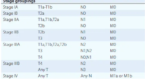
the involvement of regional lymph nodes (N), and the presence or absence of distant metastases (M). The most recent revision of the TNM staging system (7th edition) was provided by the American Joint Committee on Cancer (AJCC) and the International Union for Cancer Control (UICC) (Table 1). T, N, and M are combined to form different stage groups from I to IV, as shown in Table 2 [8].
12 Table 1. Lung cancer TNM staging.
13
Table 2. Lung cancer stage groups based on T, N, and M.
In patients with SCLC, is generally used the Veteran Administration staging system, a two-stage system that divides SCLC into limited-stage and extensive-stage disease. Limited-stage disease is restricted to the ipsilateral hemitorax and can be encompassed with a single radiation therapy port. The extensive-stage disease includes metastatic disease outside the ipsilateral hemitorax, including malignant pleural effusion, bilateral pulmonary parenchymal, and cardiac tamponade, and cannot be encompassed effectively within a single radiation port. Extensive-stage disease is diagnosed in 60 to 70% of patients with SCLC [9].
1.1.5 Clinical manifestations
Over half of the lung cancer cases are diagnosed at late stage, with locally advanced or metastatic disease. The majority of patients present with signs or symptoms that appear related to primary lesion, metastatic disease, or a paraneoplastic syndrome.
Primary tumors present with symptoms depending on its location. Central or endobronchial growth of primary tumors produce cough, hemoptysis, wheeze, dyspnea,
14
or postobstructive pneumonitis. Peripheral growth of the primary tumor may cause pain as a result of pleural or chest wall involvement, dyspnea on a restrictive basis, and symptoms of a lung abscess resulting from tumor cavitation.
Regional spread of the tumor may cause superior vena cava obstruction, paralysis of the recurrent laryngeal nerve causing hoarseness, phrenic nerve palsy with paralysis of the diaphragm, pressure on the sympathetic plexus causing Horner’s syndrome, and dysphagia resulting from esophageal compression. Other problems of regional spread include pericardial and cardiac effusion, lymphatic obstruction, and lymphangitic spread through the lungs with hypoxemia and dyspnea.
Distant metastatic disease is found in over 50% of patients with squamous cell carcinoma, 80% of patients with adenocarcinoma and large-cell carcinoma, and over 95% of patients with SCLC. Approximately one-third of patients present with symptoms as a result of distant metastases, and such symptoms are correlated with the site of metastatic involvement.
Paraneoplastic syndromes (PNS) occur in 10% of patients with lung cancer and is mainly caused by the production of cytokines or hormones by cancer or as an immune response to cancer. A wide variety of PNS is associated with lung cancer, including hypercalcemia, syndrome of inappropriate antidiuretic hormone secretion and Cushing’s syndrome. The symptoms may precede the primary diagnosis by many months, and might be, in some cases, the presenting complaint [4].
1.1.6 Diagnosis
In lung cancer, clinical outcome is related to the stage at diagnosis, thus the identification of high-risk lung cancer individuals at an early disease stage could represent the most effective way of improving survival.
15
Screening studies conducted in the 1970s based on chest x-rays (CXR) and sputum cytology reported no impact on lung cancer-related mortality in patients characterized as high risk. In contrast to CXR, low-dose computed tomography (LDCT) has emerged as an effective tool for the screening of lung cancer. In 2002, The National Lung Screening Trial (NLST) was initiated to determine whether screening with low-dose computed tomography, as compared with chest radiography, would have reduced mortality from lung cancer among high-risk persons. The NLST demonstrated a 20% reduction in lung cancer-related mortality in the low-dose CT group as compared with the radiography group [10]. Based on these findings, hospital authorities and various organizations recommend lung cancer screening with LDCT in high-risk populations. Most patients with suspected lung cancer require tissue biopsy to confirm the diagnosis. Tumor tissue may be obtained via bronchial or transbronchial biopsy during fiberoptic bronchoscopy, by needle aspiration or percutaneous biopsy, or lymph node biopsy during mediastinoscopy. Percutaneuos biopsy of a soft tissue mass, pleural or liver lesion, lytic bone lesion, or bone marrow may be obtained in patients with suspected metastatic disease. Because of their central location, squamous cell carcinomas and small-cell carcinomas are readily diagnosed by bronchoscopic biopsy, whereas for peripheral lesions, such as adenocarcinomas and large-cell carcinomas, a transthoracic fine needle aspiration biopsy is generally preferred. Diagnostic accuracy for SCLC versus NSCLC for most specimens is excellent, with lesser accuracy for subtypes of NSCLC [4].
1.1.7 Therapeutic strategies
Treatment of lung cancer may occur in three ways, such as surgery, chemotherapy, or radiotherapy, depending on the stage and the histological type of the disease.
16
In patients with small-cell lung cancer, the standard treatment is chemotherapy and chest radiotherapy, while surgical resection is not routinely recommended because even limited-stage patients may have occult micrometastases.
Patients with SCLC receive four to six cycles of chemotherapy, with cisplatin or carboplatin plus either etoposide or irinotecan. In limited-stage SCLC patients, thoracic radiation therapy (TRT) is combined with chemotherapy, improving the 3-year survival rate of approximately 5% compared with chemotherapy alone.
With current therapy, patients with limited-stage SCLC have a median survival of 17 months and a 5-year overall survival rate of 12%, while in patients with extended-stage SCLC the median survival ranges from 7 to 11 months, with a 5-year overall survival rate of 2%.
Treatment options of non-small cell lung cancer are based mainly on the stage of cancer. Surgery is the treatment of choice for patients with stage I and II NSCL, who are able to tolerate such procedure. Lobectomy is the standard treatment if the patient can tolerate it, as it offers the best chance for cure, otherwise wedge resection and segmentectomy may be reasonable surgical options. Pneumonectomy is reserved for patients with central tumors and should be performed only in patients with excellent pulmonary reserve. With surgical based therapy, the 5-year survival rates are 60–80% for patients with stage I NSCLC and 40–50% for patients with stage II NSCLC. If patients either refuse or are not suitable candidates for surgery, stereotactic body radiation therapy (SBRT) or another type of radiation therapy may be an option for the treatment of stage I and II NSCLC.
For stage IIIA non-small cell lung cancer, the distinction between non-bulky and bulky mediastinal lymph node (N2) disease is used to select the therapeutic approach. In patients with resectable stage IIIA NSCLC, surgery followed by adjuvant chemotherapy
17
may be the treatment of choice, while concurrent radiochemotherapy is the standard approach in patients with bulky mediastinal lymph node involvement. Although concurrent radiochemotherapy is associated with improved overall survival compared to sequential treatment, the latter is associated with lower toxicity (including esophagitis and pneumonitis). Therefore, for fit patients, concurrent radiochemotherapy is the preferred treatment approach, whereas sequential treatment may be applied for patients with a poor performance status. For patients who are not candidates for a combined-modality treatment approach, typically due to a poor performance status or a comorbidity that makes chemotherapy untenable, radiotherapy alone may provide a modest survival benefit in addition to symptom palliation [4,7].
18
1.2 ORAL CANCER
1.2.1 Epidemiology
An estimated 300,400 new cases (2.1% of the total cancer) and 145,400 deaths (1.8% of the total) from oral cavity cancer occurred in 2012 worldwide. The incidence and mortality rate of oral cancer is higher in men than in women. With 199,000 cases estimated in 2012, oral cancer is the 11th most common tumor worldwide among men. There is a wide geographical variation in the incidence of oral cancer, with two-thirds of the total cases occurring in less developed regions. The highest rates are found in Melanesia among both males and females (22.9 and 16.0 new cases per 100,000 person-years, respectively), followed by South-Central Asia, and Central and Eastern Europe, whereas the lowest rates are found in Western Africa and Eastern Asia. The mortality rates follow the geographical pattern of the incidence, with 77% of total deaths estimated in less developed countries [1].
The risk of developing oral cancer increases with age, and the majority of cases occur after the age of 40 years, with a peak at 60 years.
The overall 5-year survival rates for oral cancer has not improved over the past three decades, remaining at approximately 50%. The main reason of this poor prognosis is the diagnostic delay, which leads patients to be treated when the tumor has reached an advanced stage. The survival rate increases up to 80% when the tumor is detected in the early stage [11].
19
1.2.2 Risk factors
Alcohol consumption and tobacco use are the major risk factors for oral cavity cancer, and when used together, they act synergistically. Smokeless tobacco use (marijuana and occupational exposures) and HPV infection are other etiological factors for oral cancer. Each of these risk factors differently contributes to the burden across countries. Tobacco smoking is estimated to be responsible for about 71% of deaths from oral cancer in high-income regions and 37% in low- and middle-income regions. Incidence rates are increasing among both males and females in several countries of Eastern and Northern Europe as well as among females in Southern and Western Europe, whereas oral cancer rates are decreasing in Asia, Northern America, and Australia among both males and females, reflecting the uptake and consumption of tobacco [2].
Tobacco-specific nitrosamines, such as 4-(methylnitrosamino)-1-(3-pyridyl)-1-butanone (NNK) and N-nitrosonornicotine (NNN), and polycyclic aromatic hydrocarbons (PAH) have been causally linked to oral cancer. The metabolism of carcinogens contained in tobacco smoke involves oxygenation and conjugation reactions by P450 enzymes and glutathione-S-transferase, and it has been suggested that genetic polymorphisms in the genes coding for these enzymes may lead to genetic predisposition to tobacco-induced oral cancer [12]. In former smokers the risk of developing oral cancer is consistently lower than in current smokers, with a trend of decreasing risk with increasing number of years since quitting.
Betel quid has been classified as an oral carcinogen by the International Agency for Research on Cancer, with evidence for a dose–response relationship, representing one of the major risk factors for oral cavity cancer in Taiwan, India, and other neighboring countries.
20
In Western countries, the human papilloma virus (HPV) is associated with a rising incidence of oral cancer, and it is estimated to cause over 50% of oropharyngeal tumors in the United States. HPV is categorized in high-risk and low-risk subtypes, depending on their oncogenic potential. The high-risk HPVs 16 and 18 are the major viral subtypes that have been associated with precancerous and cancerous oral and oropharyngeal epithelial lesions [11].
Several epidemiological studies have shown a relationship between diet and the risk of oral cancer. Low consumption of fruits and vegetables have been correlated with an higher oral cancer rate, whereas more frequent intake of foods containing carotenoids may be protective if included in a balanced diet [12].
1.2.3 Histopathological classification
Oral squamous cell carcinoma (OSCC) is the most common malignancy of oral cavity, accounting for more than 90% of oral cancers, and is divided in well-differentiated, moderately- and poorly-differentiated categories. The assessment of OSCC tumor differentiation is based on the percentage of keratinized tumor cells, with more than 75% keratinization in well-differentiated OSCC, 25-75% keratinization in moderately-differentiated OSCC, and less than 25% in poorly-moderately-differentiated OSCC. Poorly differentiated tumors have the worst prognosis, with a 5-year survival rate of 42%, followed by moderately- and poorly-differentiated tumors with a 5-year survival rate of 62% and 74%, respectively. Tumors of the salivary glands represent 2-4% of oral neoplasms, arising from the major (parotid, submandibular, sublingual) or the minor salivary glands (dispersed throughout the upper aerodigestive submucosa). Most parotid gland tumors are benign, whereas slightly more than 50% of submandibular and
21
sublingual gland tumors, as well as most minor salivary gland tumors are found to be malignant [13].
The World Health Organization defined as potentially malignant disorder (PMD) several lesions and conditions of the oral mucosa with high potential tendency for transformation into malignancy. Conditions described as PMD include erythroplakia, leukoplakia, lichen planus, and oral submucous fibrosis. Erythroplakia and leukoplakia are respectively defined as red and white patch on the oral mucosa, and can be histopathologically classified as hyperplasia, dysplasia, carcinoma in situ, or carcinoma [11].
At molecular level, a variety of gene mutations has been associated with oral cancer. Overexpression of the epidermal growth factor receptor (EGFR) has been reported in oral cancer and has shown to correlate with poor prognosis. P53 tumor suppressor gene mutations are also found frequently, while more than 50% overexpression of p53 has been correlated with a poor prognosis in advanced oral tumors. Frequent alterations associated with oral cancer include the mitotic signaling pathway, Notch pathway, and the PI3K pathway, which is the only mutated cancer gene identified in HPV-positive oral cancer [4].
1.2.4 Staging
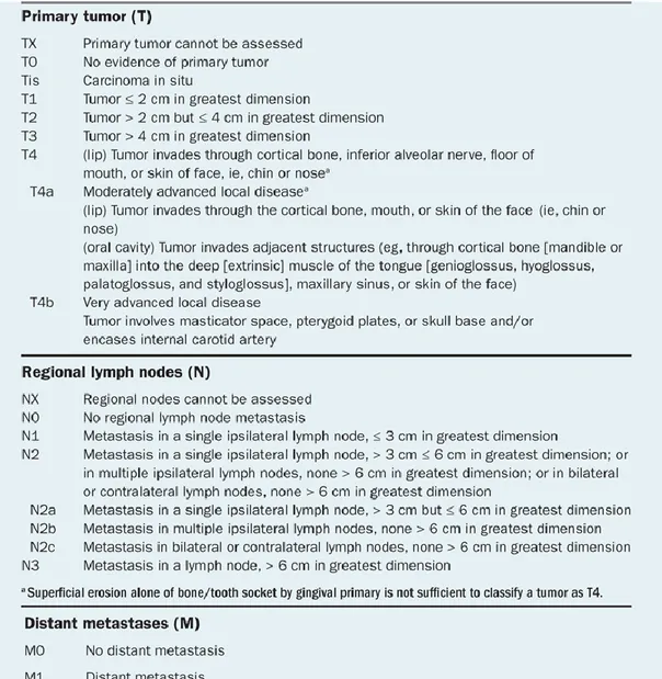
Oral cavity and oropharyngeal cancers are classified according to the tumor-node-metastasis (TNM) system of the American Joint Committee on Cancer. T indicates the size of the primary tumors (T1 to T3), whereas T4 usually represent invasion of other tissues of the oral cavity. N describes the size, number, and location on regional lymph node, while M indicates whether tumor has spread to other organs of the body. Distant metastasis are found in less than 10% of patients, mainly involving lung, liver and
22
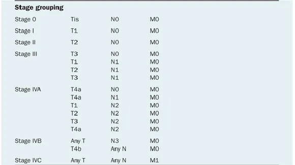
bones (Table 3). T, N, and M are combined to form different stage groups from I to IV as shown in Table 4 [14].
23
Table 4. Oral cancer stage groups based on T, N, and M.
1.2.5 Clinical manifestations and diagnosis
Clinical manifestations vary depending on the stage and primary site of the tumor. The signs and symptoms of oral cavity carcinomas can be a mouth sore that fails to heal, sudden tooth mobility without apparent cause, or painful lesions. Most early signs are painless and are difficult to detect without a thorough head and neck examination by a dental or medical professional [15].
Tumors of the tongue base or oropharynx can cause decreased tongue mobility and alterations in speech. Cancers of the oropharynx or hypopharynx rarely cause early symptoms, but they may cause sore throat and/or otalgia.
Advanced head and neck cancers in any location can cause severe pain, otalgia, airway obstruction, cranial neuropathies, trismus, odynophagia, dysphagia, decreased tongue mobility, fistulas, skin involvement, and massive cervical lymphadenopathy, which may be unilateral or bilateral [4].
24
Oral examination involves the inspection of the oral cavity and the palpitation of the floor of the mouth, tongue and neck. Changes in surface texture, loss of surface integrity, color, and size may be suspicious for oral cancer or premalignant lesions. Conventional oral examination has been shown to have high sensitivity, whereas the specificity remains low due to the limited ability to differentiate between benign and precancerous lesions with high risk of progression into invasive oral cancer.
Given the high mortality rate of late stage disease, early detection of oral cancer and premalignant lesions is of paramount clinical importance to improve patient survival and reduce treatment-related morbidity. Detection of lesions may be enhanced by the use of adjunctive aids including vital staining, light-based detection system, histological and cytological techniques, molecular analysis and imaging techniques. Vital tissue staining with toluidine blue is the most used method for early detection of oral cancer and oral potentially malignant disorders. Due to the binding ability to the DNA, toluidine blue staining is used to detect dysplastic and malignant cells, that display higher nucleic acid content than normal, and can be useful to highlight premalignant and malignant oral lesions.
Light-based detection systems work on the assumption that mucosal tissues that have undergone abnormal metabolic or structural changes have different absorbance and reflectance properties when exposed to specific wavelengths. In this light, light-based techniques represent helpful tools to enhance the oral examination process. Tissue biopsy of the suspicious lesion and histopathological examination remain the gold standard for oral cancer diagnosis. An accurate histopathologic diagnosis depends on the clinician doing an appropriate biopsy and providing adequate clinical information, and on the pathologist correctly interpreting the biopsy results. Pathologic evaluation of the presence and degree of epithelial dysplasia is used to assess the malignant risk of
25
oral premalignant lesions, as low-grade (mild or moderate) and high-grade (severe) dysplasia show a different risk of progression to cancer [16, 17]. Imaging diagnostic techniques provide useful information for screening patients for oral cancer. Computed tomography (CT) scan of the chest and upper abdomen is used to screen for distant metastasis in patients with lymph node involvement. A positron emission tomography (PET) scan may also be helpful to identify or exclude distant metastases. Endoscopy examination, including laryngoscopy, esophagoscopy, and bronchoscopy, is a common diagnostic procedure for oral cancer. Multiple biopsy samples are obtained to establish a primary diagnosis, define the extent of primary disease, and identify any additional premalignant lesions. In patients with lymph node involvement and no visible primary tumor, the diagnosis should be made by lymph node excision [4].
1.2.6 Therapeutic strategies
Treatment of oral cancer depends on the location and the stage of the tumor, as well as the treatment preferences of the individual patient.
Early stage disease is generally treated with single modality therapy by either surgery or radiotherapy (brachytherapy or external beam radiotherapy). Surgery is the traditional approach for small lesions in the oral cavity, while radiotherapy is preferred for unresectable disease, high operative risk patients due to poor performance status, or recurrent disease.
Local or regional advanced diseases are treated with a combined-modality therapy that include surgery followed by adjuvant radiation or chemoradiotherapy.
For unresectable disease, concurrent chemoradiotherapy is the treatment of choice. Meta-analysis of randomized trials has shown that the use of radiation therapy together
26
with cisplatin improve the overall survival rate in patients with oro- and hypo-pharyngeal carcinomas, as compared to radiotherapy alone.
Patients with recurrent and metastatic disease are generally treated with palliative intent. Chemotherapy using single-agent cisplatin shows a response rate of 30% and a median overall survival of 6-9 month. Combinations of cisplatin/carboplatin with 5-FU or taxane has increased response rates to around 35%, and are considered a standard treatment approach [18, 19].
27
1.3 NICOTINAMIDE N-METHYLTRANSFERASE
1.3.1 Drug metabolism
Drug metabolism consists of a series of reactions that change the chemical structure of drugs to compounds that can be readily eliminated from the body. During these biotransformation processes, lipid-soluble xenobiotic compounds are enzymatically transformed into polar, water-soluble metabolites which are excreted into urine or bile. The major site for drug metabolism is the liver, although xenobiotic metabolizing enzyme may be present in other organs such as lung, intestine, and kidney.
Biotransformation of drugs is divided into two phases. Phase I metabolism includes functionalization reactions that introduce polar chemical groups. Phase I reactions involve two groups of enzymes, such as oxidoreductases (cytochrome P450 monooxygenases, flavin monooxygenases, monoamine oxidases, aldehyde dehydrogenase, alcohol dehydrogenase) and hydrolases (amidases, esterases, epoxide hydrolase). In phase II reactions, endogenous compounds or activated xenobiotics metabolites are conjugated with endogenous polar molecules. These conjugation reactions are catalyzed by enzymes, such as glutathione S-transferase, glucuronosyltransferase, sulfotransferase, N-acetyltransferase, and methyltransferase [20].
Among phase II reactions, methyl conjugation is an important pathway in the metabolism of many drugs, xenobiotic compounds, and neurotransmitters.
Methylation of pyridine compounds was first described in 1884 by Wilhelm His. S-adenosyl-L-methionine (SAM) is the methyl donor for this reaction as well as for the methylation of most other drugs and xenobiotics. Human methyltransferases catalyze
28
the reactions of S-methylation, O-methylation and N-methylation. S-methylation is an important pathway in the biotransformation of drugs such as 6-mercaptopurine, D-penicillamine, and captopril, catalyzed by thiopurine methyltransferase (TPMT, E.C. 2.1.1.67), and thiol methyltransferase (TMT, EC 2.1.1.9). Catechol-O-methyltransferase (COMT, E.C. 2.1.1.6) and phenol O-methyltransferase (POMT, E.C. 2.1.1.25) are involved in the metabolism of several catecholamine neurotransmitters. N-methylation reactions are catalyzed by several N-methyltransferases in human, including histamine N-methyltransferase (HNMT, E.E. 2.1.1.8) and nicotinamide N-methyltransferase (NNMT, E.C. 2.1.1.1) [21, 22].
1.3.2 Nicotinamide homeostasis
The biological effects of vitamin B3, also known as vitamin PP or niacin, are mediated by two functionally related forms of such vitamin: nicotinic acid and nicotinamide. Nicotinamide is a precursor for the synthesis of NAD+ (Nicotinamide Adenine Dinucleotide) and NADP+ (Nicotinamide Adenine Dinucleotide Phosphate), coenzymes that play a key role in ATP production through redox reactions. Both NAD+ and NADP+ are also involved in non-redox mechanisms which lead to the cleavage of the β-N-glycosidic bond to free nicotinamide and include:
ADP-ribosylation of proteins catalyzed by mono-ADP-ribosyltransferases (ARTs) and poly ADP-ribose polymerases (PARPs) [23].
calcium mobilization from intracellular stores, mediated by cyclic ADP-ribose (cADPR) and nicotinic acid adenine dinucleotide phosphate (NAADP) [24].
deacetylation of histones and transcription factors catalyzed by NAD(+) -dependent histone deacetylases (sirtuins) [25, 26].
29
Niacin is absorbed in the stomach and intestine by sodium ion-dependent facilitated diffusion at low concentrations, while passive diffusion predominates at higher concentrations [27]. Niacin is found in a variety of foods, but can be also synthesized from tryptophan. In case of low niacin availability, mammals are able to synthesize nicotinamide-containing nucleotides through the kynurenine pathway [28]. The conversion of tryptophan to niacin requires nine steps, whereby 1 mg of nicotinamide was estimated to be produced from approximately 60 mg of dietary tryptophan [29]. Nicotinamide levels may be also regulated through the NAD+ storage in the liver. Hepatic regulation involves the conversion of excess serum nicotinamide to NAD+, whereas hepatic NAD glycohydrolases lead to the release of nicotinamide, which is transported to other tissues. Thus, the liver controls serum levels of nicotinamide, which plays a key role in regulating tissue pyridine nucleotide concentrations [30].
1.3.3 Characterization of human NNMT
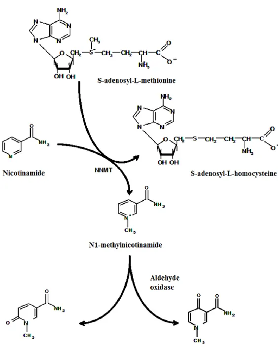
Nicotinamide N-methyltransferase (NNMT) is a phase II metabolizing enzyme which plays an important role in the biotransformation and detoxification of many xenobiotic compounds [22]. NNMT catalyzes the N-methylation of nicotinamide, pyridines and other structural analogs, using S-adenosyl-L-methionine as methyl donor, to form positively charged pyridinium ions [31]. The reaction catalyzed by NNMT yields N1-methylnicotinamide (MNA) and S-adenosyl-L-homocysteine (SAH), which is subsequently hydrolyzed to L-homocysteine and adenosine. In human, N1-methylnicotinamide is mostly excreted through urine and partly further converted via catalysis by aldehyde oxidase to 2-pyridone-5-carboxamide and N1-methyl-4-pyridone-5-carboxamide, which are also excreted into urine (Figure 1) [32]. While different N-methyltransferases can catalyze the N-methylation of aza-heterocyclic
30
compounds, NNMT is the only enzyme known to use nicotinamide as methyl acceptor, playing an important role in nicotinamide catabolism [33, 34].
Figure 1. Methylation of nicotinamide and oxidation of N1-methylnicotinamide
31
Human NNMT enzyme was first characterized from liver tissue samples, through the development of a radiochemical assay for enzyme activity. The assay was based on the conversion of nicotinamide to radioactively labelled MNA, using [14 C-methyl]-S-adenosyl-L-methionine as the methyl donor. The positively charged reaction product was then extracted into 60% isoamyl alcohol in toluene in the presence of the ion-pairing reagent 1-heptanesulfonic acid. The identification of the radioactive product of the enzyme reaction was performed by reversed-phase HPLC, and the radioactivity was measured using a liquid scintillator.
Human hepatic NNMT showed a cytoplasmic distribution with a pH optimum of 7.4. Km values determined for nicotinamide and S-adenosyl-L-methionine were 347 and 1.76 µM, respectively. Human liver NNMT activity was inhibited by both the reaction products, while was not affected by inhibitors of several other methyltransferase. The mean NNMT specific activity, measured in liver biopsy samples, was 51,5 ± 32,5 U/mg, with a large individual variation.
The distribution of activities was bimodal, with approximately 26% of the samples included in a subgroup with high NNMT activity, suggesting that such variations in NNMT activity might be responsible for individual differences in the metabolism and toxicity of pyridine compounds [35]. These observations also raised the possibility that human NNMT activity might be regulated by a genetic polymorphism within the gene sequence [36, 37].
To elucidate such hypothesis, human liver NNMT was partially purified by sequential ion exchange and gel filtration chromatography. The final step in the purification utilized SDS-PAGE, followed by photoaffinity labeling of NNMT that enabled the identification of a protein with a molecular mass of approximately 29 kDa. Partially purified NNMT was then subjected to chemical and enzymatic proteolysis, and the
32
sequence of the cDNA was obtained from the analysis of the amino acid sequence of generated fragments. The NNMT cDNA possessed a region of 792 bp encoding for a 264 amino acids protein with a calculated molecular mass of 29.6 kDa. The NNMT cDNA was subcloned into the eukaryotic expression vector p91023(B), and the construct obtained was used to transfect COS-1 cells. Substrate kinetic and enzyme inhibition experiments performed on both human liver NNMT and transfected COS-1 cell NNMT, showed a Km values for nicotinamide of 0.43 and 0.38 mM, respectively, and a Km values for S-adenosyl-L-methionine of 1.8 and 2.2 µM, respectively. IC50
values for the inhibition of NNMT by N1-methylnicotinamide were 60 and 30 µM for human liver and COS-1 cell-expressed NNMT, respectively [38].
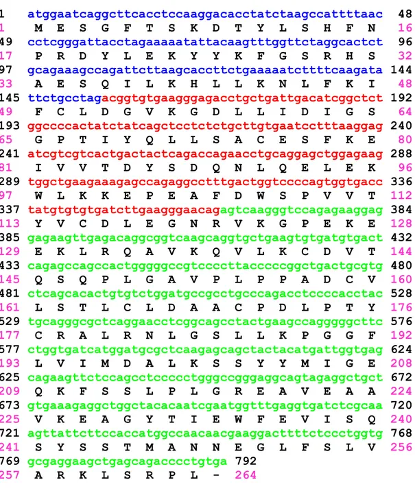
Subsequent experiments were performed to determine the chromosomal localization of the human NNMT gene. The gene is localized on the human chromosome 11q23.1, with a nucleotide sequence of approximately 16.5 kb in length, and consists of 3 exons and 2 introns. Transcription initiation for the NNMT gene occurs at or near a nucleotide located -108 bp upstream from the translation initiation codon, and approximately 30 nucleotides downstream from a TATA box element (Figure 2) [39].
33 1 atggaatcaggcttcacctccaaggacacctatctaagccattttaac 48 1 M E S G F T S K D T Y L S H F N 16 49 cctcgggattacctagaaaaatattacaagtttggttctaggcactct 96 17 P R D Y L E K Y Y K F G S R H S 32 97 gcagaaagccagattcttaagcaccttctgaaaaatcttttcaagata 144 33 A E S Q I L K H L L K N L F K I 48 145 ttctgcctagacggtgtgaagggagacctgctgattgacatcggctct 192 49 F C L D G V K G D L L I D I G S 64 193 ggccccactatctatcagctcctctctgcttgtgaatcctttaaggag 240 65 G P T I Y Q L L S A C E S F K E 80 241 atcgtcgtcactgactactcagaccagaacctgcaggagctggagaag 288 81 I V V T D Y S D Q N L Q E L E K 96 289 tggctgaagaaagagccagaggcctttgactggtccccagtggtgacc 336 97 W L K K E P E A F D W S P V V T 112 337 tatgtgtgtgatcttgaagggaacagagtcaagggtccagagaaggag 384 113 Y V C D L E G N R V K G P E K E 128 385 gagaagttgagacaggcggtcaagcaggtgctgaagtgtgatgtgact 432 129 E K L R Q A V K Q V L K C D V T 144 433 cagagccagccactgggggccgtccccttacccccggctgactgcgtg 480 145 Q S Q P L G A V P L P P A D C V 160 481 ctcagcacactgtgtctggatgccgcctgcccagacctccccacctac 528 161 L S T L C L D A A C P D L P T Y 176 529 tgcagggcgctcaggaacctcggcagcctactgaagccagggggcttc 576 177 C R A L R N L G S L L K P G G F 192 577 ctggtgatcatggatgcgctcaagagcagctactacatgattggtgag 624 193 L V I M D A L K S S Y Y M I G E 208 625 cagaagttctccagcctccccctgggccgggaggcagtagaggctgct 672 209 Q K F S S L P L G R E A V E A A 224 673 gtgaaagaggctggctacacaatcgaatggtttgaggtgatctcgcaa 720 225 V K E A G Y T I E W F E V I S Q 240 721 agttattcttccaccatggccaacaacgaaggacttttctccctggtg 768 241 S Y S S T M A N N E G L F S L V 256 769 gcgaggaagctgagcagacccctgtga 792 257 A R K L S R P L - 264
Figure 2. Human NNMT coding sequence. Exons are highlighted with different
colours. The amino acid sequence of the encoded protein is shown in single-letter code beneath the nucleotide sequence.
A number of studies reported that the NNMT promoter activity is regulated by transcription factors such as HNF-1β, STAT3, and TGF-β1.
34
A positive correlation between HNF-1β and NNMT expression levels was found in papillary thyroid cancer cell lines. A putative HNF-1β binding site was identified in the NNMT basal promoter region. Mutations in this site abolished HNF-1β binding and significantly decreased NNMT promoter activity in the HNF-1β-positive papillary thyroid cancer cell line BHP 2-7, suggesting that HNF-1β is able to function as a transcription activator of NNMT promoter [40]. Furthermore, NNMT expression was reduced at both mRNA and protein level in BHP 18-21 papillary thyroid cancer cells treated with depsipeptide, a histone deacetylase inhibitor, and was demonstrated that the inhibition of NNMT by depsipeptide occur at transcription level through the downregulation of the transcription activator HNF-1β [41].
NNMT promoter activity was also correlated to the activation of signal transducer and activator of transcription 3 (STAT3) in colorectal tumor tissues. STAT3 is activated by tyrosine phosphorylation in response to several cytokines including IL-6 and LIF. The NNMT promoter construct was transfected into 293 embryonic kidney and Hep-G2 hepatoma cells, and it was found that the promoter activity was stimulated by LIF and IL-6, respectively. In addition, inhibition of Stat3 in HT29 colon cancer cells with siRNA or curcumin, which inhibited Stat3 phosphorylation, led to a reduction of NNMT expression level [42].
NNMT was also reported to be a target of TGF-β1. Downregulation of TGF-β1 and its target genes, including NNMT, was shown at both mRNA and protein level in human insulinomas compared to normal pancreatic islets [43].
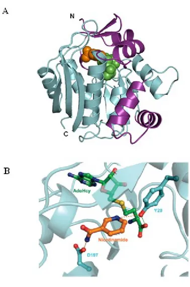
The enzyme is mainly expressed in the liver, while a lower NNMT expression was found in the kidney, lung, placenta, brain, heart, skeletal muscle, and bladder [38]. A recent study revealed the crystal structure of the human NNMT bound to both S-adenosyl-L-homocysteine and nicotinamide. Results obtained by site-directed
35
mutagenesis confirmed protein features important for the binding of nicotinamide, and the identification of several residues in the active site. Among these, Y20 and D197 residues were shown to be important for nicotinamide recognition and NNMT catalysis (Figure 3) [44].
Figure 3. Crystal structure of human NNMT bound to S-adenosyl-L-homocysteine and
nicotinamide (A). Y20 and D197 residues in the active site playing a critical role for NNMT catalysis (B).
36
1.3.4 NNMT polymorphisms
It was hypothesized that the phenotypic differences observed in nicotinamide N-methyltransferase activity may be due to a genetic polymorphism within the coding regions of the gene.
To investigate such hypothesis, the sequence of the three exons, intron 1, and 5'-flanking region of the NNMT gene were tested for the presence of genetic polymorphisms in human liver biopsy samples with low, intermediate or high levels of NNMT activity. No single nucleotide polymorphisms (SNPs) or insertion/deletion events were detected within either the exons or the 5'-flanking region. Eight SNPs were identified within intron 1, but none of these were related to the NNMT activity level. Thus, the individual variability observed in NNMT activity was not attributed to the structure of the enzyme but due to differences in the intracellular NNMT mRNA and protein levels [45, 46].
A genomewide linkage scan for genes affecting variation in plasma Hcy levels was conducted in a study on venous thrombosis, and revealed the presence of a genetic determinant at chromosome 11q23, corresponding to NNMT. Haplotype analyses for 10 single nucleotide polymorphisms (SNPs) in the NNMT gene identified one SNP (rs694539) that was strongly associated with homocysteine levels. Since the SNP is localized in the intron 1, it was hypothesized that this noncoding SNP might be related to the regulation of NNMT transcription [47].
Considering the critical role of NNMT in homocysteine metabolism, a case-control family study, investigated the effect of the NNMT polymorphism on the risk of developing congenital heart defects (CHDs), where maternal hyperhomocysteinemia is a known risk factor. Data obtained showed an eight-fold increased risk for congenital heart defects (CHDs) in children carrying the nicotinamide N-methyltransferase
37
polymorphism rs694539 (AG/AA genotype) and exposed to periconception medicines and low nicotinamide intake [48].
As maternal hyperhomocysteinemia is considered an important risk factor for neural tube defects, a work conducted in 251 infants with spina bifida and 335 controls, explored the potential correlation between genetic variations of NNMT gene and spina bifida. Although eleven SNPs of the NNMT gene were evaluated, no association between infant NNMT gene variants and risk for spina bifida was found [49].
Several studies suggested an association between abdominal aortic aneurysm (AAA) and hyperhomocysteinemia. Thus, NNMT gene variants may contribute to AAA susceptibility. Using a primer extension based microarray technology, 56 polymorphisms of genes involved in methionine metabolism were evaluated in 423 AAA patients and 423 matched controls. Results obtained showed that seven haplotypes, including one screened for NNMT, were significantly associated with abdominal aortic aneurysm. [50, 51].
A similar study was conducted in 501 young patients who survived ischemic stroke and 1,211 controls. However, no significant correlation between NNMT variants and the pathology was detected [52].
Another work investigated the effects of polymorphisms within folate pathway genes on risk of acute lymphoblastic leukemia (ALL). Results obtained from analyses performed on samples obtained from 245 pediatric ALL patients (cases) and 500 blood bank donors (controls), identified polymorphisms in MTHFR (677 C>T), NNMT (IVS-151 C>T), RFC1 (80G>A) genes. The NNMT IVS-151 TT variant was associated with increased susceptibility to ALL [53].
In a comprehensive association study, the rs694539, previously reported to be correlated with hyperhomocysteinemia, and the rs1941404 NNMT variants were significantly
38
associated with schizophrenia (SZ). In addition, post-mortem frontal cortex NNMT mRNA levels were 35% lower in schizophrenia patients compared to control subjects, suggesting the involvement of NNMT in the etiology of this disorder [54].
A correlation between the NNMT rs694539 polymorphism and schizophrenia was also found within a Han Chinese female population in a case-control study, confirming the important role of NNMT in the pathogenesis of SZ [55].
Recent studies investigated the potential role of NNMT gene rs694539 variant in the development of diseases correlated with high plasma homocysteine level. The rs694539 NNMT variant was reported to be a genetic risk factor for bipolar disorders, nonalcoholic steatohepatitis, and epilepsy. Furthermore, individuals with the GG genotype displayed protection against nonalcoholic steatohepatitis and epilepsy, whereas the individuals with the AA genotype showed an increased risk for both diseases. Although the biological significance of the rs694539 NNMT variant is unclear, dysregulation of epigenetics, elevated homocysteine levels, as well as dysregulation of the nicotinamide levels may be one of the causes for the development of such pathologies [56, 57, 58].
1.3.5 NNMT and non-neoplastic diseases
Elevated serum levels of N1-methylnicotinamide were detected in patients with cirrhosis, as compared to control subjects, although this disease does not impair the efficiency of nicotinamide methylation [59].
N1-methylnicotinamide has been considered a biologically inactive metabolite of nicotinamide for a long time. However, recent studies have found that MNA possesses anti-inflammatory properties. After topical application of MNA, beneficial therapeutic effects were observed in patients with inflammatory skin diseases, including contact
39
dermatitis and acne vulgaris [60]. In addition, it was suggested that the anti-inflammatory properties of MNA may be associated with its ability to reduce adherence of pro-inflammatory cells to the vascular endothelium [61].
MNA also displayed anti-thrombotic activity in rats with arterial thrombosis, through a mechanism involving cyclooxygenase-2 (COX-2) and prostacyclin (PGI2). These observations supported the hypothesis that endogenous MNA may regulate thrombosis as well as inflammatory processes characterizing cardiovascular diseases [62].
In a study conducted in mice with atherosclerosis, the progression of the disease was associated with increased hepatic NNMT activity and MNA plasma levels. Therefore, given the anti-thrombotic and anti-inflammatory properties of MNA, high NNMT level may play an important compensatory role in atherosclerosis [63].
Subsequent analysis revealed a significant increase of hepatic NNMT activity and MNA plasma levels during the progression of concanavalinA (ConA)-induced hepatitis in mice, suggesting a potential hepatoprotective effect of MNA through a PGI2-dependent mechanism [64].
NNMT- MNA pathway was shown to be activated during endurance exercise in mice. In addition, data obtained demonstrated that exercise-induced activation of NNMT in the liver involves IL-6, while changes in MNA plasma concentration was partially IL-6-independent. Thus, IL-6 is able to regulate hepatic NNMT activity but also other tissues may determine MNA level in plasma [65].
Changes in NNMT-MNA pathway were also examined in pulmonary arterial hypertension (PAH), a disease associated with inflammatory response. NNMT activity increased progressively in liver and lung in relation to PAH progression, and NNMT response was associated with elevated plasma MNA level. Given the vasoprotective
40
activity exerted by MNA, the activation of the NNMT-MNA pathway may play a compensatory role in PAH [66].
Upregulation of NNMT gene expression was shown in human renal allograft biopsies with histologic evidence of acute cellular rejection [67]. Furthermore, NNMT expression was detected in endometrial biopsies from patients who became pregnant after intracytoplasmic sperm injection (ICSI) cycles, suggesting a potential involvement of the enzyme in the embryo implantation process [68, 69]. Elevated NNMT expression was demonstrated in endometrial stromal cells in response to factors secreted by macrophages. Therefore, the increased expression of the enzyme observed may be related to the migration and invasion of endometrial cells that occurs during the establishment of endometriosis [70].
In several studies NNMT expression was associated with chronic obstructive pulmonary disease (COPD). High NNMT mRNA expression was observed in quadriceps muscles of patients with COPD [71]. In addition, overexpression of the enzyme in myoblasts significantly increased cell proliferation and migration, as well as reduced protein oxidation and H2O2-induced cell death. Therefore, NNMT upregulation observed in the
skeletal muscles of patients with COPD may improve myogenesis and defend against oxidative stress [72]. Elevated NNMT levels were also detected in lung tissues of patients with COPD, suggesting a potential association of the enzyme with COPD severity [73].
A recent study highlighted the protective role of NNMT against mitochondrial ROS generation in the pathogenesis of proximal tubular cell (PTC) damage and subsequent renal dysfunction, in patients with refractory proteinuria. In particular, MNA was shown to reduce lipotoxicity-induced oxidative stress and cell damage in kidney proximal tubular cells [74].
41
In contrast, increased NNMT activity levels were observed in mice treated with phenobarbital, a chemical involved in ROS generation through the induction of cytochrome P450 enzymes. The enhancement of NNMT activity was associated with the depletion of pyridine nucleotides, leading to the attenuation of the NADPH-dependent antioxidant enzymes. Thus, NNMT overexpression may reduce protective systems against oxidative stress [75].
Analysis conducted in murine adipocytes as well as in human and murine adipose tissue samples revealed increased NNMT activity levels and homocysteine release. In this light, high NNMT activity may lead to elevated plasma homocysteine levels, which is considered a risk factor for cardiovascular disease [76].
A recent study reported increased NNMT expression in white adipose tissue (WAT) and liver of obese and diabetic mice. In addition, NNMT knockdown protected mice from diet-induced obesity by augmenting cellular energy expenditure [77]. Subsequent work explored WAT NNMT expression levels in patients with type 2 diabetes and insulin-resistant individuals. Higher WAT NNMT expression was shown in human insulin resistance and type 2 diabetes compared with controls, with a positive correlation observed between NNMT expression and the degree of insulin resistance [78].
Elevated NNMT expression, at both mRNA and serum protein level, was detected in patients with peripheral occlusive arterial disease, as compared to healthy subjects, suggesting a potential use of the enzyme as a biomarker for this pathology [79]. In order to investigate the involvement of the enzyme in metabolic syndrome, NNMT expression was analyzed in the adipose tissue of Wistar Ottawa Karlsburg W (WOKW) rats, which represent an animal model for metabolic syndrome, and Dark Agouti (DA) rats, as control. High NNMT mRNA, protein, and activity levels were observed in adipose
42
tissue of WOKW rats, supporting the important role of the enzyme in the pathogenesis of this disorder [80].
Although the liver represent the tissue with the strongest NNMT expression, the role played by the enzyme in hepatocytes metabolism is still unclear. A recent study demonstrated that hepatic NNMT levels regulate glucose, lipid and cholesterol metabolism in mice by stabilizing Sirt1 protein, and that the metabolic effects of NNMT are mediated by its product MNA [81].
1.3.6 NNMT and Parkinson’s disease
N-methylation is an important pathway in the detoxification of many xenobiotic compounds. However, it has also been reported that this biotransformation increases the toxicity of some substrates. Thus, high levels of NNMT activity in brain may lead to the production of neurotoxic methylpyridinium ions, representing a factor in the etiology of Parkinson disease (PD). To investigate the involvement of NNMT in the occurrence of PD, regional expression of the enzyme were explored in normal human brain tissue. NNMT was found to be expressed solely in neurons and was demonstrated a regional variation in expression levels. High NNMT mRNA levels were shown in the spinal cord, medulla, and temporal lobe, whereas low expression levels were found in the cerebellum, subthalamic and caudate nucleus. Both NNMT protein and enzyme activity were detected in the spinal cord and temporal lobe. Expression of NNMT was compared in PD and non-PD control cerebella and caudate nucleus, resulting significantly elevated in the brains of patients who have died with PD. Furthermore, disease duration was found to be inversely correlated with the level of patient’s NNMT expression, suggesting a causative effect in the pathogenesis of the disease [82, 83]. Other studies supported such hypothesis, reporting the neurotoxic effect of N1-methylnicotinamide,
43
which causes the destruction of complex I subunits of the mitochondrial chain via the induction of free radicals [84, 85, 86, 87].
A recent study was conducted in the human neuroblastoma-derived cell line SH-SY5Y, which lacks of endogenous NNMT, and thus represents an ideal model for investigating the effects of NNMT expression upon cell viability and metabolism. Stable NNMT expression significantly decreased cell death in SH-SY5Y cells, which correlated with increased Complex I activity and ATP synthesis, thus revealing a cytoprotective effect of the enzyme. These effects were replicated by incubation of SH-SY5Y cells with N1-methylnicotinamide, suggesting that this compound is able to mediate the cellular effects of NNMT. Moreover, both NNMT expression and MNA exposure protected SH-SY5Y cells from the toxicity of the Complex I inhibitors MPP+ (1-methyl-4-phenylpyridinium ion) and rotenone. These observations raised the possibility that the NNMT overexpression observed in association with PD patients may represent a stress response of the cell to the underlying pathogenic process [88]. NNMT was also shown to confer protection against mitotoxicity induced by potassium cyanide (KCN), 2,4-dinitrophenol, and 6-hydroxydopamine in vitro, and was demonstrated that its neuroprotective effects are not mediated solely via increased N1-methylnicotinamide production, but a variety of mechanisms [89]. Another study conducted in SH-SY5Y and N27 rat mesencephalic dopaminergic cell lines reported the involvement of the enzyme on neuron morphology and differentiation, showing that NNMT expression increased neurite branching [90]. In order to explore the molecular mechanism leading to the observed cytoprotective effects, it was investigated whether sirtuins may mediates the effect of NNMT upon complex I activity. Silencing of sirtuin 3 in NNMT-expressing SH-SY5Y cells demonstrated the important role that sirtuin 3 have in mediating the NNMT-induced complex I activity and ATP synthesis [91]. Given the
44
high levels of both NNMT and N-methylated β-carbolines (BCs) in PD, in a recent study was investigated whether the enzyme may display N-methyltransferase activity towards BCs. Recombinant NNMT was able to N-methylate the BC norharman (NH) to 2-N-methylnorharman (meNH). Further analyses demonstrated that meNH was less toxic than its precursor NH, and it was shown to increase cell viability and intracellular ATP content in NNMT-expressing SH-SY5Y cells. Taken together, these results suggested the involvement of the enzyme in the detoxification pathway for BCs and that NNMT overexpression may represent a cytoprotective response to the pathogenesis of PD [92].
1.3.7 NNMT and neoplastic diseases
Gene expression profile of glioblastoma multiforme (GBM) tissue samples showed the upregulation of nicotinamide N-methyltransferase in GBMs compared to normal tissues [93]. Moreover, high NNMT activity and N1-methylnicotinamide levels were reported in human glioma cells cultured in the presence of interferon-γ (IFN- γ) [94].
In order to investigate the molecular pathogenesis of thyroid cancer, DNA microarray was used to analyze the gene expression profiles of different human thyroid cancer cell lines, including papillary, follicular, medullary, and anaplastic carcinomas. High NNMT expression was found in papillary cancer cells, whereas weak or no NNMT expression was showed in the other tested cancer cell lines. In addition, NNMT catalytic activity was correlated with the mRNA expression level. Immunohistochemical staining for NNMT of human thyroid specimens revealed strong cytoplasmic reactions in papillary and follicular carcinomas, as weak or scanty reaction was detected in follicular adenomas, colloid goiters, and normal thyroid tissues [95].
45
A study conducted in a human breast adenocarcinoma cell line revealed the expression of NNMT in adriamycin resistant MCF-7 cells, whereas enzyme level was not detected in parental MCF-7 cells [96, 97]. In a recent study, the cellular effect of NNMT expression was evaluated in different human breast cancer cell lines. NNMT downregulation in Bcap-37 and MDA-MB-231 cells significantly decreased cell growth
in vitro and tumorigenicity in vivo. In addition, NNMT silencing increased ROS
production and induced apoptosis via the mitochondria-mediated pathway. The effect of the enzyme on cell proliferation and apoptosis was confirmed by overexpressing NNMT in MCF-7 and SK-BR-3 cell lines, suggesting that NNMT could become an interesting molecular target for breast cancer therapy [98].
Gene expression profile of pancreatic cancer was analyzed in the RNA isolated from pancreatic juice. Increased NNMT expression levels were shown in the pancreatic juice of patients with pancreatic cancer compared with controls [99]. A metabolomics analysis conducted in miR-1291-expressing and control PANC-1 pancreatic cancer cells revealed elevated level of N1-methylnicotinamide in miR-1291-expressing PANC-1 cells. MNA levels were also correlated with NNMT mRNA expression. Stable expression of miR-1291 in PANC-1 cells reduced cell migration, invasion and xenograft tumorigenesis compared to controls. Furthermore, NNMT mRNA level was inversely correlated with pancreatic tumor size in the xenograft mouse model, suggesting the involvement of NNMT in pancreatic tumor progression [100]. Another study explored the biological function of NNMT in pancreatic cancer, showing that NNMT silencing significantly reduced cell proliferation, and suppressed the migration and invasion capacities of PANC-1 cells. In addition, overexpression of NNMT enhanced the survival of PANC-1 cells subjected to either glucose deprivation or glycolytic inhibition, or treated with rapamycin, indicating that NNMT plays an