zur nichtkommerziellen Nutzung auf der privaten Homepage und Institutssite des Autors
Somnologie
Schlafforschung und Schlafmedizin
Somnology
Sleep Research and Sleep Medicine
Offizielles Organ der DGSM und der ÖGSM • Official Journal of the DGSM and of the ASRA
Elektronischer Sonderdruck für
Ein Service von Springer Medizin
Somnologie 2012 · 16:125–132 · DOI 10.1007/s11818-012-0560-2 © Springer-Verlag 2012
P. Pirelli · M. Saponara · C. Guilleminault
Forcierte Gaumennahterweiterung vor und nach
Adenotonsillektomie bei Kindern mit obstruktiver
Schlafapnoe
Somnologie 2012 · 16:125–132 DOI 10.1007/s11818-012-0560-2 Received: 19 December 2011 Accepted: 4 April 2012 Published online: 11 July 2012 © Springer-Verlag 2012
P. Pirelli1 · M. Saponara2 · C. Guilleminault3
1 Department of Odontostomatological Sciences, University“Tor Vergata”, Rome 2 Department of Neurology and Otolaryngology, University“La Sapienza”, Rome 3 Division of Sleep Medicine, Stanford University Medical School, Stanford
Rapid maxillary expansion before
and after adenotonsillectomy
in children with obstructive
sleep apnea
In children adenotonsillectomy (AT) is the recommended treatment in the presence of obstructive sleep apnea (OSA) [1]. It should be performed, if needed, in association with nasal in-ferior turbinate reduction usually with radiofrequency [2]. But as shown in previous reports, upper airway soft tissues treatment is not always suc-cessful in completely controlling ab-normal breathing during sleep in chil-dren, despite the fact that subjective improvement is often reported [3, 4, 5, 6].
Recently, a randomized study performed in Caucasian prepubertal children with OSA mostly recruited from an otolaryn-gological practice showed that many chil-dren would benefit from both otolaryngo-logical and orthodontic treatment; in that report, one out of all the children treated initially with orthodontics maneuvers did not need further otolaryngological treat-ment [7]. Our investigation looks further at this interaction between otolaryngolog-ic and orthodontotolaryngolog-ic treatment in OSA. We hypothesized that orthodontic treatment alone, specifically rapid maxillary expan-sion (RME), may resolve OSA without the need for adenotonsillectomy.
Patients and methods
Study participants were recruited among children referred to our ear nose and throat (ENT) otorhinolaryngological and odontological ambulatory clinic during
an 8-year period; all children were from the Rome (Italy) urban area. The select-ed group includselect-ed 225 children (119 boys and 106 girls) ages 6–13 years (average 7.3 years).
Main inclusion criteria were the follow-ing: presence of clinical complaints usu-ally noted in children with sleep-disor-dered breathing (SDB) and signs of mouth breathing, chronic snoring; adenotonsil-lar hypertrophy (2+ or 3+); OSA demon-strated at polygraphy with a minimum ap-nea–hypopnea index (AHI) of 1 and a min-imum respiratory disturbance index (RDI) of 5 [8]; and narrow upper jaw at exami-nation, diagnosed clinically and confirmed by cephalometric assessment according to Ricketts parameters [9]. Presence of oth-er significant pathologies that could influ-ence upper airway patency was an exclu-sion criteria.
The following cases were excluded: 41 children with vasomotor rhinitis, aller-gy and turbinate hypertrophy, 19 children with mandibular retrognathia, 63 children with associated obesity based on weight and height tables for age, 16 children with macroglossia due to medical causes, and 6 children with associated chronic illness-es. A total of 80 children (43 male and 37 female) with a body mass index (BMI) <24 kg/m2 participated in the study.
Pro-tocol and informed consent were ap-proved by the Tor-Vergata University eth-ics committee, Rome (Italy).
Group allocation
All children underwent the same baseline investigation: all had a clinical interview and pediatric evaluation, additional in-formation on their prior health status was obtained from their pediatricians. Based on the current complaints, past medi-cal history, and clinimedi-cal evaluation, suspi-cion of the presence of obstructive sleep apnea led to polygraphy that demonstrat-ed an abnormal amount of SDB events. Subjects then had simultaneous EN) and orthodontic evaluations including rhinos-copy and cephalometric X-rays.
Patients were subdivided into two groups based on results of this investiga-tion:
Those with indicators of chronic ade-notonsillar inflammatory problems—di-agnosed based on clinical evaluation with history of chronic tonsillitis and later-al enlargement of tonsils based on Fried-man scale [10]—were placed in the group to be initially treated with adenotonsillec-tomy (group II), while those not clearly presenting this ENT problem were placed in the initial orthodontic treatment group (group I). The goal was to recruit a simi-lar number of subjects in both groups in order to simplify comparison of results. A total of 40 subjects per group was ob-tained.
Parents were informed that their child had two different morbidities and that both ENT and orthodontic treatment may be needed. In our community, standard of care for inflammatory tonsils is surgical
excision, but children with enlarged but noninflammatory tonsils were referred for orthodontic treatment as a first step. For each treatment modality, patients were followed by the specialist responsible for application of the selected treatment. In-dependent of treatment order, all children underwent an evaluation of the effect of treatment and impact on OSA. This eval-uation consisted of clinical interview and examination, polygraphic recording, and cephalometric X-rays analyses. A first as-sessment was made 4 months after end of the first treatment (T1).
In the AT group, patients with reso-lution of OSA did not enter the second phase of the study. For the remaining children, parents could decide to proceed with orthodontic treatment either within the research protocol or independent of any study schedule. Similarly in the RME group, children with complete resolution of SDB at T1 did not undergo follow-up AT surgery.
Patients who only had a partial ben-efit with orthodontics were referred to their pediatric ENT specialist, but some were not submitted to surgery, based on the unilateral decision of the specialist who felt that the polygraphic findings at T1 were too mild to justify adenotonsil-lectomy [11]. Thus, in this group the sec-ond treatment was reserved only to sub-jects who clearly had persistence of prob-lems at T1.
Procedures
Orthodontic evaluation
All children were examined by an ortho-dontist with a large experience in pediatric facial growth and in abnormal breathing awake and asleep. The orthodontist evalu-ated each patient on:
F facial thirds:
1 The face is normally divided in three thirds of equal length. A flattening of the area of the face located between the superior orbital margin (superior limit) and the occlusal plane (infe-rior limit) can be observed with ab-normal growth. This clinical finding can be evaluated in relation to the “aesthetical analysis of the human profile” based on the “Holyday line of harmony” (H-line) which is de-fined as the distance from the deep-est point of sulcus at the upper lip and the most prominent point of the chin (ideally −5±−2 mm).
1 The presence/absence of labial in-competence defined as absence of contact between upper and lower lips when at rest position with pres-ence/absence of hypotonia of upper lip manifested by an open mouth with a tended upper lip, resulting in an increase of the nasal labial angle (angle formed by the labial surface of the upper lip at the midline and the inferior border of the nose
(nor-mally 85–90°) with a tender upper lip increasing this nasial-labial angle.
1 The presence/absence of mouthbreather facies.
F intra-oral evaluation showed pres-ence/absence of anterior cross-bite; presence/absence of an ogival shape of the upper maxillary arch (the result from a high palatal vault and a narrow maxillary arch), related to a contrac-tion of the upper jaw at its base.
F cephalometric assessment: the narrow-ness of the upper jaw was diagnosed clinically and confirmed by cephalo-metric assessment according to Rick-etts parameters on posteroanterior cephalograms [11]. The following mea-surements were obtained from the pos-teroanterior tele-radiograms (. Fig. 1): to define a narrow maxilla, we assessed the maxillary cross-section study-ing the distance of JR and JL (bilater-al points located at the depth of the concavity of the lateral maxillary con-tour at the junction of the maxilla and the zygomatic buttress; . Fig. 1); and from the frontofacial plane, the ues ZR-AG and ZL-AG (normal val-ue at 8 years of age =10±1.5 mm, and until 14 years =8±1 mm) with a larg-er distance indicating a narrow upplarg-er jaw. We also assessed the nasal cavity width NC-CN (bilateral points locat-ed at the larger cross section of the na-sal cavities with normal value at 9 years of age =25.2±2 mm, and +0.6±1 mm/ year), with a shorter distance confirm-ing the diagnosis of a narrow upper jaw (. Fig. 2). In addition, the interincisive space A1-1A and the intermolar width A6-6A were measured.
The ENT evaluation, as mentioned above, was a comprehensive visual examination of upper airway including rhinopalatosco-py, and cephalogram analyses indicating size and location of adenoids and tonsils.
The polygraphic recording was a noc-turnal home study performed with a val-idated against polysomnography cardio-respiratory recorder, the SOMNOscreen™ plus RC (SOMNOmedics GmbH, Rand-ersacker, Germany) [12], monitoring: body position, SpO2, thoracic effort, abdominal
effort, snore, pulse rate, pulse transit time (PTT), and oral–nasal flow. The software Fig. 1 9
Cephalomet-ric assessment. See text for further clarifi-cation
we employed was DOMINO© (SOMNO-medics GmbH, Randersacker, Germany). The equipment was placed on the child in the sleep laboratory with education of par-ents about leads and potential problems re-lated to activity before bedtime. The scor-ing of the respiratory events was based on the AASM manual for scoring of respirato-ry events (using respiratorespirato-ry channels and PTT) [8]. An AHI >1 was considered
ab-normal. Apnea and hypopnea were scored using the published recommendation in the absence of an EEG recording. A respirato-ry disturbance index (RDI) was scored by adding to apnea and hypopnea, respirato-ry events-related activation (RERA). These events were defined by a drop in the am-plitude of the nasal cannula tracing curve by at least 30% compared to prior normal tracing, presence of snoring and increase
in snoring signal from beginning to end of event associated with the presence of an in-crease in respiratory efforts seen on the tho-racic and abdominal tracing curves, and in-dication of an abrupt change (20 mm from baseline) in the sympathetic tone marker derived from the PTT recording—indica-tive of an activation response—at the end of the event.
Somnologie 2012 · 16:125–132 DOI 10.1007/s11818-012-0560-2 © Springer-Verlag 2012
P. Pirelli · M. Saponara · C. Guilleminault
Rapid maxillary expansion before and after adenotonsillectomy
in children with obstructive sleep apnea
Abstract Adenotonsillectomy is not always successful in controlling obstructive sleep apnea (OSA) in children, and orthodontic treatment may be a helpful adjunction and sometime an im- portant alternative. A total of 80 nonover- weight children (37 girls) with a high and nar- row hard palate, based on an indepth orth-odontic evaluation, were subdivided into two groups based on presence/absence of chron-ic adenotonsillar inflammatory problems as determined by full otolaryngological exam-ination, with recruitment of 40 children in each group. Patients with evidence of chron- ic inflammation were treated with adenoton- sillectomy first (group II), while all other chil-dren had rapid maxillary expension (RME) first (group I). In all children, clinical inter- views and clinical evaluations, frontal and lat-eral cephalometry, and polysomnography were performed at entry. Four months after end of treatment, the initial evaluation was repeated in all children. Children incomplete-ly treated were offered to cross-over to the other treatment venue. At entry, there was no significant difference between the mon- itored respiratory polygraphic variables be- tween the two groups. At the 4-month fol-low-up, 15 subjects treated with RME were considered as cured compared to 6 patients after adenotonsillectomy, and absence of im-provement was observed in 8 children with RME and 16 with adenotonsillectomy. Af- ter cross-over to the other treatment, involv-ing 42 subjects due to 17 drop-outs between treatment 1 and 2,, three children were still having residual sleep disordered breath- ing at the 12-month follow-up, while nor-mal breathing during sleep was observed in all others. With appropriate clinical investiga- tion, prepubertal children with OSA and nar- row maxilla may have better treatment out-come when treated with orthodontics than with adenotonsillectomy; and polygraph- ic and even better polysomnographic doc-umentation of clinical impression is always needed posttreatment. Keywords Adenotonsillectomy · Rapid maxillary expansion · Prepubertal children · Obstructive sleep apnea · Treatment
Forcierte Gaumennahterweiterung vor und nach Adenotonsillektomie
bei Kindern mit obstruktiver Schlafapnoe
Zusammenfassung Die Adenotonsillektomie (ATE) zur Behand-lung obstruktiver Schlafapnoe (OSA) bei Kindern hat nicht immer Erfolg, eine kiefer-orthopädische Therapie kann eine hilfreiche Ergänzung und manchmal eine wichtige Alternative sein. Insgesamt 80 Kinder (37 Mädchen) mit hohem, engem hartem Gaumen – laut gründlicher kieferorthopädi- scher Untersuchung –, aber ohne Überge-wicht, wurden je nach Bestehen oder Fehlen chronischer adenotonsillärer Entzündungen – gemäß vollständiger HNO-ärztlicher Unter- suchung – in 2 Gruppen eingeteilt, dabei wa-ren in jeder Gruppe 40 Kinder. Bei Patienten mit Anzeichen chronischer Entzündung er- folgte zuerst die ATE (Gruppe II), bei allen an-deren die forcierte Gaumennahterweiterung („rapid maxillary expansion“, RME; Gruppe I). Zu Beginn wurden bei allen Kindern klinische Befragungen und Untersuchungen, eine frontale und laterale Zephalometrie sowie Polysomnographie durchgeführt. Die initia-len Untersuchungen wurden 4 Monate nach Behandlungsende bei allen Kindern wieder- holt. Den Kindern mit unvollständigem The- rapieerfolg wurde der Wechsel in den ande-ren Therapiearm angeboten. Zu Beginn gab es keinen signifikanten Unterschied bei den überwachten respiratorischen Polygraphie-variablen zwischen den beiden Gruppen. Bei der Untersuchung nach 4 Monaten wurden 15 Teilnehmer mit RME gegenüber 6 Patien-ten nach ATE als geheilt angesehen, keine Verbesserung wurde bei 8 Kindern mit RME und 16 mit ATE festgestellt. Nach Wechsel zu der je anderen Therapie, der wegen 17 Aus- fällen zwischen Therapie 1 und 2 also 42 Teil-nehmer betraf, wiesen 3 Kinder bei der Untersuchung nach 12 Monaten immer noch eine restliche schlafbezogene Atmungs-störung auf, während bei allen anderen eine normale Atmung im Schlaf beobachtet wur-de. Bei geeigneter klinischer Untersuchung kann das Therapieergebnis bei präpubertä-ren Kindern mit OSA und engem Oberkiefer besser sein, wenn sie kieferorthopädisch statt mittels ATE behandelt werden; dabei ist die polygraphische oder besser noch polysom-nographische Dokumentation des klinischen Eindrucks nach Therapie immer erforderlich. Schlüsselwörter Adenotonsillektomie · Forcierte Gaumennahterweiterung · Präpubertäre Kinder · Obstruktive Schlafapnoe · Therapie
Rapid maxillary expansion (RME) was performed, as previously reported [13, 14], applying orthopedic forces on the midpal-atal sutures using the first molars and per-manent premolars as anchor teeth; while in deciduous dentition, the second pri-mary molars were selected as long as they could provide the required firmness. The device was comprised a central expansion screw with four arms: two front arms and two back arms, anchored to the selected
anchor teeth. The bone distraction at the suture level enables an effective enlarge-ment of the maxillary skeletal base. En-largement was visually appreciable as the bone distraction leads to an interincisive space—a diastema—and with X-rays as the gain appears as a radiotransparency corresponding to the visually seen space. The procedure usually lasts 3 weeks with daily turning of a midline screw that al-lows enlargement (distraction) of the
space at the level of the midline suture: the transpalatal force, which exceeds the orthodontic force, produces an orthope-dic force that opens the midpalatal su-ture leading to maxillary movement with-out tipping of the teeth. Once the need-ed extension was obtainneed-ed (end of the ac-tivation phase), the midline screw was blocked and the device was kept in place at least 3–4 months more in order to al-low the newly formed bone to strengthen. Prior to distraction, child and parents had been briefed on daily care (turning the screw), diet, and oral hygiene in order to avoid any complications, which could in-terfere with the expansion [14, 15]. Based on the screw activation system that we used, the following schedule was followed: day 1 (morning and evening) three consec-utive activations at 10 min intervals, and from day 2 onward, one activation every morning and evening; one activation con-sists of one turn of the screw, i.e., 0.25 mm. All modifications induced by RME on the upper jaw and adjacent anatomical structures were studied with posteroan-terior cephalometric evaluation at T0, T1, and T2 and with CT Dentascan at T0 and T1. The Ricketts parameters were adopted for posteroanterior cephalometric evalua-tion (. Fig. 1, 2).
Adenotonsillectomy
Patients were instructed to avoid heavy exercise and to remain at home for ap-proximately 1 week.
Residual
OSA N=3 N= 39Cure
T2 N = 42
Cure
N= 6 Not cureN=34 Not CureN= 25 N= 15Cure
T1
T & A Orthodontic
Inflammatory Tonsils
N = 40 No InflammatoryTonsils N=40
ENT & Orthodontist Examination 80 Children Not Further Treated N = 14 T&A Surgery N = 8 Orthodontics N = 33 Fig. 3 8 Flow diagram for treatment. OSA obstructive sleep apnea Fig. 2 8 A shorter nasal cavity (NC-CN) distance confirms the diagnosis of a narrow upper jaw
Review
Analysis
At each evaluation, each child was seen by both the ENT and orthodontist team. Polygraphic recordings were analyzed fol-lowing the recommendation of the Amer-ican Academy of Sleep Medicine (AASM) and apnea and hypopnea were scored as recommended in the atlas [8].
Statistical analyses
The t test for repeated measure was used to compare groups at different stages of treatment. The Kruskal–Wallis analy-sis of variance (ANOVA) was performed when comparing several groups with post hoc analysis, if the ANOVA was signifi-cant. The χ2 statistics were used to
com-pare percentages.
Results
During the treatment period (. Fig. 3), no complications were observed in either group.
Polygraphic results
Entry (T0)
At T0 there was no difference between the two groups in demographics and poly-graphic variables (see Table 1). For both treatment groups, the mean AHI was 12.8 events/h, which is considered to be in the “moderate” range. There was no signifi-cant difference in age, BMI, AHI, and lowest oxygen saturation between the two groups at entry (T0).
Initial posttreatment
evaluation (T1)
In the RME group (group I), 15 subjects (37.5% of subjects) had normal clini-cal evaluation and normal polygraphy at the initial posttreatment evaluation (T1) 4 months after completion of treatment; 17 presented a significant improvement (AHI 6.5 ±3.1) and 8 minimal or no im-provement (AHI 13±3.5; ANOVA p=0.05).
Progression to the second
treatment phase (T2)
From this initial orthodontic treatment group (group I), AT was performed only on the 8 patients who show only minimal
or no improvement. The 17 children with incomplete improvement were deemed to have a too mild sleep disordered breathing problem by local standard of practice and were dropped out of the protocol.
These results contrasted with the AT group (group II) where only 6 patients (15% of subjects) presented total remis-sion, 18 presented an improvement of OSA (AHI 6±3.1) and 16 minimal or no improvement (AHI 15±2.9; ANOVA p=0.05). The second treatment with RME was performed on all 34 patients who did not have resolution of their OSA with AT treatment.
Only 42 children were submitted to the second phase of treatment. This was relat-ed not solely to the complete absence of clinical and polygraphic findings noted in 21 children, but to the decision made by the ENT to not pursue treatment in 17 cas-es, despite initial scoring of tonsils as 2+ or 3+. Widening of the palatal vault sec-ondary to orthodontic treatment chang-es scoring of lateral position of the tonsils, but the AHI score in the dropout group was still abnormal (. Fig. 3).
Evaluation after the second
treatment phase (T2)
The evaluation at T2 demonstrated the ef-ficacy of the combined treatment proce-dures: there was complete remission of symptoms in most patients and normal-ization of AHI and nocturnal SpO2 with a
minimum SpO2>96% for the total group.
However, three cases (2 patients in the ini-tial RME group and 1 patient in the iniini-tial AT group) presented incomplete positive results at the end of both treatments with scores of 2.9, 3.2, and 3.8 events/h, respec-tively. However, parents reported overall clear improvement and great abatement of clinical symptoms.
Orthopedic/orthodontic results
All modifications induced by RME on the upper jaw and adjacent anatomical struc-tures were studied with posteroanterior cephalometric evaluation at T0, T1, and T2 and with CT Dentascan at T0 and T1. The Ricketts parameters were adopted for posteroanterior cephalometric evaluation (. Fig. 1).
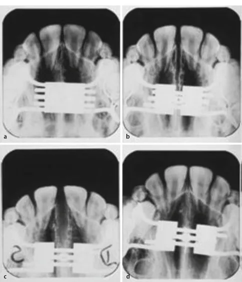
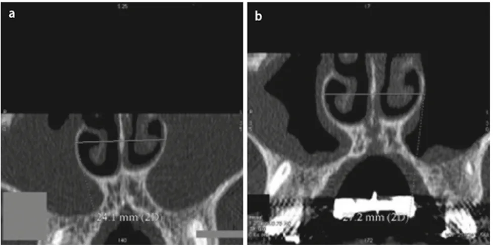
In all our treated cases, an opening of the midpalatal suture was obtained with the results being confirmed by intraoral oc-clusal radiographs (. Fig. 4) and CT Den-tascan (. Fig. 5). The objective assessment of maxillary width shows an increment, confirming that the RME maneuver
di-rectly influences the skeleton. Through the expansion of the midpalatal suture, RME produces an expansion that occurs not on-ly in the maxillary arch but also in the na-sal cavities (. Fig. 6). When considering the whole RME-treated group (n=74), the expansion of the midpalatal suture is
re-sponsible for the expansion of both maxil-las, with an average cross-sectional increase (JL-JR) of 5.91±0.7 mm. The analysis of the upper intermolar distance (A6-6A) shows an average increase of 8.18±0.3 mm. The interincisive space (A1-1A), typical of mid-palatal suture opening, was noted in all treated cases, with an average opening of 4.72±0.2 mm.
The increase in maxillary cross section is also shown by the investigation of the nasal cavities width (NC-CN) that is wid-ened by the RME treatment with an av-erage increase of the pyriform opening of 3.85±0.3 mm. The presence of an impor-tant and isolated maxilla narrowing with an important cross-bite was, in our series, the best predictor for important clinical and polygraphic gains post-RME.
Discussion
Adenoidian facies was already well de-scribed in the 19th century, and between 1960 and 1980, Swedish and UK ortho-dontists emphasized the role of mouth breathing and enlarged tonsils and ade-noid in cranial facial changes [16, 17, 18, 19, 20, 21, 22, 23, 24, 25, 26].
An animal model of the infant mon-key demonstrated the important role of abnormal nasal resistance during the de-velopmental period and its consequenc-es leading to the narrowing of dental arch-es, decrease in maxillary arch length, an-terior cross bite, maxillary overjet and crease in anterior face height [27]. The in-terplay between nasal and oral breathing is well described in the literature, but rarely investigated in clinical practice. However, this interaction has important consequenc-es on the development of the upper airway in children [18, 19, 24] and it can promote the development of obstructive sleep apnea syndrome.
Orthodontic treatment has been shown to improve abnormal breathing related to these developmental changes. Some of these previous reports present typical cases of obstructive sleep apnea in children but without having polygraphic demonstration of SDB [20, 21, 22, 25, 26]. Investigation of the role of orthodontics in nonsyndromic OSA children has been more systematic in recent times [6, 7, 14, 15, 28]. But the first line of treatment for Fig. 4 8 Intraoral occlusal radiographs before and after opening of the midpalatal suture
Fig. 5 8 CT Dentascan before and after opening of the midpalatal suture
Review
many children with OSA is still AT. Sever-al studies, however, have shown that many children subjected to AT surgery present residual SDB [3, 4, 5, 7, 29, 30, 31, 32]. The addition of orthodontic treatment in these children may help to further reduce the residual AHI, but many questions remain unresolved, i.e., role of orthodontics in the treatment of OSA and its placement com-pared to adenotonsillectomy.
In this study, we found greater effective-ness of RME as an initial treatment param-eter: a significantly larger number of pa-tients improved with RME alone, and there were fewer patients in the RME group who crossed-over to the second arm of treat-ment. We acknowledge that this was not a randomized study and that patients with greater chance at clinical inspection to re-spond to one type of treatment than the other were placed in group I or II. How-ever, selection of patients to have adeno-tonsillectomy first was not demonstrative of success. The results of our study let us affirm that, in many cases, the functional improvement observed is clearly related to the skeletal expansion caused by the ma-neuver of RME: the widening of the na-sal fossa (. Fig. 4) and septal release can restore normal airflow. This anatomical change leads to an increased patency of the upper airways and, as a consequence, to a clear improvement of SDB.
When the AHI distribution at entry was examined, independent of the group allocation, subjects with a higher AHI were more commonly in the “non-cure” group at T1. If we consider all the subjects who were denied the second phase of treatment as “failure” of both treatments,
it would mean that 20 out of 80 children failed both treatment, i.e., 25% of the stud-ied children. On the other hand, if we con-sider only the 3 children who failed after undergoing both treatments and do not include the 17 children who were denied adenotonsillectomy surgery, the failure rate would be 4.8%.
Our investigation also represents a study of beliefs and clinical practices. The decision not to perform AT as a second step indicates a dilemma faced by sleep physicians: many surgeons will easily per-form surgery on infectious tonsils, a clear-cut clinical presentation. But, as in the cas-es above, the surgical indication in the 17 children was related to a different ideol-ogy: tonsils were not “infectious” but were occupying space and had an impact on breathing during sleep with only abnormal results at a nocturnal test. Despite initial agreement to have children failing RME to undergo AT, 17 children were denied this procedure. Such a decision by treat-ing community of ENTs was based on the absence of knowledge of the international recommendations on treatment of OSA in children, the uncertainties about the low-est acceptable AHI for recognition of pa-thology, the lack of the understanding that RME will widen the palatal vault and may give a visual impression of decrease in the horizontal plane of the size of the tonsils that may not look as a 2+ tonsils, and the absence of knowledge that presence of ad-enotonsils may be associated with persis-tence of mouth breathing and within a few years, disappearance of the beneficial effect of RME with abnormal maxillary growth and re-occurrence of SDB [33].
F Our study was performed with a val-idated and commonly used home polygraphic recorder. Such home-re-cording devices are more and more commonly used considering cost-ef-fectiveness. Ambulatory monitoring does not mean necessarily low diag-nostic capabilities as some ambula-tory devices have even more capabil-ities than the common in-laborato-ry sleep systems. But there will always be the absence of the continuous sur-veillance that the in-laboratory studies provides. It is a limitation of our study. But home studies allow more frequent follow-up of treatment procedures, larger numbers of subjects investi-gated, more acceptable procedure for parents due to a lower perceived dis-comfort, and less expensive investiga-tion. We acknowledge, however, that in-laboratory polysomnography may have provided slightly different re-sults at any of the three different stud-ies, but the fact that same methodolo-gy was used at each testing period al-lows valid between-test comparisons.
F Imaging is helpful to investigate chil-dren with SDB. At least frontal and lateral cephalometric X-rays should be obtained. Depending of the number of cephalograms obtained, the amount of irradiation related to these tests, which is always a concern in children, must be considered, and newest techniques usually give better results with low-er radiation exposure. Usage of a more advance technique such as the CT Dentascan may be considered: this is the most reliable test verifying the ex-tension of the maxillary cross section. Indeed, several studies investigating the improvement of upper dental arch width on cat models showed that fi-nal data can be influenced by dental tipping, thus demonstrating a false in-crease which does not correspond to real skeletal changes and such a tip-ping may occur in children.
F Depending on the setting in which a child is evaluated, one may expect that the primary complaint may be differ-ent, and searching for symptoms con-firming OSA may require extra effort. Fig. 6 8 Expansion of the nasal cavities
In addition, the clinical focus may vary based on the dominant anatom-ical problem being evaluated. For ex-ample, large tonsils may mask the in-volvement of the skeletal structures, or skeletal structures may mask the role of soft tissues. Our study shows that both treatments have beneficial ef-fects, and that often both will be need-ed. The challenge will be to recognize children where only one treatment may be needed. Systematic evaluation by both orthodontists and ENTs may assist in deciding the first step to take. Imaging with at least frontal and later-al cephlater-alometric X-rays should be ob-tained.
F Finally, despite its limitations, this study demonstrates the importance of a multidisciplinary approach involv-ing orthodontists at the same level as ENT specialists in the diagnosis and treatment of SDB in children. Finally, patients failing treatment with AT and RME should be recognized and close-ly followed, as they could benefit from an orthognatic surgery performed in the teenage years in order to avoid re-lapse of symptoms and permanent health problems in adulthood [28, 30].
Corresponding address
P. Pirelli Department of Odontostomatological Sciences, University“Tor Vergata” Rome Italy [email protected]Conflict of interest.
On behalf of all authors, the cor-responding author states that there are no conflicts of interest.