“Amedeo Avogadro”
Dipartimento di Scienze Chimiche, Alimentari, Farmaceutiche e
Farmacologiche
Dottorato di Ricerca in Biotecnologie Farmaceutiche ed
Alimentari
XXVI ciclo a.a. 2010-2013
Functional characterization of the ESAT-6
secretion system of Staphylococcus aureus
“Amedeo Avogadro”
Dipartimento di Scienze Chimiche, Alimentari, Farmaceutiche e
Farmacologiche
Dottorato di Ricerca in Biotecnologie Farmaceutiche ed
Alimentari
XXVI ciclo a.a. 2010-2013
Functional characterization of the ESAT-6
secretion system of Staphlococcus aureus
Candidate
Giuliana Balsamo
Supervisors
Meera Unnikrishnan, PhD
Novartis Vaccines and Diagnostics, Siena
Prof. Menico Rizzi
University of Piemonte Orientale, Novara
Giuliana Balsamo was the recipient of a Ph. D. fellowship by
Novartis Vaccines and Diagnostics, Siena,
that is deeply acknowledged.
A Stefano e Leonardo
Chapter 1
1 IntroductionChapter 2
18Outline of the thesis
Chapter 3
19 “Generation and functional characterization of esxA adesxB deletion mutant of Staphylococcus aureus”
Chapter 4
47“The staphylococcal Esx proteins modulate apoptosis and release of intracellular Staphylococcus aureus from epithelial cells ”
Chapter 5
80Conclusions and references
Introduction
1. Staphylococcus aureus
In an elegant series of clinical observations and laboratory studies published in 1880 and 1882, Ogston described staphylococcal disease and its role in sepsis and abscess formation. Staphylococcus aureus (S. aureus) is a Gram positive coccus and a member of the Micrococcaceae family. The name "aureus" is derived from characteristic gold pigmentation. On microscopical examination, the organisms appear as Gram-positive cocci in clusters [1] . The staphylococcal genome consists of a circular chromosome (of approximately 2800 bp) with prophages, plasmids, and transposons. Genes governing virulence and resistance to antibiotics are found on the chromosome, as well as in the extrachromosomal elements [2]. These genes, on the extrachromosomal elements, are usually transferred between staphylococcal strains, species, or other Gram-positive bacteria [3].
2. Antibiotic resistance in Staphylococcus aureus
Meticillin-resistant S. aureus (MRSA) was discovered in 1960, within a year of the introduction of semi-synthetic anti-staphylococcal penicillins. Over the following 40 years MRSA was a problem confined largely to hospitalized patients and to occasional outpatients who had readily identifiable predisposing risk factors, such as recent hospitalization, presence of an invasive device, history of surgery, haemodialysis or residence in a nursing home [4, 5].
Invasive MRSA infections result in more deaths annually (~18,500) than any other single infectious agent in the United States, exceeding the number of deaths associated with HIV/AIDS, viral hepatitis and influenza combined [6]. MRSA infections are endemic in hospitals worldwide; in addition, community-associated MRSA (CA-MRSA) can cause infections in otherwise healthy individuals [7] and is responsible for a significant percentage of S. aureus skin and soft tissue infections in the United States (>50%), Asian countries (~17%) and Europe (ranging from <1% to 32%, depending on the country) [8, 9]. Unlike HA-MRSA, CA-MRSA is remarkably fit and able to spread within communities; it is virulent
2
and often susceptible to multiple narrow-spectrum antimicrobial agents. For example USA300 strain has emerged as the most prevalent CA-MRSA isolate in the United States and is commonly associated with skin and soft tissue infections [7].
Resistance to methicillin, and in consequence to β-lactam antibiotics, is mediated by the mecA gene, which codes for the penicillin-binding protein 2A. The mecA gene is located on a genetic island called the staphylococcal cassette chromosome mec (SCCmec), differences in which are used to categorise MRSA. HA-MRSA strains carry SCCmec types I–III, whereas CA-MRSA strains carry SCCmec IV and the more recently isolated SCCmec [10]. Vancomycin resistance was first reported for Enterococcus faecium, and transfer of vancomycin resistance from enterococci to S. aureus has been shown to occur[11]. In 1996 a vancomycin reduced sensitivity strain of S. aureus was isolated. The first highly-vancomycin-resistant strain was isolated in 2002. This strain was shown to carry a plasmid harboring, among other resistance genes, the vanA gene. The proteins encoded by these genes are responsible for replacing the C-terminal D-alanyl–D-alanine (D-Ala–D-Ala) of the disaccharide pentapeptide cell wall precursor with a depsi- peptide, D-alanyl–D-lactate (D-Ala–D-Lac), thereby lowering the cell wall affinity for vancomycin [12].
3. Virulence factors of Staphylococcus aureus
Pathogenicity of S. aureus is caused by the expression of an arsenal of virulence factors which can lead to superficial skin lesions, such as styes, furunculosis, and paronychia, or to more serious infections, such as pneumonia, mastitis, urinary tract infections, osteomyelitis, endocarditis, and even sepsis. In rare cases S. aureus causes meningitis [13] (Fig. 1).
Fig. 1 The broad spectrum of S. aureus disease. Starting from benign
colonization, throught increasingly more viulent and medically burdensome presentations. The degree of severity of these disease decreases from top to bottom of the triangle whereas the size of the affected population increases from top to bottom.
Virulence factors that S. aureus employs to cause these diseases are displayed at the surface of the staphylococcal cell or secreted into host milieu [14]. Specifically, these virulence factors include: surface proteins that promote adhesion to and colonization of host tissues, invasins that are exported to an extracytoplasmic location and promote bacterial spread in tissues (leukocidin, kinases, and hyaluronidase), surface factors that inhibit phagocytic engulfment (capsule and protein A), biochemical properties that enhance staphylococcal survival in phagocytes (carotenoid and catalase production), immunological disguises (protein A, coagulase, and clotting factor), membrane damaging toxins that disrupt eukaryotic cell membranes (hemolysins and leukotoxin), super-antigens that contribute to the symptoms of septic shock (SEA-G, toxic shock syndrome toxin [TSST], and ET) and determinants for inherent and acquired resistance to antimicrobial agents [15]. Staphylococci produce various enzymes, such as proteases, lipases, and hyaluronidases that destroy tissues. These bacterial products may facilitate the spread of infection to adjoining tissues, although their role in the pathogenesis of disease is not well defined.
4
Some of the key virulence associated factors are discussed in more details below. Capsule: More than 90% of S. aureus strains can produce in vitro capsular polysaccharide (CP), which can be divided into 11 serologically distinct capsular types, CP1 – CP11 [16]. The majority of isolates from different sources, are CP5- or CP8-positive. Expression of CP has been shown to enhance the bacterial ability to evade opsonophagocytosis, whereas CP-specific antibodies mediate type-specific opsonophagocytosis and killing by polymorphonuclear cells. S. aureus capsules also promote abscess formation, animal studies suggest that the capsule promotes bacterial colonization and persistence on mucosal surfaces. Loss of capsule expression, however, may lead to S. aureus persistence in a chronically infected host. Experimental studies conducted with a mouse model of staphylococcal mastitis demonstrated that lack of capsular polysaccharide expression results in an increased capacity for persistence in the host. This capacity was attributed to more effective interaction between unmasked staphylococcal surface ligands and cell receptors, leading to internalization into epithelial cells [17]. CPs have been shown to be protective antigens in animal models, leading to their investigation as potential vaccine targets [18, 19].
Protein A: Staphylococcal protein A (SpA) is a key virulence factor that enables S. aureus to evade innate and adaptive immune responses [20]. SpA is secreted as a precursor with an N-terminal signal peptide and a C-terminal LPXTG sorting signal [21]. SpA binds to the Fcγ portion of human and animal immunoglobulins, a defense mechanism that provides S. aureus with protection from opsonophagocytic killing. Previous works suggested that the mutated form of the protein SpAKKAA may be useful as a vaccine to prevent S. aureus disease in humans. SpAKKAA MAbs promoted opsonophagocytic killing of MRSA in mouse and human blood, provided protection from abscess formation, and stimulated pathogen-specific immune responses in the mouse model [22].
Toxins: Staphylococci produce numerous toxins that are grouped on the basis of their action's mechanisms. Lysis of red blood cells is primarily mediated by the
hemolysins known as alpha (α), beta (β) and delta (δ) toxins. The α toxin encoded by the hla gene is important for S. aureus pneumonia, sepsis, septic arthritis, brain abscess and corneal infections[23, 24]. This pore forming toxin (33-kDa) is secreted by majority of S. aureus clinical isolates and is active against a wide range of mammalian cells. In addition to its pore forming ability, α toxin induces the release of cytokines and chemokines such as IL-6, IL-1β, IL-1α, IL-8, TNF-a, KC and MIP-2 [25-27]. Immunization with inactive α toxin was recently shown to protect mice against lethal S. aureus pneumonia [28]. Certain strains of S. aureus also secrete beta (β) toxin, a 35-kDa sphingomyelinase encoded by the hlb gene. In contrast to α toxin, β toxin is highly hemolytic for sheeps but not for rabbits erythrocytes. Delta (δ) hemolysin or toxin is a 26 amino acid peptide encoded by the hld gene [29]. This toxin is produced by 97% of S. aureus isolates and lyses erythrocytes, a variety of mammalian cells and sub-cellular structures such as membrane bound organelles, spheroplasts and protoplasts. In contrast to the α and β toxins, δ toxin does not possess a cleavable signal sequence, and its secretion mechanism is not completely understood.
Phenol-soluble modulins (PSMs): PSMs are a family of amphipathic, a-helical peptides that comprise four shorter and two longer PSM-like peptides, whose genes are arranged in two gene clusters. Although the PSM genes are present in all sequenced S. aureus strains, much higher in vitro PSM production was detected in the CA-MRSA compared to HA-MRSA, raising the possibility that PSMs contribute to the enhanced virulence of CA-MRSA [30]. Within the past 5 years it has been demonstrated that PSMs have a variety of biological functions that are crucial to staphylococcal pathogenesis. Consequently, deletion mutants of the psma operon in S. aureus are severely attenuated in animal infection models, indicating a central role of PSM peptides in staphylococcal virulence [31]. Some studies revealed that all S. aureus PSMs are involved in different biological functions.
6
They efficiently lyse red and white blood cells, control biofilm development [32, 33] and trigger receptor-mediated inflammatory responses [34].
4. Secretion systems in Staphylococcus aureus
Staphylococcal proteins are exported thougth different secretion system that have been characterized experimentally or that can be deduced from sequenced genomes. Since these pathways are likely used for the export of virulence factors to the cell surface and the milieu of the host, can be regarded as a subcellular road map to staphylococcal pathogenesis. The secretion of proteins across biological membranes is in most cases mediated by translocation machinery recognising a specific sequence motif in the protein to be secreted [35].
The most commonly used pathway for bacterial proteins transport is the general secretory "Sec" pathway. Specifically, this pathway is responsible for the secretion of the majority of the proteins found in Bacillus subtilis, and other Gram-positive bacteria, including S. aureus. Proteins that are exported via the Sec pathway contain signal peptides with recognition sites for so called type I or type II SPases. Furthermore, the Sec-dependent export of proteins can be divided into the following three stages: I) targeting to the membrane translocation machinery by export-specific or general chaperones, II) translocation across the membrane by the Sec machinery, and III) posttranslocational folding and modification [15].
Another well characterized secretion system is the twin-arginine translocation "Tat" pathway that exists in many bacteria, archaea, and chloroplasts. This pathway was named after the consensus double (twin) Arg residues that are present in the signal peptides[36] . In contrast to the Sec machinery, where only unfolded proteins are translocated across the membrane, the Tat machinery is capable of translocating folded proteins. In Gram-negative bacteria, an active Tat pathway seems to require three core components, named TatA, TatB, and TatC [37]. In all Gram-positive bacteria except streptomycetes and Mycobacterium smegmatis, the Tat pathway involves only TatA and TatC [38].
Type VII secretion system. Recent evidence shows that S. aureus has a novel and
specialized secretion systems for the transport of extracellular proteins across their hydrophobic and highly impermeable cell wall [39]. The identification of this specialized secretion system began with the isolation of the tuberculosis vaccine strain Mycobacterium bovis Bacille Calmette-Guérin (BCG) at the Pasteur Institute (Lille, France) in 1921 [40]. This novel secretion secretion mechanism Type-VII secretion is responsible for the secretion of two highly immunogenic proteins: ESAT-6 (early secretory antigenic target) of 6 kDa and CFP-10 (culture filtrate protein) of 10 kDa protein. They are missing from the closely related attenuated live vaccine (BCG) due to the deletion of region of difference 1 (RD1) in which lie the two genes [41]. Strikingly, mycobacterial genomes encode five of these transport systems. Two of these systems ESX‐1 and ESX‐5, are involved in virulence: they both affect the cell‐to‐cell migration of pathogenic mycobacteria. ESX-3, a paralogous system present in all mycobacterial species, is required for growth in vitro. Was shown that mycobacteria lacking ESX-3 are defective in acquiring iron [42].
In Mycobacterium tuberculosis (M. tuberculosis), ESAT-6 (EsxA) and CFP-10 (EsxB) belong to the WXG100 family of 23 small secreted proteins that share a size of approximately 100 amino acids, a helical structure, and a characteristic hairpin bend formed by the conserved Trp-Xaa-Gly (W-X-G) motif. The genes encoding these proteins, are called esx genes in M. tuberculosis (esxA-W) and are arranged in tandem pairs at 11 genomic loci. In five of these genomic loci (ESX-1– ESX-5), the esx genes are flanked by genes coding for components of secretion machineries involved in the export of the corresponding Esx proteins [43].
ESX-1 components have been implicated in the ability of the bacterium to trigger macrophage production of IFN-β [44], activate the inflammasome, modulate macrophage cytokine production and signaling, and escape from the phagolysosome [45, 46]. The ESX-1 substrate proteins are also important targets of
8
the adaptive immune response and are recognized by both CD4+ and CD8+ T cells in a majority of infected individuals [47].
In addition to ESAT-6 and CFP-10, four other substrates of the ESX-1 locus have been reported in M. tuberculosis. This system includes a multi-transmembrane protein, Rv3877 (Snm4), and two putative SpoIIIE/FtsK adenosine triphosphatase (ATPase) family members, Rv3870 (Snm 1) and Rv3871 (Snm2). ESX-1 secretes also a transcriptional regulator, EspR (Rv3849) and two proteins of unknown function, EspA (Rv3616c) and EspB (Rv3881). EsxA, EsxB, EspA and EspB require each other for secretion[48].
The two proteins CFP-10 and ESAT-6 form a 1:1 complex. A striking feature of the complex is the long flexible arm formed by the C-terminus of CFP-10, which was found to be essential for binding to the surface of cells. The unstructured C-terminus of the CFP-10 substrate was recognized by Rv3871, a cytosolic component of the ESX-1 system that itself interacts with the membrane protein Rv3870. Point mutations in the signal that abolished binding of CFP-10 to Rv3871 prevented secretion of the CFP-10/ESAT-6 virulence factor complex.[49].
Pallen et al. discovered ESAT-6 homologues in the sequenced genomes of other Gram-positive bacteria including Bacillus subtilis, Bacillus anthracis, Clostridium
acetobutylicum, Listeria monocytogenes, and S. aureus[43]. The genes for the
ESAT-6-like proteins are clustered with at least one gene encoding an FSD-type membrane protein, suggesting that FSD ATPases may represent a universal portal for excretion of Esx-like proteins (Fig. 2)[50].
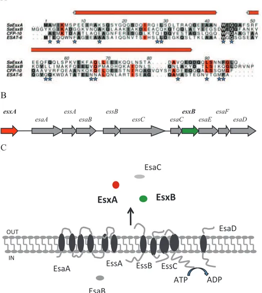
Fig. 2 Comparison of different gene clusters that encode Type-VII secretion system
S. aureus ESAT-6 secretion system. Protein sequence alignment of S. aureus EsxA
and EsxB with M. tuberculosis Esx proteins show that M. tuberculosis and S.
aureus EsxA display 20.8% identity and 25% similarity, whereas S. aureus EsxB
and M. tuberculosis EsxA are 17.8% identical and 35% similar. The peptide sequences of S. aureus EsxA and EsxB encompass the WXG motif, a signature sequence of ESAT-6-like proteins [51] (Fig. 3A). It appears that both EsxA and EsxB carry the C-terminus translocation signal, which means that each protein is transported and acts independently as a virulence factor with no requirement for heterodimerization. It may be significant that the C-terminus of EsxA adopts a helical conformation, a structural feature that may be important in the molecular recognition of EsxA by the transport machinery.
Unlike M. tuberculosis, S. aureus contains only a single Type-VII secretion sytem (Ess) gene cluster and EsxA and EsxB are encoded within a cluster comprised of eight predicted ORFs (esxA, esaA, essA, esaB, essB, essC, esxY, and esxB) as shown in Fig. 3B.
10
Missiakas et al., attributed a biological role to Esx proteins during staphylococcal infection and abscess formation. Mutants that failed to secrete EsxA and EsxB displayed defects in the pathogenesis of S. aureus murine abscesses, suggesting that this specialized secretion system may be a general strategy of human bacterial pathogenesis. Also they have shown that mutations in essA, essB, or essC, that are predicted encode for transmembrane proteins, abolished synthesis and therefore secretion of EsxA and EsxB without affecting transcription of esxA and esxB. Without secretion, EsxA and EsxB may be rapidly degraded in the bacterial cytoplasm or posttranscriptional feedback inhibition may reduce esxA and esxB expression [39]. Contrary to ESAT-6 and CFP-10 genes that are co-trascribed, altought esxA and esxB are located in the same locus, they are separated by 6 other genes and they are not co-trascribed. The esxA gene is under complex control. EsxA has been reported to be regulated by sigma factor B and σB-controlled SpoVG, and is a monocistronic transcript that is driven by a σA promoter. It was demonstrated that the transcription of esxA is controlled by a regulatory cascade involving downstream σB-dependent regulatory elements, including the staphylococcal accessory regulator SarA, the ArlRS two-component system and SpoVG. The regulation of EsxB is currently unknown [52].
A B esxA esaA essA esaB essC essB esaC esxB esaE esaF esaD C !"#$% !"#&% !"#'% !""&% !""'% !""$% &()% &*)%
!"#$%
!"#&%
+,(% -.% !"#*%Fig. 3 A Sequence alignment of S. aureus EsxA and EsxB with ESAT-6 and CFP-10 of M. tuberculosis. S. aureus EsxA shares 12% sequence identity with ESAT-6, 14% with CFP-10, and 13% with S. aureus EsxB. EsxB shares 8% sequence identity with ESAT-6 and 13% with CFP-10. B esx locus in S. aureus C Membrane topology of Ess secretion system in S. aureus
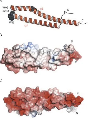
The crystal structures of EsxA was determined. The asymmetric unit of each crystal form is a dimer. The EsxA subunit forms an elongated cylindrical structure created from side-by-side α-helices linked with a hairpin bend formed by the WXG motif (Fig 4). Structural and sequence comparisons, exploiting biological data on
12
related proteins found in M. tuberculosis, suggest that this family of proteins may contribute to pathogenesis by transporting protein cargo through the secretion system exploiting a C-terminal signal that facilitate interactions with host receptor proteins[51].
A
B
C
Fig 4. Structure of EsxA
A A subunit of EsxA protein. The position of the conserved WXG motif is marked
and Trp43 is depicted as black Van der Waals spheres. also the N- and C-terminal region are marked. B The electrostatic surface of EsxA protein (blu, positive; red, negative; white, neutral) show the hydrophobic side of the protein. C Similar representation rotated 180°C show the hydrophilic side of the protein.
Secretion of EsxA and EsxB into culture medium during growth in vitro was demonstrated previously, with mutation of EsxB affecting secretion of EsxA and vice versa [39]. Very recent work further reports that deletions in either gene also
affects secretion of additional Ess substrates, EsxC and EsxD. EsxA and EsxB were reported to interact with different Ess substrates such as EsxC and EsxD respectively [53].
5. Staphylococcal Biofilms
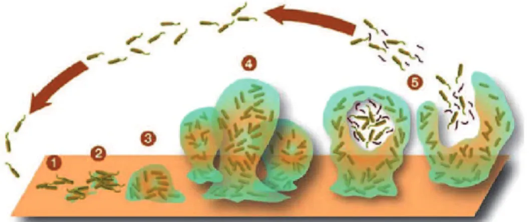
Staphylococci are recognized as the most frequent causes of biofilm-associated infections [54]. This exceptional status among biofilm-associated pathogens is due to the fact that staphylococci are frequent commensal bacteria on the human skin and mucous surfaces. S. epidermidis and S. aureus are the most frequent causes of nosocomial infections and infections on indwelling medical devices, which characteristically involve biofilms[55]. Often, S. aureus biofilm-associated infections are difficult to treat with antibiotics and devices need to be replaced [56]. In addition, they represent a reservoir for dissemination of infection to other sites in the human body. In contrast to many other medical biofilms, such as multi-species dental plaque formation, biofilm-associated infections with staphylococci are usually not mixed with other species . Research performed in many biofilm-forming organisms has revealed that the development of a biofilm is a multi step process involving an initial attachment and a subsequent maturation phase and a final dispersal phase that involves the detachment of the bacterial cells, believed to be crucial for the dissemination of the bacteria [54] (Fig 5).
Fig. 5 Schematic representation of development of a biofilm as a five-stage preocess. Stage1: Initial attachment of cells to a surface. Stage2: Production of
14
EPS resulting in more firmly adhered "irreversible"attachement. Stage3: Early development of biofilm architecture. Stage4: Maturation of biofilm architecture. Stage5: Dispersion of single cells from the biofilm.
In the human body, the attachment to human matrix proteins represents the first step of biofilm formation. S. aureus express dozens of so-called MSCRAMMs (microbial surface components recognizing adhesive matrix molecules) that have the capacity to bind to human matrix proteins such as fibrinogen or fibronectin, and often combine binding capacity for several different matrix proteins. The subsequently phase is the maturation of biofilm formation is characterized by 1) intercellular aggregation that can be accomplished by a variety of molecules such as adhesive proteins or – usually polysaccharide-based - exopolymers, and 2) biofilm structuring forces that lead to the typical 3-dimensional appearance of mature biofilms with a fluid-filled channels. Biofilm detachment is crucial for the dissemination of bacteria to other colonization sites. It may occur by the detachment of single cells or larger cell clusters. Several factors may contribute to detachment: 1) mechanical forces, 2) cessation of the production of biofilm building material and 3) detachment factors sensu strictu [57]. It has long been recognized that biofilms have dramatically increased resistance to antibiotics. Two main mechanisms contribute to biofilm resistance: 1) prevention of the antibacterial substance from reaching its target and 2) the specific physiology of a biofilm, which limits the efficacy of antibiotics, mainly of those that target active cell processes and which may also include specific subpopulations of resistant cells. Recent studies of staphylococcal biofilm development have demonstrated that there are some key structural and regulatory factors that determine the form and physiology of staphylococcal biofilms.
6. Intracellular life of Staphylococcus aureus
In addition to its armor of virulence factors, the capacity of S. aureus to successfully evade host defenses has been recently attributed to its ability to invade immune and non-immune cells. S. aureus is mainly an extracellular pathogen, but
an accumulating number of studies have shown that it can invade and replicate in many types of host cells in vitro [58].
This property potentially contributes to bacterial persistence and several benefits for the pathogen. It has been proposed that intracellular S. aureus evades exposure to antibiotics and host immunity [59]. Clinical studies have reported the presence of intracellular staphylococci from nasal epithelial cells, indicating that this could serve as a reservoir for recurrent infections[60]. The intracellular enviroment leads to the formation of small-colony variants (SCVs) characterized by slow growth and a range of morphological and metabolic changes including altered antibiotic resistance profiles [61, 62].
The intracellular process of S. aureus is mediated by the interaction between fibronectin binding protein and host-cell α5β1integrin [63]. Other bacterial surface proteins like clumping-factor A or host cell Src kinase also appear important in the mediation of S. aureus uptake and intracellular persistence [64]. After internalization, the behavior of the bacterium varies according to cell-line or bacterial strain. In order to subvert host cell functions to their benefit, bacterial pathogens have developed various strategies. In particular, intracellular bacteria have adapted mechanisms to modulate the apoptotic pathway. The resulting induction or inhibition of apoptosis is often crucial for a successful infection of the host. Several bacteria elicit an inflammatory process which, in the place of infection, leads to the disruption of tissue barriers and thus may secure efficient microbial spread in the host [65].
Some studies have reported an active intracellular bacterial replication within vacuoles. Others have described both a rapid bacterial escape from vacuole with a consequently induction of cellular apoptosis and persistence for several days before induction of escape processes[66].
In the case of S. aureus the affected apoptotic pathways appear to depend on the strain and host cell type. Several studies reported that after infection the Cell Death Program occurs trought caspases activation [67-69]. During S. aureus infection the
16
production of α-toxin appears correlated with the induction of apoptosis[70]. In contrast, inhibition of apoptosis may be essential for intracellular pathogens to establish chronic infection. Pathogen-triggered anti-apoptosis of infected host cells facilitates a slow microbial replication process and enables persistence in the infected host. S. aureus was recently reported to induce anti-apoptotic factors in epithelial cells. It seems, like for many other bacteria, S. aureus may also be able to block apoptosis [71].
7. New therapeutic and vaccine strategies for Staphylococcus aureus treatment
Mortality, morbidity, and cost from invasive S. aureus infections remain disturbingly high despite the introduction of several new antibiotics to treat methicillin-resistant S. aureus infections [72]. S. aureus infections are now the most common cause of hospitalization. To address these problems, investigators at universities, biotechnology companies, and large pharmaceutical companies have tried to develop an effective vaccine. Despite much effort, no clinical trials have succeeded to date. What has made S. aureus vaccine so difficult? First, the protective immunity against S. aureus is not completely understood and animal models, especially murine models, have not predicted success in humans as they did for the other successful vaccines. Second, the situation for S. aureus is much more complex, as this bacterium has multiple virulence factors that neutralize the host immune responses than these other bacterial pathogens. Third, S. aureus– infected patients present with a very broad range of diseases, which means that vaccine development must focus on preventing a wide spectrum of disease presentations. The need for an effective S. aureus vaccine is increasing in view of broad antibiotic resistance and apparent increasing virulence of the community strains[73, 74].
Careful consideration of the patient populations in which candidate vaccines are initially evaluated for their efficacy will also play a key role for vaccine development. So far the vaccines tested in clinical trials targeted a single S. aureus
component. Studies using antigen combinations have shown greater efficacy than single antigen vaccines in animal models. Therefore, multivalent vaccines will likely work better in humans as well so that it is able to cover different pathologies that S. aureus causes. On this basis, Bagnoli et al. have proposed a model in which vaccine efficacy is gained through three major immune responses: 1) antibodies to directly inhibit bacterial viability and/or toxicity 2) antibodies mediated opsonophagocytosis 3) cell-mediated immunity to stimulate recruitment of phagocytes at the site of the infection. A combination of staphylococcal antigens with different properties and functions is able to elicit a potent antibody production, and a proper cellular response. In that way is possible to cover different pathologies caused by S. aureus infections [75].
18
Outline of thesis
Staphylococcus aureus is a versatile pathogenic bacterium capable of rapidly developing or acquiring multiple antibiotic resistances, and is now recognized as a global major health-care associated problem. S. aureus is responsible for a wide spectrum of human diseases, ranging from skin infections to severe diseases, such as rhinosinusitis, otitis, arthritis, osteomyelitis, endocarditis or fatal sepsis.
These infections are difficult to eradicate and often relapse even after prolonged and adapted antibiotic therapy, suggesting that S. aureus has developed specific strategies for persistence such as the ability to live within biofilms or inside cells, and thereby evading antibiotics and immune responses. Due to the increasing antibiotic resistance, a new strategy for prevention the disease has become an important medical need.
During my PhD I have focused my studies on the functional characterization of two secreted proteins, EsxA and EsxB secreted by the specialyzed ESAT-6 like secretion system. These proteins are important for virulence in murine infection during kidney abscess formation and they are considered as potential vaccine candidates.
My research encompasses many areas including: 1) the genetic manipulation of S. aureus to understand the function of these proteins, 2) gene expression studies to explore environmental conditions under which the Esx proteins are induced, 3) the mechanisms invesigation of Ess-mediated secretion of EsxA and EsxB and finally 4) the functional characterization of Esx proteins during intracellular staphylococcal infection in in vitro cellular models.
Data from these studies will better characterize promising vaccine candidates and support development of an effective vaccine against S. aureus infection.
Generation and functional characterization of esxA and
esxB deletion mutants of Staphylococcus auresus
Giuliana Balsamo, Fabio Bagnoli, Davide Serruto and Meera Unnikrishnan
Unpublished Results
20
Introduction
Staphylococcus aureus (S. aureus) a Gram-positive coccus, is a major human pathogen causes a wide spectrum of nosocomial and community-acquired infections that are associated with high morbidity and mortality [1]. It causes infections ranging from superficial skin lesions to serious conditions like pneumonia and endocarditis. Systemic and chronic infections, in particular, place a major burden on healthcare systems worldwide [2, 3].
Treatment is made more difficult by the increasing number of MRSA, VRSA strains that are resistant to multiple antibiotics. One of the factors that contribute to staphylococcal resistance includes biofilm formation. Formation of biofilms or complex bacterial communities occurs in a multistep process and has been demonstrated to be important in staphylococcal virulence. Numerous reports in the past two decades have shown that especially biofilm-forming staphylococci cause a severe infections [4]. Biofilm formation by pathogenic staphylococci on implanted medical devices leads to “chronic polymer-associated infections”. Bacterial cells in a biofilm have been reported to alter metabolic activity [5, 6].
S. aureus pathogenicity is multifactorial and depends on the expression of a large class of gene products comprising cell-wall associated and extracellular proteins. These secreted virulence factors including enzymes and toxins are commonly exported by the Sec secretion system, a well-described secretion pathway [7]. An Ess secretion system similar to the Type-VII secretion system of M. tuberculosis was reported in S. aureus [8]. The ESAT-6 secretion system (Ess) consists of 12 proteins, including EsxA and EsxB that are similar to the ESAT-6 and CFP-10 of M. tuberculosis. ESAT-6 (EsxA) and CFP-10 (EsxB) are well-characterized virulence factors of M. tuberculosis [9].
The Esx substrates are typically small proteins (100 amino acids) with a conserved WXG motif. These WXG100 proteins are a class of effector molecules found in other Gram-positive bacteria. Bioinformatic analyses have shown that one WXG gene is frequently positioned near, or directly adjacent to a second related WXG
gene [10]. The identification of WXG proteins encoded by the pathogens M. tuberculosis and S. aureus has created significant interest in the proteins biological activity.
EsxA and EsxB factors, secreted by the system, are well conserved across clinical S. aureus strains and are currently being considered as potential vaccine candidates. Although well conserved between strains, expression and secretion of Ess-encoded substrates are poorly studied, both in vitro growth and during human staphylococcal infection. While the mycobacterial Esx proteins are important in several host-associated functions such as cell survival and granuloma formation[11, 12], the biological functions of the S. aureus Esx proteins are however not known and the mechanism of action during the pathogenesis is still unclear. Missiakas et al. have showed that EsxA, EsxB and other Ess proteins are important during kidney abscess formation in a murine infection and that some of the Ess proteins are important for bacterial persistence in a staphylococcal abscess [8, 13].
In M. tuberculosis the Esx substrates ESAT-6 and CFP-10 were shown to interact each other and form a 1:1 complex [14]. A striking feature of the complex is the long flexible arm formed by the C-terminus of CFP-10, which was found to be essential for the secretion [15] and for binding to the surface of cells strongly suggest a key signalling role for the complex, in which the binding to cell surface receptors leads to modulation of host cell behaviour to the advantage of the pathogen [14].
The structure of the S. aureus secretion apparatus or the mechanisms of Esx protein secretion are still unclear. The interactions between the various Ess components have not yet been mapped completely.
Sundaramoorthy et al. reported that EsxA and EsxB purified from E. coli do not form a complex. They obtained crystals of EsxA and there are no current evidence that the two proteins could interact each other [16]. Recent study shown that EsxB associates with a novel substrate, EsxD, and EsxA dimerizes with itself or EsxC (EsaC). In particular EsxA and EsxC form both homo- and heterodimers whereas
22
EsxB and EsxD appear to function as a heterodimer [17]. EsxD carries the C-terminal motif YxxxD/E that has been proposed to target Type VII substrates for secretion in mycobacteria [18]. Was shown that the deletion, in this motif prevent secretion of EsxA and EsxC but not EsxB or EsxD [19]. The genetic inactivation of esxA, esxB, esxC or esxD leads to loss of secretion of all four substrates. Moreover the deletion of esxD abrogated the production of EsxB altogether and affected the secretion, but not the production, of EsxA and EsxC.
Identifying all the bacterial interactors of the Esx proteins would help to understand how the secretion apparatus function. Futhermore, identification host proteins that may interact with these proteins could indicate a role in host-pathogen interactions. In this study genetic and biochemical approaches were used to characterize the Esx proteins of S. aureus in order to better understand their mechanisms of action during staphylococcal infection.
Material and methods
1. Bacterial RNA extraction and qRT-PCR
Over night (O/N) cultures were diluted (1/100) in 5ml of TSB and grown until specific OD600. Cells were sedimented by centrifugation, suspended in TSM (100
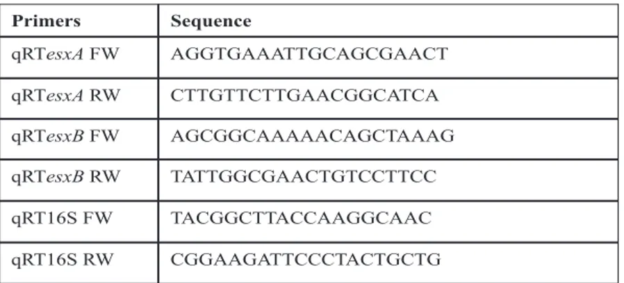
mM Tris·HCl, pH 7.0/500 mM sucrose/10 mM MgCl2) and digested with lysostaphin (5µg/ml) for 5 min. The resulting protoplasts were suspended with RLT buffer and RNA was isolated using the Qiagen RNeasy mini kits (Qiagen) using manufacturer’s protocol. For intracellular bacterial RNA extraction, after infection of cells total RNA was extracted with TRIzol (1ml/well) (Life Technologies) and purified using Zymo Quick RNA purification Kit (The Epigenetics Company). Followed by on column DNase I treatment (Qiagen) and then DNase I treatment in solution (Roche). Reverse transcription was performed using Superscript II (Invitrogen). Real-time PCR was done using TaqPlatinum Syber Green polymerase SuperMix UDG (Invitrogen). A relative quantification was performed using 16S rRNA as internal control. Primers used for qRT-PCR are listed in the table 1.
Primers Sequence qRTesxA FW AGGTGAAATTGCAGCGAACT qRTesxA RW CTTGTTCTTGAACGGCATCA qRTesxB FW AGCGGCAAAAACAGCTAAAG qRTesxB RW TATTGGCGAACTGTCCTTCC qRT16S FW TACGGCTTACCAAGGCAAC qRT16S RW CGGAAGATTCCCTACTGCTG !
24
2. Construction of bacterial mutants
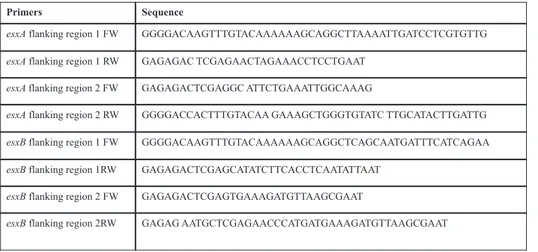
For deletion of esxA and esxB a 2-kbp DNA fragments flanking the esx genes were amplified by PCR and cloned into the Escherichia coli/S. aureus shuttle/suicide vector pKOR1 [20] with abutted XhoI restriction sites and att site using primers listed in the table 2. Constructs containing flanking genomic regions of esxA and esxB were cloned first in E. coli then into RN4220 (restriction negative, methylation positive strain). Plasmid DNA extracted was used to transform S. aureus USA300 MRSA. Integration of plasmid into the chromosome was obtained at non permissive conditions for pKOR1 replication (incubation twice at 43°C in TSB) and the selection for homologous recombination and pKOR1 integration into the bacterial chromosome was induced by growing bacteria at permissive temperature (incubation twice 2 at 30°C in TSB). Anhydrotetracycline-mediated induction of pKOR1- encoded secY antisense RNA which inhibits growth was used for selecting for chromosomal excision and loss of plasmid at 30°C.
Primers Sequence
esxA flanking region 1 FW GGGGACAAGTTTGTACAAAAAAGCAGGCTTAAAATTGATCCTCGTGTTG
esxA flanking region 1 RW GAGAGAC TCGAGAACTAGAAACCTCCTGAAT
esxA flanking region 2 FW GAGAGACTCGAGGC ATTCTGAAATTGGCAAAG
esxA flanking region 2 RW GGGGACCACTTTGTACAA GAAAGCTGGGTGTATC TTGCATACTTGATTG
esxB flanking region 1 FW GGGGACAAGTTTGTACAAAAAAGCAGGCTCAGCAATGATTTCATCAGAA
esxB flanking region 1RW GAGAGACTCGAGCATATCTTCACCTCAATATTAAT esxB flanking region 2 FW GAGAGACTCGAGTGAAAGATGTTAAGCGAAT
esxB flanking region 2RW GAGAG AATGCTCGAGAACCCATGATGAAAGATGTTAAGCGAAT
Table 2. Sequence of primers for knoching out esxA and esxB gene.
For complementation of mutant strains, the full length esxA and esxB genes were amplified using specific primers for the esx genes and cloned into plasmid pOS1CK, which was generated by cloning the P1 constitutive promoter of the sarA
gene into the pOS1 plasmid. For complementing with both genes, esxB was cloned downstream of esxA in the pOS1CKesxA construct. For site direct mutagenesis of esxA the leucine, serine double mutations were introduced into the esxA gene cloned into a pET vector by PIPE [21]. To create the mutations in glycine95 we used primers carrying the mutated amino acid sequence. esxA truncated gene was amplified using primers listed in the table 3. All mutant forms were cloned into the episomal plasmid pOS1CK.
Primers Sequence
esxALSGFW: GCGCTGCAGTT
GAGAGGAGAGAAAATGGCAATGATTAAGATG
esxALSGRW: GCGCCCGGTTATTGCAAAC CGAAATTAT
esxAtruncFW GCGCTGCAGCCGCTCGAGATGGCAATGATTAAG
esxAtruncRW TCCCCCGGGTTGTTGGTCTTGTTC
Table 3: Sequence of primers for esxA mutation
3. Tandem Affinity Purification (TAP)-Tagging and Pull Downs
esxA/B gene was cloned in-frame with the two tags: FLAG protein and the Streptavidine Binding Peptide (SBP) at N- and C-terminus into episomal plasmid pAM401. ∆esxA or ∆esxB, were transformed with the plasmid. As negative control the same strains were transformed with empty plasmid. After growing the bacteria until late log phase, the total extracts were prepared using buffer containing lysostaphin 5ug/ml, 10mM HEPES, 150 mM NaCl pH 7.5, 0.4% NP40 and complete protease inhibitor. The affinity pull down was performed as described in Schluepen et al. article [22]. After binding of the EsxA-SBP-FLAG to the streptavidine beads the sample were washed using stringent solution and after the elution step performed with biotin, the purified complex was subjected to SDS-PAGE. The sample was trypsin-digested and analyzed by LC-MS/MS in PPI group
26
in Novartis. Anti-FLAG immunoblotting was used to verify the presence of the flagged EsxA protein.
4. Preparation and analysis of bacterial fractions
Bacterial lysates and supernatants were prepared as modification of described previously [23]. Bacterial strains were grown O/N from glycerol stocks in TSB at 37°C with shaking. Cultures were diluted 1:100 in fresh broth and shaken at 37°C until they reached an OD600 1. For whole culture lysates, 5ml of cultures were
centrifuged, washed once with PBS and suspended 1ml di Tris-HCl pH 6.8 with protease inhibitors and incubated in the presence of lysostaphin (Sigma) at a final concentration of 100µg/ml for 1h at 37°C at 750 rpm, followed by freezing/thawing three times in a dry ice/thermomixer at 37°C. The bacterial lysates were centrifuged at 14,000 rpm for 10 min, the supernatants were filtered and the proteins in the lysates were precipitated with TCA at final concentration of 10%.
5. Biofilm formation in vitro
To reproduce biofilm in vitro, bacteria grown in a rich medium BHI supplemented with 1% glucose and 3% NaCl at different time points: 6, 12, 24, 48 and 72h at 37°C and in 5% of CO2 rich environment without shaking in a 24 well plate.
Biofilm formation was made visible by staining S. aureus cells with 0.2% of crystal violet for 15 minutes. After three washes biofilm was quantified by measuring OD at540nm.
Results
1. Gene expression analysis for esxA and esxB
1.2 Quantitative Real Time PCR in different in vitro conditions
In order to better identify the molecular mechanisms of action of esxA and esxB it was necessary first define the conditions in which esxA and esxB are expressed. Previous studies involving the analysis of expression of virulence factors were mainly performed in vitro during growth in rich medium [24]. However, when S. aureus infects a host, the bacterial growth conditions are quite different from those in a medium, which may be related to the different expression of virulence factors in the host [24]. It has been previously reported that stress conditions, such as growth in presence of serum, CO2, anaerobic conditions and biofilm are
responsible for the induction of different virulence factors [24].
In this study we investigated the expression of esxA and esxB during different phases of growth, in presence of serum, during biofilm formation and within eukaryotic cells after in vitro infection using quantitative real-time PCR.
We compared the expression of esxA and esxB in two relevant clinical isolate Newman a meticillin sensitive strain (MRSS) and USA300 a meticillin resistent strain (MRSA) during growth in rich medium Tryptic Soy Broth (TSB). The result obtained shown an increase of transcript levels for both genes in USA300 in comparison to Newman strain (Fig. 1A).
Then we investigated the expression kinetics of the esxA and esxB genes. We extracted the bacteria RNA at early, late and stationary phase. The data, normalized to expression during early phase showed that esxA is expressed more during the late phase of growth (Fig. 1B).
Since is known from literature that many virulence factors, are significantly increased during growth in serum compared to that in bacterial medium, we investigated the same hypotesis for esxA and esxB expression. We quantified the level of transcript of esxA and esxB during growth in DMEM medium
28
supplemented with different concentration of Fetal Bovine Serum (FBS) 2%, 10% and 20%. The bacterial RNA was extracted at late log phase and the data were normalized to transcript levels measured during growth in DMEM medium. The result showed that the transcript levels of esxA and esxB decrease in medium supplemented by 2-20% of serum (Fig. 1C). The biofilm-forming ability is an attribute of several pathogens. In particular S. aureus is one of the most frequent cause of biofilm-related infections [4]. A comparative S. aureus global gene expression transcriptome analysis has shown several genes differentially expressed under biofilm formation in comparison to planktonically growth[6]. In order to test if our genes could be expressed in biofilm, we set up an in vitro biofilm assay where the bacteria grow in a rich media Brain Heart Infusion broth (BHI) supplemented with 1% of glucose and 3% of NaCl. We detected an increase in expression of esxB gene after 24h of biofilm growth in static condition compared to planktonic growth after 24h of incubation at 37°C (Fig. 1D).
A transcriptome analysis by Garzoni et al. shows an extensive alteration of bacterial gene expression following internalization of epithelial cells [25]. These findings suggest that S. aureus extensively reprograms its transcriptome to adjust the intracellular environment. As the mycobacterial Esx proteins are known to function intracellularly [12], we wanted to investigate if esxA and esxB are differentially expressed when S. aureus is within eukaryotic cells. In order to study this, we set up an in vitro infection assay where A549 human lung epithelial cells were infected with USA300 strain (as described in Methods of the fourth chapter of the thesis). RNA was extracted from intracellular bacteria 6h after infection (p.i.). Quantitative real time PCR analysis for esxA and esxB showed that both genes, in particular esxB, are up-regulated upon internalization when normalized to expression in the infection medium, DMEM with 10% FBS (Fig. 1E).
A 0 1 2 3 4 5 Newman USA300 F ol d Inc re a s e esxA esxB * B 0 0.5 1 1.5 2 2.5
Early phase Late phase Stat phase
F ol d Inc re as e esxA esxB *
30 C 0 0.2 0.4 0.6 0.8 1 1.2 DMEM DMEM + 2% FBS DMEM + 10% FBS DMEM + 20% FBS F ol d Inc re a s e esxA esxB D 0 1 2 3 4 5 6 7 8 9 10 Planktonic Biofilm F ol d Inc re a s e esxA esxB * E 0 2 4 6 8 10 12 DMEM+10%FBS 6h p.i. F ol d Inc re as e esxA esxB *
Fig. 1 Gene expression analysis for esxA and esxB genes by quantitative real-time PCR (qRT-PCR).
A Expression analysis of esxA and esxB genes in different strains Newman and
USA300 and B in different growth of phases during growth in TSB medium: early, late and stationary. C esxA and esxB transcripts level from DMEM plus fetal bovine serum. D Expression during biofilm formation and E upon internalization of epithelial cells 6h post infection. A relative quantification was performed using 16S rRNA as internal control. * indicates significant differences as compared with reference control condition (P< 0.05). The data presented are the mean of 3 independent experiments +/- s.d. in all the experiment. Data shown in C are representative of 2 independent experiments.
32
2. Generation and characterization of esx mutants 2.1 Generation of deletion mutants
In order to characterize the functions of EsxA and EsxB we made unmarked, single deletion mutants for esxA and esxB genes in two relevant clinical isolates of S. aureus, Newman (MSSA) and USA300 (MRSA) strains. To create deletion of these genes in the genome of S. aureus we used pKOR1, an Escherichia coli/S. aureus shuttle/ suicide vector that permits rapid cloning via lambda recombination and ccdB selection (Fig 2A). A method for rapid selection of allelic replacement mutations in the chromosome of S. aureus was described by Bae et al. [20]. Staphylococci were transformed and grown at 43°C, a non-permissive condition for pKOR1 replication that allows the selection for homologous recombination and plasmid integration into the bacterial chromosome. Anhydrotetracycline-mediated induction of pKOR1-encoded secY antisense transcript via the Pxyl/tetO promoter, a condition that is not compatible with staphylococcal growth, selects for chromosomal excision and loss of plasmid. Using this strategy, allelic replacements in S. aureus were generated at frequencies that obviated the need for antibiotic marker selection (Fig. 2B).
In order to characterize the function of both proteins, double mutant for esxA and esxB was generated in both strains. ∆esxA genetic background was used to make deletion in esxB gene. ∆esxA competent cells were transformed using the same strategy describe before.
A B
1Kb
esxA/B
attB1-esxA/B-FW attB2-esxA/B-RW
1Kb
Fig. 2 pKOR1 allelic replacement generates staphylococcal chromosomal deletions
A pKOR1 vector map. B Chromosomal map of esxA/B gene and the location of
PCR primers to make esx deletion constructs. The location of primers (attB1-esxAB-FW and attB2- esxAB-RW) are shown as short black arrows.
The mutants were confirmed by PCR using two different sets of primers: one annealing to the flanking region of esxA or esxB gene and the other specific for each gene. As shown in Fig 3A we verified the different size of fragment around the esx gene for mutant (2Kb) compared to WT (2.5Kb) and the lack of esxA and esxB (Fig. 3A). The lack of the protein was verified by western blot analysis (Fig. 3B). The deletion was further confirmed by qRT-PCR and sequencing.
34
A B
Fig. 3 Genetical and biochemical analysis of esxA, esxB and esxAB deletion mutants
A PCR was performed on chromosomal DNA from WT and ∆esxA, ∆esxB or
∆esxAB mutants using primers specific for flanking region of esx gene or binding to the esx genes. PCR products were analyzed on 0.8% agarose gel. The expected size for PCR products from genomic DNA obtained from the WT or mutant strains is 2.5 or 2 Kb, respectively. B Western blot analysis anti-EsxA on total extract of WT and esx mutants generated in two different strains USA300 and Newman.
3. Characterization of esx mutant strains during biofilm formation 3.1 Role of Esx proteins in staphylococcal biofilm
It is reported in a previous transcriptome analysis on S. aureus biofilms that several virulence factors are differentially expressed during biofilm formation [4] and our gene expression analysis showed an up-regulation of esxB gene in biofilm. Hence we wondered if these two small proteins could have a role during biofilm development. We investigated the behavior of the esx deletion mutants in an in vitro biofilm assay. We found that, although the mutants have no growth defects in BHI medium (Fig. 4A), a reproducible 1.5-2 fold increase in biofilm formation for the esxB deletion mutant as compared to WT strain at later time point (Fig. 4B, C). No differences were detected at early time points 6 and 12h after incubation.
Our hypothesis is that EsxB protein could play a role at later steps of biofilm development, i.e during the dispersal phase of staphylococcal biofilms. It could be directly involved in biofilm detachment by affecting cell-cell interactions or it could act to destroy the biofilm matrix. It is also possible that EsxB plays indirect role acting as a transcriptional regulator (up-regulates proteases, adhesins etc.), or by controlling post-translationally the secretion of proteins that may be involved in biofilm formation.
36 A B C 0 0.2 0.4 0.6 0.8 1 1.2 1.4 WT DesxA DesxB O D 540 nm (10X ) Newman USA300 * *
Fig. 4 esx mutants do not display any in vitro growth defects in media tested; increase of biofilm formation for esxB and esxAB at later time points
A Growth curves of WT and the various esx mutants in Brain Heart Infusion +
1%glucose, 3%NaCl at 37°C with shaking B Image showing biofilm formed by WT Newman and USA300 S. aureus strains and the various esx mutants after 24h in BHI containing NaCl and glucose at 37°C in 5% CO2 and stained with 0.2% crystal violet C A reproducible 2-fold increase in biofilm accumulation was observed for the ∆esxB mutants as compared with the WT at later time points. * indicates significant differences as compared with WT (P< 0.05). The data are representative of 3 independent experiments.
3.2 Transcriptome analysis of esx mutants
The mycobacterial ESX-1 substrate EspR is known to regulate the expression on other esx genes [26]. It is possible that the staphylococcal Esx proteins may have regulatory roles, which may mediate the effects observed above in biofilm formation. In order to investigate if EsxA and EsxB have a role in regulating the expression of other S. aureus factors, we performed a comparative gene expression analysis between WT Newman strain and various esx deletion mutants using S. aureus oligonucleotide microarrays.
Microarrays were first validated using various genomic DNA and RNA controls. Gene expression analysis showed no significant differences in the expression profiles of WT and esx mutant during growth in BHI.
38
4. Characterization of the Esx proteins secretion mechanisms 4.1 Interactions between Staphylococcus aureus Esx components
Interactions between staphylococcal Esx components and mechanisms by which they mediate secretion of substrates are not clear, very recent work further reports that deletions in either gene also affects secretion of additional Ess substrates [19]. The homologues mycobacterium Esx proteins ESAT-6 and CFP-10 was shown to be secreted as an heterodimer and function only when interacted each other and with other ESX-1 components. In order to investigate the proteins that interact with EsxA and EsxB, we used in vitro pull down approach. We employed a Tandem Affinity Purification (TAP) method in combination with mass spectrometry to identify possible interactors of EsxA and EsxB. This method is based on the sequential utilization of two different affinity tags to purify protein assemblies [22]. esxA and esxB genes were cloned in frame with a dual tag: a streptavidin binding protein plus a FLAG-peptide epitope at either the N- or the C-terminus of the proteins, and expressed in ∆esxA and ∆esxB (Fig 5A). Eluates from the respective affinity purifications were analysed by western blot showing only the C termed FLAG tag EsxA protein (Fig 5B, C). EsxB- FLAG was not detected probably due to the probable tag cleavage. However when we analysed the eluates by Mass Spectrometry analysis we did not identify any S. aureus proteins that directly bound to EsxA. This might be because the conditions under which the pull downs were performed were not suitable to allow stable interactions. Also, it is possible that the presence of a FLAG tag in these small proteins could interfere with the interactors binding to the proteins.
A
B
Supernatant of S. aureus /cell lysate Expression of the
Tap-tagged protein in cells
First affinity column Streptavidin affinity
column
Second affinity column Incubation with
anti-FLAG
Native elution of the complex
C
Fig. 5 TAP-tag Pull down
A Schematic representation of TAP-tagged esx gene constructs. B Schematic
representations of TAP tag pull down of interacting proteins. C Western blot analysis to detect FLAG-tagged protein. A: unbound fraction B: eluate fraction after pulling down total lysate with anti-FLAG antibody Lines 1 and 3: ∆esxA expressing EsxA-FLAG protein. Lines 2 and 4: negative control, ∆esxA-empty plasmid.
40
4.2 Secretion of EsxA
The homologues mycobacterial EsxA and EsxB proteins are secreted thought the Esx secretion system with non–classical signal sequence. As the signal sequences controlling S. aureus Esx protein secretion are not known, we wanted to determine the residues that are required for secretion of the protein in vitro. Based on the homology between M. tuberculosis and S. aureus Esx proteins, we identified amino acids (a.a.) conserved between the proteins at the C-terminus: leucine90, serine91 and glycine95 (Fig. 6A). EsxA bearing mutations in 3 a.a. or with a C-terminal 8 a.a. deletion were expressed episomally in the ∆esxA mutant to create strains ∆esxApOS1CKesxA-LSG esxALSG) and ∆esxApOS1CKesxAtrunc (∆esxA-esxAT) respectively. Immunoblotting analysis showed that the triple mutation in EsxA reduced the secretion of the protein into the culture supernatant while truncation of the C-terminal tail of EsxA severely impaired secretion of the protein compared with secretion of native EsxA (Fig. 6B, C).
A B C 0 20 40 60 80 100 120 pOS1empty pOSesxA native pOSesxA LSG pOSesxA trunc %of E sxA s e c re ti on
pOS1 pOS esxA pOS esxALSG pOS esxAtrunc EsxA 11kDa Culture Supernatant Bacterial Extract Hla 38 kDa pOS1 pOS esxA
pOS esxALSG
pOS esxAtrunc
Fig. 6 C-terminal residues are important for secretion of EsxA
A Alignment of amino acid sequence of M. tuberculosis and S. aureus EsxA and
EsxB proteins. In red are shown the high conserved residues that were replaced with alanine and the blue line indicates the 8 a.a deleted in the truncated EsxA protein. B Immunoblotting analysis of total extracts (TE) and supernatants (SN). Proteins in each fraction were precipitated with TCA, separated on SDS-PAGE, and detected by immunoblotting with anti-EsxA and anti-Hemolysin (loading control). Loading was normalized by OD600 of bacterial culture. C The graph
42
Discussion
Little is known about the biological functions of EsxA and EsxB during Staphylococcus aureus pathogenesis. Missiakas's group demonstrated that the transposon mutants defective in EsxA and EsxB secretion show decreased virulence in an abscess model of staphylococcal infection indicating a key role of these two proteins in a kidney abscess formation [8]. The high conservation among the S. aureus clinical strains suggests their importance as virulence factors. Although the mechanisms of action of the two ortholologous proteins in Mycobacterium tuberculosis ESAT-6 (EsxA) and CFP-10 (EsxB) are very well characterized, the biological functions of the S. aureus counterparts during infection and the mechanisms of the secretion are not well defined.
In this study we have analyzed different aspects of EsxA and EsxB proteins including expression, secretion and role during in vitro biofilm formation.
A gene expression analysis, performed in different in vitro condition, showed that esxB is induced under selective conditions such as biofilm, and repressed in presence of serum. Both genes are more expressed in MRSA USA300 compared to MSSA Newman strain indicating differencies in regulation of the two proteins in diverse clinical strains. Expression analysis during in vitro infection of epithelial cells show and up-regulation 6h post infection suggesting that esxA and esxB could play a role in S. aureus intracellular infection.
The development of a biofilm is a multistep process involving an initial attachment to human matrix proteins, a subsequent maturation phase characterized by intercellular aggregation and a final dispersal phase that involves the detachment of single cell or cell clusters by various mechanisms [4]. We found that esxB is significantly up-regulated in bacteria within biofilms. Consequently, employing isogenic mutants we observed that ∆esxB accumulated more biofilm in vitro at later
time point. In order to understand if EsxB had a direct role in biofilm formation, we analysed the differences in gene expression profile between WT and mutant strains. We observed that none of other staphylococcal genes were significantly differential expressed in the esxB mutant strain, indicating a potential direct role of this protein in biofilm. We still do not understand the precise role of EsxB in biofilms. However our hypothesis is that EsxB may be important in dissemination of biofilms so could play a role in the dispersal of staphylococcal biofilms being directly involved in biofilm detachment by affecting cell-cell interactions. It could act to destroy the biofilm matrix or it is also possible that EsxB has post-translationally control on the secretion of proteins that may be involved in biofilm development.
Interaction between the various components of the apparatus of secretion was also analyzed. While the mycobacterial EsxA and EsxB function as a heterodimer [15], the staphylococcal EsxA was crystallized as a homodimer, and there is no current evidence suggesting that the two proteins interact [16]. Unlike ESAT-6 and CFP-10, EsxA and EsxB do not interact each other. In a very recent work using a biochemical approach to detect protein-protein interaction, EsxA and EsxB of S. aureus were reported to interact with different Ess substrates such as EsxC and EsxD respectivel[19]. We employed a Tandem Affinity Purification method (TAP) to identify any other interactors of S. aureus. It combines affinity purification and mass spectrometry analysis to identify the interactors. However our experiments performed on the total lysate of S. aureus did not reveal any interactions between Esx proteins. Hence as discussed earlier, the conditions used and the presence of tags on the proteins may have interfered with our analysis.
Staphylococcal EsxA and EsxB are small secreted proteins that lack a canonical topogenic sequence. The secretion of EsxA and EsxB into culture medium during growth in vitro was previously demonstrated [8]. C-terminal signals were reported previously to mediate secretion of CFP-10 and other Esx locus encoded substrates
44
secreted by mycobacterial ESX-1 system [15]. We show that the C-terminus of EsxA, that contains residues conserved in the Esx substrates, is required for secretion in vitro. While single or multiple changes in amino acids did not appear to affect secretion, a truncation of the C-terminus resulted in a significant decrease in secretion, as reported recently for secretion of another Ess substrate-EsxD [19]. This indicates that the C-terminus may control secretion by mediating interactions with other proteins.