UNIVERSITÀ DEGLI STUDI DI ROMA
"TOR VERGATA"
FACOLTA' DI MEDICINA e CHIRUGIA
DOTTORATO DI RICERCA IN FISIOPATOLOGIA SPERIMENTALE
CICLO DEL CORSO DI DOTTORATO XXII
Postoperative Crohn's Disease recurrence as assessed by conventional vs alternative non invasive techniques
Sara Onali
A.A. 2009/2010 Docente Guida/Tutor: Prof.ssa Livia Biancone Coordinatore: Prof. Francesco Pallone
INDEX
Background pag. 3
Aims pag. 7
Materials and Methods
Prospective study pag. 8
Retrospective study pag.22
Results
Prospective study pag. 13
Retrospective study pag. 25
Discussion pag. 30
References pag. 40
BACKGROUND
Postoperative recurrence after ileo-colonic resection is a feature of Crohn’s Disease (CD). Ileocolonoscopy currently represents the gold standard for assessing CD recurrence, graded according to the Rutgeerts’ score (1-2). Endoscopic recurrence after curative ileo-colonic resection is observed in almost 73% of patients at 1 year and in almost 90% of patients at 3 years, even in the absence of overt symptoms (1-3). Severe endoscopic recurrence (2) is observed in about one/third of patients at 3 months and in almost two/thirds of patients at 6 months after surgery (1-4). The severity of the lesions as assessed by endoscopy has been shown to predict clinical relapse, and the frequency of symptomatic recurrence parallels the follow up length (5-7). Surgical reintervention is indeed required in about 15-40% of CD patients at 10 years and in about 50-70% of patients at 20 years after initial resection (5-6). Beside the severity of endoscopic lesions after resection, risk factors for a more aggressive course after surgery include active smoking, especially in women, disease extent and a fistulizing pattern (5-9).
Identification of the subgroup of patients undergoing early symptomatic CD recurrence may be useful for a timely treatment and
procedures alternative to ileocolonoscopy may well be of use for assessing CD recurrence. Small bowel follow-through (SBFT) and enteroclysis have been proposed (10,11), although providing a high radiation exposure to the patient. These 2 techniques also show a low sensitivity in terms of visualization of minor lesions related to CD recurrence, even in experienced hands (10,11).
More recently, small intestine contrast ultrasonography (SICUS) has been proposed for detecting small bowel lesions in patients with suspected or known CD (>95%)(12-14). SICUS findings compatible with CD recurrence include an increased bowel wall thickness (BWT), thus providing the view of the extraluminal small bowel lesions. In a prospective longitudinal study we recently reported that SICUS may represent an alternative non-invasive technique useful for assessing CD recurrence after ileo-colonic resection (15,16). However, by our knowledge, no studies compared findings compatible with CD recurrence as assessed by techniques visualizing either the inner or the extraluminal small bowel surface. The possible clinical usefulness of procedures providing different views of the peri-anastomotic area after ileo-colonic resection, as also the natural history of the extraluminal lesions (i.e. BWT) associated with CD recurrence is undefined.
The outcome of CD patients after “curative” resection different from the ileo-colonic, is less well defined. The prevalence pattern and the site of
CD lesions has been reported to be comparable before and after ileo-colonic resection (4). Several studies have looked for the potential role of risk factors for CD recurrence after ileo-colonic resection, but only active smoking, especially in women and location of disease (ileocolitis) appears significant risk factors (6-9). “In vivo” studies showed that the fecal stream is required for the development of recurrence after ileo-colonic resection (17), almost invariably including neo-terminal ileum (3). In CD patients with curative resection different from the ileo-colonic, recurrence may involve other segments of the gastrointestinal (GI) tract (3). However, very few studies investigated the natural history of recurrence after curative resection for CD involving GI segments different from the ileo-colon. In a small retrospective study, 92% of patients with jejuno-colonic anastomosis showed endoscopic recurrence, suggesting that the proximity to colonic contents also predisposes to recurrence (18). The few retrospective studies investigating this issue suggest that the recurrence rate in CD patients with colo-colonic anastomosis is lower than in patients with ileo-colonic anastomosis, while the natural history of jejunal CD after surgery appears to run a more aggressive course when compared with patients resected for ileo-cecal CD (19-20).
As upper GI lesions related to CD appears more frequently in younger patients (20) and a lower age at disease onset is being observed during the last few years (19), the knowledge of the natural
history of CD after “curative” resection of the upper small intestine (jejunum or proximal ileum) may add clues for proper indication for surgery, including timing and type of surgical approach
AIMS
1. Prospective Study
We aimed to compare, in a prospective longitudinal study, findings related to CD recurrence as assessed by procedures visualizing either the luminal surface (i.e. ileocolonoscopy or SBFT) or the extraluminal surface (SICUS) in a cohort of CD patients prospectively followed up at 1, 2 and 3 years after ileo-colonic resection. (20)
2. Retrospective Study
In an different cohort of CD patients, we also aimed to assess, in retrospective analysis, the frequency, pattern and risk factors of postoperative recurrence in CD patients with “curative” resection different from ileo-colon. (21)
1. PROSPECTIVE STUDY: MATERIALS AND METHODS
Patients. In a prospective longitudinal study, 25 patients undergoing
elective ileo-colonic resection for CD (12 M , median age 35 range 16-69 years) were consecutively enrolled from July 2003 to July 2005, and followed up for 3 years. All patients were under regular follow up at the GI Unit of the Università “Tor Vergata” of Rome, Italy, and resected by the same surgical Unit. The diagnosis of CD was made according to conventional criteria (2).
Before surgery, disease assessment was performed within 6 months by using ileocolonoscopy in all 25 patients and also by SBFT in 16 out of the 25 patients.
Clinical characteristics of each patient are summarized in Table 1. Risk factors for recurrence and other characteristics were recorded, including smoking habits (yes: n=10; no: n=12; ex: n=3), familial history of IBD (yes: n=5; no: n=20), disease site (ileum alone: n=17; ileum-colon: n=8), age at diagnosis of CD (median 28; range 15-49), disease duration (median 6 years; range 0.5-29), presence of typical CD granuloma at histological examination of the surgical specimen (yes: n=1; no: n=24). Inclusion criteria included: CD patients under regular follow up undergoing elective ileo-colonic resection, age ranging from 15 to 70 years, surgical resection including all the involved tissue, written informed consent. Exclusion criteria: Patients with relevant co-morbidities
(renal, cardiovascular diseases), body mass index (BMI) >30 (not allowing a proper ultrasonographic assessment), low compliance.
Study protocol
From July 2003 to July 2005, all eligible CD patients with no complications after ileo-colonic resection, fulfilling the above reported inclusion criteria were prospectively enrolled. Reasons for drop out were reported. After surgery, all patients were treated with mesalamine (2.4 gr/day) within 14 days from resection. Treatment changes during the follow up were made according to current clinical criteria (2).
Clinical activity was assessed according to the Crohn’s Disease Activity Index (CDAI)(24) every 6 months for 3 years after surgery.
At 12 months, all patients underwent clinical assessment (CDAI) and recurrence was assessed by using ileocolonoscopy as a gold standard (2), followed by SICUS. The two procedures were performed by 2 independent gastroenterologists, unaware of previous findings. At 24 months, patients in follow up underwent clinical assessment (CDAI), and lesions compatible with CD recurrence were evaluated by using SICUS and SBFT. At 36 months, patients underwent clinical assessment (CDAI) and lesions compatible with CD recurrence were assessed by using ileocolonoscopy and SICUS.
Clinical assessment
Clinical assessment (CDAI) was performed every 6 months for 3 years, together with routine blood tests.
Ileocolonoscopy
Endoscopical assessment of recurrence was made at 1 and 3 years, and the severity of recurrence assessed according to the Rutgeerts’ score (0-4)(1,3)(Figure 1, panel a). Endoscopic findings were documented in all patients by photographic verification. Ileocolonoscopy was performed with or without sedation (ipnovel i.v.), according to patients’ request. During each endoscopy, biopsies were taken from the involved and uninvolved areas, for routine histologic assessment.
Small bowel follow through
Small bowel radiographic examination was made according to standard criteria 2 years after surgery, and lesions compatible with recurrence graded from 1 to 4, according to Hanauer et al. (18)(grade 1=Normal; grade 2= Mucosal edema/aphthoid ulcers;grade 3= Linear ulcers/cobblestoning; grade 4=Strictures/fistulas/inflammatory mass)(Figure 1, panel b)(25).
SICUS
Ultrasound examination was performed after the ingestion of 375 ml (range 250-500 ml) of oral contrast solution consisting of Polyethylen glycole (PEG) (Promefarm, Milano, Italy), by using with 3.5 MHz and 5-MHz convex and linear-array transducers.(Hitachi, EUB 6500, Japan), as described (13). All procedures were performed by the same expert gastroenterologist (>2000 examinations).
The following findings were considered compatible with CD recurrence (21-23): 1. increased BWT (>3 mm); 2. “stiff loop”, identified by the presence of small bowel loop, with increased bowel wall thickness not distended by contrast solution; 3. small bowel dilation, defined as a lumen diameter >2.5 cm; 4. bowel stricture defined as lumen diameter <1 cm, measured at the level of maximally distended loop, independently of the presence of pre-stenotic dilation; 5. fistulae defined as hypoechoic tract with or without hyperechoic content; 7. mesenteric enlargement and/or masses; 8. abscesses identified as roundish anechoic lesions, with an irregular wall, often presenting internal echoes and posterior echo enhancement.
Histological assessment
During endoscopy, biopsies were taken from the peri-anastomotic area for routine histological assessment and for the search of granuloma. All biopsy specimens were assessed by the same anatomopathologist.
Statistical analysis
Data were expressed as median and range, both in the text and in the Tables. Differences between group were assessed by the paired or unpaired Student’s t test. Correlation between quantitative scores by using different procedures was assessed by the Spearman correlation test.
1. PROSPECTIVE STUDY: RESULTS
Recurrence at 1 year
At 1 year, all the 25 patients enrolled completed the follow-up, including clinical, endoscopical and sonographic assessment. As shown in Table 2, Clinical recurrence (CDAI>150) was observed in 1 out of the 25 patients (4%), followed by steroid-induced remission (tapered from prednisone 1 mg/Kg).
Endoscopic recurrence at 1 year was detected in 24 out of 25 patients (96%). The severity of recurrence as assessed by the Rutgeerts’ score was of grade 0 in 1 (4%), grade 1 in 6 (24%), grade 2 in 6 (24%), grade 3 in 5 (20%) and grade 4 in 7 (28%) patients (associated with stenosis not passed by the endoscope in 2). At 1 year, SICUS detected lesions compatible with CD recurrence in all 25 patients (100%), associated with stricture with no bowel dilation in 10 (40%). SICUS findings compatible with recurrence included an increased BWT (median 5 mm; range 3.5-10 mm). No additional sonographic findings not related to CD recurrence were observed.
In the only patient with no endoscopic recurrence (grade 0), the BWT was at limit of normal values (3.5 mm). The median BWT in the subgroups of patients with different endoscopic scores of recurrence is reported in Figure 1 (panel a). A not significantly different median BWT was observed between the 7 patients with an endoscopic recurrence of
grade ≤1 and the 18 patients with an endoscopic score ≥2 (median in mm: 4, range 3.5-6 vs 5, range 3.5-10; p=0.16)(Figure 1 panel b).
When considering a Rutgeerts’ score ≥2 (rather than >1) as a cut-off value for defining recurrence, it was detected in 18 out of the 25 (72%) patients, showing a grade 2 in 6 (33%), grade 3 in 5 (28%) and grade 4 in 7 (38%). The number of false positive findings compatible with recurrence as assessed by SICUS was therefore higher (n=7) when considering this higher cut-off value for endoscopic recurrence.
CRP seropositivity was observed in 3 out of the 25 patients (12%) (endoscopic score: grade 4,2,2; BWT in mm: 4,6, 3.5, respectively) and anaemia (Hb < 12 gr/dL) in 3 other patients (endoscopic score: grade 2,1, 1; BWT in mm: 5, 6 3.5 mm, respectively).
Recurrence at 2 years
Among the 25 patients enrolled, 21 (80%) completed the 2 follow-up at 2 years including clinical, radiological and sonographic assessment. After the first year from surgery, 4 patients indeed dropped out, due to pregnancy (n=1) or to low compliance (n=3) (Table 2). The remaining 21 patients included 12 males, with a median age of 35 years (range 16-69).
At 2 years, clinical recurrence was observed in 6 out of the 21 (28%) patients, followed by steroid-induced remission (tapered from prednisone 1 mg/Kg). SBFT detected lesions compatible with CD
recurrence in 12 out of these 21 (57%) patients. The radiological score of recurrence was of grade 2 in 3 (14%), grade 3 in 4 (20%) and grade 4 in 5 (24%) patients, while 9 (42%) patients showed no radiological evidence of recurrence (grade 1)(Table 2)(24). Among these 9 patients with no radiological evidence of recurrence at 2 years, 8 showed endoscopic recurrence at 1 year (grade 1 in 4; grade 2 in 3; grade 3 in 1) while 1 patient (GV) showed no recurrence (1 true negative)(Figure 1, panel c). All the 6 patients with clinical recurrence at 2 years also showed recurrence by SBFT. At 2 years, the median SBFT score was significantly higher in the 6 clinically active patients when compared with the 15 patients in remission (median: 4, range 2-4 vs 1, range 1-4, respectively; p=0.001).
SICUS detected lesions compatible with CD recurrence in all 21 patients (100%) at 2 years. When comparing the median BWT with the radiologic grade of recurrence, the median BWT was ≥4 mm (range 4-10) in all patients showing a SBFT score compatible with recurrence (grade ≥2), while in the 9 patients showing no radiological recurrence the BWT was ≥3.5 mm (range 3.5-7). The higher median BWT in the 12 patients with a SBFT score ≥2 when compared with the 9 patients with no radiological recurrence (score 1) was at limit of statistical significance (p=0.05)(Figure 1, panel d).
At 2 years, the median BWT was significantly higher in the 5 patients with clinically active disease when compared with the 16 patients in remission (median BWT in mm: 6.5, range 4-10 vs 4, range 3.5-7.5, respectively; p=0.028).
When assessing the predictive value of the BWT for clinical recurrence among the 21 patients in follow up at 2 years, the median BWT at 1 year was comparable between patients developing (n=6) or not (n=19) clinical relapse at 2 years (median BWT in mm: 5, range 4-7 vs 4, range 3.5-6, respectively; p=0.19). Differently from the BWT, the median endoscopic score at 1 year was significantly higher in the 6 patients developing clinical relapse at 2 years than in the 15 patients maintaining remission (median Rutgeerts score: 3.5, range 2-4 vs 2, range 0-4, respectively; p=0.022)(Table 2). At 2 years, CRP seropositivity was observed in 5 out of 21 patients (24%), while 3 other patients (14%) showed anaemia (Hb value 12 gr/dL).
Recurrence at 3 years
At 3 years, 15 out of the 21 (71%) patients in follow up at 2 years enrolled performed the scheduled clinical, endoscopical and sonographic assessments (7 males, median age 35 years, range 16-69). Reasons for drop out at 3 years included pregnancy (n=2) or low compliance (n=4).
Clinical recurrence was observed in 5 out of 15 (33%) patients, followed by steroid-induced remission (tapered from prednisone 1
mg/Kg). Clinical relapse between 2 and 3 years from surgery was observed in 5 out of the 6 patients already showing relapse between 1 and 2 years from surgery.
At 3 years, ileocolonoscopy detected postoperative recurrence in 14 out of the 15 (93%) patients completing the follow up. Among these 15 patients, the degree of recurrence was of grade 0 in 1 (6%), grade 1 in 2 (13%), grade 2 in 4 (27%), grade 3 in 4 (27%) and grade 4 in 4 (27%). All the 4 patients with a grade 4 recurrence showed a stenosis not passed by the endoscope (2 out of these 4 patients showing the same finding before surgery).
At 3 years, SICUS detected lesions compatible with CD recurrence in all the 15 patients. As observed at 1 year, at 3 years also the BWT assessed by SICUS in the only patient with no endoscopic recurrence (grade 0) was at limit of normal values (3.5 mm). The median BWT in the subgroups of patients with different endoscopic scores of recurrence is reported in Figure 1 (panel e). A comparable median BWT was observed between the 3 patients with an endoscopic recurrence of grade ≤1 and the 12 patients with an endoscopic score ≥2 (median 4 mm, range 3.5-4 vs 5.5 mm, range 3.5-12; p=0.15)(Figure 1, panel f).
When considering a Rutgeerts’ score ≥2 (rather than >1) as a cut-off value for defining recurrence, it was detected in 12 out of the 15 (80%) patients, showing a grade 2 in 4 (27%), grade 3 in 4 (27%) and
grade 4 in 4 (27%), while the remaining 3 patients showed a grade 0 (n=1) or 1 (n=2)(Table 2). The number of false positive findings compatible with recurrence as assessed by SICUS was therefore higher (n=7) when considering this higher cut-off value for endoscopic recurrence.
As observed at 2 years, at 3 years also the median BWT was significantly higher in the 5 clinically active patients than in the 10 patients in remission (median BWT in mm: 6, range 4-12 vs 4.5, range 3.5-6, respectively; p=0.019). When considering the endoscopic score at 3 years, this difference between active (n=5) and inactive patients (n=10) was not statistically significant (median 4, range 2-4 vs 2, range 0-4, respectively; p=0.05).
When assessing the predictive value of the BWT for clinical recurrence, the median BWT at 1 year was not significantly higher in the 5 patients developing clinical relapse at 3 years when compared with the 10 patients maintaining remission (median BWT in mm: 5, range 4-7 vs 3.75, range 3.5-6, respectively; p=0.19). The BWT at 2 years was significantly higher in the 5 active patients at 3 years (median BWT in mm: 7, range 5.5-10 vs 4.5, range 3.5-7, respectively; p=0.01), although the same 5 patients were already active at 2 years (Table 2).
At 3 years, CRP seropositivity and aneamia (Hb value <12 gr/dL) was observed in 2 different patients. Risk factors for CD recurrence
considered in the analysis appeared not related to CD recurrence at both 1, 2 and 3 years.
Changes of luminal vs extraluminal findings at 1 vs 2 and 3 years
When considering the frequency of recurrence according to clinical, endoscopical, radiological and sonographic assessment only in the 15 patients performing all the procedures at 1, 2 and 3 years, results are summarized in Figure 2. As shown, at 1 year none of these 15 patients showed clinical recurrence, while endoscopical recurrence was observed in 14 (93%) and SICUS showed findings compatible with recurrence in all 15 (100%) patients. When considering recurrence an endoscopic grade ≥2 (rather than ≥1), recurrence was detected in 12 out of these 15 patients (80%) and the number of false positive findings as assessed by SICUS therefore increased from 1 to 3 (Table 2).
At 2 years, clinical recurrence was observed in 5 out of the 15 patients (33%), findings compatible with recurrence were detected by SBFT in 10 patients (67%) and by ultrasonography in all 15 patients (100%). The SBFT score was significantly higher in the subgroup of 5 patients clinically active than in the 10 patients in remission at 2 years (median G score: 4, range 2-4 vs 1, range 1-3; p=0.005).
At 3 years, clinical recurrence was observed in 5 out of the 15 patients (33%), endoscopic recurrence was observed in 14 (93%), while SICUS showed findings compatible with recurrence in all the 15 patients
(100%). Clinical recurrence was observed in no patients at 1 year and in the same 5 patients at both 2 and 3 years. Endoscopic recurrence was observed in the same 14 out of 15 patients at 1 and 3 years. As shown in Figure 3, when comparing the frequency of recurrence at different time from surgery, clinical recurrence was observed in a significantly higher proportion of patients at 2 and 3 years vs 1 year (33% vs 0%; p<0.001). Differently, when recurrence was assessed by either colonoscopy or SICUS, the same proportion of patients showed recurrence at 1 vs 3 years (93% at both times vs 100% at both times, respectively)(Figure 3). When assessing the possible role of the endoscopic score at 1 year for predicting clinical relapse, the median score was significantly higher in the subgroup of 5 inactive patients developing relapse at 2 years than in the 10 inactive patients maintaining remission (median score: 4, range 3-4 vs 2, range 0-3; p=0.003)(Figure 4, panel a). Differently, the median BWT assessed by SICUS at 1 year was not significantly higher in the subgroup of 10 patients developing clinical relapse at 2 years (median BWT: 5, range 4-7 vs 3.7, range 3.5-6; p=0.19)(Figure 4, panel b). The median SBFT score at 2 years was significantly higher in the 10 patients developing clinical relapse at 3 years than in the 5 patients maintaining remission. However, the same 5 patients were already active at 2 years (median SBFT score:4, range 2-4
Longitudinal study: findings before vs 1 and 2 years after surgery
All the 25 patients enrolled performed ileocolonoscopy also within 6 months from surgery, showing stenosis in 11 (44%) patients (involving the ileo-cecal valve in 9, the ileum in 1, the right colon in 1 patient). Both patients showing peri-anastomotic stenosis not passed by the endoscope at 1 year also showed endoscopic stenosis before surgery. In 16 out of the 25 patients, ileocolonoscopy and SBFT were performed both before (≤ 1 yr) and after surgery, including endoscopic assessment after 1 year and radiological evaluation 2 years after resection. Among these 16 patients, stenosis not passed by the endoscope was detected by ileocolonoscopy in 7 (44%) patients before surgery and in no patients (0%) at 1 year. At 1 year, among these 16 patients clinical recurrence was observed in 1 patient (25%) showing a grade 2 recurrence, and endoscopic recurrence in all patients (grade 1: n=4;grade 2 n=3; grade 3: n=5; grade 4: n=4). At 2 years, recurrence as assessed by the CDAI was observed in 5 out of these 16 patients (31%) and by SBFT in 10 patients (62%). Stenosis was detected by SBFT in all these 16 patients before surgery (G4) and in 3 out of 16 patients (19%) at 2 years (G1=6; G2=3; G3=4; G4=3). No significant correlations were observed between the SBFT score before vs 2 years after surgery (r=0.00; p=1).
2. RETROSPECTIVE STUDY: MATERIALS AND METHODS
Study protocol
In a retrospective study, clinical records of all CD patients under regular follow up in our Gastroenterology Unit from January 2001 to August 2007 were reviewed. Diagnosis of CD was made according to conventional clinical, endoscopical and radiological criteria, confirmed by the histological analysis of the surgical specimen (1-2). Clinical records, including demographic and detailed clinical characteristics were prospectively recorded for each patient. The following parameters were collected on a computer datasheet for each patient: sex, age, smoking habits, previous appendectomy, familial history of Inflammatory Bowel Disease (IBD), previous intestinal resection(s) for CD. All patients with one or more previous intestinal resection for CD were included in the analysis. Patients were subgrouped according to the type of resection (ileo-colonic vs other other anastomosis) on the basis of the last surgical resection for CD. The following parameters were recorded when considering the last resection: localization, extent and prevalence pattern of the lesions before surgery, type of resection (ileo-colonic vs all other GI resections for CD), type of anastomosis, time from surgery to diagnosis of recurrence, time from surgery to clinical recurrence, prevalent pattern of the recurrent lesions). In all patients, indication for surgery was made according to conventional clinical criteria (2-3). In
particular, clinical characteristics of patients with previous curative resection for CD different from the ileo-colonic resection were recorded and considered for the analysis. Indication for surgery in the subgroup of patients with ileo-colonic vs other types of resection was represented by: recurrent sub/occlusions (n=108 vs n=18), abdominal abscess and/or fistulae (recto-vaginal or entero-vesical) (n=25 vs n=5), refractory disease (n=12 vs n=15).
The two patients subgroups (i.e. with ileo-colonic resection vs other resections) were treated similarly in terms of both treatment and follow up. All patients received medical treatment after resection. In particular, all clinically inactive patients were treated with oral mesalazine (2.4 gr/day) after resection, with the exclusion of patients with permanent ileostomy. Differently, all clinically active patients with recurrence (including those with permanent ileostomy) were treated with corticosteroids or immunomodulators (azathioprine, 6-mercaptopurine or biologic therapies)(2).
In both subgroups, routine timing of clinical follow up and extent evaluation at the referral center was performed every 3 months, according to conventional criteria (2). Endoscopy or radiographic imaging was performed in case of new symptoms suggesting either the development of recurrence or changes of the pattern/extent of the lesions (low hemoglobin, altered bowel habits)(2).
The assessment of CD recurrence was made according to conventional criteria, including different approaches in relation to the type of anastomosis. In particular, endoscopic assessment was performed in patients with permanent ileostomy, ileo-rectal or colo-rectal anastomosis, while radiological assessment, including small bowel follow through (SBFT) was used for assessing recurrence in patients with ileo-ileal, duodenum-jejunal or jejunum-jejunal anastomosis. The development of radiologic or endoscopic recurrence at any time from surgery and the possible need of subsequent surgery were reported. The prevalent pattern of the lesions related to CD recurrence was assessed according to the Roma classification. In patients with ileo-colonic anastomosis undergoing colonoscopy, recurrence was assessed according to Rutgeerts et al. (1). Risk factors for CD recurrence, including smoking habits, previous appendectomy and familial history of IBD were considered.
Statistical analysis
All data were expressed as median and range in all Figures, Tables and along the text. Differences in terms of frequency of postoperative
recurrence between CD patients with ileo-colonic vs other intestinal anastomoses were searched by using the χ2 test.
The Odds Ratio (O.R.) for known risk factors of CD recurrence were calculated .
2.RETYROSPECTIVE STUDY: RESULTS
Clinical records showed that among 537 CD patients under regular follow up in our GI Unit from January 2001 to August 2007, 183 (34%) had at least 1 previous curative resection for the disease. Among these 183 patients, 145 (79%) had at least one previous ileo-colonic resection, while 38 (21%) patients had other types of intestinal resection for CD. When considering the whole group of 537 patients, a higher percentage of patients had a previous ileo-colonic resection (145/537; 27%) when compared with other types of resections for the disease (38/537;7%; p<0.0001)(Figure 2.1). Among the subgroup of 145 patients with ileo-colonic resection, there were 72 males and 73 females, with a median age of 47.5 (range 18-82 years), including 47 (32%) smokers, 67 (46%) no smokers and 31 (21%) ex-smokers, 17 (12%) patients with familial history of IBD and 47 (32%) patients with previous appendectomy. When considering the group of 145 patients with ileo-colonic resection, 33 (22.7%) patients had at least one previous resection for the disease. In particular, the ileo-colonic resection represented the second resection for 21 (64%) patients and the third resection for 12 (36%) patients.
Previous intestinal resection different from ileo-colonic was observed in 38 CD patients (18 males, 20 females, median age 45 years; range 23-84)(Table 2.1). Among these 38 patients, 2 patients had 2 anastomoses different from the ileo-colonic (CL, jejunum-jejunal and ileostomy; EM,
duodenum-jejunal and ileo-ileal), while 2 additional patients (DIA, DAD) had a two sequential resections different from the ileo-colonic. When considering the group of 38 patients with previous intestinal resection different from ileo-colonic, 13 (34%) patients had at least one previous resection for the disease. In particular, resection different from ileo-colonic represented the second resection for 8 (61%), the third resection for 4 (31%) and the fourth resection for 1 (8%) patient. Therefore, a comparable percentage of patients from the 2 subgroups underwent more than one intestinal resection for CD before the last resection considered in the study (ileo-colonic resection vs other resections: 22.7% vs 34%; p=n.s.).
Recurrence in patients with curative resection for CD different from
ileo-colonic
Table 2.1 summarizes for each of the 38 patients with previous curative resection for CD different from ileo-colonic, the clinical characteristics (age, gender, type of anastomosis) and risk factors for recurrence (smoking habits, familial history of IBD, appendectomy).
The median follow-up of patients after surgery was 8 years (range 1-26). Recurrence was assessed by endoscopy in 22/38 (57.8%) patients, by SBFT in 15/38 (39%) patients, while in 1 of the 2 patients with two
anastomoses different from ileo-colon (jejunum-jejunal and ileostomy), CD recurrence was assessed by both techniques.
Endoscopical or radiological recurrence was detected in 16/38 (42%) patients. Among the 22 patients with recurrence assessed by endoscopy, recurrence was detected in 11 (50%) patients, while among the 16 patients with recurrence assessed by radiology, recurrence was detected in 5 (31%)(including patients studied using both techniques). All the 16 patients with endoscopical or radiological recurrence also showed clinical recurrence at time of assessment. The median follow up from surgery to the last clinical assessment was 78 months (range 12-240), the median time from the diagnosis of recurrence to the last clinical assessment was 12 months (range 0-84) and the median time from the ileo-colonic resection to the diagnosis of endoscopical/radiological recurrence was 12 months (range 3-384).
When the frequency of CD recurrence was considered in relation to the type of anastomosis, recurrence was observed in 5/14 (35%) patients with permanent ileostomy, in 5/5 (100%) patients with ileo-rectal anastomosis, in 3/11 (27%) patients with ileo-ileal anastomosis, in 1/4 (25%) patients with colo-rectal anastomosis and in 2/6 (33%) patients with duodenum-jejunal or jejunum-jejunal anastomosis (total 40 anastomoses, as 2 patients had 2 concomitant anastomoses at time if the study)(Figure 2.2). Figure 3 shows endoscopic CD recurrence in 3
patients with permanent ileostomy (panel a) and with colo-rectal (panel b) or ileo-rectal (panel c) anastomosis. The site of recurrence included both the anastomosis and the neo-terminal ileum in all the 5 patients with ileo-rectal anastomoses and in the 3 patients with ileo-ileal anastomoses showing recurrence. Recurrence involved both the anastomosis and the neo-terminal jejunum or ileum in the 2 patients with duodenum-jejunal or jejunum-jejunal anastomoses, while recurrence involved the anastomosis only in both patients with colo-rectal anastomoses.
The prevalent pattern of CD recurrence was fibrostricturing in 8/18 (44%) and inflammatory in 10/18 (56%) anastomoses.
Among the group of 14 patients with permanent ileostomy, 3 had a previous recurrence in the ileum.
When analyzing risk factors for CD recurrence, previous appendectomy was observed in 15/38 (39%) patients, familial history of IBD in 4/38 (10%) patients and smoking in 12/38 (31%) patients. Among patients with symptomatic recurrence, 4/16 (25%) had a previous appendectomy, 2/16 (12.5%) had a familial history of IBD and 7/16 (43.7%) were smokers.
Frequency of recurrence considering the 42 anastomoses
In a subgroup analysis, the frequency of recurrence was investigated among the 38 patients, the 42 anastomoses different from ileo-colonic (2 patients had 2 anastomoses different from ileo-colonic and 2 other patients had a second resection different from ileo-colonic). These 42 anastomoses included: ileostomy (n=14), ileo-rectal (n=6), ileo-ileal (n=11; end-to-end n=4, side-to-side n=7), colo-rectal (n=5), duodenum-jejunal or jejunum-duodenum-jejunal anastomosis (n=6; duodenum-duodenum-jejunal n=3, jejunum-jejunal n=3). Among the 42 anastomoses, recurrence was assessed by endoscopy in 25/42 (59.5%) and by SBFT in 17/42 (40.4%) patients. Peri-anastomotic recurrence was detected in 18/42 (43%) patients, by using endoscopy in 11/25 (44%) or SBFT in 7/17 (41%) patients.
When CD recurrence was considered in relation to the type of anastomosis, it was observed in 5/14 (35%) patients with ileostomy, in 6/6 (100%) patients with ileo-rectal anastomosis, in 3/11 (27%) patients with ileo-ileal anastomosis, in 2/5 (40%) patients with colo-rectal anastomosis and in 2/6 (33%) patients with duodenum-jejunal or jejunum-jejunal anastomosis. The site of recurrence included both the anastomosis and the neo-terminal ileum in all the 6 patients with ileo-rectal anastomosis, the 3 patients with recurrence involving the ileo-ileal anastomoses and the 2 patients with duodenum-jejunal or
jejunum-jejunal anastomoses, while recurrence involved the anastomosis only in both colo-rectal anastomoses.
Recurrence in patients with ileo-colonic curative resection for CD
Among the 183 patients enrolled, 145 (79%) patients had a previous ileo-colonic resection.
In this subgroup of patients, endoscopical or radiological recurrence was observed in 128 out of 145 (88.3%) patients, assessed by endoscopy in 95/128 (74%) and by SBFT in 33/128 (26%) of patients. At time of endoscopical/radiological recurrence, 47 out of 128 (36.7%) patients were clinically active. The prevalent pattern of the lesions related to recurrence was fibrostricturing in 57/128 (44.5%) and inflammatory in 71/128 (55.5%). The median follow up from surgery to the last clinical assessment was 72 months (range 3-408), the median time from the diagnosis of recurrence to the last clinical assessment was 12 months (range 0-84) and the median time from the ileo-colonic resection to the diagnosis of endoscopical/radiological recurrence was 12 months (range 3-384).
When comparing the timing of recurrence between patients with ileo-colonic vs other GI resections, both the median follow up from surgery to the last clinical assessment and the median time from surgery to the
(p=0.4 and p=0.83, respectively). Differently, the median time from the diagnosis of recurrence to the last clinical assessment was higher in the subgroup of patients with other types of anastomoses (p=0.002).
Recurrence: comparison between patients with ileo-colonic vs
other types of curative intestinal resections for CD
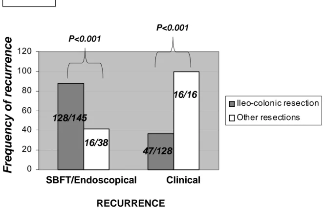
In our cohort of patients, the frequency of radiological/endoscopical recurrence was significantly higher in patients with ileo-colonic resection when compared with patients with other types of curative intestinal resections for CD (128/145, 88% vs 16/38, 42%, p<0.001)(Figure 2.4). At time of endoscopical/radiological recurrence, the frequency of symptomatic recurrence was significantly higher in patients with curative resection different from the ileo-colonic resection (16 out of 16 patients, 100%) than in patients with ileo-colonic resection (47 out of 128 patients, 37%; p<0.001)(Figure 2.4).
When considering the known risk factors for CD recurrence, comparable findings were observed when comparing patients with ileo-colonic vs other types of anastomoses (smoke: OR 1.5 vs 1.4; previous appendectomy: OR 0.32 vs 0.33; familial history of IBD: OR 0.43 vs 1.26).
DISCUSSION
Despite the growing knowledge regarding the pathogenesis of CD, postoperative recurrence after ileo-colonic resection is observed in almost two/thirds of patients as early as 1 year after surgery (1-4). The recurrence rate further increases over years, being almost ineluctable in CD, showing the same pattern before and after ileo-colonic resection (19) and a different frequency in relation to the type of intestinal resection (20). Current treatment modalities for preventing CD recurrence include oral mesalamine, although results at this regard are conflicting (1,19,20). A recent placebo-controlled pilot study reported a significant efficacy of anti-TNFs at this purpose (22). Nevertheless, recurrence of the lesions after surgery still represents a crucial issue in clinical management of patients with CD. Recurrence assessment after ileo-colonic resection is currently made at 1 year (or even at 6 months), by using ileocolonoscopy, a quite invasive technique most often performed in young patients already undergoing repeated endoscopies before surgery. The natural history of endoscopic recurrence in CD has already been described by Rutgeerts et al. (1,4). Due to the invasiveness of colonoscopy, several non-invasive techniques have been proposed for assessing CD recurrence after ileo-colonic resection (23,25-34). Although the possible role of bowel ultrasound for assessing CD recurrence has been suggested (13,35,36), very few studies investigated
the usefulness of ultrasonography using oral contrast (SICUS) at this purpose (15,16,35-36). We recently reported the possible usefulness of SICUS for assessing the postoperative recurrence of CD after ileo-colonic resection (15,16). The role of the non-invasive WCE for assessing CD recurrence has been suggested by Bourreille et al. (33), as also supported by our findings (15). However, by our knowledge no studies compared the role of endoscopy and ultrasonography (visualizing the intraluminal or the extraluminal surface, respectively), for assessing CD recurrence in patients followed up for 3 years after ileo-colonic resection. Our findings from the prospective study, suggest that ileocolonoscopy and SICUS are quite comparable for detecting lesions related to CD recurrence both at 1 and 3 years after surgery. Differently, the frequency of recurrence as assessed by SBFT at 2 years appeared to be lower than by using endoscopy or SICUS at 1 and 3 years. It is well known that SBFT may not visualize minor lesions related to CD recurrence (i.e. aphtoid ulcers) when compared to colonoscopy (2). This issue appears not to account for our findings, as in our series a high proportion of patients showed a high endoscopic score at 1 year (grade >2 in 12/25 patients, including a grade 4). Nevertheless, present data also indicate that in 8 out of the 9 patients showing no recurrence by SBFT at 2 years, the endoscopic score at 1 year was ≤2, including a true negative finding.
In the present study, all techniques were performed by one single experienced endoscopist, radiologist or ultrasonographist, thus avoiding the possible bias of inaccurate examinations affecting the results. Unfortunately, in our series a high proportion of patients showed endoscopic recurrence at 1 year (24 out of 25), thus leading to only one true negative finding. This observed high recurrence rate at 1 year is however within the upper limit reported by previous studies (93%), and appeared not related to known risk factors for early CD recurrence (6,8,39). In our study population a low proportion of patients were indeed smokers, showed a fistulizing pattern or a familial history of IBD. Reasons for the observed high rate of recurrence in our study population may rather be related to the relatively young age of patients at surgery (median age 35 years), including only one patient older than 50 years (IM, age 67). Moreover, in order to consider only established CD lesions after surgery, recent studies consider a higher endoscopic score as a cut-off value for defining CD recurrence (i.e. >2)(40). In our series, a laparoscopic or laparotomic surgical approach appeared not to influence the recurrence rate, confirming our previous findings (41).
Ultrasonography showed findings compatible with recurrence in all patients at all times (1, 2 and 3 years), thus leading to one false positive finding at both 1 and 3 years. However, in this patient showing SICUS findings compatible with recurrence not confirmed by endoscopy the
BWT was at limit of the normal value (3.5 mm: n.v. ≤3mm) at both times. No additional sonographic findings, including those compatible with recurrence (i.e. mesenteric enlargement) were observed.
Findings from the prospective study, also further suggest that SBFT at 2 years may not visualize CD recurrence detected by ileocolonoscopy at 1 year. This observation may not be related to mucosal healing induced by immunomodulatory drugs, as in our series no patients received thiopurines or anti-TNFs after surgery. The same discrepancies between techniques were observed when endoscopic, sonographic and radiologic findings were compared among the subgroup of 15 patients completing the follow up at 3 years. Differently from SBFT, in our series SICUS provided to be quite comparable to ileocolonoscopy in terms of recurrence assessment. SICUS allows the visualization of extraluminal lesions related to CD, including an increased BWT, mesenteric and lymphonode enlargement. Differently, both ileocolonoscopy and SBFT provide the intraluminal view of the bowel, thus allowing the visualization of the mucosal lesions (i.e. ulcers, strictures). Nevertheless, results from the present prospective longitudinal study suggest that although a different view of the bowel wall is provided by using ileocolonoscopy and SICUS, recurrence assessment appears more comparable by using these 2 techniques than by using ileocolonoscopy and SBFT. This finding is in agreement with our previous studies (15,16), thus supporting
that after ileo-colonic resection for CD the development of intraluminal lesions is associated with an increased BWT. This hypothesis is also supported by recent findings comparing RMN and colonoscopy for assessing colonic lesions in CD, showing a significant correlation between the two scores (16). Differently from recent findings suggesting that colonoscopy and MR enteroclysis are equivalent tools in predicting clinical recurrence in patients with CD after ileo-colic resection (38), in our series the BWT as assessed by SICUS at 1 year was comparable between patients showing or not clinical relapse at 2 years. This finding may be related to relatively low number of patients showing relapse (n=5). Nevertheless, the median endoscopic score of recurrence at 1 year was significantly higher in the subgroup of 5 patients developing clinical relapse at 2 years, confirming previous findings (3,39). We also found at 2 years a significantly higher BWT and SBFT score in the 5 clinically active patients than in the subgroup of 10 patients in remission at 2 years, as recently observed for magnetic resonance colonography (40-42).
The prospective study also suggests that although at 1 year more than two/thirds of patients show peri-anastomotic lesions as assessed by 2 techniques visualizing either the intraluminal or the extraluminal surface, the frequency of clinical recurrence is observed in a low proportion of patients at 1 year, significantly increasing at 2 and 3 years.
Early treatment with immunomodulatory treatments, including anti-TNFs has been reported to be effective for preventing clinical (22) and endoscopical recurrence after surgical resection (1,21,39). These observations, together with the here reported significant concordance between endoscopic and sonographic findings for assessing CD recurrence further supports that SICUS may be useful for a proper follow up and treatment of patients after ileo-colonic resection for CD. Although SICUS is a non-invasive technique, the accuracy of this technique is highly affected by the specific experience of the sonographist. On the basis of these observations, the main message arising from the prospective study is that in CD patients under regular follow up after ileo-colonic resection, in experienced hands SICUS represents a non-invasive, repeatable technique providing findings comparable to colonoscopy and therefore recommended for an adequate assessment and follow up of patients. Whether SICUS findings in the early post-operative period may have a role for predicting the clinical course of CD in patients with ileo-colonic resection needs further investigations.
In a different retrospective study, we aimed to assess, the frequency and pattern of CD recurrence in a cohort of CD patients under regular follow up. Indeed, despite the growing knowledge regarding the pathogenesis and treatment of CD, postoperative recurrence still remains almost ineluctable after curative ileo-colonic resection. Although
ileo-colonic resection represents the most frequent surgical procedure for CD (1-3), other surgical resections may be required in these patients. Differently from patients with previous ileo-colonic resection, the outcome and the natural history of CD after other surgical procedures is not clearly defined. By our knowledge, only few retrospective studies including a limited number of patients investigated this issue, with conflicting results . The knowledge of the natural history of the postoperative course of CD patients after surgical resection different from the ileo-colonic may be useful for proper clinical management and surgical indication.
Present findings further indicate that CD recurrence may also develop in patients with anastomosis different form the ileo-colon, including patients with permanent ileostomy. Although the limited number of enrolled patients and the different type of anastomoses do not allow conclusive statements, present findings also suggest a high frequency of symptomatic recurrence in other types of anastomoses, particularly including the ileo-rectum. Comparisons with previous studies at this regard is limited by the observed wide range of frequency of recurrence in different series. Our findings from a retrospective analysis of data recorded prospectively, indicate that CD recurrence above the anastomosis is observed also in patients with previous resections different from ileo-colonic, including permanent ileostomy. However, this
frequency appeared to be lower than after ileo-colonic resection. Prospective longitudinal studies are ongoing in order to address this issue.
References:
1. Rutgeerts P, Geboes K, Vantrappen G, et al. Natural history of recurrent Crohn’s disease at the ileocolonic anastomosis after curative surgery. Gut 1984; 25: 665-672.
2. Travis SPL, Stange EF, M Lèmann, et al. for the European Crohn’s and Colitis Organisation (ECCO) . Gut 2006 (Suppl I): i63-i35.
3. Rutgeerts P, Geboes K, Vantrappen G, et al Predictability of the postoperative course of Crohn’s Disease. Gastroenterology 1990; 99: 956-963.
4. Rutgeerts P. Strategies in the prevention of postoperative recurrence in Crohn’s Disease. Best Pract Res Clin Gastroenterol 2003; 17: 63-73. 5. Olaison G, Smedh K, Sjodahl R. Natural course of Crohn’s Disease after ileocolonic resection: endoscopically visualised ileal ulcers preceding symptoms. Gut 1992;33:331-335.
6. Shivanda S et al. Crohn’s disease: risk of recurrence and reoperation in a definite population. Gut 1989; 30: 990-995.
7. Farmer RG, Whelan G, Fazio VW. Long-term follow up of patients with Crohn’s Disease. Relationship between the clinical pattern and prognosis. Gastroenterology 1985; 88: 1818-1825.
8. Sachar DB, Wolfson DM, Greenstein AJ, et al Risk factors for postoperative recurrence of Crohn’s disease. Gastroenterology 1983; 85: 917-921.
9. D’Heans et al .Duration of recurrent ileitis after ileocolonic resection correlates with presurgical extent of Crohn’s disease. Gut 1995;715-717 10. Carter MJ, Lobo AJ, Travis SPL . Guidelines for the management of inflammatory bowel disease in adults. Gut 2004 53: 1-16.
11. Triester SL, Leighton JA, Leontiadis GI, et al A meta-analysis of the yield of capsule endoscopy compared to other diagnostic modalities in patients with no stricturing small bowel Crohn’s disease. Am J
Gastroenterol 2006;101; 954-964.
12. Parente F, Greco S, Molteni M, et al. Oral contrast enhanced bowel ultrasonography in the assessment of small intestine Crohn’s disease. A prospective comparison with conventional ultrasound, x-ray studies, and ileocolonoscopy. Gut 2004; 53: 1652-1657.
13. Calabrese E, La Seta F, Buccellato A, et al. Crohn’s Disease: a comparative prospective study of transabdominal ultrasonography, small intestine contrast ultrasonography and small bowel enema . Inflamm
Bowel Dis. 2005;11(2):139-145
14. Pallotta N, Tomei E, Viscido A, et al. Small intestine contrast ultrasonography : an alternative to radiology in the assessment of small bowel disease. Inflamm Bowel Dis 2005; 11: 146-153.
15. Biancone L, Calabrese E, Petruzziello C, et al. Wireless capsule endoscopy and small intestine contrast ultrasonography in recurrence of Crohn’s Disease. Inflamm Bowel Dis 2007; 13: 1256-1265.
16. Calabrese E, Petruzziello C, Onali S, et al. Severity of postoperative recurrence in Ctrohn’s Disease: correlation between endoscopic and sonographic findings. Inflamm Bowel dis 2009;15(11):1635-42
17. Rutgeerts P, Goboes K, Peeters M, et al. Effect of faecal stream diversion on recurrence of Crohn’s disease in the neoterminal ileum.
Lancet 1991;338:771-774.
18. Zalev A, Profipchuk E, Jeejeebhoy G, et al. Recurrent Crohn’s disease in the duodenum and jejunum following extensive small bowel resection and jejunocolonic anastomosis: radiologic findings in twenty-five patients. Abdom Imaging 1999;24:538-543
19.Greenstein A, Sachar D, Pasternack B, et al. Reoperation and Recurrence in Crohn’s colitis and ileocolitis. N Engl J Med 1975;293:685-690.
20. Onali S, Calabrese E, Petruzziello C, et al. Endoscopic vs
ultrasonographic findings related to Crohn’s disease recurrence. JCC 2010;in press (Accepted in Dec 2009 )
21. Onali S, Petruzziello C, Calabrese E, et al.Frequency, pattern, and risk factors of postoperative recurrence of Crohn's disease after resection different from ileo-colonic. J Gastrointest Surg. 2009;13(2):246-52
22. Regueiro M, Schraut W, Baidoo L, et al. Infliximab prevents Crohn’s Disease recurrence after ileal recurrence. Gastroenterology 2009: 136: 441-450.
23. Tibble JA, Sightorsson G, Bridger S, et al. Surrogate markers of intestinal inflammation are predictive of relapse in patients with inflammatory bowel disease. Gastroenterology 2000;11:15-22.
24. Best WR, Becktel JM, Singleton JW, et al. Development of a Crohn's disease activity index. National Cooperative Crohn's Disease Study.
Gastroenterology 1976; 70:439-44.
25. Hanauer SB, Korelitz B, Rutgeerts P, et al Postoperative Maintenance of Crohn’s Disease Remission With 6-Mercaptopurine, Mesalamine, or Placebo: A 2-Year Trial. Gastroenterology 2004;127:723-729
26. Pallone F, Boirivant M, Stazi MA, et al. Analysis of clinical course of postoperative recurrence in Crohn’s disease of distal ileum. Dig Dis Sci 1992; 37: 215-219
27. Tibble J, Teahon K, Thjodleifsson B, et al. A simple method for assessing intestinal inflammation in Crohn’s Disease. Gut 2000; 47: 506-513.
28. Orlando A, Modesto I, Castiglione F, et al. The role of calprotectin in predicting endoscopic postsurgical recurrence in asymptomatic Crohn’s
Disease: a comparison with ultrasound. Eur Rev Med Pharmacol Sci 2006; 10: 17-22.
95. Boirivant M, Pallone F, Leoni M, et al. Usefulness of fecal alpha-1-antitrypsin clearance as an early indicator of asymptomatic postoperative recurrence in Crohn's Disease. Dig Dis Sci 1991; 36: 347-52.
30. Biancone L, Scopinaro F, Ierardi M, et al. 99m-Tc-HMPAO granulocyte scintigraphy in the early detection of postoperative asymptomatic recurrence in Crohn’s Disease. Dig Dis Sci 1997; 42: 1549-56.
31. Biancone L, Fiori R, Tosti C, et al. Virtual colonoscopy compared with conventional colonoscopy for stricturing postoperative recurrence in Crohn's disease. Inflamm Bowel Dis 2003; 9:343-50.
32. Rispo A, Imbriaco M, Celentano L, et al. Noninvasive diagnosis of small bowel Crohn’s disease: combined use of bowel sonography and Tc-99m-HMPAO leukocyte scintigraphy. Inflamm Bowel Dis 2005; 11: 376-82.
33. Bourreille A, Jarry M, D'Halluin PN, et al. Wireless capsule endoscopy versus ileocolonoscopy for the diagnosis of postoperative recurrence of Crohn's disease: a prospective study. Gut 2006; 55: 978– 82.
34. Horsthuis K, Bipat S, Bennink R, et al. Inflammatory bowel disease diagnosed with US, MR, scintigraphy, and CT: Metanalysis of prospective studies. Radiology 2008; 247: 64-79.
35. Di Candio G, Mosca F, Campatelli A, et al Sonographic detection of postsurgical recurrence of Crohn’s Disease. Am J Roentgenol 1986;146:523-6.
36. Andreoli A, Cerro P, Falasco G, et al. Role of ultrasonography in the diagnosis of postsurgical recurrence of Crohn’s Disease. Am J
Gastroenterol 1998;93:1117-21.
37. Castiglione F, Bucci L, Pesce G, et al. Oral contrast-enhanced sonography for the diagnosis and grading of postsurgical recurrence of Crohn's disease. Inflamm Bowel Dis 2008;14(9):1240-5
38 Bourreille A, Jarry M, D'Halluin PN, et al. Wireless capsule endoscopy versus ileocolonoscopy for the diagnosis of postoperative recurrence of Crohn's disease: a prospective study. Gut 2006; 55: 978–82.
39. Cottone M, Rosselli M, Orlando A, et al Smoking habitus and recurrence in Crohn’s disease. Gastroenterology 1994; 106: 643-648. 40. Koilakou S, Sailer J, Peloschek P, et al. Endoscopy and MR enteroclysis: equivalent tools in predicting clinical recurrence in patients with Crohn’s disease after ilecolic resection. Inflamm Bowel Dis 2010;16(2):198-203.
41. Sica GS, Iaculli E, Benavoli D et al. Laparoscopic Versus Open Ileo-Colonic Resection in Crohn's Disease: Short- and Long-Term Results from a Prospective Longitudinal Study. J Gastrointest Surg 2008; 12(6): 1094-1102.
42. Rimola J, Ordás I, Rodríguez S, et al . Colonic Crohn’s Disease: value of magnetic resonance colonography for detection and quantification of disease activity. Abdom Imaging 2009 (in press)
43. Peyrin-Birulet L, Deltenre P, Ardizzone S, et al. Azathioprine and 6-mercaptopurine for the prevention of postoperative recurrence in Crohn’s Disease: a meta-analysis. Am J Gastroenterol 2009 (in press)
1.Prospective study:
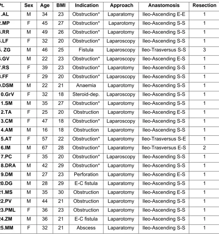
Table 1. Clinical characteristics of each of the 25 patients enrolled.
Pt. Sex Age BMI Indication Approach Anastomosis Resection
1.AL M 34 23 Obstruction* Laparatomy Ileo-Ascending E-E 1
2.MP F 45 27 Obstruction* Laparotomy Ileo-Ascending S-S 1
3.RR M 49 26 Obstruction* Laparotomy Ileo-Ascending S-S 1
4.LF F 32 20 Obstruction* Laparoscopy Ileo-Ascending S-S 1 5. ZG M 46 25 Fistula Laparoscopy Ileo-Trasversus S-S 3 6.GV M 22 23 Obstruction* Laparoscopy Ileo-Ascending E-S 1 7.RS F 39 23 Obstruction* Laparotomy Ileo-Ascending S-S 1 8.FF F 29 20 Obstruction* Laparoscopy Ileo-Ascending S-S 1
9.DSM M 22 21 Anaemia Laparotomy Ileo-Ascending S-S 1
10.GrV F 32 18 Steroid-dep. Laparoscopy Ileo-Ascending S-S 1 11.SM M 35 27 Obstruction* Laparotomy Ileo-Ascending S-S 1
12.TA F 25 20 Obstruction Laparotomy Ileo-Ascending E-S 1
13.CM F 47 18 Obstruction* Laparoscopy Ileo-Ascending S-S 1 14.AM M 16 18 Obstruction Laparotomy Ileo-Ascending S-S 1 15.AT F 57 22 Obstruction* Laparotomy Ileo-Trasversus S-E 1 16.IM M 67 28 Obstruction* Laparatomy Ileo-Trasversus E-S 2 17.PC F 35 20 Obstruction* Laparoscopy Ileo-Ascending S-S 1 18.DRA M 42 29 Obstruction* Laparotomy Ileo-Ascending S-S 1
19.DM M 27 23 Perforation Laparotomy Ileo-Ascending E-S 1
20.DG M 28 29 E-C fistula Laparatomy Ileo-Ascending S-S 1
21.MS M 35 30 Obstruction Laparotomy Ileo-Ascending E-S 1
22.PV M 44 21 Obstruction Laparotomy Ileo-Ascending S-S 1 23.PML F 36 23 Obstruction Laparotomy Ileo-Ascending S-S 1 24.ZM M 36 21 E-C fistula Laparotomy Ileo-Ascending S-S 1
25.MM F 32 21 Abscess Laparatomy Ileo-Ascending S-S 1
Abbreviations: E-C=Entero-cutaneous fistula;E-S=End-toSide;S-S=Side-to-Side; E-E=End-to end; BMI=Body Mass Index; Pt=Patient.
1.Prospective study: Table 2.*
A=Active (CDAI>150); R= remission (CDAI<150); Endoscopy: Y=Recurrence (grade ≥1); N= No recurrence (grade 0); SICUS: Y=Findings compatible with recurrence (BWT>3 mm); No=Findings compatible with no recurrence (BWT≤3 mm); stenosis= pt. IM showed an endoscopic stenosis at 1 year and therefore did not perform endoscopy at 1 year; * n.d.= not done: reasons for drop out are reported in the text.
Pt 1 YEAR 2 YEARS 3 YEARS
CDAI (A>150) ENDO ( 0-4) SICUS ( mm) CDAI (A>150) SBFT ( 1-4) SICUS ( mm) CDAI (A>150) ENDO ( 0-4) SICUS (BWT,mm) 1.AL R Y (3) Y (5) A Y (2) Y (10) A Y (4) Y (12) 2.MP R Y (4) Y (5) A Y (4) Y (5.5) A Y (4) Y (6) 3.RR R Y (1) Y (3.5) R Y (2) Y (4) R Y (3) Y (4) 4.LF R Y (2) Y (4) R Y (3) Y (5) R Y (3) Y (5) 5.ZG R Y (4) Y (7) A Y (3) Y (7) A Y (3) Y (9) 6.GV R N (0) Y (3.5) R N (1) Y (3.5) R N (0) Y (3.5) 7.RS R Y (3) Y (4) R Y (3) Y (5) R Y (2) Y (5) 8.FF R Y (1) Y (6) R N (1) Y (7) R Y (1) Y (4) 9.DSM R Y (3) Y (6) R Y (3) Y (5) R Y (4) Y (5) 10.GrV R Y (3) Y (5) A Y (4) Y (7) A Y (2) Y (6) 11.SM R Y (3) Y (6) R N (1) Y (4) R Y (3) Y (6) 12.TA R Y (1) Y (3.5) R N (1) Y (7) R Y (1) Y (4) 13.CM R Y (4) Y (4) A Y (4) Y (6) A Y (4) Y (4) 14.AM R Y (2) Y (3.5) R N (1) Y (3.5) R Y (2) Y (6) 15.AT R Y (2) Y (3.5) R N (1) Y (3.5) R Y (2) Y (3.5) 16.IM R Y (4) Y (5) R Y (4) Y (7.5) R n.d. Y (9) 17.PC R Y (4) Y (4) R Y (2) Y (5) n.d. n.d. n.d. 18.DRA R Y (1) Y (4) R N (1) Y (4) n.d. n.d. n.d. 19.DM A Y (2) Y (5) A Y (4) Y (4) n.d. n.d. n.d. 20.DG R Y (1) Y (5) R N (1) Y (4) n.d. n.d. n.d. 21.MS R Y (2) Y (6) R N (1) Y (4) n.d. n.d. n.d. 22.PV R Y (4) Y (10) n.d. n.d. n.d. n.d. n.d. n.d. 23.PML R Y (1) Y (4) n.d. n.d. n.d. n.d. n.d. n.d. 24.ZM R Y (4) Y (6) n.d. n.d. n.d. n.d. n.d. n.d.
1.Prospective study: Figures FIGURE 1a 0 2 4 6 8 10 0 1 2 3 4 ENDOSCOPIC DEGREE OF RECURRENCE
n=1 n=6 n=6 n=5 n=7 BW T ( m m) * * * * * 1 YEAR AFTER SURGERY
FIGURE 1b 0 2 4 6 8 10 ≤1 ≥2
ENDOSCOPIC DEGREE OF RECURRENCE p=0.16
BW
T
(mm)
1 YEAR AFTER SURGERY
FIGURE 1c 0 2 4 6 8 10 1 2 3 4
RADIOLOGIC (SBFT) SCORE OF RECURRENCE
BW T ( mm) * * * *
2 YEARS AFTER SURGERY
n=9 n=3 n=4 n=5 0 2 4 6 8 10 p=0.05 n=9 n=12 BW T (mm)
2 YEARS AFTER SURGERY
FIGURE 1d
1 ≥2
0 2 4 6 8 10 n=1 n=2 n=4 n=4 n=4 12 * * * * FIGURE 1e 0 1 2 3 4
ENDOSCOPIC DEGREE OF RECURRENCE
BW
T
(mm)
3 YEARS AFTER SURGERY
0 2 4 6 8 10
3 YEARS AFTER SURGERY
FIGURE 1f
≤1 ≥2
ENDOSCOPIC DEGREE OF RECURRENCE
BW T (m m) p=0.15 n=3 n=12
0 20 40 60 80 100 Pati ents wi th recur rence (% )
TIME INTERVAL FROM SURGERY (YRS) 1 2 3 Colonoscopy SICUS CDAI SBFT FIGURE2 * ** ** * ** *** *** *** (n=15) (n=15) (n=15)
0 20 40 60 80 100 120 SICUS ENDOSCOPY ENDOSCOPY CDAI CDAI
TIME FROM SURGERY
TIME FROM SURGERY
1
1
year
year
3
3
years
years
% P A TIE N T S WITH RE CU RRE NCE % P A TIE N T S WITH RE CU RRE NCE
*
* p<0.001 vs 1 year FIGURE 30 1 2 3 4 5 6
CLINICAL ACTIVITY AT 2 YEARS (CDAI)
CLINICAL ACTIVITY AT 2 YEARS (CDAI)
ACTIVE REMISSION ACTIVE REMISSION EN D O SC OPI C SCOR E A T 1 Y E A R EN D O SC OPI C SCOR E A T 1 Y E A R p=0.003 n=5 n=10 Figure 4, panel a 0 1 2 3 4 5 6 7 8 9
CLINICAL ACTIVITY AT 2 YEARS (CDAI)
CLINICAL ACTIVITY AT 2 YEARS (CDAI)
ACTIVE REMISSION
ACTIVE REMISSION
SICUS
: BW
T
A
T
2
Y
E
A
R
S
S
IC
U
S
: B
W
T
AT
2
YE
AR
S
p=0.19
Figure 4, panel bn=5
n=10
PROSPECTIVE STUD: LEGEND FOR THE FIGURES Figure 1 (panels a-f)
Panels a,b. Histograms show the median (and range) BWT in CD
patients 1 year after ileo-colonic resection subgrouped according to: each single endoscopic recurrence score (grade 0-4)(panel a) or to an endoscopic recurrence score ≤1 or ≥2 (panel b). No significant differences were observed between groups by using both criteria (*).
Panels c,d. Histograms show the median (and range) BWT in CD
patients 2 years after surgery, subgrouped according to: each single SBFT recurrence score (grade 0-4)(panel c) or to a radiologic score ≤1 or ≥2 (panel d). The observed higher median BWT in patients with a radiological score ≥2 was at limit of statistical significance (p=0.05).
Panels e,f. Histograms show the median (and range) BWT in CD
patients 3 years after surgery, subgrouped according to: each single endoscopic recurrence score (grade 0-4)(panel e) or to an endoscopic recurrence score ≤1 or ≥2 (panel f). No significant differences were observed between groups when considering both criteria (*).
Figure 2
Histograms show the frequency of recurrence assessed clinically (CDAI), by endoscopy (Rutgeert’s score 0-4), radiology (Hanauer’s score 0-4) and ultrasoography (SICUS: BWT n.v. ≤3 mm), when considering only the 15 patients performing all the procedures at 1,2, and 3 years. The frequency clinical recurrence significantly increased at both 2 and 3
years vs 1 year (0% vs 33%; p<0.001). Differently, at 1 and 3 years the same proportion of patients showed recurrence as assessed either by colonoscopy or by SICUS (93% and 100% at both 1 and 3 years, respectively; p=n.s.).
Figure 3
The lines show the frequency of recurrence as assessed clinically (CDAI), endoscopically (Rutgeerts’ score) and by ultrasonography (BWT at SICUS) at 1 at 3 years after surgery. As shown, clinical recurrence was observed in a significantly higher proportion of patients at 3 years vs 1 year after resection (33% vs 0%; p<0.001). Differently, a comparable proportion of patients showed recurrence at 1 vs 3 years when assessed either by colonoscopy or by SICUS (93% at both times vs 100% at both times, respectively).
Figure 4 (panels a, b)
Panel a. Histograms show the endoscopic score (grade 0-4)(panel a)
and the BWT (in mm; n.v. ≤3 mm) 1 year after surgery in patients developing (n=5) or not (n=10) clinical relapse 2 years after surgery. As shown, the endoscopic score at 1 year was significantly higher in the subgroup of 5 inactive patients developing relapse at 2 years than in the 10 inactive patients maintaining remission (p=0.003). Panel b. Differently, the median BWT assessed by SICUS at 1 year was not significantly
higher in the 10 patients developing clinical relapse at 2 years (median BWT: 5, range 4-7 vs 3.7, range 3.5-6; p=0.19).
2.RETROSPECTIVE STUDY:TABLE
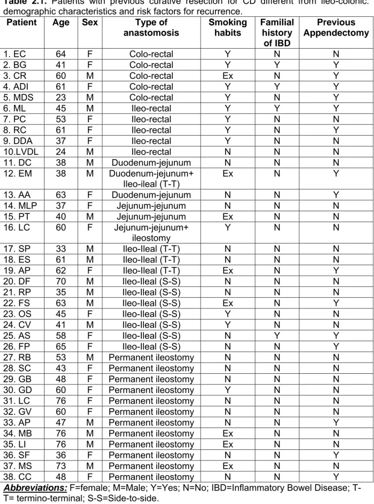
Table 2.1. Patients with previous curative resection for CD different from ileo-colonic: demographic characteristics and risk factors for recurrence.
Patient Age Sex Type of
anastomosis Smoking habits Familial history of IBD Previous Appendectomy 1. EC 64 F Colo-rectal Y N N 2. BG 41 F Colo-rectal Y Y Y 3. CR 60 M Colo-rectal Ex N Y 4. ADI 61 F Colo-rectal Y Y Y 5. MDS 23 M Colo-rectal Y N Y 6. ML 45 M Ileo-rectal Y Y Y 7. PC 53 F Ileo-rectal Y N N 8. RC 61 F Ileo-rectal Y N Y 9. DDA 37 F Ileo-rectal Y N N 10.LVDL 24 M Ileo-rectal N N N 11. DC 38 M Duodenum-jejunum N N N 12. EM 38 M Duodenum-jejunum+ Ileo-ileal (T-T) Ex N Y 13. AA 63 F Duodenum-jejunum N N Y 14. MLP 37 F Jejunum-jejunum N N N 15. PT 40 M Jejunum-jejunum Ex N N 16. LC 60 F Jejunum-jejunum+ ileostomy Y N N 17. SP 33 M Ileo-Ileal (T-T) N N N 18. ES 61 M Ileo-Ileal (T-T) N N N 19. AP 62 F Ileo-Ileal (T-T) Ex N Y 20. DF 70 M Ileo-Ileal (S-S) N N N 21. RP 35 M Ileo-Ileal (S-S) N N N 22. FS 63 M Ileo-Ileal (S-S) Ex N Y 23. OS 45 F Ileo-Ileal (S-S) Y N N 24. CV 41 M Ileo-Ileal (S-S) Y N N 25. AS 58 F Ileo-Ileal (S-S) N Y Y 26. FP 65 F Ileo-Ileal (S-S) N N Y 27. RB 53 M Permanent ileostomy N N N 28. SC 43 F Permanent ileostomy N N N 29. GB 48 F Permanent ileostomy N N N 30. GD 60 F Permanent ileostomy Y N N 31. LC 76 F Permanent ileostomy N N N 32. GV 60 F Permanent ileostomy N N N 33. AP 47 M Permanent ileostomy N N Y 34. MB 76 M Permanent ileostomy Ex N N 35. LI 76 M Permanent ileostomy Ex N N 36. SF 36 F Permanent ileostomy N N Y 37. MS 73 M Permanent ileostomy Ex N N 38. CC 48 F Permanent ileostomy N N Y
Abbreviations: F=female; M=Male; Y=Yes; N=No; IBD=Inflammatory Bowel Disease; T-