Abstract
The biological activity of extract obtained from the ascidians Styela plicata and Ascidia mentula, living in the coastal lake Faro (Messina, Italy) was ascertained with specific regard to cytotoxic effect, tested on cultured human embryonic kidney (HEK 293 Phoenix) cells after 12, 24 or 48 h, and to antimicrobial effect (10 mg/mL), assayed on both Gram positive and negative strains. Nitrosative stress, possibly induced by the extract on HEK 293 cells and assessed by NO2–/NO3–
production measurement, was also verified. With regard to cytotoxic activity, A. mentula extract was more effective than S. plicata one, as shown by 3-(4,5-dimethylthiazol-2-yl)-2,5-diphenyltetrazolium bro-mide assay, with toxic action due to nitrosative stress. S. plicata extract had a more significant antimicrobial activity than A. mentula. The present findings provide a first evidence of the biological power exhibited by both S. plicata and A. mentula extracts, thus increasing the knowledge about the biological activity of marine compounds and providing novel information to possibly correlate the different toxicity pattern displayed by both specimens and their distribution in the lake Faro (Messina).
Introduction
The biological and chemical diversity of the marine environment con-stitute a great source of new bioactive products, the knowledge of which has greatly increased the interest of researchers over the past twenty years.1,2Several marine organisms produce a variety of chemical
com-pounds for predation strategies and for defence against potential preda-tors and competipreda-tors. Such compounds are critically important for soft bodied organisms such as marine algae, sponges, ascidians (sea squirts) to deter predators and compete for space.
Amongst marine organisms, ascidians (Phylum Chordata, Class Ascidiacea) are distributed on hard bottoms from the intertidal to the subtidal area, abyssal depths, including coral reef and mangroves. Approximately 3000 living species of Ascidians have been identified so far3 and, due to their distribution, they are considered good indicators
for water quality monitoring.4In addition, they represent a potential
source of marine natural products,1 whose characterization has risen
the interest of marine biologists, chemists and toxicologists.
Marine toxicological studies have shed light on numerous com-pounds extracted from several marine animals5,6 focusing on their
structure and biological activity. In this latter regard, different biologi-cal assays were set up to verify the effect of such compounds on cell targets, thus opening the way to consider their possible use as novel tools for pathophysiological and/or therapeutical investigations.7
Amongst biological assays, the hemolytic one, together with the cyto-toxicity assay on cultured cells, are the most suitable.8
With regard to Ascidians, more than 800 different biologically active compounds exhibiting a range of biological activities, such as cytotoxic, antibiotic or immunosuppressive activities, inhibition of topoisomeras-es and cyclin kinastopoisomeras-es, have been dtopoisomeras-escribed.1,2,9Amongst ascidian
speci-mens, Trididemnum solidum, Lissoclinum patella and Styela clava have been studied (as reviewed by Aneiros and Garateix),10with Didemnin A
and B, Lissoclinamides and Styelin D as major compounds displaying antitumoral, antiviral and antimicrobial activity, respectively. Recently, Correspondence: Angela Marino, Department of Chemical, Biological,
Pharmaceutical and Environmental Sciences, University of Messina, viale F. Stagno D’Alcontres 31, Messina, Italy.
Tel: +39.090.6765214 - Fax: +39.090.394030. E-mail: [email protected]
Key words: Styela plicata; Ascidia mentula; Extract; Biological activity; Ascidians.
Contributions: SKP, RM, AR, AM performed experiments; RM, SG and SKM collected and analyzed data; GLS, SG and NS reviewed the manuscript; AM wrote the manuscript and searched for references; SKP and RM contributed equally to this work.
Conflict of interest: the authors declare no potential conflict of interest. Acknowledgements: the author SKP is grateful to University of Messina (Italy) for the award of Non-European student Doctoral Research Fellowship 2013-2015. The authors are grateful to Mr. S. De Matteo and Mr. A. Spinelli for their help in specimens collection; to Dr. S. Umamaheswari, Department of Environmental Biotechnology, Bharathidasan University, India for antimi-crobial assay; to Prof. S. Cuzzocrea, University of Messina, for providing facilities for cytotoxic assay.
Received for publication: 15 February 2016. Revision received: 27 April 2016. Accepted for publication: 11 June 2016.
©Copyright S.K. Palanisamy et al., 2016 Licensee PAGEPress, Italy
Journal of Biological Research 2016; 89:5812 doi:10.4081/jbr.2016.5812
This article is distributed under the terms of the Creative Commons Attribution Noncommercial License (by-nc 4.0) which permits any noncom-mercial use, distribution, and reproduction in any medium, provided the orig-inal author(s) and source are credited.
Biological activity of extract from
Styela plicata
and
Ascidia mentula
(Ascidiacea)
Satheesh Kumar Palanisamy, Rossana Morabito, Alessia Remigante, Nunziacarla Spanò,
Giuseppina La Spada, Salvatore Giacobbe, Angela Marino
Department of Chemical, Biological, Pharmaceutical and Environmental Sciences, University of
Messina, Messina, Italy
Non-commercial
an acetylcholinesterase inhibitor has been isolated from the sub-Arctic colonial ascidian Synoicum pulmonaria.11
Based on these premises, the aim of the present study was to provide a first characterization of the biological activity of extract from two ascid-ian specimens, Styela plicata and Ascidia mentula, never investigated so far, in an attempt to add more information about the Ascidian fauna inhabiting the ecosystem surrounding the Strait of Messina (Italy),12
one of the most complex Mediterranean area in terms of biodiversity and species distribution.
For this purpose, the extract, obtained by alcoholic extraction method from both species, has been assayed for cytotoxic activity on cultured embryonic kidney (HEK 293 Phoenix) cells and for a possible antimicro-bial activity on Gram positive and negative strains. Moreover, oxidative nitrite formation, known to be produced in response to inflammatory or mitogenic stimuli,13,14has been also evaluated on HEK 293 cells after
treatment with the extracts.
Materials and Methods
Sample collection
The specimens chosen for the present study, Styela plicata (Lesueur, 1823) and Ascidia mentula (Müller, 1776), were collected from the coastal Lake Faro (Messina Italy) at 2 to 10 m depth, during January to April 2014. The animals were then transferred in laboratory and stored at -20°C until extraction procedures.
Extraction procedures
Extracts from either S. plicata or A. mentula were obtained by MeOH/CHCl3 Soxhlet extraction, sonication method.15The extract was
then re-suspended in MeOH to precipitate salt, filtered and afterwards diluted with water in a 7:3 ratio (MeOH:H2O). A successive partition of the aqueous phase was performed with CH2Cl2(2:1, v/v, 3-fold) and then with Na2SO4. The solvent was removed under reduced pressure and the obtained extract underwent protein determination (mg/mL) according to Bradford16 and then addressed to cytotoxic and antimicrobial activity
assessment.
Cytotoxic activity assay
Cell cultureHuman renal HEK 293 Phoenix cells, kindly provided by Paracelsus Medizinische Privatuniversität (Salzburg, Austria), were cultured in minimum essential eagle medium [(MEM); Sigma Aldrich, St. Louis, MO, USA] supplemented with 10% fetal bovine serum [(FBS); Cambrex Bio Science, East Rutherford, NJ, USA], 2 mM L-glutamine, 100 µg/mL streptomycin, 100 U/mL penicillin and 1 mM pyruvic acid (sodium salt). Cells were maintained at 37°C, 5% CO2, 95% air and 100% humidity. Subcultures were routinely established by seeding the cells into 100 mm diameter Petri dishes after trypsin/ethylene diamine tetraacetic acid (EDTA) treatment.
To evaluate the cytotoxic activity of extract from either S. plicata or, alternatively, A. mentula, cells were seeded in 96-microwell culture plates at a density of 3x10⁴cells/well. Cells were allowed to grow for 24 h (37°C,
5% CO2, 95% air and 100% humidity) and incubated for various time intervals (12, 24 and 48 h) in a medium containing extract at different concentrations (1 to 10 mg/mL). Cell viability was then estimated and compared to untreated cells.
Cell viability assay
To test the cytotoxicity of extract, cell viability was estimated by 3-(4,5-dimethyl-2-thiazolyl)-2,5-diphenyl-2H tetrazolium bromide (MTT; Sigma
Adlrich) assay, which measures the levels of mitochondrial dehydroge-nase activity, using MTT reduction assay. The assay is based on the redox ability of living mitochondria to convert dissolved MTT into insoluble for-mazan by cleavage of the tetrazolium ring by dehydrogenase enzymes. Extract from both ascidian species was dissolved in dimethyl sulfoxide (DMSO, 1% v/v final concentration). DMSO alone was tested on control cells to exclude any possible damage. After treatment with extract, the medium was removed and cells incubated with MTT solution (0.5 mg/mL) at 37°C for 1 h in the dark. The incubation was stopped by removing MTT solution and adding DMSO (1% v/v final concentration) to solubilize the formazan. Optical density (OD) was spectrophotometrically measured at 540 nm by a microplate reader (SLT-Lab Instruments, Salzburg, Austria).
Measurement of nitrite/nitrate production
Nitrite (NO2–)/nitrate (NO3–) production is an indicator of NO
synthe-sis as the final product of NO reacting with molecules present in biolog-ical fluids.13To measure NO2–/NO3– levels, medium from both treated and
untreated cells was incubated with nitrate reductase (0.1 U·mL–1),
nicotinamide adenine dinucleotide phosphate (1 mM) and flavin ade-nine dinucleotide (50 mM) at 37°C for 15 min, followed by further incu-bation with lactate dehydrogenase (100 U·mL–1) and sodium pyruvate
(10 mM) for 5 min. NO2–concentration was measured by Griess reaction,
by adding 100 µL of Griess reagent [0.1% (w/v) naphthylethylenedi-amide dihydrochloride in H2O and 1% (w/v) sulphanilnaphthylethylenedi-amide in 5% (v/v) H2PO4; vol. 1:1 to the 100 µL sample]. The OD at 550 nm (OD550), meas-ured using a microplate reader (SLT-Lab Instruments), was compared with OD550of standard solutions containing sodium nitrate in saline solution to calculate NO3– concentrations.14
Antimicrobial activity assay
A first screening of the possible antimicrobial activity of ascidian extracts was performed by the standard disk diffusion method, the most used test for antimicrobial susceptibility assessment, as described by Matuschek and co-authors.17The test was performed on the following
microbial strains: Burkholderia mallei (B7), Klebsiella pneumonia (B19), Staphylococcus aureus (B23), Pseudomonas sp (B831), K. pneu-monia (B938), Escherichia coli (U743) and E. coli (U744). A volume of 25 µL of extract (10 mg/mL) from both species was tested per disk and disks were placed on plates within 15 min of inoculum. Samples were then incubated overnight at 37°C with 5% CO2and the diameter (mm) of inhibition area after treatment with both extracts was measured in trip-licate and compared with inhibition area after ampicillin application, assumed as control.
Experimental data and statistics
GraphPad Prism software (version 5.00 for Windows; GraphPad soft-ware, San Diego, CA, USA) was used and data expressed as arithmetic means±standard error of the mean (SEM) for statistical analysis. Significant differences between means were tested by paired Student’s t test or one-way analysis of variance (ANOVA), followed by Bonferroni’s post hoc test for multiple comparisons. Statistically significant differ-ences were assumed at P<0.05 (*P<0.05, **P<0.01, ***P<0.001). N rep-resents the number of independent experiments performed on different experimental days.
Results
Cytotoxic activity assay
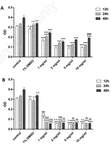
DMSO (1% v/v final concentration) did not elicit any cell damage when applied alone, cell viability being comparable to that observed in control cells (Figure 1).
Non-commercial
Styela plicata
The exposure of HEK 293 Phoenix cells to different extract concen-trations (1, 2, 5 and 10 mg/mL) for 12, 24 or 48 h provoked a significant (P<0.001) reduction in cell viability, if compared to control cells, as depicted in Figure 1A. There was no statistical difference between the different times of exposure to extract (12-24-48 h) at each dose employed, so that no time dependence of extract effect was observed. With regard to 12 h exposure (Figure 1A), no statistical difference was seen between cell viability measured at each dose. With regard to 24 h exposure to extract, cell viability after 10 mg/mL treatment was signif-icantly (P<0.001) lower with respect to both 1 and 2 mg/mL treatment (Figure 1A). Cell viability after 1 mg/mL treatment was significantly (P<0.001) higher than that measured after treatment with the other doses (Figure 1A). When cells were exposed for 48 h to the extract, no statistical difference was seen between cell viability measured at each dose (Figure 1A). Hence, a dose-dependent effect of extract was shown only in case of 24 h exposure.
Ascidia mentula
The exposure of HEK 293 Phoenix cells to different concentrations of extract (1, 2, 5 and 10 mg/mL) for 12, 24 and 48 h caused a significant (P<0.001) reduction in cell viability with respect to control cells (Figure 1B), as shown by MTT assay. There was no time dependence for extract toxicity, as cell viability was not reduced by increasing time of exposure (Figure 1B). With regard to 12 h treatment, cell viability measured at 1 mg/mL was significantly (P<0.01) higher than that measured at the other doses (Figure 1B), while there was no statisti-cally significant difference between cell viability measured at 2-5 and 10 mg/mL.
With regard to both 24 and 48 h incubation, there was no statistically significant difference between cell viability measured at each dose (Figure 1B). Hence, 1 mg/mL extract was the minimum dose capable of significant cell viability reduction.
There was no statistically significant difference between cell viabili-ty detected after 12 h treatment with A. mentula extract at each dose (Figure 1B) and the one measured after S. plicata extract treatment (Figure 1A). Moreover, cell viability measured after either 24 or 48 h exposure to A. mentula extract (Figure 1B) was significantly (P<0.01 and P<0.001) lower than that observed after S. plicata extract treat-ment, at all doses (Figure 1A), thus showing a more toxic effect of A. mentula extract (Figure 1B).
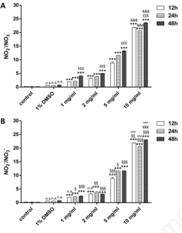
Measurement of intracellular nitrite/nitrate production
DMSO (1% v/v final concentration) did not induce NO2–/NO3–produc-tion when applied alone (Figure 2). Styela plicata
The exposure of HEK 293 Phoenix cells to different S. plicata extract concentrations (1, 2, 5 and 10 mg/mL) for 12, 24 or 48 h caused a sig-nificant (P<0.001) increase in NO2–/NO3–production, when compared
to control cells (Figure 2A). Moreover, NO2–/NO3–measured after 10
mg/mL was significantly (P<0.001) higher than that measured at the other doses, at any time of exposure (Figure 2A), thus showing a dose-dependent effect. NO2–/NO3–production measured after 48 h was
sig-nificantly (P<0.001) higher than those measured at both 12 and 24 h (Figure 2A), thus exhibiting a time-dependent effect.
Ascidia mentula
The exposure of HEK 293 Phoenix cells to different concentrations of extract (1, 2, 5 and 10 mg/mL) for 12, 24 or 48 h caused a significant (P<0.001) increase in NO2–/NO3–production, with respect to control
con-ditions (Figure 2B). Nitrite production was significantly (P<0.001)
high-er afthigh-er 10 mg/mL extract treatment than afthigh-er applying the othhigh-er doses, at any observation time (Figure 2B), showing a dose-dependent effect. Nitrite production measured after 10 mg/mL treatment was significantly (P<0.001) higher after 48 h incubation than both 12 and 24 h (Figure 2B), exhibiting a time-dependent effect of extract, not seen when lower doses were considered. Nitrate production after treatment with A. men-tula extract was significantly higher than that produced after S. plicata extract application at both 5 and 10 mg/mL after 12 h incubation (P<0.001 and P<0.05 respectively; Figure 2B), while significantly (P<0.001) higher at all doses after both 24 and 48 h (Figure 2B).
Antimicrobial activity assay
The antimicrobial activity of ascidian extracts assessed on 7 micro-bial strains is reported in Table 1. As far as S. plicata extract is con-cerned, the antimicrobial activity detected on Pseudomonas sp (B831)
Figure 1. Effect of Styela plicata (A) and Ascidia mentula (B) crude extract on cultured human embryonic kidney cells viability as determined by 3-(4,5-dimethylthiazol-2-yl)-2,5-diphenyltetra-zolium bromide assay. Cells were treated with different concentra-tions of extract (1, 2, 5 and 10 mg/mL) for the indicated times: 12, 24 and 48 h. A) ***P<0.001, significantly different vs control cells; ns, not significant vs control; ###P<0.001, significantly different vs
both 1 and 2 mg/mL; °°°P<0.001, significantly different vs 2, 5 and 10 mg/mL. B) ***P<0.001, significantly different vs control cells; ns (DMSO), not significant vs control;¥¥P<0.01
significant-ly different vs 2, 5 and 10 mg/mL; §§§P<0.001 significantly
differ-ent vs S. plicata;§§P<0.01 significantly different vs S. plicata; ns
(all doses in B) not significant vs S. plicata. DMSO, dimethyl sul-foxide; OD, optical density.
Non-commercial
and U743 was significantly (P<0.001 and P<0.05, respectively) higher with respect to what observed in control conditions.
The antimicrobial activity of S. plicata extract on MRSA
Staphylococcus aureus (B23) and on (B938) strain was not significant-ly different (ns) with respect to the control, while the antimicrobial activity on U744 and B7 strain was significantly (P<0.05) lower than that one observed in control conditions.
With regard to A. mentula extract, the antimicrobial activity detected on U743 was significantly (P<0.001) higher than what observed in control conditions. The antimicrobial activity on B938, B831 and B23 was not sig-nificantly different (ns) with respect to control conditions, while the activ-ity on B7 and U744 was significantly (P<0.05 and P<0.001, respectively) lower than that observed in control conditions. No antimicrobial activity was detected on B19 strain after treatment with extract from both species.
Discussion
The first goal of the present investigation was to add more informa-tion to the field of marine toxins by studying the biological power of extract obtained from S. plicata and A. mentula, Ascidians inhabiting the coastal lake Faro, close to the Strait of Messina (Italy).
As a general feature, the assessment of biological activity of marine compounds has long attracted the interest of many researchers,2in an
attempt to characterize substances with biological activity possibly pro-viding beneficial effects.7 To prove this latter feature, a variety of
assays has been validated so far, with the cytotoxic assay, performed on erythrocytes18,19 or on cultured cells20-22chosen as the most suitable for
in vitro studies. For the present investigation, human epithelial kidney cultured cells (HEK 293 Phoenix) were used as a model to verify the cytotoxic effect of both S. plicata and A. mentula extracts.
Our findings show that the extracts from both ascidian species exhibit a cytotoxic action on HEK 293 cells in a dose-dependent man-ner. A. mentula extract seems to be more cytotoxic since its effect, after 24 and 48 h of incubation, is higher than that of S. plicata. Such dis-crepancy could be due to the different habitats where the two species live, in the wide and complex ecosystem of the Strait of Messina and surrounding lakes. S. plicata is an introduced species which is domi-nant in harbour areas and in zones with highly modified substrates, a low rate of water renewal and excess silting and suspended matter.23It
mainly colonized the mussel farm structures in the inner part of the Lake Faro, with high benthic production and related space completion. By contrast, the native A. mentula, colonizing both natural rocks in the outer zone of the bay and vertical walls of ports, is generally absent from internal harbour areas with low water movement.12In the Lake
Faro, it settled near the canals, where sea-water exchanges dilute the local organic load, lowering benthic production and space competition. This consideration may putatively explain the different toxicological pattern exhibited by the extracts of both specimens, though the exact biological potency of each metabolite has not been studied so far.
The evidence that the toxicological profile of a marine species can be related to its habitat has been already provided.24In this regard, it has
Figure 2. Nitrite/nitrate levels measured in cultured human embryonic kidney cells after exposure to Styela plicata (A) and
Ascidia mentula (B) crude extract. Cells were treated with
differ-ent concdiffer-entrations of extract (1, 2, 5 and 10 mg/mL) for the indi-cated times: 12, 24 and 48 h. A) ***P<0.001, significantly differ-ent vs control cells; ns, not significant vs control; &&&P<0.001,
significantly different vs 1, 2 and 5 mg/mL; £££P<0.001,
signifi-cantly different vs 12 and 24 h. B) ***P<0.001, signifisignifi-cantly dif-ferent vs control cells; ns (DMSO), not significant vs control; ^^^P<0.001, significantly different vs 1, 2 and 5 mg/mL;
€€€P<0.001, significantly different vs 12 and 24 h; ns (1 and 2
mg/mL), not significantly different vs S. plicata; §§§P<0.001,
sig-nificantly different vs S. plicata; §§P<0.01, significantly different vs S. plicata; §P<0.05, significantly different vs S. plicata. DMSO,
dimethyl sulfoxide.
Table 1. Antimicrobial activity of extract from Styela plicata and Ascidia mentula.
Strain name B7 B19 B23 B831 B938 U743 U744 Ampicillin (control) 23 32 11 11 10 2 20 Styela plicata 12** - 10ns 16*** 9ns 8** 10**
Ascidia mentula 12## - 11ns 10ns 11ns 10### 13### Inhibition zone (mm) after application of 25 µL extract (10 mg/mL) per disk. B7, Burkholderia mallei: Gram negative, rod shape; B19, Klebsiella pneumonia: Gram negative, rod shape; B23, MRSA Staphylococcus
aureus: Gram positive, cocci shape; B831, Pseudomonas sp.: Gram negative, rod shape; B938, Klebsiella pneunoniae: Gram negative, rod shape; U743, Escherichia coli: Gram negative, rod shape; U744, Escherichia coli:
Gram negative, rod shape. **P<0.05; ***P<0.001 vs control; ##P<0.05; ###P<0.001 vs control; ns, not significant.
Non-commercial
been previously shown that crude venom from the scyphozoan Pelagia noctiluca (mesoplankton) induces a more powerful hemolytic effect than the anthozoan Aiptasia mutabilis (sessile), while, on the other hand, crude venom from the latter species elicits a more significant cytotoxic effect on cultured cells than P. noctiluca.25,26Therefore, it can
be hypothesized that the biological power of crude venom from some marine organisms may correlate with different strategies performed to guarantee survival in their habitat. On this basis, a similar considera-tion can be proposed here to explain the difference in the toxic power displayed by S. plicata and A. mentula, collected in the coastal lake Faro, communicating with sea water of the Strait of Messina and submitted to many significant changes in chemico-physical parameters due to both climatic and anthropic factors.
Although the action mechanism of extract from both ascidians was not the focus of the present study, a first attempt to verify whether cell viability reduction under extract treatment was due to nitrosative stress has been performed. NO• is produced by NO• synthases and its major metabolic fate is oxidation to NO2–and eventually to NO3–. NO•
plays a pivotal role in vascular homeostasis and neurotransmission, whereas unregulated NO• production induces nitrosative stress lead-ing to damage of macromolecules and potentially to disease.13In this
regard, our data clearly show that the extract from both ascidian spec-imens elicits a significant nitrosative stress on HEK 293 cells, with a dose-dependent effect and species-related differences, as A. mentula extract induced a more significant nitrosative effect than S. plicata, at each observation time. Nonetheless, since a significant cytotoxic effect of both extracts was clearly detected at low concentrations (1 mg/mL) without a significant production of nitrates, it is reasonable to suggest that cell damage is independent from nitrosative stress at that dose.
As a general feature, it has been reported that, natural toxins may damage cell target through two main mechanisms of action: i) by form-ing pores into the membrane phospholipid bilayer, with consequent ionic unbalance and, hence, osmotic lysis of the cell; ii) through the induction of oxidative stress.27Further investigations on both S. plicata
and A. mentula extract will better focus on mechanism of action. In addition to the cytotoxic effect on kidney-cultured cells, the antimicrobial activity was also assayed to add more information on the toxicological profile of S. plicata and A. mentula. The antimicrobial effect of marine drugs was already demonstrated28,29and, along with
the cytotoxic effect elicited on cultured cells, represents a good basis to suggest a possible future application of marine compounds in the field of drug development, in line with what suggested by other authors.8,30
Our findings reveal a significant antimicrobial activity elicited by crude extract of both species on different microbial strains, here used as a model, suggesting a promising anti-microbial power against human microbial pathogens. Similarly to what observed after cytotoxic assay on HEK 293 cells, also in this case a different antimicrobial effect was elicited, depending on the species. In particular, S. plicata extract induced a significant inhibition zone against the Gram negative Pseudomonas sp (B831) and Gram negative Escherichia coli (U743), while A. mentula extract provoked a significant inhibition zone only against Escherichia coli (U743). Such observations allow to suggest that the ascidian extract, namely from S. plicata, can be reasonably considered in the development of antimicrobial agents, consistently with previous studies.24,28,29These latter studies have already
identi-fied a modiidenti-fied octapeptide, plicatamide and analogues from the hemolymph of the ascidian S. plicata collected from San Diego Bay (San Diego, CA, USA), displaying a significant anti-microbial activity by causing K+efflux in Staphylococcus aureus, along with a significant
hemolytic effect on human erythrocytes, by forming cation-selective channels.24 According to Lee,28 two phenylalanine-rich antimicrobial
peptides Styelin A and Styelin B were reported from the hemocytes of solitary tunicate Styela clava collected in California coast. Both styelins
are active against Gram positive and Gram negative bacterial pathogens of humans (MIC 0.5 µm), even in the presence of 100 mM NaCl. Then, also Styelin C, D and E, detected in S. clava, have been demonstrated to elicit antimicrobial activity against Gram positive and Gram negative bacteria.31 Styelins also killed marine bacteria,
Psychrobacter immobilis and Planococcus citreus, in media containing 0.4 mM NaCl. This feature is consistent with the anti-microbial activity of S. plicata extract revealed by the present data, namely against the Gram negative bacteria strain B 831 of Pseudomonas sp.
Conclusions
Collectively, our results provide additional information on the ascid-ian fauna inhabiting the area of the Strait of Messina (Italy) and demonstrate the biological power of extract from two different ascidian specimens (S. plicata and A. mentula). The extract from A. mentula seems to be more cytotoxic than S. plicata but with a less effective antimicrobial action, putatively depending on the different habitat where each specimen lives. Further studies, including fractions sepa-ration, are recommended to better focus on the toxicological properties of both ascidians extracts, which can be hopefully included in drug development investigations. Moreover, the assessment of ascidians toxicity may be possibly considered as a tool to monitor species distri-bution and ecological interactions in the coastal lake Faro.
References
1. Davidson BS. Ascidians: producers of amino acid-derived metabo-lites. Chem Rev 1993;93:1771-91.
2. Blunt JW, Copp BR, Keyzers RA, et al. Marine natural products. Nat Prod Rep 2010;27:165-237.
3. Shenkar N, Swalla BJ. Global diversity of Ascidiacea. PLoS One 2011;6:e20657.
4. Romeo T, D’Alessandro M, Esposito V, et al. Environmental quality assessment of Grand Harbour (Valletta, Maltese Islands): a case study of a busy harbour in the Central Mediterranean Sea. Environ Monit Assess 2015;187:747.
5. Mariottini GL, Pane L. Mediterranean jellyfish venoms: a review on scyphomedusae. Mar Drugs 2010;8:1122-52.
6. Puglisi MP, Sneed JM, Sharp KH, et al. Marine chemical ecology in benthic environments. Nat Prod Rep 2014;31:1510-53.
7. Morabito R, La Spada G, Crupi R, et al. Crude venom from nemato-cysts of the jellyfish Pelagia noctiluca as a tool to study cell physi-ology. Cent Nerv Syst Agents Med Chem 2015;15:68-73.
8. Mariottini GL, Pane L. Cytotoxic and cytolytic cnidarian venoms. A review on health implications and possible therapeutic applica-tions. Toxins 2013;6:108-51.
9. Faulkner DJ. Marine natural products. Nat Prod Rep 2002;19:1-48. 10. Aneiros A, Garateix A. Bioactive peptides from marine sources: pharmacological properties and isolation procedures. J Chromatogr B 2004;803:41-53.
11. Tadesse M, Svenson J, Sep i K, et al. Isolation and synthesis of pul-monarins A and B, acetylcholinesterase inhibitors from the colo-nial ascidian Synoicum pulmonaria. J Nat Prod 2014;77:364-9. 12. Mastrototaro M, Tursi A. Revisione della checklist della fauna
mari-na italiamari-na. Biol Mar Medit 2006;13:352-3.
13. Tsikas D. Methods of quantitative analysis of the nitric oxide metabolites nitrite and nitrate in human biological fluids. Free Radical Res 2005;39:797-815.
Non-commercial
14. Bruschetta G, Impellizzeri D, Morabito R, et al. Pelagia noctiluca (Scyphozoa) crude venom injection elicits oxidative stress and inflammatory response in rats. Mar Drugs 2014;12:2182-204. 15. Jimenez PC, Wilke DV, Ferreira EG, et al. Structure elucidation and
anticancer activity of 7-oxostaurosporine derivatives from the Brazilian endemic tunicate Eudistoma vannamei. Mar Drugs 2012; 10:1092-102.
16. Bradford M. A rapid and sensitive method for the quantification of microgram quantities of protein utilizing the principle of protein-dye binding. Ann Biochem 1976;72:248-54.
17. Matuschek E, Brown DF, Kahlmeter G. Development of the EUCAST disk diffusion antimicrobial susceptibility testing method and its implementation in routine microbiology laboratories. Clin Microbiol Infec 2014;20:255-66.
18. Marino A, Morabito R, Pizzata T, La Spada G. Effect of various fac-tors on Pelagia noctiluca (Cnidaria, Scyphozoa) crude venom-induced haemolysis. Comp Biochem Phys A 2008;151:144-9. 19. Morabito R, Marino A, Romano P, et al. Sulphate and
chloride-dependent potassium transport in human erythrocytes are affected by crude venom from nematocysts of the jellyfish Pelagia noctiluca. Cell Physiol Biochem 2013;32(Suppl.1):86-95.
20. Mariottini GL, Sottofattori E, Mazzei M, et al. Cytotoxicity of the venom of Pelagia noctiluca forskål (Cnidaria: Scyphozoa). Toxicon 2002;40:695-8.
21. Morabito R, Condello S, Currò M, et al. Oxidative stress induced by crude venom from the jellyfish Pelagia noctiluca in neuronal-like differentiated SH-SY5Y cells. Toxicol in Vitro 2012;26:694-9. 22. Ponce D, Brinkman DL, Luna-Ramírez K, et al. Comparative study
of the toxic effects of Chrysaora quinquecirrha (Cnidaria: Scyphozoa) and Chironex fleckeri (Cnidaria: Cubozoa) venoms using cell-based assays. Toxicon 2015;106:57-67.
23. López-Legentil S, Legentil ML, Erwin PM, Turon X. Harbor networks as introduction gateways: contrasting distribution patterns of native and introduced ascidians. Biol Invasions 2015;17:1623-38. 24. Tincu JA, Menzel LP, Azimov R, et al. Plicatamide, an antimicrobial
octapeptide from Styela plicata hemocytes. J Biol Chem 2003;278:13546-53.
25. Marino A, Valveri V, Crupi R, et al. Cytotoxicity of toxins from nematocysts of Aiptasia mutabilis. Comp Biochem Phys A 2004;139: 295-301.
26. Marino A, Musci G, La Spada G. Hemolytic effects of crude venom from Aiptasia mutabilis nematocysts. Chem Ecol 2004;20:451-9. 27. Jouiaei M, Yanagihara AA, Madio B, et al. Ancient venom systems:
a review on Cnidaria toxins. Toxins 2015;7:2251-71.
28. Lee IH, Cho Y, Lehrer RI. Styelins, broad-spectrum antimicrobial peptides from the solitary tunicate, Styela clava. Comp Biochem Phys B 1997;118:515-21.
29. Saúde AC, Ombredane AS, Silva ON, et al. Clavanin bacterial sepsis control using a novel methacrylate nanocarrier. Int J Nanomedicine 2014;9:5055-69.
30. Gerwick WH, Moore BS. Lessons from the past and charting the future of marine natural products drug discovery and chemical biology. Chem Biol 2012;19:85-98.
31. Taylor SW, Craig AG, Fischer WH, et al. Styelin D, an extensively modified antimicrobial peptide from ascidian hemocytes. J Biol Chem 2000;275:38417-26.