Neurobiology of Disease
Long-Term, Targeted Delivery of GDNF from Encapsulated
Cells Is Neuroprotective and Reduces Seizures in the
Pilocarpine Model of Epilepsy
Giovanna Paolone,
1,3Chiara Falcicchia,
1,3Francesca Lovisari,
1X
Merab Kokaia,
2William J. Bell,
3Tracie Fradet,
3Mario Barbieri,
1Lars U. Wahlberg,
3Dwaine F. Emerich,
3and Michele Simonato
1,4,51Department of Medical Science, Section of Pharmacology, Neuroscience Center, University of Ferrara and National Institute of Neuroscience, 44121 Ferrara, Italy,2Epilepsy Centre, Department of Clinical Sciences, Lund University Hospital, 221 00 Lund, Sweden,3Gloriana Therapeutics, Inc. (formerly NsGene Inc.), Providence, Rhode Island 02905,4Laboratory of Technologies for Advanced Therapy (LTTA), Technopole of Ferrara, 44121 Ferrara, Italy, and5School of Medicine, University Vita-Salute San Raffaele, 20132 Milan, Italy
Neurotrophic factors are candidates for treating epilepsy, but their development has been hampered by difficulties in achieving stable
and targeted delivery of efficacious concentrations within the desired brain region. We have developed an encapsulated cell technology
that overcomes these obstacles by providing a targeted, continuous,
de novo synthesized source of high levels of neurotrophic molecules
from human clonal ARPE-19 cells encapsulated into hollow fiber membranes. Here we illustrate the potential of this approach for
delivering glial cell line-derived neurotrophic factor (GDNF) directly to the hippocampus of epileptic rats.
In vivo studies demonstrated
that bilateral intrahippocampal implants continued to secrete GDNF that produced high hippocampal GDNF tissue levels in a long-term
manner. Identical implants robustly reduced seizure frequency in the pilocarpine model. Seizures were reduced rapidly, and this effect
increased in magnitude over 3 months, ultimately leading to a reduction of seizures by 93%. This effect persisted even after device
removal, suggesting potential disease-modifying benefits. Importantly, seizure reduction was associated with normalized changes in
anxiety and improved cognitive performance. Immunohistochemical analyses revealed that the neurological benefits of GDNF were
associated with the normalization of anatomical alterations accompanying chronic epilepsy, including hippocampal atrophy, cell
degen-eration, loss of parvalbumin-positive interneurons, and abnormal neurogenesis. These effects were associated with the activation of
GDNF receptors. All in all, these results support the concept that the implantation of encapsulated GDNF-secreting cells can deliver GDNF
in a sustained, targeted, and efficacious manner, paving the way for continuing preclinical evaluation and eventual clinical translation of
this approach for epilepsy.
Key words: cell therapy; epilepsy comorbidity; GDNF; neurodegeneration; neuroprotection; temporal lobe epilepsy
Introduction
Epilepsy affects tens of millions of individuals across all ages,
ethnic groups, and social classes. A significant portion of
epilep-sies in adults originate focally from mesial temporal lobe
struc-tures that include the hippocampus, entorhinal cortex, and
Received Feb. 16, 2018; revised Nov. 14, 2018; accepted Dec. 14, 2018.
Author contributions: G.P., L.U.W., D.F.E., and M.S. designed research; G.P., C.F., F.L., W.J.B., T.F., M.B., and D.F.E. performed research; G.P. analyzed data; G.P., M.K., L.U.W., D.F.E., and M.S. wrote the paper.
This research was supported by a grant from the European Community FP7-PEOPLE-2011-IAPP 536 project 285827 (EPIXCHANGE) to M.S. and L.U.W.
L.U.W. and D.F.E. are employees of Gloriana Therapeutics, Inc., a for-profit biotechnology company that is de-veloping the encapsulated cell technology to treat CNS diseases. The authors declare no other competing financial interests.
Significance Statement
Epilepsy is one of the most common neurological conditions, affecting millions of individuals of all ages. These patients experience
debilitating seizures that frequently increase over time and can associate with significant cognitive decline and psychiatric
disor-ders that are generally poorly controlled by pharmacotherapy. We have developed a clinically validated, implantable cell
encap-sulation system that delivers high and consistent levels of GDNF directly to the brain. In epileptic animals, this system produced
a progressive and permanent reduction (
⬎90%) in seizure frequency. These benefits were accompanied by improvements in
cognitive and anxiolytic behavior and the normalization of changes in CNS anatomy that underlie chronic epilepsy. Together,
these data suggest a novel means of tackling the frequently intractable neurological consequences of this devastating disorder.
amygdala (
Stephen and Brodie, 2000
;
Sridharan, 2002
). These
so-called mesial temporal lobe epilepsies (mTLEs) cannot be
cured, and the currently available pharmacological options cause
significant unwanted side effects and are ineffective in up to
one-third of the patients (
Kwan and Brodie, 2000
;
Engel et al., 2012
).
These patients continue to experience seizures, and, in many
cases, their seizures increase in frequency and are associated with
significant cognitive decline and psychiatric disorders (
Alden-kamp and Arends, 2004
;
Lin et al., 2012
;
Nogueira et al., 2017
).
The focal nature of mTLE opens the opportunity for direct
ther-apeutic options including surgery, local radiation, and deep brain
stimulation. More recently, gene therapy has emerged as a
possi-ble means of direct, local delivery of potentially therapeutic
agents such as trophic proteins to the temporal lobe (
Simonato et
al., 2013
). Gene therapy typically involves injecting a viral vector
into the desired site to transduce local cells for producing the
desired agent and achieving localized high levels of the agent itself
(
Kanter-Schlifke et al., 2009
;
Eriksdotter-Jo¨nhagen et al., 2012
;
Tornøe et al., 2012
;
Nikitidou et al., 2014
;
Ledri et al., 2016
).
While promising, the limitation of this approach is its
perma-nent, irreversible, and nonregulatable nature. Nonetheless,
sev-eral studies have provided compelling proof of concept for these
approaches in TLE (
Simonato, 2014
).
Recently, glial cell line-derived neurotrophic factor (GDNF)
has emerged as a potential antiepileptic candidate. GDNF and its
receptor are expressed within the temporal lobe, particularly the
pyramidal and granule cells of the hippocampus. An association
between GDNF and epilepsy is suggested by the observations that
(1) seizures increase the expression of hippocampal GDNF
mRNA and protein (
Humpel et al., 1994
;
Kokaia et al., 1999
) and
(2) that chemically and electrically induced seizures can be
sup-pressed by local infusion of GDNF or injection into the
hip-pocampus of viral vectors expressing GDNF (
Yoo et al., 2006
;
Kanter-Schlifke et al., 2007
,
2009
). Together, these data strongly
indicate that locally increasing GDNF levels in the temporal lobe
could represent a possible way of suppressing epileptic activity.
Here, we used an encapsulated cell approach for the delivery
of GDNF (EC-GDNF) directly into the hippocampus of rats
made epileptic by a systemic injection of pilocarpine. This
ap-proach is based on enclosing ARPE-19 cells genetically modified
to secrete GDNF in an immunoprotective polymer membrane
before transplantation (
Fjord-Larsen et al., 2012
;
Tornøe et al.,
2012
;
Emerich et al., 2014
). Using this approach, we tested the
following hypotheses: (1) that EC-GDNF can provide controlled,
stable, and long-term delivery of GDNF to the hippocampus in a
well tolerated manner; (2) that the targeted delivery of GDNF can
elicit a significant and long-lasting reduction of
pilocarpine-induced seizures while normalizing changes in anxiety and
cog-nition; and (3) that these functional benefits are associated with
improvements in quantifiable immunohistochemical indices of
hippocampal morphology. The present findings are the first to
demonstrate that encapsulated GDNF-secreting cells produce
long-term and robust elevations in hippocampal GDNF that are
well tolerated, efficacious, and perhaps disease modifying across a
spectrum of epilepsy-relevant neurological measures. In
addi-tion, these neurological benefits were associated with GDNF
receptor engagement and normalization of hippocampal
mor-phology, Fluoro Jade C (FJC)-positive cells, neurogenesis, and
the number of parvalbumin-expressing GABAergic neurons.
Materials and Methods
Animals. Experiments were performed on adult male Sprague Dawley
rats (Harlan Laboratories) weighing 225–250 g upon arrival. Rats were housed in a temperature- and humidity-controlled colony room main-tained on a 12 h light/dark cycle (lights on at 7:00 A.M.). Food and water were available ad libitum throughout the experiment. All procedures were performed in adherence with the guidelines of the National Insti-tute of Health and the European Community (EU Directive 2010/63/EU) on the Use and Care of Animals. The approved protocol from the Uni-versity of Ferrara Committee on Animal Welfare was authorized by the Italian Ministry of Health (D.M. 246/2012-B). Furthermore, the Animal Research: Reporting In Vivo Experiments guidelines (Kilkenny et al., 2011) and the recommendations for improving animal welfare in rodent models of epilepsy and seizures (Lidster et al., 2016) have been followed. Following pilocarpine treatment, animals were randomly assigned to the experimental or control group.
Cell culture. ARPE-19 cells were cultured using standard procedures in
T-175 flasks with 1⫻ DMEM-GlutaMAX growth medium supplemented with 10% fetal bovine serum (catalog #10566-016, Invitrogen). Routine culture consisted of feeding the cells every 2–3 d and passaging them at 70 –75% confluence. Cells were incubated at 37°C, 90% humidity, and 5% CO2.
Cell line establishment. We generated clonal GDNF-secreting ARPE-19
cell lines using the sleeping beauty (SB) transposon expression system (Fjord-Larsen et al., 2010,2012). Briefly, ARPE-19 cells were cotrans-fected with the plasmid pT2.CAn.hopp. GDNF, containing the entire GDNF sequence, and the SB vector pCMVSB100X. clones were selected using G418 (Sigma-Aldrich), and single colonies were expanded and isolated based on their GDNF release levels. Clonal cell lines producing high and stable levels of GDNF were further characterized in vitro and in
vivo, and the GDNF clone used in the experiments was selected based on
high GDNF secretion and long-term function in devices in vivo.
Device fabrication. Cells were encapsulated into hollow fiber
mem-branes as previously described (Tornøe et al., 2012). Devices were man-ufactured from 7 mm segments of polysulfone membrane internally fitted with filaments of polyethylene terephthalate yarn scaffolding for cell adhesion. Before filling, cultured cells were dissociated and sus-pended in human endothelial (HE)-SFM (catalog #11111-044, Thermo Fisher Scientific), and 5⫻ 104cells were injected into each device using
a custom-manufactured automated cell-loading system. Devices were kept in HE-SFM at 37°C and 5% CO2for 2–3 weeks before surgical
implantation. Devices loaded with nonmodified ARPE-19 cells were treated in the same manner and included as negative controls.
Surgical implantation and retrieval. In all efficacy studies, surgery for
device implantation was performed 20 d after status epilepticus (SE), between two video monitoring sessions, as described below. Rats were placed into a stereotaxic instrument (Stoelting) and were continuously anesthetized with 1.5–2% isoflurane via a nose cone. Ophthalmic oint-ment was used to lubricate eyes. A midline incision was made in the scalp, and two bilateral holes were drilled through the skull. Devices filled with ARPE-19 GDNF cells or nonmodified ARPE-19 cells were bilaterally implanted into the hippocampus using a cannula mounted to the stereo-taxic frame. The coordinates for implantation were as follows: antero-posterior (AP) ⫺4.8 mm mediolateral (ML) ⫾4.1 with respect to bregma; and dorsoventral (DV)⫺7.0 mm below dura (Paxinos and Wat-son, 2007). After placement of the device, the skin was sutured closed. Animals received a postoperative injection of amikacin (250 mg/ml, 0.1 ml, i.p.) and buprenorphine (0.01 mg/kg/ml, i.p.), and the incision site was treated with neosporin. For retrieval, devices were removed by plac-ing the anesthetized animal into the stereotactic frame, visualizplac-ing the proximal tip of the implant, and gently removing it using microforceps. Rats used for video-EEG monitoring (experimental group, n ⫽ 7; control group, n⫽ 5) were implanted with a bipolar electrode (Plastic-sOne) in the right dorsal hippocampus during the surgery session of the device implantation. The coordinates for electrode implantation were AP ⫺3.4 mm and ML ⫺1.7 with respect to bregma; and DV ⫺3.5 mm below dura. The ground wire was connected to five screws secured to the skull, and the electrode was fixed with dental cement.
Correspondence should be addressed to Giovanna Paolone [email protected]. https://doi.org/10.1523/JNEUROSCI.0435-18.2018
ELISA. GDNF secretion from cell-loaded devices was verified before
implantation and again following retrieval from the brain. Devices were incubated at 37°C in HE-SFM. Media samples (4 h incubation) were collected the next day to quantify GDNF release using a commercially available ELISA kit (DuoSet for human GDNF; catalog #DY212, R&D Systems). Previously implanted hippocampi were dissected free, trans-ferred to vials containing 1 ml of Tissue Protein Extraction Reagent (T-PER; catalog #78510, Thermo Fisher Scientific), and flash frozen in liquid nitrogen. Supernatants from pulverized, and centrifuged tissue samples were assessed for GDNF levels using the ELISA kit mentioned above.
Long-term encapsulated cell function and tissue levels of GDNF in intact rats. We assessed the long-term (6 months) in vivo secretion of GDNF
from cell-loaded devices, and the resulting levels of GDNF in the im-planted hippocampi of intact rats (experimental group, n⫽ 36; control group, n⫽ 4). Devices were implanted as described above, and were removed at 1, 2, and 4 weeks postimplant, then monthly up to 6 months postimplant. The number of midline cage crossovers and the level of animal arousal were monitored for 60 s every 20 min for 2 h, at each time point. Behavior was rated using a modified version of the scale of El-linwood and Balster (1974)i.e., 1, asleep (lying down, eyes closed); 2, awake/inactive (lying down, eyes open); and 3, crossing (moving, sniff-ing, rearing). Body weights were monitored biweekly. Four normal, in-tact animals were used as controls at each time point.
Pilocarpine treatment. Pilocarpine (340 mg/kg; catalog #P6503,
Sigma-Aldrich) was administered intraperitoneally 30 min after a single subcu-taneous injection of methylscopolamine (1 mg/kg), and the behavior of the rats was monitored and rated for several hours thereafter, using the scale of (Racine et al., 1972;Curia et al., 2008), as follows: 1, chewing or mouth and facial movements; 2, head nodding; 3, forelimb clonus; 4, generalized seizures with rearing; and 5, generalized seizures with rearing and falling. Within the first hour after pilocarpine injection, animals developed seizures that evolved into recurrent generalized (stage 4 and higher) convulsions (SE). Of the 60 animals administered pilo-carpine, 95% (i.e., 57 animals) entered SE. SE was interrupted after 2 h by the administration of diazepam (10 mg/kg, i.p.). Twenty ani-mals died during SE or in the following 3 d. Therefore, 37 aniani-mals were included in the study (allocation in the groups described below: 2 week implant experimental group, n⫽ 9; 12 week implant experi-mental group, n⫽ 8; control group, n ⫽ 8; video-EEG monitoring: experimental group, n⫽ 7; control group, n ⫽ 5).
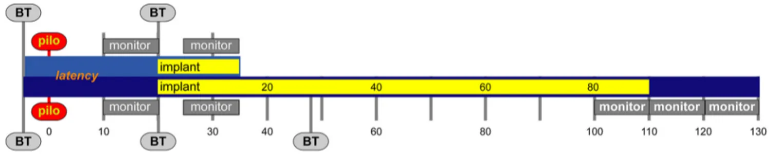
Video monitoring of seizure activity. Pilocarpine-treated animals were
continuously video recorded for the quantification of generalized (class 4 or 5) seizures (Fig. 1). At the end of the latent phase (i.e., following the first spontaneous seizure; 10⫾ 1 d after SE), the frequency and duration of generalized seizures were recorded through continuous video moni-toring for 10 d. Animals then received bilateral intrahippocampal im-plants of devices or devices plus electrodes, and video or video-EEG monitoring was resumed 5 d after implantation for an additional 10 d. At the conclusion of monitoring (⬃35 d post-SE), animals were anesthe-tized and the devices were retrieved for confirmation of GDNF secretion. In a separate experiment, we examined the long-term effects of GDNF device implantation on seizures as well as the persistence of these effects after the devices were removed. SE was induced as described above, and animals were randomly divided into the following two treatment groups: bilateral implants of GDNF devices or of control devices loaded with the
nonmodified parental ARPE-19 cell line. Animals were then monitored, and the seizure frequency and duration were recorded for two 10 d peri-ods (days 5–15 and days 80 –90 postimplantation;Fig. 1). After the sec-ond monitoring session, the devices were removed and the animals were monitored for an additional 20 d. At the conclusion of all experiments, hippocampi were removed and processed for quantification of GDNF levels.
For video-EEG monitoring, the rat headset was connected through a tripolar cable (PlasticsOne) to an EEG100C amplifier/MP150 Data Ac-quisition (Biopac) system, and signals were analyzed using AcqKnowl-edge version 5.0 software (Biopac). EEG seizures were defined as periods of paroxysmal activity of high frequency (⬎5 Hz) characterized by a more than threefold amplitude increment over baseline with progression of the spike frequency that lasted for a minimum of 5 s (Williams et al., 2009;Paradiso et al., 2011).
Open field arena and novel object recognition tests. The effects of GDNF
on anxiety-like behavior and cognition were investigated using the open field (OF) and novel object recognition (NOR) tests, respectively (exper-imental group, n⫽ 13; control group, n ⫽ 14). Each test was performed at the following three time points: before pilocarpine treatment (base-line); before device implantation (4 weeks after SE); and again 4 weeks after implant (Fig. 1). Before device implant, animals were randomly assigned to the following two groups: pilocarpine or pilocarpine ⫹ GDNF. For the OF test, animals were placed in an arena (75⫻ 75 ⫻ 30 cm) and video monitored for 30 min. Videos were analyzed (catalog #60201, ANY-maze) for distance moved, immobility, entries, and time spent in the center part of the arena. For the NOR, rats were tested in three phases. In the habituation phase (day 1), animals were allowed to freely explore the arena for 30 min. The following day, in the familiariza-tion phase, rats were transferred for 5 min to the arena, where two iden-tical objects were positioned in the central quadrant in opposite and symmetrical corners (A⫹A). In the testing phase (2 h later), animals were returned to the arena with one of the objects replaced with a novel one (A⫹B). The time spent exploring the objects was recorded by a blind investigator. To control for seizure effects in behavioral performance, rats were always tested at least 2 h after the last seizure.
Immunohistochemistry. A separate cohort of animals (N⫽ 25; 2 week
implant study: naïve, n⫽ 3; pilocarpine, n ⫽ 4; pilo⫹GDNF, n ⫽ 5; 12 week implant study: naïve, n⫽ 3; pilocarpine, n ⫽ 4; pilo⫹GDNF, n ⫽ 6) was unilaterally implanted with GDNF devices 20 d after pilocarpine-induced SE. After either 2 or 12 weeks, rats were deeply anesthetized and transcardially perfused with 200 ml of 0.9% ice-cold saline. Following saline perfusion, rats were decapitated and the devices removed. Brains were placed in Zamboni’s fixative (catalog #NC9335034, Thermo Fisher Scientific) for 1 week, then were embedded in paraffin. The 8-m-thick sections were cut coronally using a Leica RM2125RT microtome through the hippocampus (⫺2.3 to ⫺5.8 mm from bregma;Paxinos and Watson, 2007). Sections were dewaxed and rehydrated as previously described (Paradiso et al., 2009): two 10 min washes in xylene (catalog #1330-20-7, Sigma-Aldrich), 5 min in 100% ethanol, 5 min in 95% ethanol, 5 min in 80% ethanol, and then rehydration in distilled water for 5 min. All anti-gens were unmasked using a commercially available kit (Unmasker; cat-alog #T0010, Diapath), according to the manufacturer instructions. After washing in PBS, sections were incubated at room temperature with Tri-ton X-100 (0.3% in PBS; catalog #9002-93-1, Sigma-Aldrich) for 10 min, washed twice in PBS, and incubated with 5% bovine serum albumin and
5% serum of the species in which the secondary antibody was produced for 30 min. Sections were incubated overnight at 4°C in a humid atmo-sphere with a primary antibody specific for the following: glial fibrillary acid protein (GFAP; mouse polyclonal, catalog #AMAb91033, Atlas An-tibodies; PRID:AB_2665775), 1:100; doublecortin (DCX; rabbit poly-clonal; catalog #3973S, Cell Signaling Technology; PRID:AB_2276960), 1:400; parvalbumin (mouse monoclonal; catalog #235, Swant; PRID:
AB_10000343), 1:100; phosphorylated receptor tyrosine kinase RET (rab-bit polyclonal, catalog #sc-20252-R, Santa Cruz Biotechnology; PRID:
AB_2179766), 1:100; and neural cell adhesion molecule (NCAM; mouse monoclonal; catalog #MAB5324, Millipore; PRID:AB:_11210572), 1:400. After 5 min rinses in PBS, sections were incubated with Triton (as above; 30 min), washed in PBS, and incubated with a goat anti-mouse Alexa Fluor 594 (catalog #A-11058, Invitrogen; PRID:AB_142540) or Alexa Fluor 488 (catalog #A-21125, Invitrogen; PRID:AB_141593) sec-ondary antibody (1:250) for mouse primary antibodies, or with a goat anti-rabbit, Alexa Fluor 488 (catalog #A-11094, Thermo Fisher Scien-tific; PRID:AB_221544) or Alexa Fluor 594 secondary antibody (catalog #A-11012, Invitrogen; PRID:AB_141359, 1:250) for rabbit primary anti-bodies, at room temperature for 3 h. After staining, sections were washed in PBS, counterstained with 0.0001% 4⬘,6⬘-diamidino-2-phenylindole dihydrochloride (DAPI; catalog #D1306, Thermo Fisher Scientific; PRID:AB_2629482) for 15 min, and washed again before coverslipping. Omission of the primary antibody resulted in no specific staining.
Fluoro-Jade C staining. Slides were dewaxed and immersed for 5 min in
a solution of 1% sodium hydroxide in 80% ethanol, then rinsed for 2 min in 70% ethanol, for 2 min in distilled water, and incubated in 0.06% potassium permanganate solution for 10 min. Slides were then trans-ferred for 20 min into a 0.001% solution of FJC (catalog #AG325-30MG, Millipore) followed by three 1 min washes in distilled water. Slides were air dried on a slide warmer at 50°C for 5 min and cleared in xylene for 1 min. Coverslips were mounted using DPX (catalog #06522, Sigma-Aldrich) mounting media.
Morphological analyses. Quantitation of FJC, DCX, GFAP, PV, NCAM,
and pRET-positive cells was performed by an investigator who was blinded to the experimental condition, using a Leica DMRA2 Micro-scope. Sections were collected for each stain at 500m intervals through-out the hippocampus (⫺2.3 to ⫺5.3 mm relative to bregma;Paxinos and Watson, 2007), and the total numbers of immunopositive cells were counted bilaterally.
Hippocampal volumetry. FJC-stained sections were selected at three
levels in the dorsal hippocampus (⫺2.8, ⫺3.3, ⫺3.8 mm relative to bregma; N⫽ 25), and the area of the hippocampus was calculated (in square micrometers) on images obtained using the scan slide module of the MetaMorph software using a 6.3⫻ objective. For volumetric analysis of the hippocampus, values were obtained by applying the formulaV⫽ h
3 ⫻ 共Bmax⫹ bmin⫹
冑
Bmax⫻ bmin), where h⫽ 500m.Statistical methods. Mixed-design ANOVAs were used to determine
the effects of treatment, time, and any treatment⫻ time interactions.
Table 1statistical analyses performed. Where appropriate, post hoc com-parisons were conducted using t test and Fisher’s LSD test. Statistical analyses were performed using SPSS Statistics software (SPSS). All data were found to have a normal distribution, based on the Shapiro–Wilk test. In cases of the violation of the sphericity assumption, Huynh–Feldt corrected F values are given. The␣ value was set at 0.05, and exact p values are reported for significant results (Greenwald et al., 1996). Mean values and SEMs were calculated for each group and were expressed as the percentage of the naive group to facilitate statistical and graphic comparisons.
Results
Long-term encapsulated cell function and tissue levels of
GDNF in normal rats
GDNF-secreting devices were implanted in naive rats and
re-trieved at different time points (1, 2, 4, 8, 16, or 24 weeks after
implantation). All implanted devices were easily retrieved from
the brain with no host tissue adhering to the capsule wall. All
implants were located within the hippocampus and remained
intact, with no evidence that any capsule broke either in situ or
during the retrieval procedure. Quantification of device output
confirmed a robust and sustained secretion of GDNF from the
implanted devices (
Fig. 2
A). Before implantation, devices
se-creted 277.59
⫾ 10.72 ng/d GDNF. Explanted devices showed an
initial increase in secretion peaking at
⬃2–8 weeks (28,451.50 ⫾
192.84 ng/24 h), then tapering to a sustained amount that
re-mained well above preimplant levels for the 6 months of analysis
(
Fig. 2
A; 566.79
⫾ 192.47 ng/24 h). The continued delivery of
GDNF to the hippocampus significantly elevated local tissue
con-centrations of GDNF in a manner that paralleled device secretion
(
Fig. 2
B). Tissue levels of GDNF were highly increased within 1
week following implantation and reached peak levels at 2– 8
weeks (53.74
⫾ 8.89 ng), gradually decreasing while remaining
elevated for at least 6 months postimplantation (9.04
⫾ 2.82 ng).
The prolonged and elevated levels of GDNF within the
hip-pocampus were not associated with any changes in body weight
or general behavioral activity. Indeed, over the 6 months of
ob-servation all rats gained weight at a typical rate that was
indistin-guishable between intact rats and those implanted with GDNF
devices (
Fig. 2
C; t
(6)⫽ 0.27; p ⫽ 0.81). Similarly, no differences
were noted when animals were evaluated for time spent sleeping
(
Fig. 2
D; U
⫽ 15.50, z ⫽ ⫺0.45; p ⫽ 0.65), awake/inactive (U ⫽
15.00, z
⫽ ⫺0.51; p ⫽ 0.65), or moving across the cage (U ⫽
15.50, z
⫽ ⫺0.45; p ⫽ 0.65).
Seizures
Short-term (10 d) intrahippocampal implantation of GDNF
de-vices dramatically reduced the frequency of motor seizures in
pilocarpine-treated rats. Before GDNF treatment (first 10 d after
the first spontaneous seizure; i.e., 10 –20 d after SE), rats
exhib-ited 2.34
⫾ 0.05 motor seizures/d. Five to 15 d following
implan-tation (i.e., 25–35 d after SE), the frequency of motor seizures was
reduced to 0.62
⫾ 0.14 seizures/d (
Fig. 3
A; main effect of
treat-ment: F
(1,8)⫽ 189.16; p ⬍ 0.001). This effect had a relatively rapid
onset, as it was observed within the first week of implantation and
was confirmed by the significant time
⫻ treatment interaction
(F
(9,72)⫽ 2.14; p ⫽ 0.037). While the frequency of motor seizures
was reduced by
⬃75%, their duration was not altered relative to
preimplantation (
Fig. 3
B; t
(8)⫽ 1.29, p ⫽ 0.23). As expected, EEG
monitoring detected a greater number of seizures (i.e., animals
were experiencing nonmotor as well as motor seizures). Again,
the frequency of EEG seizures was strongly reduced in animals
implanted with GDNF devices compared with those implanted
with nonmodified parental ARPE-19 cells (
Fig. 4
A), but their
duration was identical in both groups (
Fig. 4
B). Representative
EEG traces from nonmotor and motor seizures are shown in
Figure 4
, C and D. The magnitude of GDNF secretion from
ex-planted devices (
Fig. 3
C; preimplant, 252.28
⫾ 7.34 ng/24 h;
postimplant, 2339.26
⫾ 194.86 ng/24 h) and the levels of GDNF
within the tissue (41.17
⫾ 3.69 ng; data not shown) were
compa-rable to those observed in the intact, normal brain.
We then extended these observations to investigate longer
lasting implantations and the postimplantation persistence of the
effects. We implanted the devices as in the previous experiment,
but left them in the brain much longer (90 d), while monitoring
seizures in the following two epochs: 5–15 d (as above) and 80 –90
d postimplantation. We then retrieved the devices and continued
monitoring for 20 additional days. We found that the reduction
in seizure frequency increased in magnitude over time, from 84%
at 5–15 d postimplantation to 93% in the same animals at 80 –90
d postimplantation (
Fig. 5
A; F
(1,13)⫽ 698.62, p ⬍ 0.001).
Implan-tation with control devices loaded with parental ARPE-19 cells
did not alter seizure frequency at any time point. No change in
seizure duration was observed at any time point in GDNF-treated
animals and controls (F
(1,13)⫽ 0.57; p ⫽ 0.46).
Interestingly, a persistent effect was observed when the GDNF
devices were retrieved 90 d following implantation (
Fig. 5
A).
Although the frequency of spontaneous seizures did increase over
preremoval values, it was still significantly lower than in control
epileptic animals (65% lower;
Fig. 5
A). Incidentally, it should be
noted that, as previously reported (
Soukupova´ et al., 2014
), the
frequency of seizures tended to increase in time in untreated
animals.
At explant, the secretion of GDNF from devices remained
high (
Fig. 5
B; preimplant, 283.36
⫾ 8.82 ng/24 h; postexplant,
4500.50
⫾ 410.93 ng/24 h). Relative to tissue levels measured
Table 1. Effects of GDNF treatment on seizure frequency and histological analyses
Experiment Treatment Group size Main effect of treatment Treatment⫻ time
Seizure frequency
2 week implant Pilo⫹ GDNF 9 F(1,8)⫽ 189.16; p ⬍ 0.001 F(9,72)⫽ 2.14;p ⫽ 0.037
12 week implant Pilo⫹ Parental 8 F
(1,13)⫽ 698.62; p ⬍ 0.001 F(4,52)⫽ 240.56; p ⬍ 0.001
Pilo⫹ GDNF 8
Behavioral testing
Open field Pilo 14 F
(1.25)⫽ 4.46; p ⫽ 0.04 F(2,50)⫽ 11.12; p ⬍ 0.001
Pilo⫹ GDNF 13
Novel object recognition Pilo 14 F
(1.25)⫽ 2.07; p ⫽ 0.04 F(2,50)⫽ 11.82; p ⬍ 0.001
Pilo⫹ GDNF 13
Hippocampal volume
2 week implant Naive 3
Pilo 4 F(2,12)⫽ 7.23; p ⫽ 0.011 NA
Pilo⫹ GDNF 5
12 week implant Naive 3
Pilo 4 F(2,11)⫽ 96.87; p ⬍ 0.001 NA
Pilo⫹ GDNF 5
Histology
2 week implant Treatment⫻ hippocampal level
FJ C Naive 3 Pilo 4 F(2,23)⫽ 17.59; p ⬍ 0.001 F(10,115)⫽ 2.51; p ⫽ 0.009 Pilo⫹ GDNF 5 PV Naive 3 Pilo 4 F(2,23)⫽ 17.54; p ⬍ 0.001 F(10,115)⫽ 3.42; p ⬍ 0.001 Pilo⫹ GDNF 5 GFAP Naive 3 Pilo 4 F(2,23)⫽ 0.14; p ⫽ 0.88 NA Pilo⫹ GDNF 5 DCX Naive 3 Pilo 4 F(2,23)⫽ 6.67; p ⫽ 0.005 F(10,115)⫽ 3.88; p ⬍ 0.001 Pilo⫹ GDNF 5 NCAM Naive 3 Pilo 4 F(2,23)⫽ 22.41; p ⬍ 0.001 F(10,115)⫽ 2.06; p ⫽ 0.034 Pilo⫹ GDNF 5 pRET Naive 3 Pilo 4 F(2,23)⫽ 158.59; p ⬍ 0.001 F(10,115)⫽ 6.71; p ⬍ 0.001 Pilo⫹ GDNF 5 12 week implant FJ C Naive 3 Pilo 4 F(2,21)⫽ 9.71; p ⫽ 0.09 NA Pilo⫹ GDNF 6 PV Naive 3 Pilo 4 F(2,21)⫽ 54.71; p ⬍ 0.001 F(10,105)⫽ 2.43; p ⫽ 0.012 Pilo⫹ GDNF 6 GFAP Naive 3 Pilo 4 F(2,21)⫽ 0.11; p ⫽ 0.90 NA Pilo⫹ GDNF 6 DCX Naive 3 Pilo 4 F(2,21)⫽ 53.96; p ⬍ 0.001 F(10,105)⫽ 7.833; p ⬍ 0.001 Pilo⫹ GDNF 6 NCAM Naive 3 Pilo 4 F(2,19)⫽ 46.63; p ⬍ 0.001 F(10,95)⫽ 2.63; p ⫽ 0.04 Pilo⫹ GDNF 6 pRET Naive 3 Pilo 4 F(2,21)⫽ 205.86; p ⬍ 0.001 F(10,105)⫽ 22.70; p ⬍ 0.001 Pilo⫹ GDNF 6
immediately after device retrieval, GDNF levels in the
hippocam-pus were reduced by
⬎98% when quantified 20 d after device
removal (1.57
⫾ 1.01 ng; data not shown). This observation
sug-gests that the persistent reduction in seizure frequency was not
related to residual GDNF in the hippocampus.
Open field and novel object recognition
Pilocarpine-induced epilepsy produced significant impairments
in the open-field and novel object recognition tests.
Pilocarpine-treated rats alternated periods of hyperactivity and of freezing,
spending significantly more time in the central part of the testing
arena (F
(2,50)⫽ 11.12; p ⬍ 0.001). In contrast, GDNF treatment
prevented the development of this phenotype, with treated rats
performing comparably to naive ones (
Fig. 6
A; t
(26)⫽ 10.35; p ⫽
0.004). Importantly, this change in behavior could not be
attrib-uted to alterations in general activity because, overall, the
dis-tance traveled and the immobility time did not change (F
(2,50)⫽
0.54, p
⫽ 0.59; and F
(2,50)⫽ 0.05; p ⫽ 0.95; respectively; data not
shown).
GDNF also reversed the cognitive impairment associated with
pilocarpine-induced epilepsy. At baseline, all rats displayed a
clear exploratory preference for the novel object, but this
prefer-ence completely disappeared 4 weeks after SE (F
(2,50)⫽ 11.82;
p
⬍ 0.001). As shown in
Figure 6
B, pilocarpine-treated rats spent
equal amounts of time exploring the new and the previously
presented objects both 4 and 8 weeks after SE (i.e., 4 weeks after
the implant of a control device; t
(26)⫽ 0.89; p ⫽ 0.38). GDNF
treatment reinstated the ability to distinguish between the novel
and the familiar object (
Fig. 6
B; no GDNF: t
(13)⫽ 1.33, p ⫽ 0.21;
GDNF: t
(12)⫽ 7.07, p ⬍ 0.001).
The findings described above were confirmed in the animals
used for these behavioral experiments: the frequency of seizures
was reduced by 74% relative to preimplantation (t
(8)⫽ 13.66, p ⬍
0.001), and the magnitude of GDNF secretion was monitored
preimplantation and after retrieval of the device from the
hip-pocampus were 144.03
⫾ 11.81 and 3641.70 ⫾ 300.30 ng/24 h,
respectively.
Immunohistochemistry
Neuroprotection
To analyze the morphological effects of GDNF on the epileptic
hippocampal tissue, we implanted a single device in one
hip-pocampus 20 d after pilocarpine-induced SE, and sacrificed the
rats 2 and 12 weeks thereafter. GDNF treatment exerted a
pro-nounced neuroprotective effect, as evidenced by the quantitation
of hippocampal volume, the counts of degenerating neurons, and
the counts of parvalbumin-positive cells. These effects were
ob-served at both 2 and 12 weeks postimplantation and were
com-parable in magnitude between both the implanted and
nonimplanted hemispheres. A generalized atrophy of the
hip-pocampus was observed in epileptic animals. Compared with
naive controls, the hippocampal volume of pilocarpine-treated
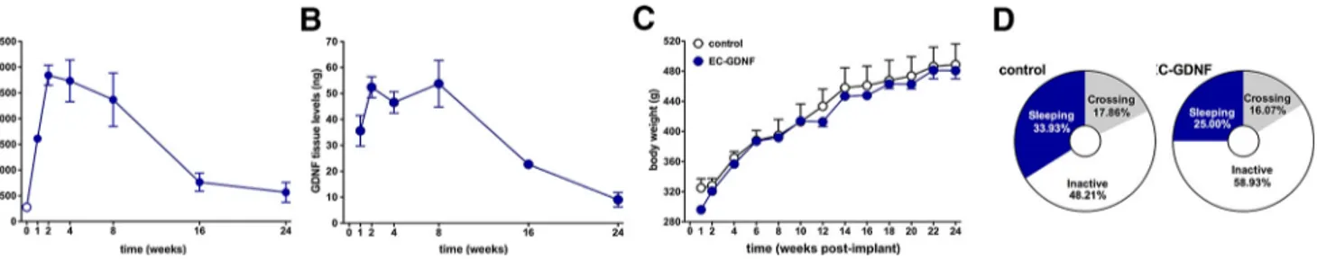
Figure 2. Encapsulated cell function, tissue levels of GDNF, body weight, and general behavioral activity in intact rats following bilateral intrahippocampal implantation of GDNF devices. A, GDNF secretion from devices explanted from the hippocampus. Devices were implanted in intact rats and removed after 1, 2, 4, 8, 16, and 24 weeks. GDNF secretion dramatically increased over preimplant values at 2– 8 weeks and then slowly declined a sustained amount that remained above preimplant levels for the 6 months of analysis. B, Hippocampal tissue levels of GDNF during device implantation. Tissue levels were elevated within 1 week, reached and maintained a peak at 2– 8 weeks, and then slowly decreased, remaining elevated for the duration of the experiment. C, D, Device implantation and elevation of tissue GDNF levels did not alter body weight (C) or affect the general pattern of behavioral activity (D; Mann–Whitney U test). All data are expressed as the mean⫾ SEM of 4 animals per time point in A and B, and 40 animals in C and D.
Figure 3. GDNF significantly reduces seizure frequency in pilocarpine-treated rats. Rats were video monitored before (10 –20 d after SE) and following intrahippocampal GDNF device implan-tation (25–35 d after SE). A, B, The frequency of seizures was reduced by 84% relative to preimplanimplan-tation levels (A), whereas seizure duration was not altered (B). C, Levels of GDNF secretion before implantation and after retrieval at the conclusion of the video monitoring. Data are the mean⫾ SEM of nine animals monitored before and after GDNF treatment. ***p ⬍ 0.001 based on paired t test.
rats was reduced by
⬃20% 2 weeks after pilocarpine treatment,
and by 33% after 12 weeks (2 weeks: F
(2,12)⫽ 7.23; p ⫽ 0.011; 12
weeks: F
(2,11)⫽ 96.87; p ⬍ 0.001;
Figure 7
). Treatment with
GDNF reversed this loss of hippocampal volume (
Fig. 7
).
FJC staining was used to identify degenerating neurons and to
determine the ability of GDNF to attenuate damage (
Ehara and
Ueda, 2009
) (
Fig. 8
). Quantitative analysis revealed a significant
increase in degenerating cells in CA1, CA3, and hilus of the
den-tate gyrus following pilocarpine treatment compared with naive
cells (597% and 160% at 2 and 12 weeks after postpilocarpine
treatment, respectively). Similar to the GDNF-induced
preven-tion of hippocampal atrophy, a significant attenuapreven-tion of
neuro-nal degeneration was observed in treated rats, with a much
smaller increase in FJC-positive cells (187% and 32% at 2 and 12
weeks, respectively) compared with cells from naive animals
(F
(2,23)⫽ 17.59, p ⬍ 0.001; and F
(2,21)⫽ 9.71, p ⫽ 0.09,
respectively).
Finally, because epilepsy has been consistently linked to
changes in GABAergic neurotransmission, including a loss of PV
and somatostatin cells (
Mazzuferi et al., 2010
;
Soukupova´ et al.,
2014
), we quantified the ability of GDNF to protect this cell
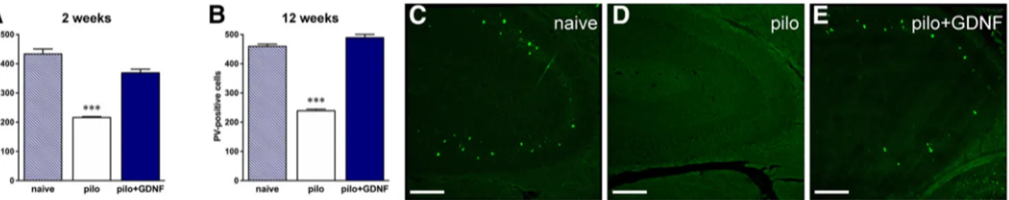
pop-ulation. As shown in
Figure 9
, pilocarpine significantly reduced
the number of PV-positive neurons in the hippocampus relative
to naive rats (2 weeks: F
(2,23)⫽ 17.54, p ⬍ 0.001; 12 weeks:
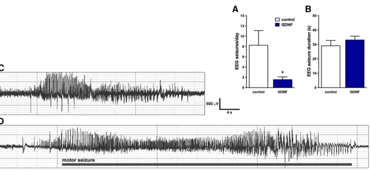
Figure 4. A, B, Electrical seizure frequency (A) and duration (B) were recorded 5–15 d after implantation with the nonmodified parental ARPE-19 cells or GDNF devices in chronically epilepticanimals. C, D, Representative EEG patterns in the hippocampus during nonmotor (C) and motor (D) seizures in GDNF device-implanted animals. Identical patterns were observed in animals implanted with empty devices. The horizontal bar in D indicates the motor part of the seizure. All data are expressed as the mean⫾ SEM of seven animals per group. *p ⬍ 0.05. Student’s t test for unpaired data.
Figure 5. Long-term and persistent effects of GDNF on seizure frequency. Animals were implanted either with devices loaded with the nonmodified parental ARPE-19 cell line (N⫽ 8) or with GDNF-secreting devices (N⫽8),asinFigure 3. A, Seizure frequency and duration were recorded before implantation (10 –20 d after SE) and for two 10 d periods postimplantation (5–15 and 80 –90 d after implantation; see text for details). At the conclusion of the second monitoring session (days 80 –90), devices were retrieved and animals were monitored for an additional 20 d. Note that the effects of GDNF persisted even after device removal (65% decrease in seizure frequency relative to epileptic controls). B, Levels of GDNF secreted before implantation and immediately following retrieval. All data are expressed as the mean⫾SEMof16animals***p⬍0.001comparedwithcontroldevices;●●●p⬍0.001comparedwithdevicepreremovalvalues(80–90dpostimplant).
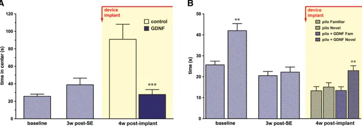
Figure 6. A, B, Effect of GDNF in the open field (A) and in the novel object recognition test (B). Rats (N⫽27)weretestedbeforeanymanipulation(baseline)and4weeksafterpilocarpine-induced
SE. Following the post-pilocarpine test session, a subgroup of animals (n⫽ 13) was implanted with GDNF devices. Pilocarpine-induced epilepsy progressively reduced the natural tendency of the rat to avoid the central region of the arena and the exploratory preference for the novel object. A, B, GDNF restored a normal behavior both as measured by the time spent in the central region of the arena (A) and as the ability to distinguish new from familiar objects (B). All data are expressed as the mean⫾ SEM. **p ⬍ 0.01; ***p ⬍ 0.001 of 27 animals. Multiple comparisons were based on significant main effects or interactions resulting from the ANOVAs described in the Materials and Methods and Results. w, Weeks.
Figure 7. GDNF reverts the loss of hippocampal volume that occurs following pilocarpine. A, B, Sections were taken from the dorsal hippocampus of naive (n⫽ 3), pilocarpine-treated (n ⫽ 4), and pilocarpine-treated rats treated with GDNF (n⫽ 5–6) for either for 2 (A) or 12 (B) weeks. Data are presented as absolute values of hippocampal volumes. Because no differences were noted between the right and left hemispheres in any group, data from both hemispheres were combined. All data are expressed as the mean⫾ SEM. *p ⬍ 0.05; ***p ⬍ 0.001 of 25 animals. Multiple comparisons were based on significant main effects resulting from the ANOVAs described in the Materials and Methods and Results. pilo, Pilocarpine.
Figure 8. GDNF significantly reduces ongoing neuronal degeneration. A, B, The effects of pilocarpine and 2 (A) or 12 (B) weeks of treatment with GDNF on the total numbers of FJC-positive cells in the hippocampus. While pilocarpine significantly increased the numbers of degenerating FJC-positive neurons at both time points, this effect was significantly attenuated by GDNF. Data are expressed as the mean⫾ SEM of three to six animals per group. *p ⬍ 0.05, ***p ⬍ 0.001 vs naive; °p ⬍ 0.05 vs pilo. Multiple comparisons were based on significant main effects resulting from the ANOVAs described in the Materials and Methods and Results. Representative images taken from naive, pilocarpine-treated, and pilocarpine-treated rats treated with GDNF at 2 weeks are shown in C–E. Scale bar, 500m. pilo, Pilocarpine.
F
(2,21)⫽ 54.71, p ⬍ 0.001). GDNF reversed this loss of
PV-positive cells.
Epilepsy-associated astrocytosis was evaluated using GFAP
immunofluorescence (
Vezzani et al., 2000
). The quantification of
GFAP-positive cells revealed that treatment with neither
pilo-carpine nor GDNF in pilopilo-carpine-treated animals altered the
number of astrocytes (
Fig. 10
A, B; 2 weeks: F
(2,23)⫽ 0.14, p ⫽
0.88; 12 weeks: F
(2,21)⫽ 0.11, p ⫽ 0.90). However, many of the
GFAP-positive cells in epileptic controls displayed short, thick
processes, an indication of active astrocytosis (
Fig. 10
D, insert),
whereas GFAP-positive cells of GDNF-treated rats were similar
to those of naive animals, with a small cell body and thin
pro-cesses (
Fig. 10
C,E, inserts).
Neurogenesis
As shown in
Figure 11
, GDNF treatment reversed the loss of
DCX-positive cells that occurred following pilocarpine. In naive
control rats, DCX-positive cells were present in the subgranular
zone with notable elongations extending across the granular layer
of the dentate gyrus region (
Fig. 11
C). In line with previous
re-ports (
Hattiangady et al., 2004
), chronic epilepsy significantly
decreased the numbers of these cells (by 48% and 70% at 2 and 12
weeks, respectively) and led to shorter and ectopic elongations. In
contrast, treatment with GDNF restored the number of
DCX-positive cells (2 weeks: F
(2,23)⫽ 6.67, p ⫽ 0.005; 12 weeks:
F
(2,21)⫽ 53.96, p ⬍ 0.001). Qualitatively, these cells had their
typical morphology with increased length and number of
elon-gations, together with reduced cluster formation and reduced
numbers of ectopic cells. The effects of GDNF were bilateral and
more pronounced following 12 weeks of treatment.
Target engagement
Quantitative immunohistochemistry confirmed GDNF receptor
engagement following GDNF device implantation. GDNF signals
through a multicomponent receptor, first binding the
Glyco-sylphosphatidylinisotol-anchored receptor
␣1 [GDNF family
re-Figure 9. A, B, GDNF reverts pilocarpine (pilo)-induced degeneration of hippocampal parvalbumin (PV)-positive cells after both 2 weeks (A) and 12 weeks (B) of treatment. Data refers to the total
number of positive cells and are expressed as the mean⫾ SEM of three to six animals per group.***p ⬍ 0.001. Multiple comparisons were based on significant main effects resulting from the ANOVAs described in the Materials and Methods and Results. C–E, Representative images taken from the CA3 area of naive, pilocarpine-treated, and pilocarpine-treated rats treated with GDNF at 2 weeks are shown. Scale bar, 100m.
Figure 10. A, B, Astrocyte density in the hippocampus is not altered by treatment with either pilocarpine or GDNF for 2 (A) or 12 (B) weeks. Data refer to the total number of positive cells and are
expressed as the mean⫾SEMofthreetosixanimalspergroup.C–E,Representativeimagestakenfromthehilusofthedentategyrusareaofnaive,pilocarpine-treated,andpilocarpine-treatedrats treated with GDNF at 2 weeks are shown. Scale bar, 100m. Higher-magnification inserts illustrate the changes in the morphology of GFAP-positive cells.
Figure 11. A, B, GDNF normalizes neurogenesis after 2 (A) and 12 (B) weeks of treatment. Data refer to the total number of positive cells and are expressed as the mean⫾ SEM of three to six
animals per group. ***p⬍0.001.MultiplecomparisonswerebasedonsignificantmaineffectsresultingfromtheANOVAsdescribedintheMaterialsandMethodsandResults.C–E,Representative DCX immunofluorescence (in red) images taken from the granular zone of the dentate gyrus of naive, pilocarpine-treated, and pilocarpine-treated rats treated with GDNF at 2 weeks are shown. Nuclei are marked by DAPI (blue). DCX-positive cells in the naive dentate gyrus are located in the subgranular zone and present notable elongations across the granular layer. Pilocarpine causes a decrease in DCX-positive cells, which is associated with fewer elongations in the remaining cells. Treatment with GDNF reverses the loss of DCX-positive cells and normalizes cellular morphology. Scale bar, 20m. pilo, Pilocarpine.
ceptor
␣1 (GFR␣1)], with the resulting complex recruiting the
transmembrane receptor tyrosine kinase Ret or the NCAM to
initiate downstream activation of FAK (focal adhesion kinase)
and FYN (proto-oncogene tyrosine-protein kinase) signaling
pathways (
Airaksinen and Saarma, 2002
;
Paratcha et al., 2006
;
Duveau and Fritschy, 2010
). To explore the role of
NCAM-mediated GDNF effects on seizure frequency and
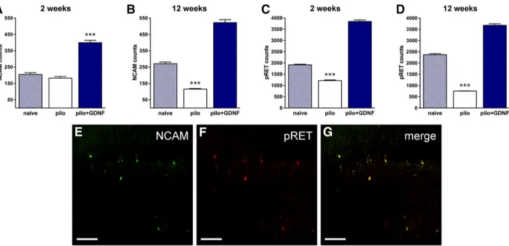
neuroprotec-tion, we quantified the numbers of NCAM-positive cells (
Fig.
12
A, B). In line with previous studies (
Ledergerber et al., 2006
),
we observed a decrease in NCAM-positive cells in the
hippocam-pus of pilocarpine-treated animals at both 2 and 12 weeks
com-pared with naive animals (12% and 57%, respectively). In
contrast, NCAM expression was dramatically increased by GDNF
treatment (195% at 2 weeks and 193% at 12 weeks: 2 weeks: F
(2,23)⫽ 22.41, p ⬍ 0.001; 12 weeks: F
(2,19)⫽ 46.63, p ⬍ 0.001). A
similar pattern was observed when the expression of
phosphory-lated Ret was assessed (
Fig. 12
C,D). Pilocarpine treatment
signif-icantly reduced the numbers of pRET-positive cells (by 37% and
68%, respectively, 2 and 12 weeks following administration).
GDNF treatment resulted in a marked increase (100% and 55%,
respectively, after 2 and 12 weeks of treatment; 2 weeks: F
(2,23)⫽
158.59, p
⬍ 0.001; 12 weeks: F
(2,21)⫽ 205.86, p ⬍ 0.001).
Discussion
Current pharmacological therapies for epilepsy are palliative at
best, frequently produce adverse effects, and are commonly
com-pletely ineffective. There is, accordingly, an urgent need for novel
therapies in the treatment of epilepsy. Neurotrophic factors such
as GDNF may have the capacity to provide therapeutic benefits,
and encapsulated cell technologies might be able to provide a safe
means of selectively targeting and continuously delivering GDNF
to the epileptic area (
Lindvall and Wahlberg, 2008
;
Eriksdotter-Jönhagen, 2012
;
Wahlberg et al., 2012
;
Orive et al., 2015
). For
these reasons, we engineered ARPE-19 cells to produce high
lev-els of GDNF and enclosed them in a semipermeable capsule for
implantation into the brain. The basic principle of this system is
that the membrane allows oxygen and nutrients to enter and
nourish the encapsulated cells while also allowing GDNF to leave
the capsule and diffuse into the surrounding brain tissue, all the
while eliminating exposure to the host immune system. This
study provides important new data regarding the translation of
this approach for continued development and human use. We
report that GDNF devices can be implanted in the temporal lobe
for prolonged periods of time while significantly elevating tissue
levels of GDNF. The sustained delivery of GDNF was efficacious,
as demonstrated by a pronounced and lasting reduction in
sei-zure frequency. Importantly, nonmotor EEG seisei-zure frequency
was also reduced, indicating that the treatment actually
sup-presses seizures and does not merely attenuate their severity. This
finding is in line with another study, in which we report that
identical GDNF implants can also reduce the frequency of EEG
seizures in the kainic acid model (
Nanobashvili et al., 2018
). In
the present study, these favorable effects were rapid and
progres-sive, with the seizure frequency reduced by 75% within 2 weeks
after treatment and by 93% after 3 months of treatment. These
effects are truly dramatic, considering that, in the pilocarpine
model, only part of the seizures originates from the hippocampus
(
Toyoda et al., 2013
). Therefore, this observation prompts the
speculation that the hippocampus may also be implicated in
sei-zures originating in other areas, and that treating the
hippocam-pus may produce effects that surpass those expected (e.g., that the
response to GDNF is nonlinear). The benefits of GDNF also
ap-peared to extend beyond a simple symptomatic effect as it
ex-tended, even if in a less robust manner, well beyond removal of
the devices. The concept of a potential disease-modifying effect is
Figure 12. GDNF receptor engagement. A, B, Immunohistochemical quantification of NCAM expression after 2 (A) or 12 (B) weeks of GDNF treatment. Pilocarpine treatment significantly reduces the number of NCAM-positive cells at both time points. In contrast, GDNF increases NCAM expression above the levels observed in both the naive and pilocarpine-treated groups. C, D, Immunohis-tochemical quantification of phosphorylated Ret expression after 2 (C) or 12 (D) weeks of GDNF treatment. Pilocarpine treatment significantly reduces the number of pRet-positive cells at both time points. In contrast, GDNF increases pRET expression above the levels observed in both the naive and pilocarpine-treated groups. Data are expressed as the mean⫾ SEM of three to six animals per group. ***p⬍ 0.001. Multiple comparisons were based on significant main effects resulting from the ANOVAs described in the Materials and Methods and Results. E, F, Representative images of NCAM and pRET double staining in GDNF-treated animals are shown. G, The merged image. Similar patterns (although quantitatively different) were observed in naive and pilocarpine-treated animals. pilo, Pilocarpine.
further supported by the observation that the reduction in seizure
frequency was accompanied by improvements in cognition and
anxiety, both significant comorbidities of epilepsy (
Strzelczyk et
al., 2017
;
Michaelis et al., 2018
). These benefits occurred without
classic signs of mistargeted neurotrophic factor delivery such as
alterations in food consumption, changes in daily activity, or
other overt neurobehavioral changes. The observation that
GDNF alleviated behavioral alterations, an expression of
comor-bidities of pilocarpine-induced epilepsy, is of particular
impor-tance. Although these comorbidities are well known and can even
predate the diagnosis of epilepsy itself, no effective treatment has
yet been developed. Once spontaneous seizures begin to occur
and the diagnosis of epilepsy is made, the disease often progresses
with increased severity of seizures and the appearance of
neuro-logical impairments. The etiology underlying these comorbid
changes in humans is complex and includes numerous factors,
like age of onset, seizure type/severity/duration, use of
antiepilep-tic medications, and neuroanatomical changes (
Pitka¨nen and
Sutula, 2002
;
Aldenkamp and Arends, 2004
;
Elger et al., 2004
;
Lin
et al., 2012
). These neurobehavioral changes can also be either
chronic, as part of the underlying etiology of the disease itself, or
in constant dynamic evolution, due to recurrent seizures and
interictal spikes. The complex interplay of chronic and dynamic
underlying mechanisms has made it difficult to develop therapies
capable of treating the broad spectrum of behavioral deficits in
epilepsy, and the data provided here suggest that direct CNS
de-livery of trophic molecules such as GDNF can, at least in part,
fulfill this requirement. In fact, pilocarpine SE induces a
progres-sive increase in seizure frequency as well as significant
impair-ments in the normal exploratory behavior (
Brandt et al., 2006
;
Tchekalarova et al., 2017
; avoidance of open space) and in the
ability of the rat to use learning and recognition memory
capa-bilities (
Ainge et al., 2007
;
Wood et al., 2000
), and all these signs
of disease are strongly attenuated by GDNF.
The potential disease-modifying benefits of GDNF might be
explained, at least in part, by long-lasting anatomical
adapta-tions. A number of morphological changes can occur in epilepsy,
including overt cell loss, synaptic reorganization, and
neurogen-esis. First, we applied volumetric and immunohistochemical
analyses to both confirm the effects of pilocarpine on cellular
degeneration and investigate whether GDNF can alter this
pat-tern (
Niessen et al., 2005
). Consistent with reports of
morpho-logical changes in hippocampal volume and shape in TLE (
Van
Paesschen et al., 1995
;
Bernasconi et al., 2003
;
Hibar et al., 2016
),
we found that pilocarpine-induced SE produced a severe loss in
hippocampal volume together with neurodegeneration and
as-trocytosis. Treatment with GDNF reversed both of these
patho-logical changes and also reversed the loss of GABAergic
parvalbumin-positive hippocampal cells that was previously
found to continue for weeks after pilocarpine-induced SE (
Sou-kupova´ et al., 2014
). The roles of GABAergic transmission in
epilepsy are complex, with some data indicating that increases in
GABAergic activity occur during the interictal phase and just
before seizure onset (
D’Antuono et al., 2004
;
Ellender et al., 2014
;
Yekhlef et al., 2015
;
de Curtis and Avoli, 2016
). Elevated
GABAe-rgic activity leads to increased extracellular potassium, which
supports hyperexcitability and epileptiform synchronization
(
Zuckermann and Glaser, 1968
;
Fertziger and Ranck, 1970
;
de
Curtis and Gnatkovsky, 2009
). On the other hand, there are
stud-ies suggesting that the impaired GABAergic inhibition, related to
a selective loss of inhibitory interneurons, accounts for
epilepti-form activity (
Wendling et al., 2002
;
Forte et al., 2016
). Although
electrophysiological validations are required, it is tempting to
speculate that the normalization of GABAergic transmission by
GDNF prevents the broad spatial hypersynchronous recruitment
of neurons and interneurons observed at the transition from
in-terictal to ictal activity (
Schevon et al., 2012
;
Fujita et al., 2014
).
GDNF can also promote the functional and morphological
dif-ferentiation of GABAergic neurons via GFR␣1 (
Pozas and
Iba´n
˜ez, 2005
;
Paratcha et al., 2006
;
Perrinjaquet et al., 2011
).
Because defects in cortical GFR
␣1 signaling increase excitability
and sensitivity to subthreshold doses of epileptogenic agents
(
Canty et al., 2009
), it can be hypothesized that GDNF, via
GFR␣1 activation, restores inhibitory neurotransmission in
epi-leptic animals by supporting the survival of GABAergic neurons.
As brain insults can induce neurogenesis of GABAergic cells
(
Magnusson et al., 2014
), it is possible that exogenous
GDNF-GFR
␣1 may redirect hippocampal granule cell neurogenesis after
seizures toward inhibitory GABAergic cells (
Marks et al., 2012
).
The direct dependence of all the effects observed after GDNF
treatment on GDNF receptors is difficult to assess with the
cur-rently available tools. Therefore, we attempted to at least
demon-strate that the procedures led to target (GDNF receptor)
engagement. To pursue this aim, we investigated the changes in
NCAM and pRET. We found that GDNF reversed the decreased
density of NCAM receptors and the expression of pRET in
pilocarpine-treated animals, leading to levels well above those
found in naive animals. These findings suggest a superactivation
of GDNF receptors. In the adult hippocampal formation, NCAM
is highly expressed in newly generated granule cells (
Seki and
Arai, 1993
). The increased expression may reflect its role in
reg-ulating axonal outgrowth, synapse formation (
Muller et al.,
1996
), and cell survival. The pattern was similar, with even more
robust differences, when we assessed the expression of pRET. It
has been previously reported that mRNA levels for GDNF and
GDNF receptors (GFR
␣1 and RET) are region, cell, and insult
specific (
Reeben et al., 1998
;
Kokaia et al., 1999
;
Kanter-Schlifke
et al., 2007
). Here, we observe a robust increase of pRET
expres-sion indicating the activation of NCAM-independent GDNF
sig-naling pathways.
The results described here form part of a program aimed at
developing the use of polymer-encapsulated GDNF-secreting
cells for direct and local delivery of GDNF to the brain of patients
with epilepsy. These studies consistently demonstrated
long-term and stable bioactive effects at doses shown to be safe in
preclinical safety studies. To our knowledge, this is the first
cel-lular delivery system capable of establishing the essential
prereq-uisites of sustained, targeted, long-term delivery of sufficient
quantities of GDNF to the temporal lobe. Based on the safety and
efficacy of this platform technology, it represents a potentially
novel and effective treatment for epilepsy.
References
Ainge JA, van der Meer MA, Langston RF, Wood ER (2007) Exploring the role of context-dependent hippocampal activity in spatial alternation be-havior. Hippocampus 17:988 –1002.
Airaksinen MS, Saarma M (2002) the GDNF family: signalling, biological functions and therapeutic value. Nat Rev Neurosci 3:383–394. Aldenkamp A, Arends J (2004) The relative influence of epileptic EEG
dis-charges, short nonconvulsive seizures, and type of epilepsy on cognitive function. Epilepsia 45:54 – 63.
Bernasconi N, Bernasconi A, Caramanos Z, Antel SB, Andermann F, Arnold DL (2003) Mesial temporal damage in temporal lobe epilepsy: a volu-metric MRI study of the hippocampus, amygdala and parahippocampal region. Brain 126:462– 469.
Brandt C, Gastens AM, Sun Mz, Hausknecht M, Lo¨scher W (2006) Treat-ment with valproate after status epilepticus: effect on neuronal damage,