Alma Mater Studiorum – Università di Bologna
DOTTORATO DI RICERCA IN
Biologia Cellulare e Molecolare
Ciclo XXVI
Settore Concorsuale di afferenza: 05/D1 Settore Scientifico disciplinare: BIO/09
TITOLO TESI
I
N VITRO STUDY OF THE OSTEOCYTES RESPONSE TO HYPOXIA AND THEIRREGULATION OF BONE HOMEOSTASIS
Presentata da: Monica Montesi
Coordinatore Dottorato Relatore
Prof. Vincenzo Scarlato Prof. Antonio Contestabile Dr.ssa Alina Beraudi
Dr.ssa Susanna Stea
3 INDEX
1.
ABSTRACT
5
2.
INTRODUCTION
7
2.1
Bone tissue
7
2.2
Bone cells
11
2.2.1 Osteocytes12
2.2.2 Intercellular Cross-Talk and bone homeostasis
14
2.3
Bone unloading and hypoxia
18
2.4 Osteocyte hypoxia
19
3.
AIMS OF THE PROJECT
23
4.
MATERIALS and METHODS
25
4.1
Cells culture
25
4.2
Hypoxia conditions
26
4.3
Conditioning of Osteoblasts and Osteoclasts
28
4.3.1 RAW 264.7 conditioning
28
4.3.2 MC3T3-E1 conditioning
28
4.4
Evaluation of Cell Viability and Apoptosis
29
4.5
TRAP staining of conditioned RAW 264.7
31
4.6
Alkaline phosphatase assay of conditioned MC3T3-E1
31
4.6 Dosage of soluble factors RANKL, OPG , PGE
2and
Sclerostin (ELISA assay)
32
4.7 Quantitative Real-Time PCR
33
4.8
Western blotting
34
4
5.
RESULTS
37
5.1 Definition of the best culture conditions for MLO-Y4 cells 37
5.2
Hypoxia induction
38
5.3
Osteocyte proliferation, viability and apoptosis
40
5.4
Expression of ORP150
42
5.5
Dosage of soluble factors PGE
2,
RANKL, OPG, and
Sclerostin
44
5.6
Trap staining and induction of Osteoclastogenesis
45
5.6.1 Multinucleated Trap positive cells (MNC TRAP+ cells) results 46 5.6.2 Pixel area covered by Trap positive cells47
5.7
Osteoblasts response to the hypoxic osteocytes conditioning
media
49
5.7.1 Osteoblast viability (MTT assay)
49
5.7.2 Alkaline Phosphatase (ALP activity) and β-catenin mRNAexpression
50
6.
DISCUSSION
53
6.1 Osteocyte response to Hypoxia condition
54
6.2 Hypoxic osteocytes regulation on bone homeostasis
56
7.
CONCLUSIONS
59
5
Chapter 1. ABSTRACT
Bone remodelling is a fundamental mechanism for removing and replacing bone during adaptation of the skeleton to mechanical loads. During periods of skeletal unloading, as seen with bed rest condition, the mechanism of bone remodelling is not in equilibrium and a loss in bone mass is observed. It was suggested that the loss of bone observed in disuse models was the result of osteocyte hypoxia, caused by deprivation of loading induced oxygen transport. Hypoxia, in vivo, is a physiological condition for osteocytes, in fact a concentration of approximately 5% O2 is more likely physiological for osteocytes than a
concentration of 20% O2, as osteocytes are embedded deep inside the mineralized bone
matrix and their nutrient availability is greatly dependent upon diffusion.
It has been hypothesized that severe oxygen deprivation, that hypothetically could be 1% of oxygen as in other pathological conditions, due to decreased mechanical loading could have an effect on osteocyte apoptosis and that this leads to osteoclast recruitment and bone resorption.
Since is well known that osteocytes orchestrate bone homeostasis by regulating both bone-forming osteoblasts and bone-resorbing osteoclasts, this study proposes to understand the molecular and cellular mechanism of hypoxia in osteocytes, using the MOL-Y4 osteocyte-like cell line.
Hypoxia and oxidative stress increase 150-kDa oxygen-regulated protein (ORP 150) expression in different cell types. ORP 150 is a novel endoplasmic-reticulum-associated chaperone induced by hypoxia/ischemia. It well known that ORP 150 plays an important role in the cellular adaptation to hypoxia, in fact it has been proposed to be involved in the prevention of apoptosis. It has been reported that the expression of ORP 150 is higher in osteocyte-like MLO-Y4 cells than in osteoblast-like MC3T3-E1 cells and is possible that
6 ORP 150 is required for osteocytes to survive in their physiological and not-physiological condition of oxygen deficiency.
The aims of the present study are 1) to determine the cellular and molecular response of the osteocytes at two different conditions of oxygen deprivation, 1% and 5% of O2 compared
to the atmospheric oxygen concentration at several time setting points, 8, 16, 24, 48 and 72 hours. 2) To clarify the role of hypoxic osteocytes in bone homeostasis, through the detection of releasing of soluble factors principally involved in a crosstalk with osteoclast and osteoblast (RANKL, OPG, PGE2 and Sclerostin). 3) To detect the activation of
osteoclast and osteoblast induced by condition media collected from hypoxic and normoxic osteocytes.
The data obtained in this study shows that hypoxia compromises the viability of osteocytes and induces apoptosis. Unlike in other cells types, ORP 150 in MLO-Y4 does not seem to be regulated early during hypoxia. The release of soluble factors and the evaluation of osteoclast and osteoblast activation shows that osteocytes, grown under severe oxygen deprivation, play a role in the regulation of both bone resorption and bone formation.
7
Chapter 2. INTRODUCTION
2.1
Bone tissue
The bone is a specialized connective tissue derived by mesenchyme with principal functions of locomotion, support for muscles and protection of vital organs and soft tissues; it is also involved in the maintaining of electrolyte balance, especially maintaining the calcium and phosphate ions homeostasis, and in the haematopoiesis that occurs in the marrow or medullary cavities of bone.
The bone tissue consists of organic and an inorganic matrices; the organic matrix, called
osteoid, represents about 30% of the weight of the tissue and it is composed primarily of
collagen fibres; type. Type I collagen fibres represent approximately 90% of the whole organic materials, but also type III and V collagen are present in bone. Due to the organisation in fibres, the hydroxylation and mature crosslinks found with type I collagen, it is essential for the bone strength and mechanical properties (Beraudi et al., 2010; George and De Crombrugghe, 2008; Nair et al., 2013). Although collagen is the most abundant protein of the bone, a huge group of proteins, called non-collagenous proteins, also plays an important role in the bone structure and biology. Proteoglycans, glycosaminoglycans, glycoproteins, leucine-rech pepeat preteins as well as fibronectin, osteopontin, osteocalcin, bone sialoprotein, dentin matrix protein 1 are involved in a broad kind of activity, regulating bone matrix organization,growth factor activity, cell attachment and proliferation, other molecular pathways, cellular signalling and mediation of hydroxyapatite deposition (Ritter et al., 1992; Robey, 2008). The inorganic matrix of the bone consists of 70% of the total weight. It is rich in calcium and phosphorus that form a complex structure with formula Ca10(OH)2(PO4)6, called hydroxyapatite. The bone
8 properties of each component. Mechanical and biological properties of bone depend on the interaction taking place across all levels of organization (Noor, 2013).
On the basis of the hierarchical organization of the bone tissue components, it is possible to distinct two kinds of tissue: cortical and trabecular bone. Cortical bone, also known as compact bone, constitutes the diaphysis of long bones and the thin shell that surrounds the metaphysis. It is extremely dense, hard and contributing 80% of the weight of a human skeleton.
Cortical bone consists of a functional structure, called osteons or haversian system, a quasi-cylindrically shaped element. The individual osteon is composed of concentric lamellae, about 3 to 7 μm thick, constructed from wrapped collagen fibres impregnated at regularly spaced sites with hydroxyapatite and other mineral crystals. The lamellae are arranged in a concentric way around a central channel called Haversian; a smaller size channel, Volkmann, runs transversely to the axis of the bone channels; the interconnection of this channel structure accommodate small arteries, arterioles, capillaries, and venues of the microcirculation system (Buckwalter and Cooper, 1987). The relationship between mechanical stress and the orientation of osteonal collagen in the various quadrants of each bone has been shown by Beraudi and collaborators that classified the different types of osteons in the cortical bone of a human fibula and identified a correlation between the orientation of the collagen within the osteons and the anatomical location (figure 1) (Beraudi et al., 2010).
9 Figure 1. Cortical bone analysed by Circular Polarized Light Microscopy (CPL).
Beraudi et.al., 2010 classified three different osteons on the basis of collagen fibres orientation. LO: LONGITUDINAL-HOOPED; T: TRANSVERSAL; A: ALTERNATE
Trabecular bone (also known as cancellous, or spongy) is a type of osseous tissue with a
low density and strength but very high surface area, that fills the inner cavity of long bones, as a three-dimensional, interconnected network of trabecular rods and plates.
The cancellous bone is the typical structure of the bones short, typically of the vertebral bodies but also of the epiphysis and metaphysis of long bones. Here the lamellae are organized in structures flattened and irregularly shaped trabeculae, osteons are often incomplete and lacking of Volkman channels (figure 2) (Kragstrup, 1985).
The skeleton is a dynamic tissue that is constantly being remodelled in response to alterations in physical activity, dietary calcium levels, hormonal changes, and local paracrine signals within the bone microenvironment. Bone remodelling, in fact, is a fundamental mechanism of removing and replacing bone during adaptation of the skeleton that occurs through all the time life. Bone homeostasis is maintained via an equilibrium of bone resorption and bone formation (Sims and Walsh, 2012)(Sims and Walsh, 2012).
10 Figure 2. Representation of bone tissue structure.
Femur
Cortical bone
Trabecular bone
Osteon
Trabeculae
11
2.2 Bone cells
The bone cell population consists of osteocytes, osteoclasts, osteoblasts and lining cells. The intercellular cross-talk that occurs among bone cells is a critical process for the maintenance of normal bone structure. Osteocytes are considered the mechanosensors of bone, and they are actively involved in the orchestration of both bone-forming osteoblasts and bone-resorbing osteoclasts (Bonewald, 2011; Nakashima et al., 2011). Osteoblasts derive from mesenchymal stem cells and probably based on a predetermined fate, can become osteocytes or lining cells. The transcription factors that control osteogenesis require the activation of runt-related transcription factor 2 (Runx2), essential for the cascade of transcriptional factors/cofactors involved in the osteoblast differentiation. Osterix (Osx), a zinc finger-containing protein, acts downstream of Runx2 to induce mature osteoblasts that express osteoblast markers, including osteocalcin, collagen type I, alkaline phosphatase (Harada and Rodan, 2003). Due to their effect on Runx2 and Osx expression, the bone morphogenetic proteins (BMPs) are consider very potent inducers of mesenchymal progenitor cell differentiation into osteoblasts; for these reasons, recombinant human BMP-2 and BMP-7 have been approved for clinical use in orthopaedic surgery (Fakhry et al., 2013).
Osteoclasts are multinucleated bone resorbing cells formed by cytoplasmic fusion of their mononuclear precursors derived from hematopoietic stem cells, through monocyte lineage progenitor cells (Miyamoto and Suda, 2003). The transformation to osteoclast requires expression in osteoclast precursors of different molecules, as c-Fos, receptor activator of nuclear factor-κB ligand (RANKL) and many others; it has been also showed that the RANKL/RANK/osteoprotegerin (OPG) signalling system is the fundamental molecular pathway involved in proliferation and activation of osteoclasts (Boyce and Xing, 2007).
12
2.2.1 Osteocytes
Osteocytes are the most abundant cells in bone and they are actively involved in the maintenance of bone homeostasis (Bianconi et al., 2013; Bonewald, 2007; Busse et al., 2010). Osteocytes lie embedded deep inside the mineralized bone matrix, in holes called osteocyte lacunae, and their dendritic processes occupy tiny canals called canaliculi (figure 3). The so formed cellular network allows osteocytes to communicate with each other and with other cells on the bone surface, moreover their dendritic processes are in contact with the bone marrow giving them the potential to recruit osteoclast precursors to stimulate bone resorption and to regulate mesenchymal stem cell differentiation (Fakhry et al., 2013). Osteocytes are considered the principal cell type responsible for integrating the mechanical and chemical signals that govern bone modelling and remodelling. Recently, it has been demonstrated that osteocytes exert their regulatory role by participating in endocrine pathways that regulate phosphate metabolism (Harada and Rodan, 2003).
Figure 3. Transversal section of cortical bone; the image shows the osteocytes inside the lacunae
13 Osteocytes originate from mesenchymal stem cells, as they are derived from a subpopulation of osteoblasts that undergo terminal differentiation (Miyamoto and Suda, 2003). However, so far, the osteocytogenesis is a process that is not well understood and several different theories exist about this process. On one side, osteocytogenesis has been proposed to be a passive process whereby osteoblasts become passively encased in a mineralized matrix (Boyce and Xing, 2007). With the increasing of knowledge about the osteocyte, it is becoming clear that osteocytogenesis is an active and controlled invasive process requiring cleavage of collagen and other matrix proteins, a strong change in morphology from a polygonal to a dendritic cells, and a fine regulation of molecular pathway (Gross et al., 2005; Rubin and Lanyon, 1984).
Bonewald and collaborators (Bonewald, 2011) defined the transitional stages of the osteocytogenesis process; i) pre-osteoblasts derive from mesenchymal stem cells that express markers such as Stro1, CD29, CD105, CD166, ii) once committed to a mature stage, osteoblasts able to produce mineralized bone matrix. It remains unclear if each osteoblast has a predefined cellularfate, yet, at the end of the bone formation phase osteoblasts can become embedded in bone as iii) osteocytes, or become iv) bone lining cells, or undergo to programmed cell death (apoptosis) (Dallas and Bonewald, 2010). Matrix-producing osteoblasts express Runx2 and Osterix, necessary for osteoblast differentiation, followed by alkaline phosphase and collagen, necessary for the production of osteoid. Osteocalcin is produced by the late osteoblast and continues to be expressed by the osteocyte. Some designated cells begin to embed in osteoid (osteoid osteocytes) and begin to extend dendritic projections, keeping connections with already embedded cells and cells on the bone surface. Molecules such as E11/gp38 and MT1-MMP appear to play a role in dendrite formation, whereas molecules such as destrin and capping protein (actin filament) gelsolin-like (CapG) regulate the cytoskeleton, and phosphate regulating
14 endopeptidase homolog X-linked (PHEX), matrix extracellular phosphoglycoprotein (MEPE), and dentin matrix protein 1 (DMP-1) regulate biomineralization and mineral metabolism, and fibroblast growth factor 23 (FGF-23) that regulating renal phosphate excretion. All of these, are the typical markers of the later stage of differentiation, the mineralizing osteocytes. Sclerostin is a marker of the mature osteocyte and is a negative regulator of bone formation. Mature osteocytes seem to be also enriched in proteins associated with resistance to hypoxia, for example ORP 150, as one would expect from their location embedded within bone and the potential for a restricted oxygen supply (Hirao et al., 2007). It has been shown that oxygen tension may regulate the differentiation of osteoblasts into osteocytes, and osteocyte hypoxia may also play a role in disuse-mediated bone resorption (Gross et al., 2001).
In contrast to osteoclasts and osteoblasts, osteocytes are defined as mechanosensor of bone (Knothe Tate et al., 1998) because of their location deep within the bone matrix and their dendritic network that permits detection of variations in the levels of strain placed on bone (Bonewald, 2011; Gortazar et al., 2013). For this reason they represent the main model adopted in our study.
2.2.2Intercellular Cross-Talk and bone homeostasis
Intercellular communication within the bone microenvironment is a critical process for the maintenance of normal bone structure, and osteocytes are considered the “master orchestrators of bone” (Schaffler et al., 2014).
15 Figure 4. Mechanism of cross-talk among the bone cells that occurs through the release of factors
(α) and cell-cell contact and interaction (β)
As above-mentioned, osteocytes carry out their role of biomechanical sensor thanks to their intricate network, that allow them to communicate to each other and to the other cells on bone surfaces; these cells use several mechanisms, both direct and indirect communication, to accomplish these signalling tasks (figure 4). Direct cell–cell communication through gap junction (the most common in osteocytes are connexin 43) allow the movement of molecules of about 1 kDa among the cells and the transmission of electrical potential within the osteocytic network. Another mechanism of osteocyte communication is the paracrine signalling by small metabolites, like prostaglandins, nitric oxide (NO), and ATP, and by macromolecules such Wnt, Sclerostin, FGF23 and RANKL involved in the cellular cross-talk (Schaffler et al., 2014).
Wnt/β-catenin signalling, also known as canonical Wnt signalling, is a key pathway required for normal bone formation and for bone homeostasis and its activation is crucial for osteocyte viability and adaptation of different stimuli, including mechanical loading (Kramer et al., 2010).
16 Wnt/β-catenin initiated by secreted Wnt ligands binding to a dual-receptor complex formed by low-density lipoprotein receptor-related protein 5 or 6 (Lrp5/6) and the seven-transmembrane domain receptor frizzled. The downstream signalling cascade leads to the β-catenin accumulatio in the cytoplasm and translocation into the nucleus where β-catenin acts as a transcription factor to regulate expression of genes involved in the control of osteoblastogenesis. Thereby, iducing mesenchymal progenitor cells to differentiate into osteoblasts and in mature osteoblasts promoting proliferation and mineralization, while blocks apoptosis (Kubota et al., 2009).
Osteocyte to osteoblast communication involve specifically the Wnt/β-catenin pathway (Javaheri et al., 2013) and the production of Sclerostin, an inhibitor of osteoblast activity in response to unloading (Robling et al., 2008). In addition to sclerostin, osteocytes express the Wnt inhibitors i.e. Dkk1 and secreted frizzled-related protein 1 (sFRP1), that inhibit osteoblast differentiation and bone formation by binding to Lrp5/6 (Burgers and Williams, 2013). Santos and collaborators in 2009 showed that MLO-Y4 osteocytes are also able to respond to fluid shear stress in vitro by modulating the expression of Wnts ligand, especially WNT3a, and the activation of Wnt responsive genes (Santos et al., 2009). Osteoclast differentiation is also driven by Wnt/β-catenin signalling, via the reduction of OPG expression, the osteoclasts differentiation inhibitor, produced by both osteoblasts and osteoclasts (Baron and Kneissel, 2013).
Recently, it has been shown that osteocytes express RANKL, a pre-osteoclastogenic cytokine involved in bone resorption activation (Nakashima et al., 2011; Paszty et al., 2010; Xiong and O'Brien, 2012). It is well known that osteoclast precursors require supporting cells for osteoclast formation and that cell-to-cell interactions play a pivotal role in regulation of osteoclast formation and bone resorption (Zhao et al., 2002). Lau E. and collaborators report that osteocytes release RANKL in response to mechanical stimulation
17 (Lau et al., 2010). The balance between RANKL and OPG defines the number of osteoclasts formed and their activity and, consequently, determines the rate of bone resorption. Moreover it is well known that the requisite for osteoclast formation and activation is the binding of RANKL to the RANK receptor on osteoclast precursor cells and that OPG, also secreted by osteoblastic cells, is a decoy receptor of RANKL. By blocking the RANKL–RANK interaction, OPG acts to antagonize the formation and survival of osteoclasts (Busse et al., 2010).
Another important signalling molecule that osteocytes release in response to mechanical stimuli is Prostaglandin E2 (PGE2) that has been reported to act on both osteoblasts and
osteoclasts, and have both stimulatory and inhibitory effects. In particular, PGE2 promotes
the differentiation of osteoclasts in bone marrow cultures and the stage of osteoclast maturation. PGE2 released by bone cells has been found to increase upon fluid flow
stimulation and mediate downstream responses such as increased expression of gap junction protein connexin (Cx) 43 and decreased expression of OPG (Tanabe et al., 2005; Zhang et al., 2007).
In recent years it has become clear that the regulation of bone homeostasis is not a process regulated by unidirectional pathway, but that it implicates many coupling factors involved in a bone cellular cross-talk. In support of this, it has been shown that osteoclasts express cardiotrophin-1, a cytokine that stimulates bone formation in vivo, and EphrinB2 expressed in the osteoclast lineage and provided an intracellular signal that inhibits osteoclast formation by cell to cell contact. It has been also shown that osteoclasts are able to expressed different inhibitory factors such as Semaphorin 4D, which is consider the first negative regulator of osteoblasts function.
Also the osteoblast seems to be involved in the regulation of osteoclast activation; in fact, a cytokine signal, that involve interleukin (IL)-33, from the osteoblast lineage has been
18 shown to inhibit osteoclast formation through an indirect mechanism that leads with the induction of other osteoclast inhibitors, like granulocyte-macrophage colony-stimulating factor (GM-CSF), IL-14, IL-13, and IL-10 (Sims and Walsh, 2012).
2.3
Bone unloading and hypoxia
Bone remodelling is a fundamental mechanism for removing and replacing bone during adaptation of the skeleton to mechanical loads. Mechanical loading placed on bone can result in mechanosensation by osteocytes via several potential mechanisms, which include changes in whole tissue strain, hydrostatic pressure, and streaming potentials generated by bone fluid flow through a charged bone matrix (Klein-Nulend et al., 2013).
During periods of non-load bearing, as seen with bed-rest condition, the mechanism of bone remodelling is not in equilibrium and a loss in bone mass is observed (Bikle and Halloran, 1999). It was suggested that the loss of bone observed in disuse models was the result of hypoxic osteocytes, caused by deprivation of loading, inducing a decreased oxygen transport (Stevens et al., 2006). Oxygen and other gases are transported to the nearest vascular canal by diffusion (due to Brownian motion) and by convection (due to the interstitial fluid flow). The later is produced in part by the pressure oscillations due to the cardiac output, and in part by the acceleration that the interstitial fluid experience due to the deformation of the mineralized extracellular matrix. Therefore, it is possible assume that:
ocy
pO2 = SpO2 + dpO2
where ocypO2 is the partial pressure of oxygen in the osteocyte lacunae, SpO2 is the fraction
of pressure due to diffusion and to cardiac output, and dpO2 is the fraction due to bone
matrix deformation. The actual concentration of oxygen occurring inside of the osteocyte lacunae during skeletal unloading is remaining still unknown, because of the fluid pressure
19 and the oxygen concentration inside an osteocyte lacuna cannot be measured experimentally.
Most studies on bone microcirculation in unloading condition are done on mice and show that hindlimb suspension reduces mice femoral intramedullary pressure of 20-25% (Stevens et al., 2006; Zhang et al., 2007).
By using another classic disuse model (turkey ulna immobilization) originally developed by Lanyon and Rubin (Rubin and Lanyon, 1984), it has been shown that osteocytes become hypoxic when the bone is totally shielded from biomechanical deformation (Dodd et al., 1999). Also this observation suggests that a possible model for disuse osteopenia could be mediated by the osteocytes hypoxia (Gross et al., 2001).
In conclusion, skeletal loading is fundamental to the maintenance of the interstitial fluid flow necessary for nutrient and gaseous exchange and, consequently, for maintaining of osteocyte viability (Knothe Tate, 2003; Ontiveros et al., 2004).
2.4 Osteocyte hypoxia
Several studies showed that hypoxia is a physiological condition for osteocytes compared to other cell types. There is evidences that a pO2 value of about 6% was measured in the
normal human bone marrow aspirates (Harrison et al., 2002) and a concentration of approximately 5% O2 is more likely to be physiological for osteocytes than a concentration
of 20% O2, as osteocytes are embedded deep inside the mineralized bone matrix and their
nutrient availability is greatly dependent on diffusion (Al Hadi et al., 2013; Arnett, 2010). Cellular oxygen concentrations are normally maintained within a physiological range; as on one site the lack of oxygen can result in a production of ATP insufficient to maintain essential cellular functions, while on the other side the excess of oxygen may result in the generation of damaging reactive oxygen intermediates. Cells respond to changes in oxygen
20 tension (pO2) via oxygen-dependent degradation of hypoxia inducible transcription factors
(HIFs), a heterodimer containing α and ß subunits (Riddle et al., 2011; Zahm et al., 2008). In the absence of sufficient oxygen, HIF-α becomes stabilized and is able to heterodimerize with its transcription partner HIF-ß. This heterodimer binds hypoxia response elements (HREs) in target gene promoter sequences and initiates transcription of hypoxia-regulated genes involved in a variety of cellular processes including angiogenesis, by increasing of vascular endothelial growth factor (VEGF), energy metabolism, cell proliferation and survival (Semenza, 2009); the inhibitory actions of HIF-1α on Wnt signalling by directly sequester of β-catenin in hypoxia, seems to be one of the mechanism involved in decreasing cell proliferation (Choi et al., 2010).
“Physiological hypoxia” might be involved in the transformation of osteoblasts to
osteocytes although it is unclear what is the cellular mechanism regulating this process in response to altered oxygen tension. It has been reported that hypoxia promotes the synthesis of mineralized matrix by pre-osteoblast cells line in vitro, and increase the expression of late osteoblast and osteocyte markers, as connexin 43, dentin matrix protein 1 (Dmp-1), matrix extracellular phosphoglycoprotein (Mepe), and fibroblast growth factor 23 (Fgf23) expression, and increase the expression of protein that mediate adaptive responses to hypoxia and oxidative stress as the ORP 150 protein (150-kDa oxygen-regulated protein), a novel endoplasmic-reticulum-associated chaperone induced by hypoxia/ischemia (Hirao et al., 2007; Kuwabara et al., 1996).
However, acute disuse has been reported to induce severe osteocyte hypoxia, hypothetically pO2 of about 1% can be reached inside of osteocyte lacuna during
unloading condition, leading to a severe environmental stress for the cells. It has been reported that severe hypoxia condition induces expression of molecules acting as chemotaxants for osteoclasts, as demonstrated with the direct upregulation of osteopontin
21 (OPN) induced by acute disuse in osteocytes both in vivo and in vitro (Gross et al., 2005). Moreover, some evidence shows that hypoxic osteocytes are also involved in human mesenchymal stem cells (MSCs) migration, through an OPN/CD44-mediated pathway (Raheja et al., 2008).
Furthermore, it has been postulated that severe oxygen deprivation could have an effect on osteocytes apoptosis (Dufour et al., 2008; Plotkin et al., 2005), leading to osteoclastic recruitment and bone resorption, probably by RANKL signal pathway (Aguirre et al., 2006; Noble et al., 2003; Plotkin, 2014).
Advancements show that osteocyte apoptosis plays a key role in orchestrating bone homeostasis, in fact, osteocyte apoptosis is elevated in bone with high rates of remodelling (Noble et al., 1997). Like many other cell types that undergoing to apoptosis or necrosis, osteocyte-like MLO-Y4 cell line release high mobility group box 1 (HMGB1) protein that stimulates the synthesis of RANKL, TNFα, and IL6, but inhibits the production of OPG (Bidwell et al., 2008). Apoptotic bodies produced during the last stages of apoptosis represent another potential means of communication with neighbouring viable cells; the diameters of apoptotic body generated from osteocyte-like cell line MLO-Y4, or apoptotic osteocyte-like cells derived from neonatal calvaria, allow them to circulate within the fluid space that surrounds the dendritic processes of viable osteocytes. Interestingly, apoptotic bodies derived from MLO-Y4 cells stimulated osteoclast differentiation in a RANKL-independent manner both in vitro and in vivo. Apoptotic bodies, isolated from dying osteoblastic cells, were unable to do so, suggesting the existence of one or more osteocyte-specific osteoclastogenic factors (Jilka et al., 2013).
Conditions of severe hypoxia, also directly affect osteoblasts and osteoclasts. In fact reduced oxygen tension is known to act as a direct stimulator of osteoclastogenesis; moreover the inhibition of osteoblastogenesis in hypoxia has shown to be due to decreased
22 cell proliferation, reduced differentiation of osteoblasts, as demonstrated by the downregulation of the osteoblast transcription factor Runx2 and expression of collagen type I and alkaline phosphatase (Arnett, 2010).
Chen and collaborators showed using an in vitro model of murine osteoblasts (MC3T3-E1), that a possible mechanism for hypoxia to inhibit osteoblast proliferation involves hypoxia/HIF-1a inhibition of Wnt pathway, and they found an upregulation in Sost (gene encoding for sclerostin) RNA level after 48 hours of severe hypoxya (1% O2), suggesting
that hypoxia activates Sost gene expression (Chen et al., 2013; Chen et al., 2012).
These findings suggest that hypoxia associated regulation pathways of osteocytes could be of fundamental importance in the biology and pathology of bone tissues.
23
Chapter 3. AIMS OF THE PROJECT
The loss of bone observed in disuse models was the result of osteocyte hypoxia, caused by deprivation of loading induced oxygen transport (Stevens et al., 2006).
It has been hypothesized that oxygen deprivation, due to decreased mechanical loading, could have an effect on osteocytes apoptosis (Noble et al., 2003; Plotkin et al., 2005) and that it could lead to osteoclast recruitment and bone resorption (Aguirre et al., 2006). Since it is well known that osteocytes orchestrate bone homeostasis by regulating both bone-forming osteoblasts and bone-resorbing osteoclasts (Nakashima et al., 2011), this in vitro study proposed to understand the molecular mechanism exerted by hypoxia in osteocytes using the MOL-Y4 osteocyte-like cell line (Bonewald, 1999; Kato et al., 1997) as cellular model.
Considering that hypoxia is a physiological condition for osteocytes, and a concentration of approximately 5% O2 is more likely physiological for osteocytes than a concentration of
20% O2, and that 1% of oxygen can be considered a severe hypoxia condition occurring
during disuse (Ritter et al., 1992; Xiong and O'Brien, 2012 Hirao et al., 2007; Arnett, 2010), we chose to test two hypoxia conditions :
- 1% O2 (SEVERE)
- 5% O2 (PHYSIOLOGICAL)
together with 20% O2 considered as the NORMOXIC condition (representing the
atmospheric oxygen level); although it is well known that a pO2 of 20% actually corresponds to hyperoxia (Arnett, 2010).
Purposes of the study were:
to investigate the osteocyte response to the hypoxic condition; specifically we investigated
24 - the trigger of hypoxia condition,
- the osteocyte viability and proliferation, - the presence of apoptosis;
- the expression of a protein probably involved in the osteocyte adaptation to oxygen deprivation called 150-kDa oxygen-regulated protein (ORP150). Therefore, we have attempted to delineate the pivotal functional role of osteocytes
in regulation of bone remodelling under hypoxia conditions and whether the
RANKL/OPG signalling axis is the relevant mechanism in hypoxic osteocyte
regulation of osteoclastogenesis, by using murine monocyte/macrophage cell line (RAW 264.7) as in vitro model for osteoclasts.
Moreover, we aimed to investigate if hypoxic osteocytes, through the release of soluble factors, can also regulate osteoblasts viability and activity (Alkaline
Phosphatase activity –ALP activity- and β-actin mRNA), in order to lay the basis
to understand the intricate mechanism involved in the regulation of bone homeostasis orchestrated by severely oxygen deprived osteocytes.
Furthermore we investigated if sclerostin and prostaglandin 2 (PGE2) released by
osteocytes in hypoxia/normoxia conditions, exerted an effect on regulation of osteoblasts activity.
25
Chapter 4. MATERIALS and METHODS
4.1
Cell culture
Murine long bone osteocyte Y4 (MLO-Y4) cell line was used and cultured as previously described (Bonewald, 1999). This cell line was derived from a transgenic mouse in which the immortalizing T-antigen was expressed under control of the osteocalcin promoter. MLO-Y4 cells exhibit properties of osteocytes including high expression of osteocalcin, connexin 43, the antigen E11/gp38. In contrast, expression of the osteoblast marker, alkaline phosphatase, is low. In addition, the dendritic morphology of MLO-Y4 cells is similar to that of primary osteocytes (Kato et al., 1997).
Briefly, the cells were cultured on collagen - coated (rat tail type I collagen, Becton Dickson Bioscience, MA, USA) plastic ware and grown at 37°C, 5% CO2, 95% air using
α-MEM containing ribonucleosides, deoxyribonucleosides, and L-glutamine, (Invitrogen Corporation, Carlsbad, CA, USA) supplemented with 2.5% fetal bovine serum (FBS) (PAA The Cell Culture Company, NJ, USA), 2.5% bovine calf serum (CS) (HyClone, Utah, USA) and penicillin/streptomycin at 100 U/ml (Invitrogen Corporation, Carlsbad, CA, USA). In order to define the best culture condition for hypoxic experiments, five different MLO-Y4 cell densities, 2000, 3000, 4000, 5000, 6000 and 7000 cells/cm2 and two different total serum (FBS +CS) concentration 5% and 1% (v/v) were tested. For these experiments the cells were cultured for 24, 48 and 72 hours in standard conditions, 37°C in an atmosphere of 5% CO2 and controlled humidity.
Pre-osteoblast cell line MC3T3-E1 Subclone 4, obtained from ATCC cell bank (Manassas, VA, USA), was used as model of osteoblasts; they were well-characterized murine osteoblasts, which express osteoblast-specific proteins including alkaline phosphatase, collagen, osteocalcin and they are capable of mineralization (Wang et al., 1999).
MC3T3-26 E1 were cultured in αMEM containing ribonucleosides, deoxyribonucleosides, and L-glutamine, 10% FBS and 100U/ml penicillin/streptomycin.
Murine monocyte/macrophage cell line Raw 264.7 obtained from ATCC cell bank (Manassas, VA, USA), was used as model of osteoclastogenesis (Collin-Osdoby et al., 2003); they were established from a tumor induced by Abelson murine leukemia virus. Raw 264.7 were cultured as suggested by the supplier, in Dulbecco's Modified Eagle's Medium high glucose (ATCC Manassas, VA, USA), 10% FBS and 100U/ml penicillin/streptomycin.
4.2
Hypoxia conditions
For the hypoxia experiments, 5000 cells/cm2 (MLO-Y4 and MC3T3-E1) were seeded onto collagen-coated multi-well dishes, incubated in αMEM w/o phenol red, 2.5% FBS + 2.5% CS and 100U/ml penicillin/streptomycin. The time 24 hours post seeding, was the designated time 0, when the cells were culture under normal conditions (normoxia 20% O2), for 8, 16, 24, 48 and 72 hours or cultured under hypoxia conditions (hypoxia 1% O2
and 5%O2).
For hypoxic conditions the cells were placed inside a Billups-Rothenberg Chamber (San Diego CA, USA) (figure 5), where a mixture of gas (95% N2 and 5% CO2) was injected
resulting in 1% and 5% O2; the oxygen percentage was controlled by the use of an Oxygen
27 Figure 5. Billups-Rothenberg hypoxia Chamber
Pimonidazole hydrochloride (HypoxyprobeTM-1, Chemicon, Temecula, CA, USA) (Raleigh et al., 2001) was used to evaluate hypoxia of osteocytes,. Pimonidazole Hydrochloride is a substance with low-molecular weight that binds only the cells that have an oxygen tension of 10 mm Hg or lower at 37°C, equivalent to oxygen concentration less than 14 µM (pO2 ~1,4%) (Genetos et al., 2010).
A monoclonal antibody IgG1 (HypoxyprobeTM_1Mab1), provided by the kit, that recognizes pimonindazole hydrochloride bound to the cells, has been used to detected the hypoxic cells, according to the manufacture’s protocol.
Following having supply to the cells culture, pimonidazole hydrochloride reachs all the cells and it is reductively activated only in the hypoxic ones; the activated intermediate forms stable covalent adducts with thiol (sulphydryl) groups in proteins, peptides and amino acids only in those cells that have an oxygen concentration less than 14 micromolar, equivalent to a partial pressure pO2 = 10 mm Hg at 37°C.
28
4.3
Conditioning of Osteoblasts and Osteoclasts
The culture media of the MLO-Y4 grown in both the hypoxic conditions (1% and 5% of oxygen) and normoxic condition (20%) were collected at all the time setting points. The culture media were centrifuge at 2 g for 7 minutes at +4°C and the supernatant was collected; therefore, only soluble factors secreted by hypoxic and normoxic osteocytes were present in the conditioned media (CMs). 1 ml of each CMs was stored at - 20°C for ELISA assay, the remaining CM were collected in cryovials, rapidly frozen in liquid nitrogen and stored at -80°C.
4.3.1 RAW 264.7 conditioning
To induce osteoclast formation, RAW264.7 cells were seeded onto tissue culture plates and cultured at 2500 cells/cm2 density in presence of 20 ng/ml Mouse RANK-Ligand soluble factor (sRANKL) (Miltenyi Biotec GmbH, Bergisch Gladbach, Germany) in serum-supplemented DMEM. Three days after the seeding, hypoxic 1% O2 and 5% O2
conditioned medium (1% O2-CM and 5% O2-CM) and normoxic conditioned media
(N-CM) collected at all the time points (time 0, 8, 16, 24, 48 and 72 hours (h)) were added to the cells in a 1:1 ratio with 20 ng/ml sRANKL in serum-supplemented DMEM. This concentration of sRANKL was the optimized minimum stimulus to induced osteoclast formation for supplementing but without masking the effect of CM. The conditioning was performed for 7 days and the culture media, mixed 1:1 with CM, was changed every two days.
4.3.2 MC3T3-E1 conditioning
The cells were seeded onto tissue culture plates and cultured at the density of 5000 cells/cm2 and supplied of normal growth media, before mentioned. After 24 hours hypoxic
29 1% O2 and 5% O2 conditioned medium (1% O2-CM and 5% O2-CM) and normoxic
conditioned media (N-CM) collected at all the time points (time 0, 8, 16, 24, 48 and 72 hours (h)) were added to the cells in a 1:1 ratio with normal growth media. The conditioning was performed for 7 days and the culture media , mixed 1:1 with CM, was change every two days.
4.4
Evaluation of Cell Viability and Apoptosis
Osteocytes viability and total cells number were determined by trypan blue staining (Ahuja et al., 2003; Jilka et al., 1998). Only blue-stained cells were considered dead, because the Trypan blue is a vital stain used to selectively color dead cells in blue; living cells with intact cell membranes appeared not colored. All 9 big squares of the hemocytometer chamber were used for cell counting. The results were obtained by counting the number of positive cells relative to the total number of cells.
Three experiments performed were done in quadruplicate.
Cells viability and total cells number with trypan blue method:
- collect all the supernatant in a tube - wash in PBS and collected the solution
- add 400µl of trypsin and incubated at 37°C for 2-3 minutes - add 1ml Calf serum
- centrifuge at 500 g for 5 minutes - discard the supernatant
- resuspend the cells in 100µl of PBS +0.1% of BSA - 10 µl of tripan blue + 30 µl of cells suspension
- count in a hemocytometer = total number in all the field of the chamber and the cells dead.
30 Osteoblast viability was assessed by MTT assy.
The MTT reagent (3-(4,5-dimethylthiazol-2-yl)-2,5-diphenyltetrazolium bromide) was prepared at 5 mg/ml in 1x PBS. Cell were incubated with the MTT reagent 1:10 for 2 h at 37°C. Medium was collected and cells incubated with 1 ml of Dimethyl sulfoxide for 15 min.
In this assay, the metabolically active cells react with the tetrazolium salt in the MTT reagent to produce a formazan dye that can be observed at 570 nm, using a Multiskan FC Microplate Photometer (Thermo Scientific). This absorbance is directly proportional to the number of metabolically active cells. Mean values of absorbance of 5 replicates were determined.
Osteocytes apoptosis was assessed, after DAPI nuclear staining, by counting cells exhibiting chromatin condensation or nuclear blebbing (Plotkin et al., 1999). Positive cells were quantified by visualizing random fields of view per sample under 20X magnification and counting the number of positive cells versus the total number of cells per image.
Apoptosis assessment using nuclear fragmentation:
- wash the cells in PBS
- fix in PAF 2% in PBS for 5 minutes at +4°C - wash in PBS
- stain with 100 µl (per well) DAPI (1:250 in bd water) for 5 minutes at R.T. in gentle agitation
31
4.5
TRAP staining of conditioned RAW 264.7
The TRAP positive cells detection is a method to determine the presence of osteoclasts. After 7 days of conditioning with 1% O2-CM, 5% O2-CM and n-CM collected at all the
time setting-points of hypoxia and normoxa experiments, the cells were fixed and stained for tartrate-resistant acid phosphatase (TRAP) following the Kit (Sigma- Aldrich, St Louis, MO, USA) protocol instruction.
Briefly, cells were fixed in a solution containing formalin (37% v/v), acetone (99.5% v/v) and citrate solution (27 mM) for 1 minute at room temperature (RT), and rinsed thoroughly in deionized water. Than the cells were incubated for 1 hour in 37°C in controlled humidity and protected from light, in a solution containing Naphthol AS-Bl phosphoric acid, 12.5 mg/ml, Fast garnet GBC base, 7.0 mg/ml, in 0.4 mol/l hydrochloric acid with stabilizer, Acetate buffer, 2.5 mol/l, pH 5.2, Sodium nitrite, 0.1 mol/l and L(+)-Tartrate buffer, 0.335 mol/l, pH 4.9. The cells were then rinsed thoroughly in deionized water and counterstained for 2 minutes in Hematoxylin Solution. Osteoclasts were quantified by imaging five fields of view under 20x magnification and counting the number of TRAP-positive cells with three or more nuclei.
Osteoclast area was also manually traced and quantitated using Adobe Photoshop software, following the criteria of TRAP positivity and multinuclearity (Huang et al., 2003).
4.6
Alkaline phosphatase assay of conditioned MC3T3-E1
After 7 days of conditioning with 1% O2-CM, 5% O2-CM and n-CM collected at all the
time setting-points of hypoxia and normoxia experiments, cell Alkaline Phosphatase (ALP) activity was quantified using an enzymatic assay based on the hydrolysis of p-nitrophenyl phosphate (pNP-PO4) to p-nitrophenol (pNP) (Teixeira et al., 1995).
32 Briefly, 25 µl of cell lysate, obtained after cell incubation with 150 µl 1x PBS with 0.1% (v/v) Triton-X, were added to pNP-PO4 solution and allowed to react at 37 °C. Absorbance was read at 0, 30, 60, 90 min and 24 hours at λmax of 405 nm, using a microplate reader (Tecan, Research Triangle Park, NC) and ALP activity calculated by cross-reference to a standard curve of nanomoles of p-nitrophenol liberated.
4.6 Dosage of soluble factors RANKL, OPG , PGE
2and Sclerostin
(ELISA assay)
For all the dosages the experiments were performed in duplicate. One hundred microliters of conditioned media were coated onto each the respective high-binding ELISA plates at 4°C overnight. Culture media collected from hypoxic 1% and 5% O2) and normoxic
osteocytes at all the time points tested were used for the dosage of the soluble factors. Briefly, the enzyme-linked immunosorbent assay (ELISA) is a common method used to quantified the presence of soluble factors, and it is based on capacity of specific antibodies of recognise selectively a sequence of the factor detected. The presence of the factor is detected by using a secondary antibody peroxidase conjugated; the adding of the enzymatic substrate (OPD) leads to the production of a visible signal, detected with spectrophotometry, which is directly proportionated with the quantity of factor in the sample. The absorbance was read at 450 nm. The total amounts of soluble factors production were converted to picograms per milliliter using the standard curve (4-PL). RANKL was detected by using Mouse Trance/RANKL Immunoassay (R&D, Minneapolis 55413, USA).
OPG was detected by using Mouse OPG Immunoassay (R&D, Minneapolis 55413, USA). PGE2 was investigated by using High Sensitivity PGE2 enzyme immunoassay (Arbor
33 Sclerostin was detected by using Mouse Sclerostin, SOST ELISA Kit (Wuhan EIAab Science Co, Ltd Wuhan 430079, China).
4.7 Quantitative Real-Time PCR
RNA isolation has been performed following the method of TRI Reagent-RNA/DNA/Protein isolation Reagent, Molecular Research Center, Inc. The amount and purity of the isolated RNA were then determined using a Nano Drop spectrophotometer. Total RNA (500 ng) was reverse transcribed (RT-PCR) using the High-Capacity cDNA Reverse Transcription Kit (Applied Biosystems) in a 20 µL reaction volume:
Revers Transcription cycles:
Step 1: 25°C for 10’
Step 2: 37°C for 2 hr Step 3: 85°C for 5’’
Step 4: 4°C forever
The amplification was performed using TaqMan Assay probes (Applied Biosystems) and specific primers included in a StepOne Plus Real Time PCR System (Applied Biosystems) with the 40 cycle amplification protocol (Stage 2):
q-PCR cycles: STAGE 1: Step1: 50°C for 2’ Step2: 95°C for 10’ STAGE 2: Step3: 95°C for 15’’ Step4: 60°C for 1’
34 Primers (Applied Biosystems): target gene ORP 150 (Mn00491279) and β-catenin (Mm00499427); housekeeping gene β2macroglobulin (Mn00437762) and GAPDH (Mm99999915).
All 5 experiments for the quantification of ORP 150 mRNA were done in triplicate, using β2 macroglobulin as an internal reference (housekeeping gene). All 2 experiments for the quantification of β-catenin mRNA were done in triplicate, using glyceraldehyde 3-phosphate dehydrogenase (GAPDH) as an internal reference (housekeeping gene). The threshold cycle (Ct) value was determined by the default settings by using StepOne Software v.2.1. We calculated the relative expression of ORP 150 in hypoxic MLO-Y4 cells and compared this expression to the expression of MLO-Y4 cells grown for 8 h under normoxic conditions and calculated the 2-∆∆Ct (Livak and Schmittgen, 2001).
4.8
Western blotting
Cells cultured under hypoxia or normoxia were lysed in a RIPA Buffer complete with protease inhibitor cocktail (Biomol International, Pennsylvania, USA). Four experiments on MLO-Y4 were performed to compare the ORP 150 expression in hypoxia and normoxia conditions at all different time points. Three different experiments were conducted to compare ORP 150 expression in normoxic osteocytes and pre-osteoblasts. Protein concentration was determined in each cell lysate supernatant by a colorimetric assay (Bio-Rad, Hercules, CA, USA). Equal amounts of protein (17 µg) were used for the analysis. Briefly, western blot methods consist in four different phases, Gel electrophoresis, Transfer, Blocking and Immunodetection. 1) Gel electrophoretic separation of protein by molecular weight was performed using 10% polyacrylamide gels and buffers loaded with sodium dodecyl sulphate (SDS-PAGE), the electrophoresis was done at 100 V for 2 hours at room temperature; 2) to make the proteins accessible to antibody detection the separated
35 proteins were transferred to a nitrocellulose membrane through a "blotting" process; the transfer was done at 34 V at +4°C overnight. 3) The membranes were blocked with 2,5% non-fat dry milk (Bio-Rad, Hercules, CA, USA) in PBS 1X at room temperature for 30 minutes and 4) incubated 1 hour and 30 minutes at room temperature with primary antibodies Anti-ORP 150 (Epitomics, California, USA) and Anti-βActin (AbFrontier, Seoul, Korea) as internal control. A horseradish peroxidase-linked secondary antibody was used to cleave a chemiluminescent agent, and the reaction product providess luminescence in proportion to the amount of protein. A sensitive sheet of photographic film is placed on the membrane, and the exposure to the light from the reaction creates an image of the antibodies bound to the blot.
4.9 Statistical analysis
Data were analyzed for normal distribution using Kolmogorov–Smirnov test. Differences between the two oxygen conditions were assessed using Mann–Whitney U-test, and differences between the experimental times were assessed using Kruskal-Wallis test for non-normally distributed data. A p value of ≤ 0.05 was considered significant. Differences between the data obtained by western blot analysis for the comparison of ORP150 expression in MC3T3-E1 to MLO-Y4 cells were assessed using T-Test.
All the statistical analyses were performed by using SPSS v. 14.0 software (SPSS Inc., IBM, Chicago, IL, USA).
37
Chapter 5. RESULTS
5.1 Definition of the best culture conditions for MLO-Y4 cells
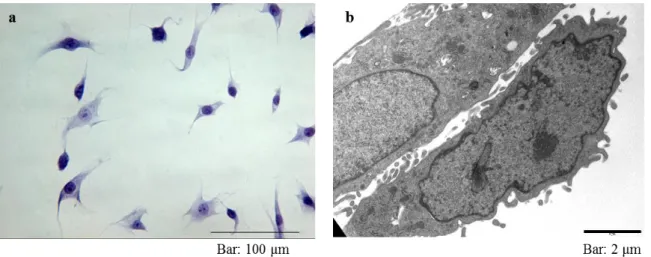
Toluidine blue staining and transmission electron microscopy (TEM) evaluation showed the characteristic morphology of the MLO-Y4 cell line (Fig. 6).
Figure 6. Figure a) shows an optical microscope image of MLO-Y4 after toluidine blue staining;
figure b) shows the morphological detail obtained by TEM analysis.
The morphological analysis (Fig. 7) showed that the cells maintained their characteristic dendrites and cellular body morphology only in presence of 5% of total serum. The cells, grown in 1% of total serum, showed a very different morphology: decrease in cell body dimensions, decrease in dendrites number and extensions; in addition, the pictures of the cells grown in 1% of total serum showed a higher number of dead cells compared to cells grown in 5% of total serum. Moreover, it was evident that a cells density of 5000 cells/cm2 was the optimal seeding density to guarantee the maintenance of cell morphology and avoidance of over-confluences (> 70%) for all experimental time points.
38 Figure 7. Morphological analysis to determinate the best culture condition for the execution of the
experiments.
5.2
Hypoxia induction
MLO-Y4 cells cultured at 1% O2 showed a significant increase in Hypoxyprobe staining
compared to cells cultured at normoxia condition at 16, 24, 48, and 72 hours, while the cells grown at 5% O2 showed a significant increase in Hypoxyprobe staining compared to
cells cultured at normoxia condition at 24 hours and 48 hours, and at 16 hours compared to 1% O2 (Fig. 8). The number of Hypoxyprobe positive cells was significantly increased with
the time of culture under both the hypoxic conditions and also under normoxic conditions suggesting that the cells became more sensitive to hypoxia (Table 1).
39 Figure 8. Effect of oxygen deprivation on MLO-Y4 detected by Hypoxyprobe staining.
Image a. Representative image of Hypoxyprobe staining of (%) hypoxic MLO-Y4; pimonidazole in green stains hypoxic cells and DAPI in blue stains cell nuclei. Graph b shows the mean and standard error of percentage of Hypoxyprobe positive MOL-Y4 cells at different time points cultured in both hypoxia and normoxia conditions.
40
Table 1. Hypoxyprobe staining. Statistical differences existing over the times for each oxygen
condition. P value obtained with Kruskal- Wallis Post Hoc test.
1%O2 5%O2 Normoxia
Times 48h 72h 24h 48h 72h 48h 72h
8h p<0.001 p<0.001 p<0.001 p<0.001 p<0.001 p<0.001 p<0.001 16h - - - p<0.001 p<0.001 p<0.001 p<0.001
24h - - - p<0.001 p<0.001
5.3
Osteocyte proliferation, viability and apoptosis
Couting of cells showed an increase in cellular proliferation with duration of the culture (Table 2), but the presence of 1% and 5% of oxygen significantly decreased the proliferation of MLO-Y4 after 8, 24 and 48 hours compared to the same time points of normoxic condition (Fig. 9 a).
Osteocyte viability, as determined by trypan blue staining, was compromised by severe oxygen deprivation (1% O2) and resulted in a significant increase in the number of dead
cells at 24 and 72 hours (Fig. 9 b). In both the normoxia and 1% O2 hypoxic conditions, the
increase in cell death over the time of culture showed a clear trend, with a significant difference between 16 and 72 hours of culture (p value= 0.012 in normoxia and p value= 0.032 in 1% O2). No difference exists comparing 5% oxygen and the other conditions;
41 Figure 9. Effect of oxygen deprivation on MLO-Y4 proliferation and viability.
Graph a shows the proliferation by counting the total cells number grown in our experimental conditions. Graph b shows the quantification of MOL-Y4 dead cells at different time points cultured in hypoxia and normoxia conditions. For both the graphs, mean and standard error are indicated.
Table 2. Osteocyte proliferation. Statistical differences existing over the time for each oxygen
conditions. P value obtained with Kruskal- Wallis Post Hoc test
1%O2 5%O2 Normoxia
Times 48h 72h 48h 72h 48h 72h
8h p<0.004 p<0.004 p<0.001 p<0.001 p<0.001 p<0.001
16h - - - - p<0.001 -
24h - - - p<0.001 p<0.001 -
The number of apoptotic osteocytes, as determined by nuclear fragmentation (Fig. 10 a, b and c), was increased by both the hypoxic conditions compared to the normoxic one and this increase was found to be statistically different at 16, 24 and 48 hours. Besides, significant increase in apoptotic MLO-Y4 cells was found in 5% O2 compared to 1% O2 at
24 hours. Surprising, at 72 hours, the number of apoptotic cells cultured under 1% O2
hypoxic conditions was decreased compared to normoxia, but this difference was found to be not significant.
42 This decrease in apoptotic cell number at 72 hours was found to be also lower than at 48 hours, although, this difference was not statistically significant (Fig. 10 d).
Figure 10. Evaluation of Apoptosis by nuclear fragmentation.
Images of DAPI stained MLO-Y4 cells under hypoxia (%) (a) and normoxia (b) conditions after 48 hours of culture. Image c shows a detail of nuclear fragmentation at higher magnification. Arrows indicate apoptotic nuclei.
Graph c. shows the average and standard error of the means of apoptotic cells grown in hypoxia and normoxia conditions.
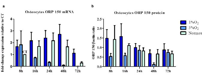
5.4
Expression of ORP150
No significant difference was found in MLO-Y4 ORP 150 mRNA expression over the time in all the tested conditions. No significant changes were observed in mRNA levels under
43 the tested conditions, 1% O2 and 5% O2 and normoxia (Fig. 11 a). Nevertheless, it was
possible to observe a trend that 5% O2 induced the expression of ORP150.
Western blot analysis, conducted on MLO-Y4 grown in 1%, 5% of oxygen and normoxic conditions, showed no statistically significant differences in ORP 150 expression at protein level at all the time points, confirming the results of the ORP 150 at mRNA level (Fig. 11 b).
Moreover, western blot analysis showed that the expression of ORP150 was significantly lower in normoxic MC3T3 (ORP150/ACT ratio=0.531±0.03 average ± standard deviation) compared to normoxic MLOY4 (ORP150/ACT ratio=0.845±0.06 average ± standard deviation), with a p-value= 0.001, as reported in literature (Guo et al., 2010).
Figure 11. Effect of oxygen deprivation on ORP 150 expression in osteocytes.
Graph a. shows the fold-change expression of ORP 150 mRNA compared to the control (CT), analysed by the 2-∆∆Ct methods. Graph b shows the level of ORP 150 protein normalized for β-Actin. For both the graphs, mean and standard error are indicated.
44
5.5
Dosage of soluble factors PGE
2,
RANKL, OPG, and Sclerostin
Although no significant difference exists in the level of each soluble factor measured in the hypoxic and normoxic osteocytes culture media, it is possible to identify a trend of expression.
PGE2 seems to be released at higher levels by osteocytes grown at 1% of oxygen compared
to the other conditions, especially after 24, 48 and 72 hours of culture (Fig. 12 a).
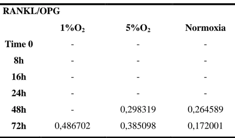
The level of RANKL in our experimental conditions, becomes detectable only after 48 hours of culture; otherwise, an increase in OPG release over the time of culture was detected, without any difference among the oxygen conditions (Fig. 12 b and c). Resulting in an overall decrease in the RANKL/OPG ratio as shown in Table 3.
The levels of Sclerostin, released in all the experimental conditions, were lower respect to the limit of detection of the kit assay.
45 Figure 12. Effect of oxygen deprivation on MLO-Y4 release of the selected soluble factors.
The graph shows the mean and standard error of the soluble factors quantified by ELISA (pg/ml). PGE2 (a), RUNKL (b) and OPG (c).
Table 3. RANKL/OPG ratio in the culture media of hypoxic and normoxic MLO-Y4 RANKL/OPG
1%O2 5%O2 Normoxia
Time 0 - - - 8h - - - 16h - - - 24h - - - 48h - 0,298319 0,264589 72h 0,486702 0,385098 0,172001
5.6
Trap staining and induction of osteoclastogenesis
After culturing Raw 264.7 cells for 7 days with conditioned media (CM) collected from osteocytes grown in all our experimental conditions (1% O2 –CM, 5% O2 –CM and N-CM
at T0, 8, 16, 24, 48 and 72 hours), the detection of TRAP positive cells was performed (Fig. 13) following two different methods:
1) by counting the multinucleated Trap positive cells (MNC TRAP+ cells) and
2) by evaluating the pixel area covered from Trap positive cells, as described by Huang and collaborators (Huang et al., 2003) .
I chose to describe the results obtained from the two analyses separately because, although the results showed the same trend, some differences in the statistical analysis were identified.
46 Figure 13. Representative image of multinucleated TRAP positive cells, specific markers of
osteoclast (red arrows). Scale bar: 50µm.
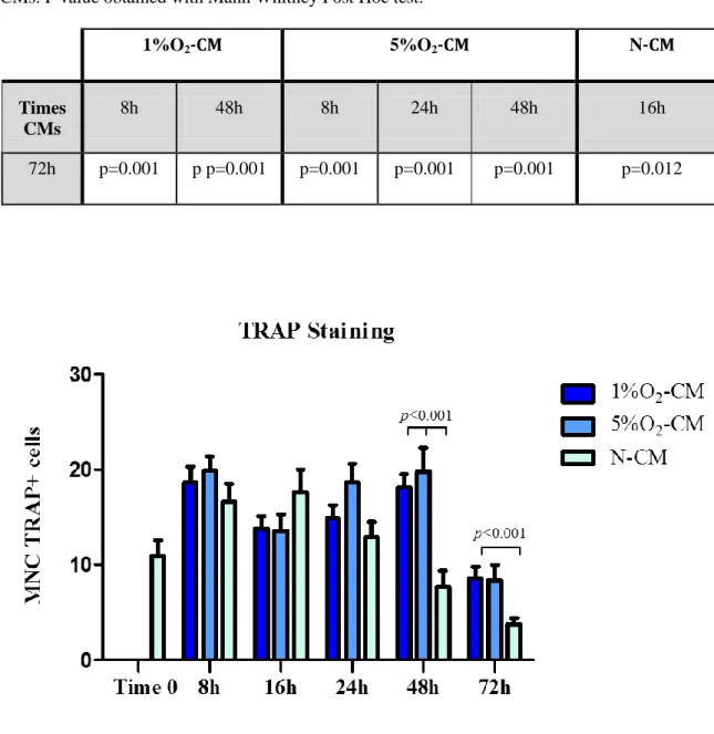
5.6.1 Multinucleated Trap positive cells (MNC TRAP+ cells) results
Looking separately at each conditions, the analysis showed a significant decrease in MNC Trap positive cells in the culture conditioned for 72h with 1% O2-CM compared to the 8,
16 and 48 hours 1% O2-CM, and the same results were found in N-CM (Table 4).
Regarding the difference in Trap positive cells induced by the three CMs derived from our oxygen conditions, I found that the conditioning of RAW cells for 48h with 1% and 5% O2-CM induced a significant increasing of MNC Trap+ cells compared to 48h N-CM.
While 72h 1% O2-CM lead to asignificantly increased osteoclastogenesis compared to 72H
47
Table 4. Multinucleated TRAP positive cells. Statistical differences existing in the different times
CMs. P value obtained with Mann Whitney Post Hoc test.
1%O2-CM 5%O2-CM N-CM
Times CMs
8h 48h 8h 24h 48h 16h
72h p=0.001 p p=0.001 p=0.001 p=0.001 p=0.001 p=0.012
Figure 14. Effect of the CMs (from hypoxic/normoxic osteocytes) on the osteoclastogenesis.
The graph shows the mean and standard error of the number of MCN Trap positive cells induced by the conditioning with the CMs tested.
5.6.2 Pixel area covered by Trap positive cells
The results showed a decrease of the area covered by osteoclast related to the time of conditioning, in detail all the 72h CMs tested (1% O2-CM, 5% O2-CM and N-CM) had
48 lower inductive effect on osteoclastogenesis compared to the effect exerted by the CMs collected at the other time points (Table 5).
However, CMs collected after 24, 48 and 72 hours showed a significant difference in inducing osteoclast formation; in particular the 24, 48 and 72h 1% O2-CM significantly
increased the osteoclasts formation compared to 24, 48 and 72h N-CM; while only 24h 5% O2-CM was found significantly different compared to 24h N-CM (Fig. 15).
Table 5. Multinucleated TRAP positive cells. Statistical differences existing in the different times
CMs. P value obtained with Mann Whitney Post Hoc test.
1%O2-CM 5%O2-CM N-CM Times CMs 8h 24h 48h 8h 16h 24h 8h 16h 24h 16h - - - p<0.001 - - - - - 48h - - - p<0.001 - - p<0.001 - - 72h p<0.001 p<0.001 p<0.001 p<0.001 p<0.001 p<0.001 p<0.001 p<0.001 p<0.001
49 Figure 15. Effect of the CMs (from hypoxic/normoxic osteocytes) on the osteoclastogenesis.
The graph shows the quantification of osteoclast formation by counting of pixel area covered by MCN Trap positive cells induced by the conditioning with the CMs tested. Mean and standard are indicated.
5.7 Osteoblasts response to the hypoxic osteocytes conditioning media
After conditioning of MC3T3-E1 cells for 7 days with CM collected from osteocytes grown at either 1% O2, 5% O2 and normoxia at T0, 8, 16, 24, 48 and 72 hours, osteoblast
viability, ALP activity and the quantification of β-catenin mRNA were performed.
5.7.1 Osteoblast viability (MTT assay)
The results showed that the conditioning for 7 days with all the CMs tested did not compromise the MC3T3-E1 viability, compared to time point 0. Looking separately at the 1% O2-CM, 5% O2-CM and N-CM, I saw a significant decrease in cell viability between