Università degli Studi di Ferrara
DOTTORATO DI RICERCA IN
FARMACOLOGIA E ONCOLOGIA MOLECOLARE
Settore Scientifico Disciplinare BIO/14
CICLO XXVI
COORDINATORE Prof. Antonio Cuneo
The HIV-Tat protein favors the activation of T lymphocytes.
Implications for new therapeutic strategies against HIV and
other viral infections
Dottorando Tutore
Dott. SFORZA FABIO Prof. GAVIOLI RICCARDO
“WE’LL FIND A WAY, WHEN ALL HOPE IS GONE”
1
INDEX
LIST OF ABBREVIATIONS 4 1. INTRODUCTION 7 1.1 Immune system 7 1.1.1 Innate immunity 8 1.1.2 Adaptive immunity 9 1.1.3 CD4+ T cells 9 1.1.4 CD8+ T cells 111.2 CD4+ and CD8+ T cells activation 12
1.2.1 CD4+ and CD8+ T cells programming 15
1.2.2 Memory CD4+ and CD8+ T cells 17
1.3 The HIV infection 20
1.3.1 Effects of HIV on CD4+ and CD8+ T cells 22
1.4 HIV-1 Tat protein 23
1.4.1 Effects of HIV-Tat protein on CD4+, CD8+ T cells and APCs 25
2. AIMS 28
3. RESULTS 30
3.1Tat enhances the production of IL-2 in PBLs 30
3.2 Tat enhances IL-2 and IFNγ production in CD8+ and CD4+ T cells 32 3.3 Tat does not affect the proliferation of activated CD8+ and CD4+ T cells 35 3.4 Tat affects the expression of key transcription factors in
2
activated CD4+ T cells 40
3.6 αvβ3 and α5β1 integrins blocking affects Tat-mediated effect
on the transcriptional profile in activated PBLs 42 3.7 Tat down regulates CD127 expression and modulates T cells fate 44 3.8 Tat favors the activation of antigen-specific naïve and
memory CD8+ T cells 45
3.9 Tat does not affect the expression of IL-2 and IFNγ in
unstimulated CD8+ and CD4+ T cells 48
3.10 Tat does not modulate the transcriptional profile of
unstimulated CD8+ and CD4+ T cells 49
3.11 Tat modulates T-bet and Eomes mRNA expression in unstimulated CD8+ and CD4+ T cells after 24 hours of
treatment (Preliminary data) 50
3.12 Immunization with an attenuated HSV1 vector expressing Tat increases the number of HSV1-specific CD8+ T cells and
favors the development of effector memory T cells 51 3.13 Immunization with an attenuated HSV1 vector expressing Tat favors
the control of HSV1 infection 56
4. DISCUSSION 58
5. MATERIALS AND METHODS 62
5.1 Human cells and culture conditions 62
3 5.4 Intracellular staining 64 5.5 Bio-Plex assay 64 5.6 MTT assay 65 5.7 CFSE assay 65 5.8 Western blotting 66 5.9 Flow cytometry 66 5.10 Generation of CTL cultures 66 5.11 Cytotoxicity assay 67 5.12 Elispot assay 67 5.13 Viruses 68
5.14 Mice immunization, challenge and samples collection 68
6. REFERENCES 70
7. PUBLICATIONS 80
4
aa amino acid
Ab antibody
Ag antigen
AIDS acquired immunodeficiency syndrome
AP-1 activating protein 1
APC allophycocyanin
APC antigen presenting cell
APC-Cy 7 allophycocyanin covalently bound to Cyanin 7
BSA bovine serum albumine
CD cluster of differentiation
CFSE 5(6)-carboxy-fluorescein diacetate succinimidyl ester
CT cycle threshold
CTL cytotoxic T lymphocyte
DAMP damage-associated molecular pattern
DC dendritic cell
DMSO dimethylsulfoxid
DMF dimethylformamide
dNTP desoxy-nucleoside triphosphate
EBV Epstein-Barr virus
ELISPOT enzyme-linked immunospot assay ERK extracellular receptor-activated kinase FACS fluorescence activated cell sorting
FCS fetal calf serum
FITC fluoresceinisothiocyanat
gp glycoprotein
ART anti-retroviral therapy
HIV human immunodeficiency virus
HLA human leukocyte antigen
HPLC high performance liquid chromatography
5
IFN interferon
Ig immunoglobulin
IL interleukin
JNK c-Jun N-terminal kinase
KLRG1 killer cell lectin-like receptor subfamily G member 1
LCL lymphoblastoid cell line
LPS lipopolysaccharide
LTR long terminal repeat
LvTat HSV1 attenuated replication-competent vector expressing Tat LvLacZ HSV1 attenuated replication-competent vector expressing LacZ
mAb monoclonal antibody
MHC major histocompatibility complex MPEC memory precursor effector cells
MTT 3-(4,5-dimethylthiozol-2-yl)2,5-diphenyltetrazolium bromide
nAb neutralizing antibody
NFAT nuclear factor of activated T cells
NF-kB Nuclear factor-kappaB
NK natural killer cells
PAMP pathogen-associated molecular pattern
PBL peripheral plood lympohocytes
PBMC peripheral blood mononuclear cells
PBS phosphate-buffered saline
PCR polymerase chain reaction
PE phycoerythrin
PerCP-Cy5 peridinin Clorophyll protein covalently bound to Cyanin 5
PHA phytohämagglutinin
PMA phorbol 12-myristate 13-acetate PRR pathogen recognition receptors
PTD protein transduction domain
qPCR quantitative polymerase chain reaction
SDS sodium dodecyl sulfate
SEM standard error of the mean
6
SLEC short lived effector cells
STAT signal transducers and activators of transcription TAR trans-activation response element
Th T helper
TCM central memory T cell
7
1. INTRODUCTION
The human immunodeficiency virus (HIV) is one of the major plague in the world for new cases of infection, number of people infected and deaths.
In 2012, 2.3 million of new HIV infections (1.9 million–2.7 million) and 35.3 million of HIV-positive people have been reported all over the world [1].
Since its isolation, in the 1983, HIV has represented a social, economic and sanitary issue. Despite the massive scale up of antiretroviral therapy (ART) has dramatically prolong the life of HIV-infected individuals and the number of new infections is decreasing, for every 10 people starting antiretroviral therapy 16 people are newly infected [2]. Today, a winning HIV/AIDS strategy does not exist, and the escalating cost for treatment will become increasingly difficult for undeveloped countries to meet. Strategies for either eradicating the virus from infected individuals or boosting their immune response so that antiviral drugs can be discontinued are urgently needed.
The most devastating damages caused by HIV infection are observed at level of cellular immunity and include the depletion of CD4+ T cells and important dysfunctions of both CD8+ and CD4+ T cells as impairment of functionality [3, 4], exhaustion [5], increased T cell proliferation [6], susceptibility to apoptosis [7, 8] and expansion of memory T cells [9-11]. This status of chronic immune activation and immune senescence involves the whole T cell compartment, including uninfected and non-HIV specific T cells [12], is also present during ART and contributes to the appearance of AIDS-defining and non-defining diseases [13]. Different mechanisms contribute to these phenomena including CD4+ T cells loss, viral replication and effects of HIV proteins such as gp120, Nef and Tat [6, 12].However, how HIV can modulate the CD8+ and CD4+ T cells responses is not completely clear.
1.1 IMMUNE SYSTEM
The immune system is a rapid, specific and powerful mechanism of defense against viruses, bacteria and other potential dangerous microorganisms.
It is divided in two major sectors, the innate immunity and the adaptive immunity (Figure 1.1).
8
Figure 1.1. The innate and the adaptive immunity. Adapted by [14]
1.1.1 Innate immunity
The innate immunity system consists of multiple distinct subsystems that perform different functions in host defense.
The first defense consists in preformed soluble molecules like complement and cytokines, present in blood, extracellular fluid, and epithelial secretions that can kill or inhibit the proliferation of some classes of pathogens. The second defense is mediated by cells like Natural Killers (NKs), eosinophils, basophils, and mast cells specialized in the recognition of specific microbial structures and molecules such as PAMPs (pathogen associated molecular patterns) and DAMPs (damage associated molecular patterns) [14, 15], identified by cellular pattern recognition receptors (PRRs).
PRRs recognition of microbial structures causes a cascade of intracellular events that leads to pro-inflammatory cytokines and chemokines release, and to phagocytosis mediated by macrophages and dentritic cells (DC). On the whole these responses cooperate with co-stimulatory molecules, soluble or expressed on the cells surface, to activate adaptive immune cells.
9 1.1.2 Adaptive immunity
Adaptive immunity is carried out by cells called lymphocytes which act through two classes of responses: antibody response and cell-mediated immune response.
The first is mediated by B lymphocytes (B cells) which are responsible of the release of antibodies such as IgG, IgM, IgA, IgE. The latter is triggered by T lymphocytes (T cells), which are divided in two subpopulations: CD4+ T cells, also known as T helper lymphocytes (Th), and CD8+ T cells, also known as cytotoxic T lymphocytes (CTLs).
1.1.3 CD4+ T cells
CD4+ T cells are the main orchestrators of immune responses to infections. This subset of T cells is extremely important for the optimal activation and development of memory CD8+ T cells [16], for the secretory activity of B and NK cells and for the antigen metabolism of professional antigen presenting cells (APC).
CD4+ T helper subsets include Th1, Th2, Th17 and Treg cells [15, 17] (Figure 1.2 and Table 1.1). There are evidences that each of these subsets is involved in the defense against specific microorganisms.
10
Th1 cells mediate immune responses against intracellular pathogens [19]. Concerted with
antigen recognition, interleukin-12 (IL-12) is the cytokine that drives naïve CD4+ T cells into Th1 subset type. IL-12Rβ1 is constitutively expressed on naïve CD4+ T cells and its expression is increased in Th1 cells. The most important cytokine products of Th1 cells are interferon-γ (IFNγ), lymphotoxin α (LTα), and IL-2. IFNγ is important to activate macrophages and CD8+ T cells, and increases antigen processing and presentation to T cells [20]. IL-2 production is critical for CD4+ and CD8+ T-cell memory maintenance since it promotes cell survival, inducing the expression of the anti-apoptotic protein Bcl-2 [19, 21]. IL-2 can also stimulate the activation and proliferation of CD8+ T cells and enhances the expression of co-stimulatory molecules on APCs surface and the production of other cytokines in favor of CD8+ T cells. Moreover, IL-2 induces the release of other cytokines like IL-4, important for the development of other Th subsets and stimulates the proliferation of NK and B cells. LTα activates neutrophils and lymphoid organogenesis.
Th2 cells mediate host defense against extracellular parasites including helminthes
[22-24]. The induction of Th2 subset is regulated by IL-4 that acts with a mechanism of feedback to produce itself and IL-5. Those cytokines produced by Th2 cells, promote the production of IgE and the differentiation and activation of eosinophils [20]. Th2 cells also support B lymphocytes stimulating the production of large amount of IgM. IL-4 antagonizes the macrophage-activating actions of IFNγ and, in association with IL-5, can inhibit acute and chronic inflammation, limiting consequences due to persistent Th1 immune responses.
Th17 cells mediate immune responses against extracellular bacteria and fungi [25]. Their
cytokine products are IL-17, IL-21, and IL-22. IL-17 recruits and activates neutrophils during immune responses [26], while IL-21 is a stimulatory factor for Th17 differentiation and serves as positive feedback, as does IL-2 for Th1 and IL-4 for Th2 cells. In addition, IL-21 acts on CD8+ T cells, B cells, NK cells, and dendritic cells [27]. IL-22 mediates host defense against bacterial pathogens, and these functions may largely depend upon IL-23 stimulation of innate cells.
Treg cells play a critical role in maintaining self-tolerance as well as in regulating immune
11
some of which require cell-to-cell contact or the production of cytokines, including TGF-β and IL-10. High levels of CD25 and IL-2Rα expression on Treg cells suggest the importance of IL-2 to regulate proliferation and induction of Treg suppressor activity.
T helper lineage Cytokines production Inductive cytokines Role Th1 IFNγ IL-2 IL-12 IL-2 IFNγ
Mediate immune responses against intracellular pathogens: NK,
macrophages, CTLs activation and IFNγ secretion Th2 IL-4 IL-5 IL-6 IL-4 IL-2
Mediate defense against extracellular parasites; Promotion of IgE and eosinophil/basophil/mast cell-mediated immune reaction Th17 IL-17 IL-21 IL-22 IL-6 TGF-β
Mediate immune responses against extracellular bacteria and fungi; Release of proinflammatory cytokines, activation of T cells, NK and neutrophils
Treg TGF-β
IL-10
TGF-β IL-10 IL-2
Regulation and suppression of immune response
Table 1.1. The CD4+ T cells subsets
1.1.4 CD8+ T cells
CD8+ T cells are critical to protect the organism against intracellular pathogens and tumor cells. In contrast to CD4+ T cells, CD8+ T cells are the direct executioners of pathogen-infected cells.
During the immune response to viruses and intracellular microbes, naïve CD8+ T cells are activated in cytotoxic T lymphocytes (CTL) to undergo a massive clonal expansion, with acquisition of new effector functions.
12
After antigen recognition, CTLs proliferate and kill the cellular target mainly with three mechanisms:
- Exocytosis of granules containing perforin that polymerize on target cell membrane causing osmotic cell lysis.
- Secretion of granzymes (serine proteases) that induce apoptosis in target cells [29]. - Expression of Fas ligand (FasL) that induces apoptosis by a specific and direct interaction with Fas molecules present on target cells [30]. Target killing by CTLs is antigen specific and contact dependent.
1.2 CD4+ AND CD8+ T CELLS ACTIVATION
T lymphocytes mount specific responses against antigens (Ags) which are peptides generated by the cellular proteolytic system (“antigen processing”) and are presented through MHC molecules to the TCR. MHC class I and the MHC class II molecules, also known as Human Leukocyte Antigen (HLA), are controlled by genes located on chromosome 6, that encodes cell surface molecules specialized to present antigenic peptides to T cells. All MHC-I molecules consist of two polypeptide chains, a heavy chain α and a monomorphic light chain β2-microglobulin (β2m).
The two chains are linked non-covalently via interaction of β2m and the α3 domain. Like MHC class I molecules, MHC-II molecules are also heterodimers, but in this case consist of two homologus peptides, an α and β chains, both of which are encoded in the MHC (Figure 1.3).
13
The MHC-I system draws its spectrum of peptides from proteins in the cytosol of all nucleated cells. In contrast to MHC-I, MHC-II molecules are expressed on a more limited set of cells called APC cells such as somatic cells, B cells, macrophages and dendritic cells. Thereby, MHC molecules are a molecular reflection of the health of cells that synthesize them (for MHC-I molecules) or of the local environment in which cells reside (for MHC-II molecules).
MHC-I and MHC-II molecules also show a different interaction with T cells. In particular, the nonpolymorphic α3 domain of MHC-I binds the CD8 co-receptor of T cells while the membrane proximal domains of MHC-II interacts exclusively with the CD4 molecule. CD8 and CD4 molecules serve as co-receptors on the surface of T lymphocytes, providing both adhesion and specific activation signals that modulate T cells. Epitope associated to the MHC molecules migrate to the cell surface to be recognized by the specific TCR.
The TCR is a disulfide-linked membrane-anchored heterodimer normally consisting of the highly variable α and β chains expressed as part of complex with the invariant CD3 chain molecules [31] (Figure 1.4).
Figure 1.4. T cell Receptor
The generation of TCR diversity is based on somatic recombination of the DNA encoded segment in individual T cells.
The combinatorial rearrangement of multiple gene segments, the error-prone process of joining these different fragments, and the various combinations of α with β chains or the
14
less represented γ with δ chains lead to big TCR diversity and, thereby, enables receptors to respond to a broad spectrum of antigens that pathogens show.
It is known that activation of T cells requires multiple signals: signal 1, antigen-specific delivered via TCR/MHC interaction, signal 2, delivered by co-stimulatory molecules (including IL-2), and signal 3, delivered by pro-inflammatory cytokines and chemokines [32].
Several co-stimulatory receptors are exposed on T cells surface; among them, CD28 and CTLA-4 co-stimulatory molecules bind B7 receptor on APCs surface. CTLA-4-mediated inhibitory signals dominate when T cells encounter antigen on APCs expressing low levels of B7, while CD28 activating signals dominate when antigen is presented by APCs with high B7 levels [33-35]. CD28 co-stimulation is responsible for the activation of c-Jun kinase and PI3K/AKT/mTOR axis, thus contributing to anti-apoptotic effects due to the up-regulation of Bcl-2. Physiologically, a balance among MAP kinase family enzymes, in particular between ERK and c-Jun, is important for T cell survival and, while ERK is activated through TCR stimulation, c-Jun depends mostly by CD28 signalling [36].
In addition, CD3 seems to be responsible for signal transduction following the antigen recognition events. This idea was suggested by the observation that antibodies directed against CD3 proteins generate the same T cell activation events as stimulation of T cells with peptide antigen plus MHC [37, 38].
Co-stimulators are required for the full maturation of responses in T cells. Furthermore, the combination of these signals induces cytokines secretion, clonal expansion and differentiation of precursor CD4+ and CD8+ T cells into effector (Figure 1.5).
15
In concert with antigen recognition, the cytokine environment modulates the polarization of T-cell homeostasis. In particular, IFNγ, IL-2, IL-7, IL-4, IL-9, IL-12 and IL-15 supply survival and expansion of CD4+ and CD8+ T cells.
The T cell activation provided by TCR, co-stimulatory molecules and cytokines determine the change of transcription factors (TFs) expression that act as master regulators by controlling the expression of a panel of genes and conferring a specific phenotype. A large number of TFs participate in initial T cell activation, such as nuclear factor of activated T cells (NFAT), activating protein-1 (AP-l), and nuclear factor-kappaB (NF-kB), that interact with transcriptional partners determining the activation of T cells.
NFAT is required for the expression of IL-2, IL-4, TNF, and other cytokine genes. Interestingly, productive T cells activation not only requires prolonged nuclear NFAT, but also depends on physical interactions between NFAT and AP-1 on composite promoter elements. Activation of AP-1 typically involves synthesis of Fos protein and phosphorylation of c-Jun protein.
NF-kB is a transcription factor that is activated in response to TCR signals and is essential for cytokine synthesis. NF-kB fulfills a central role in the cellular stress response and in inflammation by controlling the expression of a network of inducers and effectors that define responses to pathogens and other classes of danger signals [39].
Following NFAT, AP-1 and NF-kB dependent T cell activation, a subsequent phase of clonal expansion ensues.
1.2.1 CD4+ and CD8+ T cells programming
CD4+ Th1/Th2 balance is regulated by T-bet and GATA3, both induced by TCR stimulation and by several cytokines. T-bet enhances responsiveness to 12 favoring IL-12Rβ2 expression and is regulate by or regulate IFNγ production, thus supporting a Th1 phenotype; while GATA3 is the TF responsible for the acquisition of a Th2 lineage promoting IL-4 secretion [40, 41]. T-bet and GATA3 are regulated by mTOR kinase through activation of the PI3K-Akt pathway [42]. STAT proteins also regulate the expression of many genes required for the differentiation of various CD4+ T helper cell lineages.
Therefore, STAT1 and STAT5 contribute to Th1 differentiation by enhancing T-bet and IFNγ expression, respectively, while STAT6 mediates IL-4 production and Th2 differentiation [41, 43].
16
During CD8+ T cell-mediated immune response, Eomesdermin (Eomes), another T-box transcription factor like T-bet, is essential for the development of effector and memory phenotype.
Eomes is crucial in first phase of CD8+ effector development and along with T-bet it regulates CD8+ cytotoxic functions [44]. Otherwise, Eomes and T-bet expression are inversely related in central memory (TCM) cells [45]; while T-bet decreases in the contraction phase, Eomes expression is necessary. The level of inflammation and the availability of certain cytokines (e.g. IL-12) influence the levels of T-bet and Eomes available to the effector cell and that this in turn influences which cells will remain CD8+ effector T cells and which will transition to the memory CD8+ T cell pool [46].
Other two transcription factors crucial for the activation and the development of CD4+ and CD8+ T cells are Blimp-1 and Bcl-6, that are reciprocally antagonist. Blimp-1 is a transcriptional factor encoded by Prdm1 and correlated to effector and memory functions of both CD8+ and CD4+ T cells. Blimp-1 in CD8+ T cells negatively regulates Bcl-6 and, as in CD4+ T cells, it is activated by IL-2 driving CD8+ T cells to CTLs differentiation. Bcl6 is a transcriptional repressor of granzyme B and it is involved in CD4+ and CD8+ memory T cells formation [47]. Although many aspects of Blimp-1 and Bcl-6 regulation in CD4+ T cells are still unclear, some studies suggest a key role of Bcl-6 in CD4+ T cells survival and proliferation as well as Tfh formation. In addition, it has been demonstrated the significant influence of Blimp-1 in terminal effector cells differentiation and in Th1 maturation where it interacts with T-bet [48] (Table 1.2 and Figure 1.6).
Activated CD8+ T cells Activated CD4+ T cells Th1 Th2 Th17 Treg TFs expression Blimp-1 T-bet Eomes T-bet Blimp-1 STAT1 STAT5 Gata3 STAT6 Bcl-6 RORγt Foxp3
17
Figure 1.6. Transcriptional signaling after activation in CD4+ (a) and CD8+ (b) T cells. Adapted by [49]
Intensity of stimuli through TCR-MHC signaling, cytokines production and presence of different subsets of immune cells can deeply modulate transcription factors programming in both CD4+ and CD8+ T cells, driving the immune response to different fates.
The modulation of T cell responses to antigen and the improvement of activation are goals extremely important for the development of new therapies and vaccines against tumors and pathogens.
1.2.2 Memory CD4+ and CD8+ T cells
During a response to infection, there are three characteristic phases: a period of initial cell activation and expansion, a contraction or death phase, and the establishment and maintenance of immunological memory (Figure 1.7).
18
There are two competing models of memory T cell development: linear and not-linear divergent differentiation. In the linear model, activated naïve T cells develop initially into effector cells and, a small fraction of these cells, survives at contraction phase and becomes memory cells. In the not-linear divergent model, a fraction of activated naïve T cells differentiates directly into memory cells. Both these theories are supported by different reports however, recently some studies indicate the linear model as the most consistent [50, 51].
Memory cells are divided into two different subpopulations: “central memory” (TCM) and “effector memory” (TEM) (Table 1.2).
Table 1.3. Memory phenotype and secretion capacity
TCM reside mostly in lymph nodes and are responsible for the clonal expansion after re-exposure to antigen, while TEM are disseminated within peripheral tissues where they display immediate effector functions. The role and the homing capacity of memory cells is determined by their phenotype. For instance, the expression of some cellular markers like CCR7 and CD62L on memory cells surface regulate their susceptibility to antigen and consequently cells proliferation and cytokines release. Cells proliferation and cytokines production represent the two main aspects of this population. Indeed, while TCM are extremely proliferative but have a low cytokine secretion capacity after antigen recognition, TEM are not so efficient in proliferation but display high secretory potential. During acute viral infection, antigen-driven differentiation of naive CD8+ T cells leads to the expression of cytolytic molecules and cytokines that facilitate the control of infections.
Phenotype Molecules secreted
Central memory (TCM) CCR7 High CD62L High CD44 IL-7 Receptor (CD127) IL-15 Receptor (CD122) CXCR5 IFNγ low IL-2 high IL-4 low CD40 ligand IL-17A Effector memory (TEM) CD62L Low CD44 IL-7 Receptor (CD127) IL-15 Receptor (CD122) CD45RA IFNγ IL-4 IL-5
19
After the expansion and contraction phases, during which the infection is cleared, some antigen-specific CD8+ T cells survive and gain the capacity of self-renewal in lymphoid and non-lymphoid tissues, and a heightened ability to recall effector functions relative to their naïve precursors. Molecular studies of CD8+ T cells differentiation during acute viral
infection have demonstrated that at the effector stage, memory precursor cells destinated to survive into the memory phase, can be identified by some transcriptional factors such as Eomes, Blimp-1 and cellular marker like CD127 (IL-7 receptor), CD62L (homing in lymphoid organs) and CD122 (IL-15 receptor) [52, 53]. The long term maintenance of memory CD8+ T cells does not require the antigen stimulation, but it is dependent on IL-7 and IL-15 signalling that mediates homeostasis and survival, up-regulating anti-apoptotic molecules such as Bcl-2 [54]. TCM CD8+ T cells seem to originate from a subset of effector cells called MPEC, memory precursor effector cells, in contrast to SLEC, short lived effector cells. The balance of MPEC/SLEC among a population of effectors depend by the overall amount and duration of activation signals, thus the first 48-96 hours of stimulation determine the fate of the future memory population. In particular, the development of TCM cells is dictated by a short and reduced antigenic stimulation [55, 56]. Naïve CD4 T cells must usually interact with mature antigen-loaded dendritic cells to be successfully activated. This interaction takes place in the T-cell areas of secondary lymphoid organs, locations that have evolved to facilitate contact between antigen-specific T cells and DCs. CD4+ T cells require prolonged and high TCR signaling stimulation in order to differentiate into effectors and subsequently form the memory pool [57]. Memory CD4+ T cells express much lower levels of CD122, that correlates with a lower dependence on IL-15 compared to CD8+ T cells. However, IL-7 appears to be the main survival cytokine for CD4+ memory cells, as cells cannot survive in its absence. Moreover, memory CD4+ T cellslife tends to be shorter than that of memory CD8+ T cells [58]. Memory CD4+ T cells are important during the “clinical latency” phase of HIV which is accompanied by the establishment of cellular reservoirs, resting memory CD4+ T cells that host the integrated virus with low expression of viral antigens, not to be eliminated by the adaptive immune system [59]. In the case of chronic or latent infections, and particularly during the HIV infection, the persistence of the pathogen misleads the physiological development of the cellular response, affecting the functionality of memory cells, that can exhibit poor recall proliferation, exhausted phenotype, loss of effector functions and a skewed composition of T cell memory subpopulations [60].
20 1.3 THE HIV INFECTION
In 1981 appeared, in Unites States, a new disease, characterized by a deficiency in the immune system [61] which consists in a marked reduction in CD4+ cell numbers and enhanced B-cell proliferation and hypergammaglobulinemia. After two years of research, this disease known as acquired immune deficiency syndrome (AIDS), was connected to a new human retrovirus named HIV [62]. The AIDS viruses, HIV-1 and HIV-2, consist of different virus groups and a lot of different clades subtype for every virus group. Based on their genetic differences, four groups of HIV-1 (M, N, O, P) have been identified and inside these groups it is possible to distinguish 9 subpopulations or clades (A, B, C, D, F, G, H, J, K). The four groups of HIV-1 isolated differ from each other by the chemokine receptor they use for cell entry. HIV viruses that use to enter cells the chemokine receptor 4 (CXCR4) are termed X4, the ones that use chemokine receptor 5 (CCR5) are termed R5, and the others that use both CXCR4 and CCR5 are termed R5X4 [63].
HIV viral particles have a diameter of about 100 nm and are built in a concentric structure consisting in an envelope, a matrix and a capsid. The capsid contains the genome, composed by two identical copies of single stranded RNA that codifies for three major genes (gag, pol, and env) and six accessory genes (tat, rev, nef, vpr, vif, and vpu) (Figure 1.8).
21
Structural proteins composing the viral particles are encoded by env and gag, while the pol gene encodes for enzymes crucial for viral replication (reverse transcriptase, integrase, protease). The regulatory proteins deriving from accessory genes exert functions important for viral infectivity and replication, as well as for the immune system impairment.
HIV-1 is one of the most polymorphic viruses known and exists as a swarm of genetically related variants. The polymorphic nature of HIV-1 can be directly attributed to its error prone reverse transcriptase and complexity of its cDNA formation. Together with other host factors, the evolution of the viral genome underlies all of the changes in the biological characteristics of HIV-1 including cytopathic ability, immune evasion, co-receptor usage and tropism.
CD8+ T cells seems to play a key role in the control of HIV infection, as the detection of HIV-specific CTLs coincides with a viral load decline [64], and some HLA class I alleles are associated with the control of the infection [65]. HIV mutates the dominant epitopes to avoid cellular immunity, and protective CTL responses put pressure on mutations that result in a loss of replication competence. This dynamic process also leads to continues changes in the virus and, thus, to the development of genetically diverse viruses in the single individual [66]. This great variability makes extremely difficult the search for a vaccine that can induce protective antibodies and both innate and adaptive cellular immune anti-HIV responses capable of controlling the disease. Up to now, the development of vaccines against HIV has been explored, in addition to prevent HIV acquisition, also to control viral replication and disease progression in HIV-infected individuals, with the aim to discover therapeutic vaccines that may substitute ART.
Vaccine candidates able to induce cellular response were developed but the results were not promising, and the trial was stopped for an increased risk of HIV acquisition in vaccines [67], due to some pre-existent immunity to the viral vector used, and to an inappropriate bias of CTL responses towards less conserved epitopes.
However, nowadays no candidates have been shown to be highly immunogenic and, at the same time, confer protection from viral rebound after ART interruption [68, 69]. Thus, the research on preventive and therapeutic vaccines against HIV is still ongoing.
One of the most important barriers to the elimination of HIV is its persistence in cellular reservoirs [70], that consist in latently infected central memory and transitional memory CD4+ T cells [71]. Their homeostatic proliferation, low proliferation rates and long term maintenance sustained by IL7 and IL15 make them a very stable viral reservoirs [71]. Latently infected CD4+ T cells may persist in the body for many years and are supposed to
22
derive from infected activated CD4+ T cells that switch to a resting memory phenotype and reduce the transcription factors required for HIV replication.
1.3.1 EFFECTS OF HIV ON CD4+ AND CD8+ T CELLS
As mentioned above, the most devastating damages caused by HIV infection are observed at level of cellular immunity.
Depletion of CD4+ T cells is a primary reason for the opportunistic infections and cancers associated with HIV infection. Several factors can be involved in this CD4+ T cells loss: effects on permeability of cell membrane due to virus replication, induction of apoptosis via immune activation or viral replication, direct cytopathic effects of HIV, anti-CD4+ autoantibodies, anti-CD4+ T cells cytotoxic activity [72, 73] and destruction of bone marrow [74]. CD4+ T cells depletion can be also increased by cytokines, like TNF-α and several HIV protein such as Nef, gp120, Vpr and Tat may also affect CD4+ T cells viability. Moreover, autophagy has been noted as a possible cause of bystander CD4+ T cells death following HIV infection [73].
CD8+ T cells play a pivotal role in the suppression of viral replication and in the clearance of virally infected cells during HIV infection. Besides, CTLs from HIV-infected patients appear to degenerate into a functionally impaired state, irrespective to ART treatment [75]. HIV down-regulates surface MHC-I expression from infected cells, by altering the pattern of cytokine production, T cell signaling and engagement of cellular receptors [76] and the down-regulation of CD3 and CD28 has been associated with defects in TCR stimulation. Indeed, the HIV accessory genes Vpu and Tat play an important role in MHC-I down-regulation [77].
Dysfunctionality of T lymphocytes in HIV-positive patients has been linked to the deep modification of their transcriptional profile [78, 79]. In particular, it has been shown that CD8+ T cells from HIV-positive patients display an effector phenotype and a simultaneous increased expression of two T-box transcription factors, T-bet and Eomes [79].
High HIV virus levels correlate with higher level of Eomes in CD8+ T cells memory. Also T-bet may play a role in increasing the cytotoxic potential observed in HIV-specific CD8+ T cells. Besides, it has been found that a high level of T-bet correlates with a better control of HIV virus replication. T-bet expression is strongly correlated with perforin and granzyme B production and may influence the effector status of HIV-specific CD8+ T cells [80, 81].
23
HIV also enhances the secretion of the anti-inflammatory cytokine transforming growth factor (TGF-β) by CD8+ T cells, thus inhibiting the IFNγ response to HIV antigens. In CD8+ T cells from HIV-positive patients, activation of the mitogen activated protein kinase (MAPK) results in the activation of transcription factor cascade that regulates such cellular processes as cell cycle, stress response, apoptosis, differentiation and proliferation [76] (Figure 1.9).
Figure 1.9. HIV effects on CD8+ T cells. Adapted by [76]
1.4 HIV-1 TAT PROTEIN
HIV-Tat is a small protein whose length varies between 80 and 103 residues (14 KDa) depending on the different position of transcriptional stop codons in the second coding exon [82]. The predominant and most common form is 101 residues long (Figure 1.10).
24
Tat is required for HIV-1 transcription and is therefore able to reach the nucleus. During the earlier (Tat-independent) initiation phase of HIV replication, a short RNA, trans-activation response element (TAR), is produced from the HIV-1 long terminal repeat (LTR).
In the elongation phase, Tat, cyclin T1 and CDK9 form a ternary complex that binds TAR and then induces the phosphorylation of RNA polymerase II, enabling the production of full-length HIV-1 transcripts and their release.
When Tat is present, 99% of the transcripts are transcribed to their full length. However, in the absence of Tat, 87% of the initiated transcripts terminate prematurely at positions +55 to +59 [84].
Besides its involvement in viral replication, Tat is implicated in HIV spread. Indeed, its expression prior viral integration, can strongly increase the number of the HIV-1 co-receptors CXCR4 and CCR5 at the surface of CD4+ T cells, favoring viral infection [85]. After HIV infection, Tat is early produced and secreted by infected T cells and in the absence of cell lysis and can alter functions of both infected and uninfected T cells. Tat reaches the cytosol using a pathway that relies on endocytosis, low pH-driven membrane insertion and Hsp90-catalysed translocation. Intriguingly, the Tat concentration detected in supernatant during in vitro experiments of infection reaches the same levels observed in sera of HIV-1-infected individuals.
After the release in sera, Tat can bind heparan sulphate proteoglycans (HSPGs) [86] and the integrin receptors α5β1 and αVβ3 through its Arg-Gly-Asp (RGD) motif (residues 78– 80) and enters in uninfected cells [87-89].
Six distinct functional domains have been characterized in the Tat protein: 1. N-terminal (amino acids 1–21),
2. Cysteine-rich (amino acids 22–37) 3. Core (amino acids 38–48)
4. Basic (amino acids 49–57) 5. Glutamine rich region (58-72) 6. C-terminal (amino acids 73–86/101)
The N-terminal domain binds bivalent ions able to mediate interactions among Tat monomers; the cysteine-rich and core domains are highly conserved and are necessary for LTR transactivation (the absence cause the loss of Tat transactivation activity); the basic domain consists of a stretch of basic amino acids necessary for nuclear localization,
25
binding to TAR-RNA and to with heparan sulfate; the C-terminal domain of Tat is encoded by the second exon and contains an RGD motif typical of extracellular matrix proteins, which mediates the binding with integrins [90, 91].
Several studies have reported that HIV-1 Tat protein activates CD4+ T cells and increases pro-inflammatory cytokine production in both HIV-infected and uninfected cells [92]. Tat protein, possesses several immunomodulatory features making it an attractive molecule to be exploited for vaccination strategies and therapeutic interventions aimed at modulating antigen-specific immune responses in different types of human diseases [92]. In particular, the biologically active clade B Tat protein targets very actively immature dendritic cells, induces their maturation and polarizes the immune response to the Th1 pattern through transcriptional activation of TNFα gene expression, leading to a more efficient presentation of both allogeneic and heterologous antigens [93]. In addition, Tat possesses intrinsic adjuvanticity attributed to its capacity to dimerize [169], increases the number of Treg cells [67] and induces protective immunity against Leishmania major [94].
Moreover, it has been demonstrated that anti-Tat immunity is important to control the disease and to restore immune functions [82, 95], suggesting that Tat may contribute to immune activation.
1.4.1 Effects of HIV-Tat protein on CD4+, CD8+ T cells and APCs
As mentioned before, Tat can be released by infected cells and be internalized by other cells through binding heparan sulfate [86], integrin receptors [96] and via endocytosis [97]. Tat displays several effects on T cells, including activation, proliferation [92, 98], apoptosis [98, 99] and also favors the expression of chemokines and cytokines [100, 101]. Tat activates CD4+ T cells after anti-CD3/CD28 stimulation, in a mechanism dependent by CD28 co-stimulation that enhances IL-2 secretion [92] and may result in an increased susceptibility to HIV-1 infection [102]. Moreover, Tat inhibits the NAD-dependent deacetylase sirtuin-1 (SIRT1) activity [103] potentiating NF-κB transcriptional activity and unveils a molecular mechanism by which hyperactivation of immune cells is promoted during HIV infection. In addition, it has been shown that Tat induces the release of other pro-inflammatory cytokines involved in T cells activation and differentiation, such as IL-8, IL-12 and TNFα [104, 105]. Along with IFNγ secretion induced by CD4+ T cells, it has been observed a strong up-regulation of T-bet expression which is mainly implicated in generating Th1 type of immune response [105].
26
CD4+ and CD8+ T cells hyper activation is a feature of chronic immune activation, and Tat-mediated enhanced proliferation of CD4+ T lymphocytes has been proposed as a mechanism of pathogenesis [106] and associated to tumorigenic potential [107, 108]. On the contrary, suppressive effects have been ascribed to Tat and taken as explanation of the immune impairment occurring during HIV-infection. Tat-mediated inhibition of proliferation [109, 110] would be due to the inhibition of CD26 activity [111] and the enhanced release of the suppressive cytokine IL-10 [112]. Thus, it has been proposed that Tat may induce pro-apoptotic signals through JNK and it mediates cell proliferation through the ERK pathway, whose activation is required for cell growth [113, 114]. The pleiotropic effects of Tat on these two pathways are confirmed by the observation that rapamycin, which inhibits mTor activity, abolished Tat-mediated activation of JNK, while it enhanced Tat-mediated activation of ERK/MAPK, suggesting the involvement of mTOR in Tat-induced signalling [91] (Figure 1.11).
Figure 1.11. Differential activation of MAP kinases by distinct Nox pathways. Adapted by [114]
While Tat inhibits cells proliferation and induces apoptosis at high concentrations [98, 99, 115, 116], at lower physiological concentrations (nM) Tat promotes CD4+ T cells survival [117, 118].
This is confirmed by the observation that Tat, expressed on the surface of heterologous cells, activates and induces proliferation of human peripheral blood mononuclear cells (PBMCs), in a mechanism dependent by CD3 stimulation [105].
Several authors have suggested that the generalized decrease in CD127 expression on CD8+ T cells in HIV-infected patients is the result of chronic antigen stimulation and immune activation [119, 120]. Indeed, HIV-Tat protein and IL-7 seem to act
27
synergistically to down-regulate CD127 on CD8+ T cells isolated from healthy volunteers. This synergism appears to be mediated, at least in part, by JAKs as addition of JAK inhibitor 1 completely blocked IL-7 ability to down-regulate CD127 and abolished synergy with Tat. Importantly, Tat has no effect on CD8+ T cells viability [121].
In addition to these effects on CD4+ and CD8+ T cells programming and homeostasis, Tat can modulate antigen presentation at different levels. Indeed, Tat modifies the composition and the activity of the proteasome, affecting the generation and recognition of CTL peptide epitopes [122, 123]. In particular, Tat increases the presentation of subdominants epitopes at the expense of the immunodominant ones [123]. Furthermore, some studies demonstrate that Tat enhances the release of several cytokines in monocytes, macrophages and DCs [124-127], as well as to up regulate costimulatory molecules such as CD40, CD80, CD83 and CD86 [124]. However, recently, a Tat-mediated enhancement of ABC and HLA-DR expression on DCs has been demonstrated [124].
DC are professional APCs actively involved in CTLs development, and CD4+ T cells support CD8+ T lymphocytes in their generation and maintenance of effector and memory subsets; thus, it is reasonable to think that Tat-mediated effects on these cell types could also affect the CD8+ T cell response and, thus, the control of infection.
The immunomodulatory properties displayed by Tat make this molecule an attractive adjuvant for other antigens. Indeed, since Tat plays a key role in HIV life and its progression, it can be considered a perfect candidate to preventive and therapeutic AIDS vaccine.
Safety and immunogenicity data collected in several animals models [128, 129], in addition with results obtained in Tat-vaccinated monkeys, that showed a viremia at undetectable levels and absence of CD4+ decline after challenge with SHIV viruses [130, 131], supported the development of a Tat-based vaccine.
A preventive and a therapeutic phase I trials have already been performed in parallel with the recombinant biologically active HIV-1 Tat (86aa) to evaluate safety and immunogenicity [132]. After the successful achievement of the end-points, Tat vaccine was administered to 87 ART-treated HIV-positive individuals, both in Italy and South Africa, during phase II clinical trial [133]. Promising results are obtained such as a restoration of immune functions by the reversion of CD4+ T cells and B lymphocytes loss in Tat-immunized individuals. In 2013, a second Tat-based vaccine that utilizes a Tat variant isolated from a group of HIV controllers African patients (Tat Oyi) has entered in phase I clinical trial in France [134].
28
2. AIMS
The HIV infection is associated with a state of chronic immune activation that drives the general de-regulation of the immune system. T cells of HIV positive people are indeed characterized by an active proliferation, loss of functionality and susceptibility to apoptosis. These phenomena do not involve only HIV-infected or HIV-specific T cells, but are generalized to the all T lymphocyte compartment. The causes of this phenomena are not yet understood, although the contribution of HIV viral proteins (alone or in combination with other cellular factors) may play a key role in the modulation of T cell activities. It is known that the HIV-Tat protein, the transactivator of HIV gene expression, is essential for viral replication [135-137] and, therefore, for establishment of infection and virus reactivation [138-140]. Upon virus entry into cells, Tat is expressed by proviral DNA prior to virus integration [141], and it is released extracellularly by a leaderless secretory pathway [138, 139]. Upon release, Tat binds heparan sulphate proteoglycans of the extracellular matrix and integrins, and via this receptors binding, could enter cells very efficiently.
In previous studies it has been demonstrated that the Tat protein modulates cellular responses to heterologous antigens [142] suggesting that Tat displays immunomodulatory features, only partially characterized at molecular level, affecting CD4- and CD8-mediated cellular responses. In addition, it has been found that Tat increases the activation and maintenance of antigen specific T cells in mice [143]. These preliminary studies suggest that Tat may affect CD4+ and CD8+ T cells. Thus, the identification of the role of Tat on signals and transcriptional programs of T cells differentiation, survival and proliferation, is a key to clarify the role of this HIV protein on disease progression and immune dysfunctions caused by HIV infection. In addition, immunomodulatory proprieties of Tat may be used to enhance vaccines efficacy.
The specific aims of my study are:
1. The evaluation of the effects of Tat on cytokines production by T cells. 2. The evaluation of the effects of Tat on proliferation and viability of T cells. 3. The evaluation of the effects of Tat on transcriptional profile of T cells. 4. The evaluation of the effects of Tat on phenotype of T cells.
5. The evaluation of the effects of Tat on the expansion of antigen-specific T cells. 6. The evaluation of the effects of Tat on the expansion of HSV1-specific CD8+ T
29
7. The evaluation of the effects of Tat on HSV1 specific CD8+ T cells memory subpopulations after immunization with recombinant Herpes Simplex vector. In the table below are summarized the methods for each specific aim; details in “Material and Methods” section.
Effect of Tat on T cells Methods Specific aims
Cytokines release Elispot - Bio-Plex 1
Protein expression Western Blotting - Intracellular
Staining 1-3
CTLs cytotoxicity 51Cr-release assay 5
Phenotype FACS 4-6-7
mRNA expression SYBR green quantitative PCR 3
30
3. RESULTS
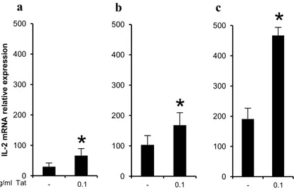
3.1 Tat enhances the production of IL-2 in PBLs
Several studies have reported the capacity of the HIV-1 Tat protein to activate PBLs and CD4+ T cells resulting in an increase of IL-2 production when cells were exposed to different stimuli, including antibodies specific for the CD3 and CD28 (anti-CD3/CD28) receptors that mimic physiological T cell activation [92, 102-104, 144]. To confirm these results, we first sought to determine whether the amounts of secreted Tat usually found in
vivo may account for this effect. To this aim, PBLs from healthy donors were activated
with anti-CD3/CD28 in the absence or presence of different doses of Tat (from 0.001 µg/ml to 1 µg/ml), and IL-2 mRNA levels were measured after 4 hours by qPCR (Scheme 1).
Scheme 1
As shown in Figure 3.1a, a 75 fold-increase of IL-2 mRNA was observed in PBLs activated in the absence of Tat compared to untreated PBLs, while the presence of Tat induced a 150-200 fold-increase of IL-2 mRNA expression. This effect was observed at similar levels for all Tat doses except at 0.001 µg/ml. Similar results were obtained at 24 hours after activation (Figure 3.1b), demonstrating that this effect is long lasting. As the highest fold-increase was observed at 0.1 µg/ml of Tat, this dose was chosen to perform the subsequent experiments. Of note, this concentration is within the range of the physiological concentration of Tat found in sera of HIV-positive individuals. Moreover, to assess whether the increased mRNA levels resulted in increased cytokine release, IL-2 secretion from PBLs was evaluated by Bio-Plex at 24 hours after activation.
As shown in Figure 3.1c, a 2 fold increase of the production of IL-2 was detected in PBLs activated in the presence of Tat.
PBLs Anti-CD3/CD28;
Tat
4 or 24 hours
31
To further confirm that the increase of the production of IL-2 was mediated by the presence of Tat, similar experiments were performed in the presence of anti-Tat antibodies obtained from sera of immunized mice. As shown in Figure 3.2, the incubation with anti-Tat antibodies during the treatment, abolished the anti-Tat-mediated effect.
Figure 3.1. Tat enhances IL-2 production in activated PBLs. (a, b) PBLs from healthy donors (n=6)
activated with anti-CD3/CD28 were cultured in the absence or presence of the indicated concentrations of Tat for 4 (a) or 24 (b) hours and IL-2 mRNA levels were quantified by qPCR and normalized to untreated cells. (c) PBLs from healthy donors (n=6) unstimulated or activated with anti-CD3/CD28 were cultured in the absence or presence of Tat (0.1 µg/ml). After 24 hours IL-2 release was quantified by Bio-Plex and normalized to untreated cells. Data are presented as mean ± SEM. For statistical analysis two-tailed Wilcoxon signed rank test was used. *P<0.05: Tat-treated cells compared to Tat-untreated control cells.
Figure 3.2. Tat-mediated increase of IL-2 production in activated PBLs is abolished by anti-Tat antibodies. PBLs from healthy donors unstimulated or activated with anti-CD3/CD28 were cultured in the
absence or presence of Tat (0.1 µg/ml) and anti-Tat immune antibodies. IL-2 mRNA levels were quantified by qPCR and normalized to untreated cells. The results of one representative experiment out of three are shown. + + -+ -+ 0 20 40 60 80 100 120 140 IL -2 mRN A re la tiv e e x pre s s ion CD3/CD28 0.1mg/ml Tat Anti-Tat Ab -+ -+ + + 0 100 200 300 400 500 - 0.001 0.01 0.1 1 IL -2 m RN A r e la tiv e e x p re s s io n 0 100 200 300 400 500 - 0.1 1 mg/ml Tat
*
*
*
*
*
0 200 400 600 800 1000 Rel a tiv e IL -2 r e le a s e CD3/CD28 0,1 μg/ml Tat -- +- ++*
a . b . c .32
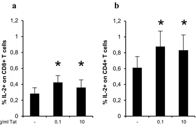
3.2 Tat enhances IL-2 and IFNγ production in CD8+ and CD4+ T cells
We sought to determine whether Tat specifically affects cytokine production in activated CD4+ and CD8+ T cells. To this aim, first the expression of IL-2 mRNA was evaluated in activated CD4+ and in CD8+ T cells activated alone or purified from activated PBLs (Scheme 2a and 2b).
Scheme 2
Interestingly, Tat significantly increased IL-2 mRNA in both CD8+ T cells activated alone (Figure 3.3a) or purified from activated PBLs (Figure 3.3b). Moreover, consistently with literature results [102], the presence of Tat during the stimulation induced a significant increase of IL-2 mRNA expression in CD4+ T cells (Figure 3.3c).
PBLs CD8+ Anti-CD3/CD28; Tat PBLs Anti-CD3/CD28; Tat CD8+/CD4+ 4 hours
IL-2 and IFNγ mRNA and protein expression
4 hours
IL-2 and IFNγ mRNA and protein expression
33
Figure 3.3.Tat enhances IL-2 production in CD8+ and CD4+ T cells.(a)CD8+ T cells were purified from PBLs from healthy donors (n=6) and activated with anti-CD3/CD28 in the presence or absence of 0.1µg/ml of Tat for 4 hours. (b) PBLs from healthy donors (n=6) were activated with anti-CD3/CD28 in the presence or absence of 0.1µg/ml of Tat. After 4 hours of stimulation, CD8+ T cells were purified. (c) CD4+ T cells
were purified from PBLs from healthy donors (n=6) and activated with anti-CD3/CD28 in the presence or absence of 0.1µg/ml of Tat for 4 hours. IL-2 mRNA levels were quantified by qPCR and normalized to untreated cells. Data are presented as mean ± SEM. For statistical analysis two-tailed Wilcoxon signed rank test was used. *P<0.05: Tat-treated cells compared to control cells.
To confirm these results, the IL-2 production was also evaluated by intracellular cytokine staining (ICS). To this aim, PBLs from healthy subjects were activated with anti-CD3/CD28 in presence or absence of Tat (0.1 and 10 µg/ml), and the secretion of IL-2 was measured in CD8+ and CD4+ T cells at 6 and 18 hours after treatment (Figure 3.4a and 3.4b). There was no effect of Tat on IL-2 production by CD8+ and CD4+ T cells at 6 hours after stimulation (data not shown). However, an average of 1.5-time fold increase of IL-2 secretion was observed after 18 hours of stimulation in both CD8+ and CD4+ T cells activated in the presence of 0.1 and 10 µg/ml of Tat compared to CD8+ and CD4+ T cells activated without Tat.
Taken together, these data demonstrate for the first time that Tat increases the
production of IL-2 in CD8+ T cells.
0 100 200 300 400 500 - 0.1
*
0 100 200 300 400 500 - 0.1 0 100 200 300 400 500 - 0.1 IL -2 m R N A re la ti ve e x pre ss ion*
mg/ml Tat*
a b c34
Figure 3.4. Tat enhances percentage of IL-2+ CD8+ and IL-2+ CD4+ T cells. PBLs from healthy donors
(n=7) were activated with anti-CD3/CD28 in the presence or absence of different concentrations of Tat for 18 hours. Percentages of CD8+ (a) or CD4+ (b) T cells secreting IL-2 were determined by ICS. Data are
presented as mean ± SEM. For statistical analysis two-tailed Wilcoxon signed rank test was used. *P<0.05: Tat-treated cells compared to control cells.
Thus, we next investigated whether the presence of Tat during the activation of T cells could also modulate IFNγ production. To this aim, first the expression of IFNγ mRNA was evaluated in activated CD4+ andin CD8+ T cells activated alone or purified from activated PBLs. The presence of Tat during the stimulation dramatically enhanced IFNγ production in CD8+ T purified from activated PBLs and in CD4+ T cells activated alone but not in CD8+ T cells activated alone (Figure 3.5 a-c).
Taken together, these results demonstrate that physiological concentrations of Tat
enhance the production of IL-2 and IFNγ in both CD8+ and CD4+ T cells activated
with anti-CD3/CD28. 0 0,2 0,4 0,6 0,8 1 1,2 - 0.1 10 % I L -2 + o n CD8+ T c e ll s 0 0,2 0,4 0,6 0,8 1 1,2 - 0.1 10 % I L -2 + o n CD 4 + T c e ll s mg/ml Tat
*
*
*
*
a b35
Figure 3.5. Tat enhances IFNγ production in CD8+ and CD4+ T cells.(a) CD8+ T cells were purified from
PBLs from healthy donors (n=6) and activated with anti-CD3/CD28 in the presence or absence of 0.1µg/ml of Tat for 4 hours. (b) PBLs from healthy donors (n=6) were activated with anti-CD3/CD28 in the presence or absence of 0.1µg/ml of Tat. After 4 hours of stimulation, CD8+ T cells were purified. (c) CD4+ T cells were purified from PBLs from healthy donors (n=6) and activated with anti-CD3/CD28 in the presence or absence of 0.1µg/ml of Tat for 4 hours. IFNγ mRNA levels were quantified by qPCR and normalized to untreated cells. Data are presented as mean ± SEM. For statistical analysis two-tailed Wilcoxon signed rank test was used. *P<0.05: Tat-treated cells compared to control cells.
3.3 Tat does not affect the proliferation of activated CD8+ and CD4+ T cells
We next evaluated whether the presence of Tat during the activation of T cells resulted in an enhanced proliferation. To this purpose, PBLs activated with different stimuli (anti-CD3/CD28, PHA, PMA and LPS) in the absence or presence of Tat were cultured up to six days, and PBLs, CD8+ and CD4+ T cell proliferation was measured by CFSE staining and MTT assay (Scheme 3). Scheme 3 a b c 0 300 600 900 1200 - 0.1
*
0 300 600 900 1200 - 0.1 IFN γ m R N A re la ti ve e x pre ss ion mg/ml Tat 0 300 600 900 1200 - 0.1*
PBLs Anti-CD3/CD28, PHA, PMA and LPS ;
Tat
6 days Proliferation and viability
36
As shown in Figure 3.6, the presence of Tat did not affect the proliferation of PBLs activated with the indicated stimuli. In addition, Tat did not affect proliferation of CD4+ and CD8+ T cells activated with CD3/CD28 (Figure 3.7). This is consistent with previous reports showing that soluble Tat was ineffective in enhancing anti-CD3/CD28 induced proliferation [102, 115].
Figure 3.6. Tat does not affect T cell proliferation. PBLs from healthy donors were activated with
different kind of stimuli: PHA, PMA + Ionomycin, LPS and anti-CD3/CD28, in the presence or absence of different concentrations of Tat, and cultured up to six days. Cell proliferation was measured by MTT assay. One representative experiment out of three is shown.
0 0,05 0,1 0,15 0,2 0,25 0,3 0,35 0,4 0,45 0,5 - - + Tat 0.1 µg/ml + Tat 1 µg/ml - + Tat 0.1 µg/ml + Tat 1 µg/ml - + Tat 0.1 µg/ml + Tat 1 µg/ml - + Tat 0.1 µg/ml + Tat 1 µg/ml
NT PHA PMA LPS Anti-CD3/CD28
O.
D
37
Figure 3.7. Tat does not affect CD4+ and CD8+ T cells proliferation. PBLs from healthy donors were
activated with anti-CD3/CD28 in the absence or presence of 0.1 µg/ml of Tat and cultured up to six days. Proliferation of CD4+ and CD8+T cells was assessed by CFSE staining. One representative experiment out of
three is shown.
3.4 Tat affects the expression of key transcription factors in activated CD8+ T cells
T-bet and Eomes are transcription factors (TFs) that are up-regulated during HIV infection [79] and that control IFNγ production [45, 145]. Since we have shown here that Tat enhances IFNγ production in human CD8+ T cells stimulated by TCR engagement, we next characterized the expression of T-bet and Eomes in CD8+ T cells activated alone (Scheme 4a) or purified from activated PBLs (Scheme 4b) cultured in the absence or presence of Tat. Moreover, the expression of other TFs important for T cell functionality, survival and programming, as Blimp-1, Bcl-6 and Bcl-2, was analyzed.
Scheme 4 NT CD3/CD28 CD3/CD28 + Tat CD4+T cells CD8+T cells PBLs CD8+ Anti-CD3/CD28; Tat PBLs Anti-CD3/CD28; Tat CD8+ 4 hours
T-bet , Eomes, Blimp-1, Bcl-6 and Bcl-2 mRNA expression.
4 hours
T-bet and Eomes mRNA and protein expression. Blimp-1, Bcl-6 and Bcl-2 mRNA expression.
38
As shown in Figure 3.8a the presence of Tat during the stimulation did not change at a significant level the transcriptional profile of CD8+ T cells activated alone, as the expression of all the TFs investigated was not affected by Tat-treatment. Conversely, mRNA levels of all the five TFs taken into consideration were significantly increased in CD8+ T cells purified from PBLs activated in the presence of Tat (Figure 3.8b). Notably, Tat up-regulated not only genes required for effector functions (as T-bet, Eomes, Blimp-1), but also TFs important for memory development (Bcl-6 and Eomes) and T cell survival (Bcl-2). Moreover, these results demonstrate that Tat does not directly affect CD8+ T cells, but requires other lymphocyte subpopulations to modulate the TFs expression in CD8+ T cells. Finally, to assess whether the increased mRNA expression correlated with an increased protein expression, T-bet and Eomes proteins were evaluated by western blotting in CD8+ T cells at 24 and 48 hours after activation. Notably, CD8+ T cells purified from PBLs activated in the presence of Tat exhibited an increase of T-bet and Eomes expression 48 hours after the activation (Figure 3.9).
Taken together, these data demonstrate that Tat favors the activation of CD8+ T cells
affecting the expression of TFs crucial for T cell programming and functionality. Of note, this Tat-mediated effect required the help of other lymphocyte subpopulations.
39
Figure 3.8. The effect of Tat on the transcriptional profile of CD8+ T cells. (a) CD8+ T cells were purified
from PBLs of healthy donors (n=8), activated with anti-CD3/CD28 and cultured in the absence or presence of 0.1µg/ml of Tat. After 4 hours, mRNA levels of the indicated molecules were quantified by qPCR and normalized to untreated cells. (b) PBLs from healthy donors (n=8) activated with anti-CD3/CD28 were cultured in the absence or presence of Tat (0.1 µg/ml). After 4 hours, CD8+ T cells were purified and mRNA
levels of the indicated molecules were quantified by qPCR and normalized to untreated cells. Data are presented as mean ± SEM. For statistical analysis two-tailed Wilcoxon signed rank test was used. *P<0.05: Tat-treated cells compared to Tat-untreated control cells.
Figure 3.9. Tat enhances T-bet and Eomes protein expression in CD8+ T cells. PBLs from healthy donors
unstimulated or activated with anti-CD3/CD28 were cultured in the absence or presence of Tat (0.1 µg/ml). (a) CD8+ T cells were purified after 24 and 48 hours, and expression of T-bet and Eomes proteins was
assessed by western blotting. The results of one representative experiment out of three are shown.
0 2 4 6 8 10 0 1 2 3 4 5 m RN A r e la tiv e e x p re s io n 0 1 2 3 4 5 0 1 2 3 4 5 0 1 2 3 4 5 0 1 2 3 4 5 6 m RN A r e la tiv e e x p re s s io n T-bet 0 1 2 3 4 5 6 Eomes 0 1 2 3 4 5 6 Blimp-1 0 1 2 3 4 5 6 Bcl-6 0 1 2 3 4 5 6 Bcl-2 -+ -+ + CD3/CD28 0,1mg/ml Tat - -+ -+ + -+ -+ + -+ -+ + -+ -+ +
*
*
-+ -+ + CD3/CD28 0,1mg/ml Tat - -+ -+ + -+ -+ + -+ -+ + -+ -+ +*
*
*
48h T-bet Eomes -actin CD8 24h -CD3/CD28 0,1 mg/ml Tat +- ++ -- +- ++ a b40
3.5 Tat affects the expression of key transcription factors in activated CD4+ T cells
As we have shown that the Tat-mediated up-regulation of T-bet, Eomes, Blimp-1, Bcl-6, Bcl-2 in CD8+ T cells requires the presence of other T lymphocyte subpopulations, we sought to determine whether Tat could also modulate the transcriptional profile of CD4+ T lymphocytes which are known to contribute to the activation of CD8+ T cells. To this aim, purified CD4+ T cells were activated, in the absence or presence of Tat, with anti-CD3/CD28, and the expression of the above mentioned TFs was evaluated 4 hours after activation (Scheme 5).
Scheme 5
Notably, a 15.5-fold increase of T-bet mRNA was observed in CD4+ T cells activated in the presence of Tat as compared to a 7-fold increase in CD4+ T cells activated without Tat. Moreover, Tat-treatment significantly increased the expression of Blimp-1, 6 and Bcl-2 but not of Eomes (Figure 3.10). Finally, to assess whether the increased mRNA expression correlated with an increased protein expression, T-bet protein was evaluated by western blotting in CD4+ T cells at 24 and 48 hours after activation. Notably, CD4+ T cells activated in the presence of Tat exhibited an increase of T-bet expression after 48 hours after the stimulation (Figure 3.11).
Thus, these data provide evidence that Tat favors the activation of CD4+ T cells
up-regulating the expression of key TFs.
PBLs
Anti-CD3/CD28; Tat
CD4+
4 hours
T-bet and Eomes mRNA and protein expression. Blimp-1, Bcl-6 and Bcl-2 mRNA expression.
41
Figure 3.10.The effect of Tat on the transcriptional profile of CD4+ T cells. CD4+ T cells were purified from PBLs of healthy donors (n=8), activated with anti-CD3/CD28 and cultured in the absence or presence of 0.1µg/ml of Tat. After 4 hours, mRNA levels of the indicated molecules were quantified by qPCR and normalized to untreated cells. Data are presented as mean ± SEM. For statistical analysis two-tailed Wilcoxon signed rank test was used. *P<0.05: Tat-treated cells compared to Tat-untreated control cells.
Figure 3.11. Tat enhances T-bet and Eomes protein expression in CD4+ T cells. CD4+ T cells were purified from PBLs of healthy donors and were activated with anti-CD3/CD28 were cultured in the absence or presence of Tat (0.1 µg/ml). After 24 and 48 hours, the expression of T-bet protein was assessed by western blotting. The results of one representative experiment out of three are shown.
-+ -+ + CD3/CD28 0,1mg/ml Tat -+ -+ + -+ -+ + -+ -+ + -+ -+ + 0 5 10 15 20 25 m RN A r e la tiv e e x p re s s io n T-bet 0 1 2 3 4 5 6 7 Eomes 0 1 2 3 4 5 6 7 Blimp-1 0 1 2 3 4 5 6 7 Bcl-6 0 1 2 3 4 5 6 7 Bcl-2
![Figure 1.2. CD4 + T cells generation. Adapted by [18]](https://thumb-eu.123doks.com/thumbv2/123dokorg/4718732.45552/11.892.271.640.650.1057/figure-cd-t-cells-generation-adapted.webp)


![Figure 1.9. HIV effects on CD8+ T cells. Adapted by [76]](https://thumb-eu.123doks.com/thumbv2/123dokorg/4718732.45552/25.892.235.658.277.548/figure-hiv-effects-cd-t-cells-adapted.webp)
![Figure 1.11. Differential activation of MAP kinases by distinct Nox pathways. Adapted by [114]](https://thumb-eu.123doks.com/thumbv2/123dokorg/4718732.45552/28.892.246.668.500.791/figure-differential-activation-map-kinases-distinct-pathways-adapted.webp)