https://doi.org/10.1007/s00405-021-06723-7 LARYNGOLOGY
Validation of the European Laryngological Society classification
of glottic vascular changes as seen by narrow band imaging
in the optical biopsy setting
Francesco Missale1,2 · Stefano Taboni3,4 · Andrea Luigi Camillo Carobbio1,5 · Francesco Mazzola6 ·
Giulia Berretti7 · Andrea Iandelli1,5 · Marco Fragale1,5 · Francesco Mora1,5 · Alberto Paderno7 · Francesca Del Bon7 ·
Giampiero Parrinello1 · Alberto Deganello7 · Cesare Piazza7 · Giorgio Peretti1,5 Received: 7 December 2020 / Accepted: 23 February 2021
© The Author(s) 2021
Abstract
Purpose In 2016, the European Laryngological Society (ELS) proposed a classification for vascular changes occurring in glottic lesions as visible by narrow band imaging (NBI), based on the dichotomic distinction between longitudinal vessels (not suspicious) and perpendicular ones (suspicious). The aim of our study was to validate this classification assessing the interobserver agreement and diagnostic test performance in detecting the final histopathology.
Methods A retrospective study was carried out by reviewing clinical charts, preoperative videos, and final pathologic diag-nosis of patients submitted to transoral microsurgery for laryngeal lesions in two Italian referral centers. In each institution, two physicians, independently re-assessed each case applying the ELS classification.
Results The cohort was composed of 707 patients. The pathologic report showed benign lesions in 208 (29.5%) cases,
papil-lomatosis in 34 (4.8%), squamous intraepithelial neoplasia (SIN) up to carcinoma in situ in 200 (28.2%), and squamous cell carcinoma (SCC) in 265 (37.5%). The interobserver agreement was extremely high in both institutions (k = 0.954, p < 0.001 and k = 0.880, p < 0.001). Considering the diagnostic performance for identification of at least SIN or SCC, the sensitivity was 0.804 and 0.902, the specificity 0.793 and 0.581, the positive predictive value 0.882 and 0.564, and the negative predic-tive value 0.678 and 0.908, respecpredic-tively.
Conclusion The ELS classification for NBI vascular changes of glottic lesions is a highly reliable tool whose systematic use allows a better diagnostic evaluation of suspicious laryngeal lesions, reliably distinguishing benign ones from those with a diagnosis of papillomatosis, SIN or SCC, thus paving the way towards confirmation of the optical biopsy concept.
Keywords Narrow band imaging · European Laryngological Society classification · Optical biopsy · Laryngeal cancer · Vascular changes · Endoscopy
Francesco Missale and Stefano Taboni have equally contributed to the manuscript.
Cesare Piazza and Giorgio Peretti have equally contributed as senior authors.
* Andrea Luigi Camillo Carobbio [email protected]
1 IRCCS Ospedale Policlinico San Martino, Genoa, Italy 2 Department of Molecular and Translational Medicine,
University of Brescia, Brescia, Italy
3 Section of Otorhinolaryngology, Head and Neck Surgery,
Azienda Ospedaliera di Padova, University of Padua, Padua, Italy
4 Department of Clinical and Experimental Sciences,
University of Brescia, Brescia, Italy
5 Department of Surgical Sciences and Integrated Diagnostics
(DISC), University of Genoa, Genoa, Italy
6 Department of Otolaryngology, Head and Neck Surgery,
IRCCS Regina Elena National Cancer Institute, Rome, Italy
7 Unit of Otorhinolaryngology, Head and Neck Surgery, ASST
Introduction
Early detection and diagnosis of laryngeal squamous cell carcinoma (SCC) are crucially involved not only in reducing mortality, but also to optimize therapeutic approaches aimed at achieving the best organ and functional preservation [1,
2]. Fortunately, glottic SCC, the most common laryngeal tumor localization, is more frequently detected at an earlier stage than tumors originating in other subsites of the head and neck due to its early (albeit highly non-specific) symp-toms [3]. Laryngeal SCC examination is usually performed by flexible (video)endoscopy under white light (WL) and relies on the analysis of superficial characteristics (size, color, location, single or multifocal appearance) and visible morphological features (smoothness, irregularity, keratini-zation, ulceration, submucosal growth), per se non-pathog-nomonic and possibly overlapping each other in malignant and benign pathologies, especially when diagnosed at early stages. This implies the frequent need to obtain an incisional biopsy before deciding on the therapeutic approach, with an increase in costs, anesthetic risks, and potential undue dam-age to the vocal cords.
Computed tomography and magnetic resonance imaging definitively play a major role in diagnosis of more advanced diseases, providing information about the involvement of laryngeal structures and lateral neck lymph nodes, while, on the other hand, they fall shortly in identifying and character-izing superficial mucosal lesions. By contrast, the judicious use of high definition and better contrasted videoendoscopic images now offer staggering details in evaluation of epithe-lial and superficial vascular patterns. Moreover, clinicians can increasingly benefit from novel optical diagnostic meth-ods, providing information even closer to those obtained by formal histopathological examination, thus differentiating between normal mucosa and discrete lesions and, among the latter, between those with benign versus malignant behaviors [4]. In this context, narrow band imaging (NBI) is a well-established bioendoscopic technique using filtered wavelengths to enhance microvascular alterations associ-ated with preneoplastic and neoplastic transformation of the upper aerodigestive tract (UADT) mucosa [5–8]. Since its first introduction in the late 1990s, the use of NBI has considerably upgraded physicians’ ability for non-invasive detection and delineation of suspicious mucosal lesions, and is thus beneficial in the diagnosis of a variety of benign and malignant lesions [9]. However, the need for a common language to be shared among clinicians to describe NBI-enhanced vascular patterns led to the proposal of different classifications during the last decades [10–12]. In 2016, the Working Committee on Endoscopic Laryngeal Imaging of the European Laryngological Society (ELS) published a new proposal for a simplified (dichotomic) description
of vocal fold vascular changes as seen under NBI [12]. In this system, the authors distinguished between normal and pathologic vascular patterns of the vocal folds. The latter, in turn, were divided into longitudinal and perpendicular vascular changes. Longitudinal vessels characterize benign lesions, while perpendicular ones (i.e. dot-like intrapapillary capillary loops [IPCL], enlarged and worm-like vessels) are considered signs of benign neoplasms (such as papilloma-tosis), squamous intraepithelial neoplasia (SIN), or frankly malignant lesions.
The present study aims to assess the performance of the ELS classification of vascular changes in a broad multicenter cohort, testing its interobserver agreement as primary end-point, and analyzing its accuracy in predicting the final path-ological results in an optical biopsy setting, i.e. by evaluat-ing the diagnostic accuracy of NBI by comparevaluat-ing it with the final histopathologic diagnosis obtained after complete removal of the glottic lesion.
Methods
Study population
A retrospective study was carried out enrolling patients treated at the Departments of Otorhinolaryngology—Head and Neck Surgery of the Universities of Genoa (Center A; from January 2012 to December 2016) and Brescia (Center B; from January 2015 to December 2018), Italy.
All patients enrolled were affected by laryngeal lesions; a pre-treatment videoendoscopic evaluation with both WL and NBI was performed in the office as well as in the operatory theater, and the records were saved in his/her medical chart; the laryngeal lesion was treated by a transoral microsurgical approach by either cold instrumentation and carbon dioxide (CO2) laser; postoperative histopathologic assessment was
obtained to classify the resected tissue as benign, dysplastic or malignant. Histopathological diagnosis was performed according to the WHO classification system [13].
Clinical diagnostic work‑up
All patients were preoperatively evaluated by high-defi-nition television (HDTV)-WL and HDTV-NBI through a videorhinolaryngoscope ENF-VQ or ENF-VH coupled to an Evis Exera II HDTV camera connected to an Evis Exera II CLV-180B light source (Olympus Medical System Corp., Tokyo, Japan). Just before surgery, in the operating room, with patient under general anesthesia, intraoperative HDTV-WL and HDTV-NBI rigid endoscopy with 0° and 70° tel-escopes (Karl Storz, Tuttlingen, Germany) was also system-atically performed. On the basis of this diagnostic work-up, laryngeal lesions were subsequently removed by either a
phonomicrosurgical approach (in case of benign lesions) or excisional biopsy (in case of papillomatosis, SIN, carcinoma in situ [CIS] or invasive SCC) by type I–III cordectomies according to the ELS classification of cordectomies [14]. Clinical evaluation applying the ELS classification Clinical records of the study population, including demo-graphic features and information on previous treatments in terms of laryngeal surgery, head and neck radiotherapy,
or other treatments before the index transoral microsurgi-cal procedure were retrieved from the hospital databases. Two independent physicians from each institution with at least a 3-year-experience in the use of NBI, blinded to the final histopathologic result, retrospectively and inde-pendently reviewed the intraoperative videoendoscopic recordings. Applying the ELS classification for laryngeal vascular changes [15], each case was categorized as sus-picious for malignancy (presence of perpendicular vas-cular abnormalities as shown in Fig. 1) or non-suspicious
Fig. 1 Endoscopic picture of three representative cases of SCC (a–d) or CIS (e, f) correctly identified as suspicious by the presence of perpen-dicular vascular abnormalities (* in all panels) evaluating the NBI endoscopic appearance (b, d, f) and applying the ELS classification
Fig. 2 Endoscopic picture of three representative cases of benign glottic lesions: keratosis without atypia (a, b), Reinke’s edema (c, d) and polyp (e, f) correctly identified as benign lesions without iden-tifying any perpendicular vascular abnormalities evaluating the NBI
endoscopic appearance (b, d, f) and applying the ELS classifica-tion. The ° in all panels points to non-suspicious longitudinal vascu-lar abnormalities that can be observed inside the lesion (d) of at its boundary (b, f)
(undetectable perpendicular vascular changes or longitudinal ones as shown in Fig. 2). In case of interobserver disagree-ment, consensus was reached by direct comparison between the examiners. The identification of features of respiratory
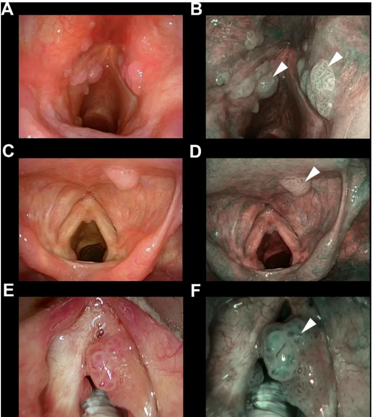
papillomatosis (i.e., wide angle IPCL) was also considered as a secondary endpoint (Fig. 3).
Fig. 3 Endoscopic pictures of three representative cases (a, b; c, d; and e, f) of recurrent laryngeal papillomatosis correctly identified detecting wide angle IPCLs (arrowheads in all panels) evaluating the NBI endoscopic appearance (b, d, f)
Statistical analysis
Clinical data were reported as absolute and relative fre-quencies. The reliability of the ELS classification was assessed for each independent cohort measuring the Cohen’s k statistic and the strength of agreement graded according to Altman et al. [16, 17], as reported in Table 1. Considering the final consensus of the evaluation, we assessed the performance of the diagnostic test for pre-diction of the final pathologic result (at least SIN1, up to SCC) in an optical biopsy setting. For better understand-ing of the clinical utility of applyunderstand-ing the classification and
detecting papillomatosis-like features, the Clinical Util-ity (CU) indexes were also derived, taking into account the measures of occurrence (sensitivity or specificity) together with the possibility of discrimination (positive [PPV] or negative predictive values [NPV]), and their qualitative grading were judged accordingly [18, 19] (Table 1). The Positive Clinical Utility Index (CU + Ve) is defined as sensitivity*PPV, and a high CU + Ve results should characterize “case finding” tests. By contrast, good Negative Clinical Utility Index (CU−Ve), defined as specificity*NPV, should be ideal for “screening” tests [18, 19].
In all analyses, a two-tailed p value < 0.05 was consid-ered significant. GraphPad Prism (San Diego, CA, USA), Stata (version 13.0, College Station, Texas, USA) and R (version 3.6.2) were used for statistical analysis and ren-dering graphs.
Results
Clinical data
A total of 707 patients met enrolment criteria: 434 (61.3%) had been evaluated and treated at the center A, and 273 (38.7%) at the center B. Five-hundred and fifty six (78.6%) were males and 151 (21.4%) females, with a mean age of 61.8 years (range 18–91). Four-hundred and seventy-eight
Table 1 Definition of interrater agreement qualitative scores accord-ing to Altman et al. [15, 16] and Clinical Utility indexes grading according to Mitchell [18]
CU + Ve positive clinical utility index, CU−Ve negative clinical utility index
Agreement classification Clinical Utility Index classification
κ Strength of
agreement CU+Ve or CU−Ve Grading
< 0.21 Poor < 0.49 Poor utility
0.21–0.40 Fair 0.49–0.63 Satisfactory utility 0.41–0.60 Moderate 0.64–0.80 Good utility
0.61–0.80 Good 0.81–1.00 Excellent utility
0.81–1.00 Very good
Table 2 Clinical features of the cohort
The sum of rows is 723 since 16 patients had two different previous treatments
RT radiotherapy, SIN squamous intraepithelial neoplasia, CIS in situ carcinoma, SCC squamous cell carci-noma
Variables All Longitudinal vessels Perpendicular
vessels n % n % n % All 707 100.0 283 40.0 424 60.0 Gender Male 556 78.6 184 26.0 372 52.6 Female 151 21.4 99 14.0 52 7.4 Previous treatments° No 478 67.6 221 31.3 257 36.4 Previous surgery 174 24.6 45 6.4 129 18.2 Previous RT 17 2.4 4 0.6 13 1.8 Previous biopsy 54 7.6 8 1.1 46 6.5 Histology Benign 208 29.4 184 26.0 24 3.4
Papillomatosis without atypia 34 4.8 0 0.0 34 4.8
SIN1 46 6.5 24 3.4 22 3.1
SIN2 61 8.6 26 3.7 35 5.0
SIN3/CIS 93 13.2 15 2.1 78 11.0
(67.6%) patients were submitted to endoscopic evalua-tion and surgical procedures without previous treatments, whereas 174 (24.6%) had been already surgically treated,
17 (2.4%) received head and neck radiotherapy, and 54 (7.6%) had been previously biopsied elsewhere. The final pathologic report was consistent with a benign lesion in 208
No Yes Longitudinal Perpendicular Benign SIN1 SIN2 SIN3 SCC Papillomatosis No Yes
Previous_treatments ELS_NBI Histology WA_IPCL
Fig. 4 Alluvial chart showing frequency distribution of previous treatment, ELS classification results, histology, and presence of wide angle IPCL features (WA IPCL). Color code according to different matching of previous treatments and ELS classification results
Table 3 Agreement analysis by Cohen’s k test
Institution Type of lesion N % Agreement (%) κ 95% CI (κ) p
University of
Genoa (Center A) AllUntreated 434284 10065.4 97.797.5 0.9540.949 0.86–1.00.833–1.0 < 0.0001 < 0.0001
Previous biopsy/surgery/RT 150 34.6 98.0 0.945 0.785–1.0 < 0.0001
University of
Brescia (Center B) AllUntreated 273194 10071.1 94.994.3 0.8720.864 0.754–0.9910.723–1.0 < 0.0001 < 0.0001
(29.5%) cases, papillomatosis without atypia in 34 (4.8%), mild SIN (SIN1) in 46 (6.5%), moderate SIN (SIN2) in 61 (8.6%), severe SIN (SIN3) or CIS in 93 (13.1%), and SCC in 265 (37.5%). Full details are summarized in Table 2, Fig. 4. ELS classification interobserver reliability
Cohen’s k statistic was used to assess the agreement between judgment of each lesion by two independent raters in each institution applying the ELS classification. Accord-ing to the criteria by Altman et al. [16, 17], reported in Table 1, for the entire cohort the result was satisfactory and showed a very good agreement between observers at both the center A (κ = 0.954; 95% confidence interval [CI] 0.86–1.0, p < 0.0001) and the center B (κ = 0.872; 95% CI 0.754–0.991, p < 0.0001) (Table 3). The agreement was consistent and significant (p < 0.0001) for both Institu-tions, as well as for untreated (κ = 0.949; 95% CI 0.833–1.0 and κ = 0.864; 95% CI 0.723–1.0, respectively) and previ-ously treated patients (κ = 0.945; 95% CI 0.785–1.0 and
κ = 0.894; 95% CI 0.674–1.0, respectively) (Table 3). Diagnostic performance
Considering the final score in the entire cohort (24 cases with initial disagreement were resolved between the exam-iners), performance of the diagnostic test was assessed investigating the detection of at least SIN1-SCC (Table 4). The best sensitivity and NPV were obtained for detection of SCC (0.90 and 0.91, respectively) and, accordingly, the best specificity and PPV for diagnosis of at least SIN1 (0.79
and 0.88, respectively). Considering previous treatments as a potential source of bias, for untreated patients the ELS classification reached the best performance with sensitivity and NPV for detection of SCC of 0.93 and 0.95, respectively, and specificity and PPV for diagnosis of at least SIN1 of 0.88 and 0.91, respectively. In previously treated patients, the performance of endoscopic evaluation was still satisfac-tory in terms of sensitivity (from 0.82 to 0.86), while it was poorer in terms of specificity (from 0.34 to 0.49), NPV (from 0.46 to 0.76), and PPV (from 0.45 to 0.84) (Table 4, Fig. 5).
The measurement of the CU indexes confirmed this observation with a good CU + Ve and CU−Ve for all out-comes except one in untreated patients, whereas no more than satisfactory or even poorer results were obtained for most outcomes in previously treated or biopsied patients, as shown in Table 5.
Diagnostic performance in respiratory papillomatosis
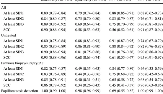
Among perpendicular vascular changes, the ELS classifica-tion well defines the vascular pattern of recurrent respiratory papillomatosis lesions, characterized by vessel loops with wide angle turning point, embedded in a three-dimensional warty structure [15]. We tested the identification of these features by NBI in our cohort, confirming their value for correct identification of this disease with a sensitivity of 1.0 (95% CI 0.90–1.0), specificity of 0.98 (95% CI 0.96–0.99), PPV of 0.69 (95% CI 0.55–0.82), and NPV of 1.0 (95% CI 0.99–1.0), as shown in Table 4. Moreover, the measure of CU indexes confirmed the excellent performance of NBI
Table 4 Diagnostic test results
PPV positive predictive value, NPV negative predictive value, CI confidence interval, SIN squamous intraepithelial neoplasia, SCC squamous cell carcinoma
Sensitivity (95% CI) Specificity (95% CI) PPV (95% CI) NPV (95% CI) All
At least SIN1 0.80 (0.77–0.84) 0.79 (0.74–0.84) 0.88 (0.85–0.91) 0.68 (0.62–0.73) At least SIN2 0.84 (0.80–0.87) 0.75 (0.70–0.80) 0.83 (0.79–0.87) 0.76 (0.71–0.81) At least SIN3 0.89 (0.85–0.92) 0.69 (0.64–0.74) 0.75 (0.70–0.79) 0.86 (0.81–0.89) SCC 0.90 (0.86–0.94) 0.58 (0.53–0.63) 0.56 (0.52–0.61) 0.91 (0.87–0.94) Untreated
At least SIN1 0.80 (0.75–0.84) 0.88 (0.83–0.93) 0.91 (0.87–0.95) 0.74 (0.67–0.79) At least SIN2 0.85 (0.80–0.89) 0.86 (0.81–0.90) 0.88 (0.84–0.92) 0.82 (0.76–0.87) At least SIN3 0.90 (0.86–0.94) 0.81 (0.75–0.86) 0.81 (0.76–0.86) 0.90 (0.86–0.94) SCC 0.93 (0.88–0.96) 0.68 (0.63–0.74) 0.61 (0.55–0.67) 0.95 (0.91–0.97) Previous biopsy/surgery/RT
At least SIN1 0.82 (0.75–0.87) 0.49 (0.35–0.63) 0.84 (0.77–0.89) 0.46 (0.33–0.59) At least SIN2 0.83 (0.76–0.89) 0.44 (0.33–0.56) 0.75 (0.68–0.82) 0.56 (0.42–0.69) At least SIN3 0.85 (0.78–0.91) 0.40 (0.31–0.51) 0.65 (0.58–0.72) 0.68 (0.54–0.79) SCC 0.86 (0.77–0.92) 0.34 (0.26–0.43) 0.45 (0.41–0.57) 0.76 (0.63–0.86) Papillomatosis detection 1.00 (0.90–1.00) 0.98 (0.96–0.99) 0.69 (0.55–0.82) 1.00 (0.99–1.00)
with a CU–Ve of 0.98 (95% CI 0.97–0.99) and good perfor-mance in terms of CU + Ve of 0.69 (95% CI 0.56–0.83), as reported in Table 5.
Discussion
Among the several bioendoscopic techniques now avail-able for routine evaluation of the UADT, NBI appears to be the most effective in evaluation of the larynx, hypophar-ynx, oral and oropharyngeal cavities [1, 20, 21]. The easy use of NBI and other bioendoscopic tools based on similar principles, which aims to enhance the vascular features of tissues (e.g. SPIES [22] or iSCAN [20]), is mainly due to
full integration of high-definition videoendoscopes, easily activated by pressing a button during in-office endoscopic examination or during pre- and intraoperative assessment. Interestingly, the superior in-depth evaluation of the bioen-doscopic features of a given lesion may pave the way to the proof of concept of the optical biopsy, i.e. the capability to understand the nature of a given vocal fold mucosal lesion before its removal, thus modulating its excisional biopsy and optimizing hospitalization time, costs, and undue damage to surrounding healthy structures [23].
The need for a common language to categorize and share the findings from NBI evaluation led to a number of different classification systems. The first to have widespread diffusion in the head and neck scientific community was proposed
Fig. 5 Radar charts showing the diagnostic test applying the ELS classification for the detection of different histologic targets in the whole cohort (a), in the untreated group (b), and in the previously
surgical or RT treated group (c). Diagnostic test results referred to the detection of wide angle IPCLs for the diagnosis of laryngeal papil-lomatosis (d)
by Ni et al. [11]. These authors divided the different IPCL changes in five types (I–V), judging them as benign (from types I–IV), suspected malignant, and frankly malignant (type V). However, apart from its intrinsic complexity, this classification clearly showed a lack of a clear-cut threshold between benign and malignant diseases. In fact, different authors proposed different cut-offs for the worst endoscopic feature of each lesion to be considered suspicious, ranging from type III [24], to type IV [25–27], and type V [9, 11,
28].
Therefore, in 2016 the ELS proposed a new classifi-cation system for the interpretation of glottic vascular abnormalities detected during NBI-guided endoscopies [15]. This classification considers vascular abnormalities as IPCL perpendicular to the epithelium surface as suspi-cious, whereas longitudinal vascular changes (e.g. dilated or tortuous vessels, increased vessels numbers) are consid-ered as not suspicious to harbor respiratory papillomatosis, pre-malignant, or cancerous lesions. The first attempt to apply this dichotomic classification was in the study by Šifrer et al. [29] who analyzed 80 vocal cords lesions in which the identification of a perpendicular vascular pattern was diagnostic for CIS-SCC with a sensitivity of 100%, specificity of 95%, PPV of 88%, and NPV of 100%. Fur-ther analysis evaluating a larger cohort of 288 vocal cords gave similar results (sensitivity 98%, specificity 95%, PPV 88%, and NPV 99%) [30].
Our results, obtained in two of the European pioneer cent-ers applying NBI for evaluation of the UADT since 2007, herein confirm the intrinsic value of the ELS classification for laryngeal vascular changes in the identification of lesions
harboring pre-cancerous or frankly neoplastic alterations. In particular, we applied this diagnostic tool to demonstrate its possible role in performing a so-called optical biopsy. In fact, our policy has always been, for early glottic lesions, a one-stage modulated excisional biopsy based on a number of pre- and intraoperative diagnostic tests in which WL and NBI rigid endoscopy under general anesthesia has always played a paramount role [23]. Moreover, as asserted by many authors, NBI is capable of enhancing small lesions that are undetectable by WL alone, thus ameliorating the treatment of laryngeal SSC, as well as assessing the poten-tial multifocality of the disease and correct evaluation of intraoperative margins [31], as well as early identification of small recurrences during follow-up that may still allow application of minimally invasive treatments such as laser office-based procedures or second-look microlaryngoscopic operations [32–35]. Of note, the present study demonstrated a lower diagnostic accuracy of NBI in the previously treated patients compared to the untreated ones, thus confirming the potential confounding factor played by invasive sampling procedures when not directed to the full removal (excisional biopsy) of the entire visible lesion within safe margins.
The excellent interobserver reliability of the ELS classifi-cation with a k > 0.81 in all scenarios tested and reproducible in two independent centers confirms the reproducibility of the operators’ findings in applying this classification tool. The high interobserver reliability of the ELS classifica-tion can be explained by its intrinsic simple applicaclassifica-tion and dichotomic arrangement, providing better performance compared to other proposed classification systems such as that by Ni, which is complicated by a 5-tier structure and
Table 5 Clinical Utility indexes and utility grading results according to Mitchell [18]
CU + Ve positive clinical utility index, CU−Ve negative clinical utility index, CI confidence interval, SIN squamous intraepithelial neoplasia, SCC squamous cell carcinoma
CU+Ve (95% CI) CU+Ve Judgment CU−Ve (95% CI) CU−Ve Judgment All
At least SIN1 0.71 (0.67–0.75) Good 0.54 (0.49–0.58) Satisfactory At least SIN2 0.70 (0.65–0.74) Good 0.57 (0.53–0.62) Satisfactory At least SIN3 0.66 (0.61–0.71) Good 0.59 (0.55–0.63) Satisfactory
SCC 0.51 (0.45–0.57) Satisfactory 0.53 (0.49–0.57) Satisfactory
Untreated
At least SIN1 0.73 (0.68–0.78) Good 0.65 (0.60–0.70) Good
At least SIN2 0.75 (0.69–0.80) Good 0.70 (0.66–0.74) Good
At least SIN3 0.73 (0.68–0.79) Good 0.73 (0.69–0.77) Good
SCC 0.57 (0.50–0.64) Satisfactory 0.65 (0.61–0.69) Good
Previous biopsy/surgery/RT
At least SIN1 0.68 (0.61–0.75) Good 0.23 (0.11–0.34) Poor
At least SIN2 0.63 (0.55–0.70) Satisfactory 0.25 (0.14–0.35) Poor At least SIN3 0.56 (0.47–0.64) Satisfactory 0.27 (0.17–0.38) Poor
SCC 0.42 (0.32–0.52) Poor 0.26 (0.16–0.36) Poor
associated with moderate/substantial interrater accordance, with a k ranging from 0.55 to 0.69 [36, 37].
On the other hand, it has to be noted that all the observers involved in this study had a minimum experience of 3 years in the use of NBI technology. Even though application of the ELS Classification on vocal fold vascular changes as observed by NBI is no more subjective than any other diag-nostic performance, evaluation of certain subtle and some-times ambiguous neoangiogenic patterns still may require a higher level of expertise, for which a learning curve is inevi-tably necessary. However, data derived from the gastrointes-tinal field show that less than a year of training evaluating 200 cases is enough to guarantee an accurate evaluation of NBI frames and that the motivation of the trainer itself can significantly improve the overall performance [38].
Investigating the diagnostic test, having as a target all the possible grades of pre-malignant or malignant transforma-tion, allowed us to depict the capability of the ELS classifi-cation in helping to correctly identify pre-malignant cases with the highest PPV and specificity for at least SIN1 diag-nosis. The lower performance of such parameters observed for the final diagnosis of glottic SCC can be explained by the presence, and progressively increase, of perpendicular vascular changes at early stages of pre-malignant transfor-mation (SIN1-SIN2). By contrast, for diagnosis of laryngeal SCC, the ELS classification had good performance in terms of sensitivity and NPV, with a low rate of false negative cases and good confidence in a negative result (absence of perpendicular vascular changes).
Furthermore, several authors have underlined the utility of NBI for detection of recurrent respiratory papillomatosis and its ability to increase the detection rate of small lesions that invisible by WL alone [29, 33, 39–42]. The excellent performance in terms of CU + Ve and CU−Ve searching for wide angle IPCLs in the identification of respiratory papil-lomatosis mandates, as previously suggested by the recent literature [33, 39–44], the use of biologic endoscopy tools like NBI, and should be considered the endoscopic gold standard for optical biopsy and follow-up of patients affected by laryngeal papillomas.
The main limits of our study are represented by its ret-rospective design, balanced by analyzing a broad bicentric cohort. Nevertheless, among the estimator analyzed, the suboptimal performance in terms of specificity, negative predictive value, and CU−Ve could has been underesti-mated having chosen among the inclusive criteria the need for a histopathological diagnosis: several patients without any suspicious lesion at the first evaluation and along time could be considered as true negatives too, thus improving the values of such estimators.
Further developments in this field might include the anal-ysis of a prospective cohort of patients, implementing the enrollment of true negative cases and developing a real-time
software applicable in the head and neck, based on artificial intelligence algorithms already tested on retrospective stud-ies [45, 46], thus improving the objectivity and detection rate of these diagnostic tools, as already devised for gastrointes-tinal tract tumors [47, 48].
Conclusion
The ELS classification for NBI vascular changes of laryn-geal lesions, herein validated in a large multicenter cohort, is a highly reliable tool with good diagnostic performance in the optical biopsy setting, confirming its overall value. The systematic use of this classification seems to allow better (and purely endoscopic) diagnostic capability of suspicious glottic lesions, reliably distinguishing benign ones from those with a diagnosis of papillomatosis, SIN, or invasive SCC. The excellent performance of NBI for correct identifi-cation of respiratory papillomatosis also confirms its useful-ness in this clinical setting.
Funding Open access funding provided by Università degli Studi di Genova within the CRUI-CARE Agreement.
Availability of data and materials Full dataset will be available at: “ELS_NBI_Classification_Validation_Dataset”, Mendeley Data, V1,
https ://doi.org/10.17632 /txzzw 9n7xs .1https ://doi.org/10.17632 /txzzw 9n7xs .1 (Embargo date: 6th December 2021).
Declarations
Conflict of interest The authors certify that they have no affiliation with or involvement in any organization or entity with any financial interest.
Ethical approval The research did not involve any animal models; the research involved human participants in accordance with the ethical standards of the institutional and/or national research committees and with the 1964 Helsinki Declaration and its later amendments or com-parable ethical standards.
Informed consent Informed consent for disclosure of privacy in man-aging personal data for scientific purposes was obtained from all par-ticipants included in the study.
Open Access This article is licensed under a Creative Commons Attri-bution 4.0 International License, which permits use, sharing, adapta-tion, distribution and reproduction in any medium or format, as long as you give appropriate credit to the original author(s) and the source, provide a link to the Creative Commons licence, and indicate if changes were made. The images or other third party material in this article are included in the article’s Creative Commons licence, unless indicated otherwise in a credit line to the material. If material is not included in the article’s Creative Commons licence and your intended use is not permitted by statutory regulation or exceeds the permitted use, you will
need to obtain permission directly from the copyright holder. To view a copy of this licence, visit http://creat iveco mmons .org/licen ses/by/4.0/.
References
1. Sun C, Han X, Li X et al (2017) Diagnostic performance of nar-row band imaging for laryngeal cancer: a systematic review and meta-analysis. Otolaryngol Head Neck Surg 156:589–597. https ://doi.org/10.1177/01945 99816 68570 1
2. Watanabe A, Taniguchi M, Tsujie H et al (2009) The value of narrow band imaging for early detection of laryngeal cancer. Eur Arch Otorhinolaryngol. https ://doi.org/10.1007/s0040 5-008-0835-1
3. Chu EA, Kim YJ (2008) Laryngeal cancer: diagnosis and preop-erative work-up. OtolaryngolClin North Am 41:673–695 4. Green B, Cobb ARM, Brennan PA, Hopper C (2014) Optical
diag-nostic techniques for use in lesions of the head and neck: review of the latest developments. Br J Oral MaxillofacSurg 52:675–680.
https ://doi.org/10.1016/j.bjoms .2014.06.010
5. Muto M, Nakane M, Katada C et al (2004) Squamous cell carci-noma in situ at oropharyngeal and hypopharyngeal mucosal sites. Cancer 101:1375–1381. https ://doi.org/10.1002/cncr.20482
6. Piazza C, Dessouky O, Peretti G et al (2008) Narrow-band imag-ing: a new tool for evaluation of head and neck squamous cell carcinomas. Review of the literature. ActaOtorhinolaryngolItalor-ganoUffdellaSocItal di Otorinolaringol e ChirCerv-facc 28:49–54 7. Piazza C, Del Bon F, Peretti G, Nicolai P (2012) Narrow band
imaging in endoscopic evaluation of the larynx. CurrOpinOto-laryngol Head Neck Surg 20:472–476
8. Cohen J (2008) Comprehensive atlas of high resolution endoscopy and narrowband imaging (1st edn)
9. Kraft M, Fostiropoulos K, Gurtler N et al (2016) Value of narrow band imaging in the early diagnosis of laryngeal cancer. Head Neck 38:15–20. https ://doi.org/10.1002/hed.23838
10. Lin YC, Wang WH, Lee KF et al (2012) Value of narrow band imaging endoscopy in early mucosal head and neck cancer. Head Neck 34:1574–1579
11. Ni X-G, He S, Xu Z-G et al (2011) Endoscopic diagnosis of laryn-geal cancer and precancerous lesions by narrow band imaging. J LaryngolOtol. https ://doi.org/10.1017/S0022 21511 00020 33
12. Takano JH, Yakushiji T, Kamiyama I et al (2010) Detecting early oral cancer: narrowband imaging system observation of the oral mucosa microvasculature. Int J Oral MaxillofacSurg 39:208–213. https ://doi.org/10.1016/j.ijom.2010.01.007
13. Barnes L, Eveson JW, Reichart P, Sidransky D (2005) World Health Organization Classification of Head and Neck Tumours. WHO Classif Tumour
14. Remacle M, Eckel HE, Antonelli A et al (2000) Endoscopic cor-dectomy. A proposal for a classification by the working commit-tee, European Laryngological Society. Eur Arch Otorhinolaryn-gol 257:227–231. https ://doi.org/10.1007/s0040 50050 228
15. Arens C, Piazza C, Andrea M et al (2016) Proposal for a descriptive guideline of vascular changes in lesions of the vocal folds by the committee on endoscopic laryngeal imaging of the European Laryngological Society. Eur Arch Otorhinolaryngol 273:1207–1214. https ://doi.org/10.1007/s0040 5-015-3851-y
16. Altman DG (1991) Practical statistics for medical research. Chapman and Hall. Stat Med. https ://doi.org/10.1002/sim.47801 01015
17. Landis JR, Koch GG (1977) The measurement of observer agreement for categorical data. Biometrics. https ://doi. org/10.2307/25293 10
18. Mitchell AJ (2008) The clinical significance of subjective memory complaints in the diagnosis of mild cognitive impairment and dementia: a meta-analysis. Int J Geriatr Psychiatry 23:1191–1202.
https ://doi.org/10.1002/gps.2053
19. Mitchell AJ (2011) Sensitivity x PPV is a recognized test called the clinical utility index (CUI+). Eur J Epidemiol 26:251–252.
https ://doi.org/10.1007/s1065 4-011-9561-x
20. Hawkshaw MJ, Sataloff JB, Sataloff RT (2013) New concepts in vocal fold imaging: a review. J Voice 27:738–743. https ://doi. org/10.1016/j.jvoic e.2013.05.011
21. Deganello A, Paderno A, Morello R et al (2020) Diagnostic accu-racy of narrow band imaging in patients with oral lichen planus: a prospective study. Laryngoscope. https ://doi.org/10.1002/ lary.29035
22. Staníková L, Walderová R, Jančatová D et al (2018) Compari-son of narrow band imaging and the Storz Professional Image Enhancement System for detection of laryngeal and hypopharyn-geal pathologies. Eur Arch Otorhinolaryngol 275:1819–1825.
https ://doi.org/10.1007/s0040 5-018-4987-3
23. Mora F, Carta F, Missale F et al (2020) Laryngeal mid-cord erythroleukoplakias: how to modulate the transoralCO(2) laser excisional biopsy. Cancers (Basel). https ://doi.org/10.3390/cance rs120 82165
24. Rzepakowska A, Sielska-Badurek E, Cruz R et al (2018) Narrow band imaging versus laryngovideostroboscopy in precancerous and malignant vocal fold lesions. Head Neck 40:927–936. https ://doi.org/10.1002/hed.25047
25. Ni XG, Zhu JQ, Zhang QQ et al (2019) Diagnosis of vocal cord leukoplakia: the role of a novel narrow band imaging endo-scopic classification. Laryngoscope 129:429–434. https ://doi. org/10.1002/lary.27346
26. Vilaseca I, Valls-Mateus M, Nogués A et al (2017) Usefulness of office examination with narrow band imaging for the diag-nosis of head and neck squamous cell carcinoma and follow-up of premalignant lesions. Head Neck 39:1854–1863. https ://doi. org/10.1002/hed.24849
27. De Vito A, Meccariello G, Vicini C (2017) Narrow band imaging as screening test for early detection of laryngeal cancer: a prospec-tive study. ClinOtolaryngol 42:347–353. https ://doi.org/10.1111/ coa.12728
28. Rzepakowska A, Sielska-Badurek E, Żurek M et al (2018) Nar-row band imaging for risk stratification of glottic cancer within leukoplakia. Head Neck 40:2149–2154. https ://doi.org/10.1002/ hed.25201
29. Šifrer R, Rijken JA, Leemans CR et al (2018) Evaluation of vascu-lar features of vocal cords proposed by the European Laryngologi-cal Society. Eur Arch Otorhinolaryngol 275:147–151. https ://doi. org/10.1007/s0040 5-017-4791-5
30. Šifrer R, Šereg-Bahar M, Gale N, Hočevar-Boltežar I (2020) The diagnostic value of perpendicular vascular patterns of vocal cords defined by narrow-band imaging. Eur Arch Otorhinolaryngol.
https ://doi.org/10.1007/s0040 5-020-05864 -5
31. Fiz I, Mazzola F, Fiz F et al (2017) Impact of close and positive margins in transoral laser microsurgery for Tis-T2 glottic cancer. Front Oncol 7:245. https ://doi.org/10.3389/fonc.2017.00245
32. Ivancic R, Iqbal H, deSilva B et al (2018) Current and future man-agement of recurrent respiratory papillomatosis. Laryngoscope InvestigOtolaryngol 3:22–34. https ://doi.org/10.1002/lio2.132
33. Ochsner MC, Klein AM (2015) The utility of narrow band imag-ing in the treatment of laryngeal papillomatosis in awake patients. J Voice 29:349–351. https ://doi.org/10.1016/j.jvoic e.2014.08.002
34. Zeitels SM, Burns JA, Franco RA et al (2004) Office-based treat-ment of glottal dysplasia and papillomatosis with the 585-nm pulsed dye laser and local anesthesia. Ann OtolRhinolLaryngol 113:265–276. https ://doi.org/10.1177/00034 89404 11300 403
35. Motz KM, Hillel AT (2016) Office-based management of recur-rent respiratory papilloma. CurrOtorhinolaryngol Rep 4:90–98.
https ://doi.org/10.1007/s4013 6-016-0118-0
36. Nogués-Sabaté A, Aviles-Jurado FX, Ruiz-Sevilla L et al (2018) Intra and interobserver agreement of narrow band imaging for the detection of head and neck tumors. Eur Arch Otorhinolaryngol 275:2349–2354. https ://doi.org/10.1007/s0040 5-018-5063-8
37. Zwakenberg MA, Dikkers FG, Wedman J et al (2016) Narrow band imaging improves observer reliability in evaluation of upper aerodigestive tract lesions. Laryngoscope 126:2276–2281. https ://doi.org/10.1002/lary.26008
38. Dias-Silva D, Pimentel-Nunes P, Magalhães J et al (2014) The learning curve for narrow-band imaging in the diagnosis of pre-cancerous gastric lesions by using Web-based video. Gastroin-testEndosc 79:910–920. https ://doi.org/10.1016/j.gie.2013.10.020
(quiz 983-e1, 983.e4)
39. TjonPianGi REA, Halmos GB, Van Hemel BM et al (2012) Nar-row band imaging is a new technique in visualization of recurrent respiratory papillomatosis. Laryngoscope 122:1826–1830. https ://doi.org/10.1002/lary.23344
40. Lukes P, Zabrodsky M, Lukesova E et al (2014) The role of NBI HDTV magnifying endoscopy in the prehistologic diagnosis of laryngeal papillomatosis and spinocellular cancer. Biomed Res Int. https ://doi.org/10.1155/2014/28548 6
41. Dippold S, Becker C, Nusseck M et al (2015) Narrow band imag-ing: a tool for endoscopic examination of patients with laryngeal papillomatosis. Ann OtolRhinolLaryngol 124:886–892. https :// doi.org/10.1177/00034 89415 59065 6
42. Jackowska J, Klimza H, Winiarski P et al (2018) The usefulness of narrow band imaging in the assessment of laryngeal papil-lomatosis. PLoS ONE 13:1–9. https ://doi.org/10.1371/journ al.pone.02055 54
43. Imaizumi M, Okano W, Tada Y, Omori K (2012) Surgical treat-ment of laryngeal papillomatosis using narrow band imaging. Otolaryngol Head Neck Surg (United States) 147:522–524. https ://doi.org/10.1177/01945 99812 44816 2
44. Adachi K, Umezaki T, Kiyohara H, Komune S (2015) New tech-nique for laryngomicrosurgery: narrow band imaging-assisted video-laryngomicrosurgery for laryngeal papillomatosis. J Lar-yngolOtol 129:S74–S76. https ://doi.org/10.1017/S0022 21511 40024 36
45. Moccia S, De Momi E, Guarnaschelli M et al (2017) Confident texture-based laryngeal tissue classification for early stage diag-nosis support. J Med Imaging (Bellingham, Wash) 4:34502. https ://doi.org/10.1117/1.JMI.4.3.03450 2
46. Moccia S, Vanone GO, De ME et al (2018) Learning-based clas-sification of informative laryngoscopic frames. Comput Meth-ods Programs Biomed 158:21–30. https ://doi.org/10.1016/j. cmpb.2018.01.030
47. Mori Y, Kudo S-E, Misawa M et al (2018) Real-time use of arti-ficial intelligence in identification of diminutive polyps during colonoscopy: a prospective study. Ann Intern Med 169:357–366.
https ://doi.org/10.7326/M18-0249
48. Guo L, Xiao X, Wu C et al (2020) Real-time automated diagno-sis of precancerous lesions and early esophageal squamous cell carcinoma using a deep learning model (with videos). Gastroin-testEndosc 91:41–51. https ://doi.org/10.1016/j.gie.2019.08.018
Publisher’s Note Springer Nature remains neutral with regard to jurisdictional claims in published maps and institutional affiliations.


![Table 5 Clinical Utility indexes and utility grading results according to Mitchell [ 18 ]](https://thumb-eu.123doks.com/thumbv2/123dokorg/5574511.66733/10.892.265.812.99.416/clinical-utility-indexes-utility-grading-results-according-mitchell.webp)
