Lancet Oncol 2016; 17: e109–17
Radiation Oncology Department, Sacro Cuore Hospital, Negrar-Verona, Italy (N Giaj-Levra MD, A Fiorentino MD, S Fersino MD, R Mazzola MD, F Ricchetti MD, F Alongi MD); and Center of Research of Immunopathology and Rare Diseases-Coordinating Center of Piemonte and Valle d’Aosta Network for Rare Diseases, Department of Clinical and Biological Sciences, University of Torino, Italy, Turin, Italy (S Sciascia PhD, Prof D Roccatello MD) Correspondence to: Dr Niccolò Giaj-Levra, Radiation Oncology Department, Sacro Cuore Hospital, Negrar-Verona 37024, Italy
Radiotherapy in patients with connective tissue diseases
Niccolò Giaj-Levra, Savino Sciascia, Alba Fiorentino, Sergio Fersino, Rosario Mazzola, Francesco Ricchetti, Dario Roccatello, Filippo Alongi
The decision to off er radiotherapy in patients with connective tissue diseases continues to be challenging. Radiotherapy might trigger the onset of connective tissue diseases by increasing the expression of self-antigens, diminishing regulatory T-cell activity, and activating eff ectors of innate immunity (dendritic cells) through Toll-like receptor-dependent mechanisms, all of which could potentially lead to breaks of immune tolerance. This potential risk has raised some debate among radiation oncologists about whether patients with connective tissue diseases can tolerate radiation as well as people without connective tissue diseases. Because the number of patients with cancer and connective tissue diseases needing radiotherapy will probably increase due to improvements in medical treatment and longer life expectancy, the issue of interactions between radiotherapy and connective tissue diseases needs to be clearer. In this Review, we discuss available data and evidence for patients with connective tissue diseases treated with radiotherapy.
Introduction
Connective tissue diseases are a heterogeneous group of autoimmune rheumatic diseases characterised by immune system dysregulation and the development of autoantibodies. Patients typically alternate between active or symptomatic periods and non-active or quiescent phases. Connective tissue diseases have historically been considered an absolute or relative contraindication to radiotherapy because of the hypothesis of a greater risk of severe radiotherapy-related acute and late complications.
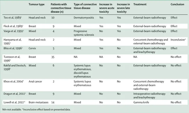
Few reports have been made of the outcomes of patients with newly diagnosed connective tissue diseases (or exacerbation of pre-existing disease) who need
radiotherapy (table 1, 2).1–21 Although an analysis of the
little available data shows that risk of radiotherapy toxicity in patients with connective tissue diseases seems to be based largely on anecdotal evidence, radiation oncologists remain hesitant. In 1998, the American
College of Radiology22 concluded that, “a history of
collagen vascular disease is a relative contraindication to breast conservation treatment because published reports indicate that such patients tolerate irradiation poorly. Most radiation oncologists will not treat patients with scleroderma or active systemic lupus erythematosus, considering either an absolute contraindication.” Thus, radiotherapy has been under used in patients with
connective tissue diseases who have cancer.16
With improved medical treatments, prognosis for patients with connective tissue diseases has improved. The 5-year survival in systemic lupus erythematosus has increased from about 40% in the 1950s, to 90% in the
1980s, to more than 90–95% nowadays.23 Therefore, a
higher number of patients with connective tissue diseases are expected to be diagnosed with cancer and will potentially be eligible for oncological treatment, including radiotherapy. Substantial improvements have been made in radiation technology, including the development of intensity-modulated radiotherapy and image-guided radiotherapy. These techniques are available in clinical practice, potentially minimising acute and late local side-eff ects. Thus, new radiotherapy
techniques could be considered feasible even in patients with connective tissue diseases who have cancer. In this Review, we analyse evidence and discuss the available data for radiotherapy in patients with connective tissue diseases.
Connective tissue diseases, cancer environments,
and radiation interactions
Connective tissue diseases are chronic and debilitating autoimmune disorders that cause substantial morbidity and mortality and disproportionately aff ect women. These diseases include rheumatoid arthritis, systemic sclerosis, scleroderma, systemic lupus erythematosus,
derma tomyositis, and vasculitis. Connective tissue
diseases often develop after environmental triggering via cellular pathways in genetically susceptible individuals
with disease-associated polymorphisms.24 However, the
specifi c cellular and molecular mechanisms leading to connective tissue diseases, and factors that establish involved organs are involved, are poorly understood.
Associations between connective tissue diseases and cancer are being increasingly investigated. Links between them are multifaceted and have diff erent relationships in terms of frequency, timing, and type of cancers. Several studies have highlighted the dynamic and bidirectional interactions occurring at the cancer–immune system interface that might be relevant to the origins of
autoimmunity.25 Data for patients with systemic sclerosis
and concomitant cancer suggest that, in some cases, autoimmunity might be triggered by an autoantigen
mutation in the patient’s cancer.26,27 Also, connective tissue
diseases might cause changes in immune function that
could be aff ected by immunosuppressive therapy.24
Although the evidence was not overwhelming, some investigators have reported that these changes in immune
function did aff ect radiotherapy toxicity.28 This bidirectional
hypothesis was based on the idea that some connective tissue diseases share a common pathological pathway of vascular obliteration and fi brosis due to heightened infl ammation and a clinical pattern of possible systemic involvement. The potential for radiotherapy to augment these pathological changes became a topic of investigation.
Radiotherapy acutely aff ects early responding tissues, such as the basal dermis and oral and gastric mucosa, by reducing proliferation. Radiation-induced obliteration of
capillaries and small vessels is also well documented.28
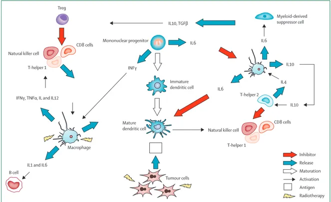
In patients with connective tissue diseases, these acute eff ects might act in conjunction with immune-related damage caused by immune complex deposition, complement cascade activation, and infi ltrating infl am-matory cells (fi gure 1). Such common targeting might be
additive to typical radiation-induced acute tissue injuries.11
The additive injury induced by both radiation and the pre-existing connective tissue diseases might also help to explain the potentially increased late eff ects noted in some
of these patients after radiotherapy.3 Radiotherapy might
trigger the onset of connective tissue diseases by enhancing the expression of self-antigens (eg, from apoptotic cell debris), diminishing regulatory T-cell activity, and activating eff ectors of innate immunity such as dendritic cells through Toll-like receptor-dependent mechanisms, all of which could potentially lead to a break of immune
tolerance.25 This potential mechanism has raised a debate
among radiation oncologists about whether patients with connective tissue diseases tolerate radiation as well as
people with no connective tissue disease.29
Experimental evidence supports the hypothesis that the immune system is able to repress tumour cells and that immune surveillance has a key role in the identifi cation
and elimination of cancer cells.30 Three diff erent phases
have been described in the interaction between cancer cells and the immune system: elimination (which is still
considered the cornerstone in the immune surveillance process), equilibrium between the immune system and
cancer cells, and escape.30 Immune surveillance is
considered a complex process involving diff erent immune system cells—ie, CD8 cells, natural killer cells, CD4 cells,
macrophages, and B lymphocytes.30 After radiotherapy,
the disruption of the tissue architecture is associated with changes in blood fl ow (zones with hyperperfusion and hypoxia) and lymphatic function and an increase in
interstitial pressure.31 Additionally, irradiation of the
tumour and its microenvironment is associated with the proliferation of infl ammatory signals detected by the
immune system.32 The resulting production of cytokines
and chemokines then attracts antigen-presenting cells (dendritic cells) that, after uptake of tumour-associated antigens, cause CD8 activation involved in tumour killing (fi gure 1).33,34
Evidence is also increasing that infl ammation contributes to cancer development and that cancer cells use infl ammatory mechanisms to prevent immune-system activation and to protect the tumour from immune attack
(equilibrium and escape phases).35 Moreover, infl ammatory
elements (such as chemokines and interleukins) released by tumour cells promote infi ltration, progression of
disease, and metastases (fi gure 2).36
Various mechanisms might exist that exacerbate the
patho physiological response induced by radiation
exposure in patients with connective tissue diseases. One potential mechanism includes the overexpression of profi brotic cytokines, such as transforming growth factor β
Tumour type Patients with connective tissue disease (n) Type of connective tissue disease Increase in severe acute toxicity Increase in severe late toxicity Treatment Conclusion
Teo et al, 19891 Head and neck 10 Dermatomyositis Yes Yes External-beam radiotherapy Eff ect
Fleck et al, 19892 Breast 9 Mixed Yes Yes External-beam radiotherapy Eff ect
Varga et al, 19913 Mixed 4 Progressive
systemic sclerosis
No Yes External-beam radiotherapy Eff ect
Hareyama et al, 19954
Head and neck 2 Mixed Yes No Concurrent chemotherapy and external-beam radiotherapy
Inconclusive*
Bliss et al, 19965 Cervix 5 Mixed Yes No External-beam radiotherapy
and brachytherapy
Eff ect
Turesson et al, 19966
Breast 35 NA NA No NA No eff ect
Rakfal and Deutsch, 19987
Mixed 6 Systemic lupus
erythematosus, discoid lupus erythematosus
No No External-beam radiotherapy No eff ect
Khoo et al, 20048 Anal cancer 2 Systemic lupus
erythematosus
No No Concurrent chemotherapy and external-beam radiotherapy
No eff ect
Dragun et al, 20119 Breast 9 Mixed No No Intraoperative radiotherapy
and brachytherapy
No eff ect
Lowell et al, 201110 Brain metastases 14 Mixed No No Gamma knife No eff ect
NA=not available. *Inconclusive eff ect based on presented data.
(TGFβ) and interleukin 1. Radiation injury in healthy tissues is usually characterised by the appearance of a fi brinous exudate within the stroma and by deposition of extracellular matrix components, including collagen, through myo fi broblasts produced by fi broblast activation
and diff erentiation.37 In some connective tissue diseases
(such as systemic sclerosis) in which TGFβ concentrations are already increased, late eff ects after radiotherapy might
be more evident.3 Another potential mechanism involves
radiation microvascular damage in a context of vasculitis, leading to increased late eff ects and reduced tolerance to treatment. After radiation, endothelial cell injury and tissue hypoxia stimulate the recruitment into the tissue of infl ammatory circulating cells, such as macrophages,
which are a source of profi brotic mediators, including
TGFβ1.38,39 Additionally, increased concentrations of
proangiogenesis factors (eg, VEGF) as a result of vascular damage and leakage of vessels in response to radiotherapy could exacerbate late eff ects such as dermal atrophy,
telangectasia, necrosis, and fi brosis.40 Finally,
radiation-induced damage to basement membranes causes this to become a target tissue, leading to increased
autoimmunity.12,28
Preclinical studies and case reports
Some studies have used in-vitro sensitivity to radiation in lymphocytes from patients with connective tissue diseases to assess risk indicators for radiation-related
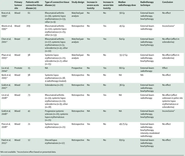
Primary tumour site Patients with connective tissue disease (n)
Type of connective tissue disease (n)
Study design Increase in severe acute toxicity Increase in severe late toxicity Median radiotherapy dose Radiotherapy technique Conclusion Ross et al, 199311
Mixed 61 Rheumatoid arthritis (n=39), systemic lupus erythematosus (n=13), other (n=9) Matched pair analysis No No 56 Gy External-beam radiotherapy, brachytherapy No eff ect Morris et al, 199712
Mixed 209 Rheumatoid arthritis (n=131), systemic lupus erythematosus (n=25), other (n=53)
Retrospective No Yes 45 Gy External-beam radiotherapy
Inconclusive*
Chen et al, 200113
Breast 36 Rheumatoid arthritis (n=17), systemic lupus erythematosus (n=5), scleroderma (n=4), other (n=10) Matched pair analysis
Yes Yes 64 Gy External-beam
radiotherapy, brachytherapy
No eff ect (eff ect in scleroderma)
Phan et al, 200314
Mixed 38 Systemic lupus erythematosus (n=21), scleroderma (n=2), other (n=15) Matched pair analysis No No 55·17 Gy External-beam radiotherapy, brachytherapy
No eff ect (eff ect in scleroderma)
Liu et al, 200415
Prostate 15 NA Prospective No Yes 66 Gy External-beam
radiotherapy
Eff ect
Benk et al, 200516
Mixed 38 Systemic lupus erythematosus (n=38; 4 radiotherapy treated)
Retrospective No No NA NA No eff ect
Gold et al, 200717
Mixed 20 Scleroderma (n=20) Retrospective No No 36 Gy External-beam radiotherapy, brachytherapy
No eff ect
Lin et al, 200818
Mixed 73 Rheumatoid arthritis (n=33), systemic lupus erythematosus (n=13), scleroderma (n=9), other (n=18)
Retrospective No Yes NA External-beam radiotherapy
No eff ect (eff ect unknown in pelvic site systemic lupus erythematosus or scleroderma) Gold et al,
200819
Mixed 41 Progressive systemic sclerosis (n=20), systemic lupus erythematosus (n=21) Retrospective NA No NA External-beam radiotherapy, brachytherapy Inconclusive* Pinn et al, 200820
Mixed 21 Systemic lupus erythematosus (n=21)
Retrospective Yes No 49·75 Gy External-beam radiotherapy, brachytherapy, intensity-modulated radiotherapy No eff ect Patel et al, 201221
Mixed 12 Discoid lupus erythematosis (n=12)
Retrospective No No 69 Gy External-beam radiotherapy, brachytherapy
No eff ect
NA=not available. *Inconclusive eff ect based on presented data.
side-eff ects.41–43 Carrillo-Alascio and colleagues41 used
pulsed-fi eld gel electrophoresis to quantify the initial radiation-induced DNA double-strand breaks in peripheral lymphocytes from 52 patients with systemic lupus erythematosus. Systemic lupus erythematosus did
not confer a higher intrinsic risk of radiosensitivity when compared with 48 healthy participants without
connective tissue diseases.41 In another study,43 the same
investigators carried out an in-vitro evaluation of the repair of mainly single-stranded DNA breaks after peripheral blood radiation of 48 children with systemic lupus erythematosus, systemic sclerosis, juvenile rheumatoid arthritis, and dermat omyositis. Greater DNA damage and a delay in DNA repair were noted in the children with connective tissue diseases group than in
healthy children.43 Another in-vitro study that used
tritiated thymidine incorporation assays showed that patients with active systemic lupus erythematosus had increased radiotherapy-related lymphocytic sensitivity when compared with healthy patients when irradiated with ⁶⁰Co-γ photons between 0 Gy and 10 Gy, resulting
in a potentially higher probability of radiation toxicity.42
Similarly, immune system changes, which can aff ect radiosensitivity, are being investigated. Among others,
Budach and colleagues44 investigated the possibly
abnormal reaction to high radiation doses in two groups of germline mutation-carrying mice, one with severe combined immunodefi ciency (SCID; even though it is not classifi ed as a connective tissue disease) and one that had normal radiation sensitivity (C3H). The lethal dose for 50% of the irradiated animals after single-dose whole-body irradiation was lower for SCID mice than for C3H mice, as was the radiation dose that was needed to achieve 50% local control and tumour growth delay,
Figure 1: Main immune cells, interleukins, and cytokines involved in immune surveillance TGF=transforming growth factor. IFN=interferon. IL=interleukin. TNF=tumour necrosis factor.
Mature dendritic cell Immature dendritic cell Macrophage B cell Mononuclear progenitor Tumour cells Treg
Natural killer cell
CD8 cells
T-helper 1
Natural killer cell
CD8 cells
T-helper 1 T-helper 2
Myeloid-derived suppressor cell
IFNγ, TNFα, IL and IL12
INFγ
IL10, TGFβ
IL6 IL6
IL6
IL1 and IL6
IL4 IL10 IL10 Inhibitor Release Maturation Activation Antigen Radiotherapy
Figure 2: Tumour-cell mechanisms against the immune system
TGF=transforming growth factor. CXC=CXC chemokine. IFN=interferon. IL=interleukin. TNF=tumour necrosis factor.
Tumour cells
Inhibitor Release Result
Release by tumour cell Positive feedback Tumour cell and infiltration
IFNγ production decreased in lymphocytes and natural killer cells
Activation of natural killer cells
Proliferation of lymphocyte T cells Proliferation of lymphocyte T cells IL12 IL8 IL10 IL12
IL2 and IL12 TGFβ
TGFβ
IL1, IL6, and IL12 TNFα Metastases Metastases CXC chemokine Proliferation and differentiation of B lymphocytes
thus confi rming that abnormal radiation sensitivity
was observed in SCID mice.44 A possible mechanism
correlated with increased sensitivity of SCID tumour cell lines is the inability of the tumour cells to overcome their genetic defi ciency in DNA double-strand break repair in
SCID fi broblasts.45
More than 300 cases involving patients with connective tissue diseases have been published reporting toxicity after radiotherapy and several early and late radiotherapy-related complications, including some deaths, have
also been reported.2,5,7,10,46 The fi rst two severe events
in patients with connective tissue diseases given
radiotherapy were noted in the late 1960s.47,48 In one case,
a patient with systemic lupus erythematosus who had lymphoma died of heart failure 1 year after radiotherapy to the mediastinal and retroclavicular nodes (20 Rad
[20 Gy] and 39 Rad [39 Gy], respectively, with ⁶⁰Co),47
whereas the second patient, who had facial lupus, developed radiotherapy-correlated osteomyelitis of the
maxilla.48 However, no data about radiotherapy dose or
modality were provided. Teo and colleagues1 assessed the
radiation toxicity profi les of ten patients with a diagnosis of early-stage nasopharyngeal carcinoma and dermato-myositis (table 1). At a median follow-up of 51·8 months, all patients had subcutaneous fi brosis and xerostomia, two patients had radiation skin necrosis, and one patient
had a VI and XII cranial nerve defi cit.1 However, no
information was provided about radiotherapy dose and techniques.
Fleck and colleagues2 published a study of nine patients
with breast cancer (four women with a pre-existing connective tissue disease and fi ve who developed a connective tissue disease after radiotherapy). Eight received radiotherapy using ⁶⁰Co with a prescription dose of 40–50 Gy and an electron boost on the tumour bed of 5–15 Gy. Three patients with a pre-existing connective tissue disease reported a severe toxicity profi le: the fi rst
case involved moist desquamation and brachial
plexopathy; the second case showed soft-tissue necrosis needing chest-wall resection, rib fractures, and pulmonary fi brosis; and the third patient had soft-tissue necrosis, bronchopleural–cutaneous fi stula, and osteonecrosis of the clavicle, sternum, and rib. None of the patients with a new diagnosis of connective tissue diseases after
radiotherapy had severe complications.2
According to McCormick,49 to reduce the side-eff ects in
patients with connective tissue disease and breast cancer, a more aggressive local surgery and systemic therapy, in particular for younger women (<40 years), was better than breast-conserving surgery followed by radiation. More recently, accelerated partial breast irradiation by either brachytherapy or intraoperative radiotherapy has been considered an alternative experimental option for the treatment of early-stage breast cancer in women with a history of connective tissue diseases. Dragun and
colleagues9 published a report of nine patients with
connective tissue diseases with breast cancer given
accelerated partial breast irradiation via high-dose brachytherapy; toxicity and cosmetic profi les were reported as satisfactory. Indeed, the authors concluded that it might not be necessary to exclude patients with connective tissue diseases from clinical trials of accelerated partial breast irradiation. As confi rmation,
Turesson and colleagues6 reported that autoimmune
disease did not increase the risk of skin teleangectasia in 35 patients who received radiotherapy for breast cancer.
Finally, Lowell and colleagues10 published data on the
use of a very high dose of radiation delivered with gamma knife for brain metastases in 14 patients with connective tissue diseases, and reported no grade 3 or 4 toxicity (table 1).
In conclusion, in-vitro studies and clinical case reports describe a narrow and heterogeneous picture for patients with connective tissue diseases who receive radiotherapy. Despite these data limitations, more recently published data show that patients with connective tissue diseases seem to be less aff ected by toxicity than are healthy individuals and case reports (table 1).
Retrospective and controlled studies
To our knowledge, no randomised controlled study has assessed whether patients with connective tissue diseases are more likely to develop acute or late
radiotherapy-related toxicity. However, we retrieved 11 case series.11–21
In a retrospective analysis, Morris and Powell12 reported a
large series of 209 patients with connective tissue diseases given radiotherapy with a median radiation dose of 45 Gy (range 13–82) between 1960 and 1995. After a median follow-up of 6 years, clinically signifi cant acute side-eff ects (Radiation Therapy Oncology Group/Eastern Cooperative Oncology Group RTOG/ECOG Early Morbidity Scoring Scale of more than three) were similar in patients with and without rheumatoid arthritis (both 12%). At 5 years, the risk of late morbidity for patients with rheumatoid arthritis was 6%, similar to the rate for the healthy population generally, whereas for patients without rheumatoid arthritis it was 21% (p=0·0002). The most highly represented connective tissue disease after rheumatoid arthritis was systemic lupus erythematosus, with 25 patients (12%). No correlation between dose, fraction size, irradiated volume, and late eff ects
were reported.12
Similar results were reported in a matched-control
study of 61 patients with connective tissue diseases.11
The number of acute reactions after radiotherapy in the connective tissue diseases group was only slightly higher than in the matched-control group, with grade 3 or greater acute toxicity noted in seven patients in the connective tissue diseases group and four in the matched-control group. Patients with systemic lupus erythematosus had an increase in the number of acute reactions due to radiation (36% of patients with systemic lupus erythematosus vs 18% in the control group, p=0·5), whereas patients with rheumatoid arthritis had
an increase in late complications (24% vs 5%; p=0·125). Nevertheless, the study showed no signifi cant diff erences
in acute and late toxicity complications between groups.11
Chen and colleagues13 reported no signifi cant diff erences
in acute complications after breast cancer radiotherapy between a group of 36 women with connective tissue diseases and a matched-control group (14% vs 8%, respectively; p=0·40), but did note a signifi cant diff erence in late toxicity in those patients with connective tissue diseases (17% vs 3%; p=0·0095). However, when the investigators stratifi ed patients by specifi c autoimmune disease, they found a signifi cant diff erence only in four
patients with scleroderma.13 Phan and colleagues14
assessed 76 patients who received radiation for cancer (38 patients with connective tissue diseases and 38 in the control group) and did not show any signifi cant diff erences in terms of acute or late complications between groups. However, increased risk of radiation complications was reported in patients with scleroderma (n=4).
In another study, Lin and colleagues18 reported toxic
eff ects in 73 patients with connective tissue diseases given radiotherapy. No diff erences were noted in acute toxicity between patients with connective tissue diseases and those in the control group. However, patients with a diagnosis of connective tissue diseases had a signifi cantly higher incidence of late toxicity compared with the control group (29% vs 14%, respectively; p=0·001), with a non-signifi cant increase in severe late toxicity (9% vs 4%; p=0·079). Patients with diagnosed connective tissue diseases who received radiation to the pelvis had a higher probability of severe toxicity reactions (grade 3 or higher); furthermore, the incidence of severe late toxicity was higher in patients with a diagnosis of systemic lupus erythematosus and scleroderma than in the
control group.18
Gold and colleagues19 retrospectively analysed the toxicity
profi le of 41 patients with connective tissue diseases given radiation for cancer (20 patients with systemic sclerosis and 21 patients with systemic lupus erythematosus). Patients were divided into high-severity and low-severity connective tissue diseases on the basis of the number of involved organs. Univariate analysis showed a signifi cant increase in the risk of any grade toxicity for patients with high-severity connective tissue diseases compared with those with low-severity connective tissue diseases (p=0·006), although no diff erences in grade 3 or higher toxicity were found between the two groups (p=0·56). Despite the small number of enrolled patients, the severity of connective tissue diseases could be considered as an important factor in the prediction of treatment tolerability. Nonetheless, the severity of connective tissue diseases was
not a clear contraindication to radiotherapy.19
Varga and colleagues3 reported on the toxicity profi le
of four patients with systemic sclerosis who were
given radiotherapy.3 All patients had cutaneous and
subcutaneous late toxicity, visceral fi brotic reactions at the radiation site, and severe skin toxicity and fi brosis
extending beyond the radiation fi eld involving internal organs. Three of the four patients subsequently died,
two from bowel obstruction and one from pneumonia.3
Liu and colleagues15 planned a prospective study to
investigate the eff ect of neoadjuvant androgen-deprivation therapy and radiotherapy in men with prostate cancer. A subanalysis showed that 15 of the men had a connective tissue disease and that these patients had a greater frequency of late genitourinary grade 2 toxicities
compared with healthy men (relative risk 3·98; p=0·007).15
As previously stated, several studies have reported radiotherapy-related toxicity profi les in patients with a range of connective tissue diseases (tables 1, 2).
Nevertheless, only a few of the studies7,8,17,20 focused on
patients with scleroderma and systemic lupus erythematosus, with contentious conclusions about radiotherapy toxicity.
Gold and colleagues17 assessed the toxicity profi les of
20 patients with scleroderma and cancer who had been treated with radiotherapy or brachytherapy or both, with or without concurrent chemotherapy. Univariate analysis showed a signifi cant association between acute toxicity, radiotherapy dose, and increased scleroderma involvement of organs. For late side-eff ects, negative antinuclear antibody serology was correlated with a higher probability of toxicity. None of the analysed pretreatment and treatment variables were correlated
with severe acute and late toxicity.17 There have been no
further reports to confi rm severe acute and late
complication profi les in this specifi c setting.7,8,10
Rakfal and Deutsch7 described data for six patients
who had a diagnosis of systemic lupus erythematosus and diff erent malignancies with various radiotherapy doses, reporting no unexpected severe acute or late
side-eff ects. Khoo and colleagues8 reported no relevant
acute or late complications in two patients with anal cancer with systemic lupus erythematosus taking concomitant immunosuppressive therapy who were treated with combined chemoradiotherapy (⁶⁰Co and external-beam radiotherapy).
One of the most important reports was published by
Pinn and colleagues,20 which included 21 patients with
systemic lupus erythematosus who received a total of 35 consecutive courses of radiotherapy. Of the 17 patients who were evaluable for late toxicity, four patients (24%) had a grade 3 or higher toxicity. The presence of renal involvement according to the American Rheumatism Association criteria was correlated with an increased risk of any grade of late toxicity (p<0·006). Univariate analysis established a correlation between acute toxicity and total dose (>49·8 Gy), treatment sites, and curative intent for treatment. Brachytherapy was used in one treatment course, 2D radiotherapy in 30 courses, 3D conformal radiotherapy in three, and intensity-modulated radiotherapy in one. Moreover, absence of photosensitivity (p<0·02), absence of arthritis (p<0·03), and presence of a malar rash (p<0·04) were correlated
with an increased risk of grade 3 or greater acute toxicity. No specifi c association between technique and late toxicity was noted. Radiation dose prescription, radiation techniques, and anatomical site (ie, abdomen, pelvis, breast, brain, neck, and chest) were associated with a high risk of any late toxicity.
In conclusion, the small number of described cases and the heterogeneity of the connective tissue disease seem to strongly aff ect the statistical power of these studies, thus limiting the possibility to show any robust association between radiation toxicity and connective tissue diseases, and confi rming that radiotherapy is frequently withheld unjustly to treat patients with
connective tissue diseases.16,19,21
Clinical solutions and future perspectives
Various treatment strategies have been considered for patients with connective tissue diseases to reduce the risk of toxicity during or after radiotherapy such as avoiding concomitant treatment or reducing dose prescription. Although the use of chemoradiotherapy is considered the gold standard in many cases, multimodality treatment in patients with connective tissue diseases could be correlated with a more severe toxicity profi le than single-modality treatment, thereby
aff ecting its feasibility.4,12,19,50 In radiotherapy, the radiation
dose could be reduced to lower the toxicity profi le, but
this could impair eff ectiveness.12,28,44,51 However, Delanian
and colleagues52 reported that reducing radiation dose
(from 65 Gy to 40 Gy) in patients with connective tissue diseases (one with lung cancer and two with anal–rectal cancer) resulted in complete remission, although side-eff ects were observed at the radiation site. Some investigators have postulated that hyperactivation of the immune system by tumour cells makes patients with connective tissue diseases more sensitive to radiation
than others.53,54 Another strategy is changing dose
fractionation schedules or reducing treatment volume,
which might decrease toxicity complications.2,12,28,40,51,52,54
Nevertheless, a crucial question still remains—is it really necessary to modify radiotherapy features to decrease toxicity in patients with connective tissue diseases?
The most common radiotherapy approach is to use external beams to deliver ionising radiation. In the past few decades, most departments have replaced their ⁶⁰Co machines with the more precise linear accelerator. Despite modern radiotherapy now being available, most reports of patients with connective tissue diseases involve obsolete and unsatisfactory technologies including 2D radiotherapy. Intensity-modulated radiotherapy and stereotactic ablative radiotherapy have allowed radiation oncologists to prescribe higher dose prescriptions to targets when useful or required. Intensity-modulated radiotherapy is considered an advancement of 3D-conformal radiotherapy that targets the radiation dose into the tumour, thus minimising the exposure of healthy tissue in several anatomical regions. Intensity-modulated
radiotherapy is considered the most appropriate technique in head and neck cancers and in most pelvic tumours, including prostate cancer. In this disease, intensity-modulated radiotherapy decreased long-term toxicity with no negative eff ect on overall survival when
compared with 3D-conformal radiotherapy.54–66
Stereotactic ablative radiotherapy is a novel radiotherapy method that delivers a very high dose of radiation (in a single or a few fractions) with high precision to the tumour, thus maximising the sparing of surrounding normal tissue. Several retrospective and prospective stereotactic ablative radiotherapy studies have shown promising results in terms of local tumour control and survival in some settings, including in early non-small-cell
lung cancer.67 Moreover, image-guided radiotherapy based
on daily patient set-up position verifi cation allowed better defi nition of the tumour target to reduce and ultimately eliminate uncertainties. To our knowledge, no randomised controlled trials using image-guided radiotherapy have
assessed toxicity and effi cacy in patients with connective
tissue disease. Hence, the promising, modern techniques could improve radiotherapy tolerability, especially in challenging clinical situations, as well as in patients with
connective tissue diseases and cancer.68,69
Conclusion
The data that are currently available from case series and a few retrospective studies are still not enough to support a specifi c contraindication for radiotherapy in patients with connective tissue diseases. Nevertheless, a cautious approach for patients with active connective
Search strategy and selection criteria
We searched Medline, Google Scholar, PubMed, and the ProQuest Dissertation, and Theses databases for reports published in English from June, 1946, to Jan 1, 2015. Our detailed search algorithm is shown in the appendix. We identifi ed additional references with a manual review of the reference lists of included articles.
Two independent reviewers (NGL and SS) identifi ed potential studies and exported them to an electronic reference management software program (RefWorks version 2.0). NGL and SS determined eligibility by reviewing fi rst the title and abstract and then the full paper. Disagreements were resolved by consensus; if consensus was not achieved, then a third author (FA) provided an assessment of eligibility. Because the data for eligibility were dichotomous (yes vs no), we established inter-rater agreement at both the title and abstract review and the full article review stages by calculating Cohen’s κ coeffi cient. A study was included when it reported on cancer-related radiotherapy and included patients with connective tissue diseases. A study was excluded when no detailed information (eg, outcome of radiotherapy, clinical manifestations related to the underlying connective tissue diseases, solid evidence of diagnosis of connective tissue diseases) was reported. Review articles were excluded from the analysis. For data extraction, all the papers were scrutinised for the following information: study design (retrospective, prospective, case-control, cross-sectional and case series, or case report); number of patients, sex, and age (mean, range); type of radiotherapy; type of underlying connective tissue disease; type of underlying cancer; defi nition of radiotherapy acute and late toxicity profi le; outcome in terms of toxicity profi le; and timing of connective tissue diseases onset or exacerbation.
For more on Cohen’s κ coefficient see http://facultyvassaredu/lowry/ kappa.html
tissue diseases seems to be reasonable. Moreover, the recent implementation of new radiotherapy approaches could be promising to improve the feasibility and tolerability of radiotherapy in some patients with cancer, including those with connective tissue diseases. Further well designed prospective studies, which also assess the most appropriate total dose and fractionation schedules, will probably help to overcome the unresolved concerns about radiotherapy indication for patients with connective tissue diseases.
Contributors
FA, NGL, and SS searched the literature, assisted with the organisation of the manuscript, interpreted and collected data, and wrote and edited the Review. AF and DR assisted with the organisation of the manuscript, interpreted and collected data, and wrote and edited the Review. SF, RM, and FR interpreted and collected data, helped to design the fi gures and panel, and wrote and edited the Review.
Declaration of interests
We declare no competing interests.
References
1 Teo P, Tai TH, Choy D. Nasopharyngeal carcinoma with dermatomyositis. Int J Radiat Oncol Biol Phys 1989; 16: 471–74. 2 Fleck R, McNeese MD, Ellerbrook MA, et al. Consequences of
breast irradiation in patients with pre-existing collagen vascular diseases. Int J Radiat Oncol Biol Phys 1989; 17: 829–33.
3 Varga J, Haustein UF, Creech RH, et al. Exaggerated radiation-induced fi brosis in patients with systemic sclerosis. JAMA 1991; 265: 3292–95. 4 Hareyama M, Nagakura H, Tamakawa M, et al. Severe reaction after
chemoradiotherapy of nasopharyngeal carcinoma with collagen disease. Int J Radiat Oncol Biol Phys 1995; 33: 971.
5 Bliss P, Parsons CA, Blake PR. Incidence and possible aetiological factors in the development of pelvic insuffi ciency fractures following radical radiotherapy. Br J Radiol 1996; 69: 548–54. 6 Turesson I, Nyman J, Holmberg E, Odén A. Prognostic factors for
acute and late skin reactions in radiotherapy patients.
Int J Radiat Oncol Biol Phys 1996; 36: 1065–75.
7 Rakfal SM, Deutsch M. Radiotherapy for malignancies associated with lupus: case reports of acute and late reactions. Am J Clin Oncol 1998; 21: 54–57.
8 Khoo VS, Saunders MP, Gowda R, et al. Anal canal cancer and chemoradiation treatment in two patients with systemic lupus erythematosus treated by chronic therapeutic immunosuppression.
Clin Oncol (R Coll Radiol) 2004; 16: 1–5.
9 Dragun AE, Harper JL, Olyejar SE, et al. The use of adjuvant high-dose-rate breast brachytherapy in patients with collagen vascular disease: a collaborative experience. Brachytherapy 2011;
10: 121–27.
10 Lowell D, Tatter SB, Bourland JD, et al. Toxicity of gamma knife radiosurgery in the treatment of intracranial tumors in patients with collagen vascular diseases or multiple sclerosis.
Int J Radiat Oncol Biol Phys 2011; 81: e519–24.
11 Ross JG, Hussey DH, Mayr NA, Davis CS. Acute and late reactions to radiation therapy in patients with collagen vascular diseases.
Cancer 1993; 71: 3744–52.
12 Morris MM, Powell SN. Irradiation in the setting of collagen vascular disease: acute and late complications. J Clin Oncol 1997;
15: 2728–35.
13 Chen AM, Obedian E, Haff ty BG. Breast-conserving therapy in the setting of collagen vascular disease. Cancer J 2001; 7: 480–91. 14 Phan C, Mindrum M, Silverman C, Paris K, Spanos W.
Matched-control retrospective study of the acute and late complications in patients with collagen vascular diseases treated with radiation therapy. Cancer J 2003; 9: 461–66.
15 Liu M, Pickles T, Agranovich A, et al. Impact of neoadjuvant androgen ablation and other factors on late toxicity after external beam prostate radiotherapy. Int J Radiat Oncol Biol Phys 2004; 58: 59–67.
16 Benk V, Al-Herz A, Gladman D, et al. Role of radiation therapy in patients with a diagnosis of both systemic lupus erythematosus and cancer. Arthritis Rheum 2005; 53: 67–72.
17 Gold DG, Miller RC, Petersen IA, Osborn TG. Radiotherapy for malignancy in patients with scleroderma: The Mayo Clinic experience. Int J Radiat Oncol Biol Phys 2007; 67: 559–67. 18 Lin A, Abu-Isa E, Griffi th K, Ben-Josef E. Toxicity of radiotherapy
in patients with a collagen vascular disease. Cancer 2008;
113: 648–53.
19 Gold DG, Miller RC, Pinn ME, et al. Chronic toxicity risk after radiotherapy for patients with systemic sclerosis (systemic scleroderma) or systemic lupus erythematosus: association with connective tissue disorder severity. Radiother Oncol 2008;
87: 127–31.
20 Pinn ME, Douglas G, Gold G, et al. Systemic lupus erythematosus, radiotherapy, and the risk of acute and chronic toxicity: the mayo clinic experience. Int J Radiat Oncol Biol Phys 2008; 71: 498–506. 21 Patel AB, Hallemeier CL, Petersen IA, et al. Acute and late toxicities
of radiotherapy for patients with discoid lupus erythematosus: a retrospective case-control study. Radiat Oncol 2012; 7: 22. 22 Winchester DP, Cox JD. Standards for diagnosis and management
of invasive breast carcinoma. American College of Radiology. American College of Surgeons. College of American Pathologists. Society of Surgical Oncology. CA Cancer J Clin 1998; 48: 83–107. 23 Pons-Estel GJ, Alarcón GS, Scofi eld L, et al. Understanding the
epidemiology and progression of systemic lupus erythematosus.
Semin Arthritis Rheum 2010; 39: 257–68.
24 Goldblatt F, O’Neill SG. Clinical aspects of autoimmune rheumatic diseases. Lancet 2013; 382: 797–808.
25 Schreiber RD, Old LJ, Smyth MJ. Cancer immunoediting: integrating immunity’s roles in cancer suppression and promotion.
Science 2011; 331: 1565–70.
26 Joseph CG, Darrah E, Shah AA, et al. Association of the autoimmune disease scleroderma with an immunologic response to cancer. Science 2014; 343: 152–57.
27 Shah AA, Rosen A, Hummers L, et al. Close temporal relationship between onset of cancer and scleroderma in patients with RNA polymerase I/III antibodies. Arthritis Rheum 2010; 62: 2787–95. 28 Chon BH, Loeffl er JS. The eff ect of non malignant systemic disease
on tolerance to radiation therapy. Oncologist 2002; 7: 136–43. 29 Lee CE, Prabhu V, Slevin NJ. Collagen vascular diseases and
enhanced radiotherapy-induced normal tissue eff ects—a case report and a review of published studies. Clin Oncol (R Coll Radiol) 2011;
23: 73–8.
30 Kim R, Emi M, Tanabe K. Cancer immunoediting from immune surveillance to immune escape. Immunology 2007; 121: 1–14. 31 Jain RK. Transport of molecules, particles, and cells in solid tumors.
Annu Rev Biomed Eng 1999; 1: 241–63.
32 Matzinger P. Tolerance, danger, and the extended family.
Annu Rev Immunol 1994; 12: 991–1045.
33 Shankaran V, Ikeda H, Bruce AT, et al. IFNgamma and lymphocytes prevent primary tumour development and shape tumour immunogenicity. Nature 2001; 410: 1107–11.
34 Demaria S, Formenti SC. Sensors of ionizing radiation eff ects on the immunological microenvironment of cancer.
Int J Radiat Oncol Biol Phys 2007; 83: 819–25.
35 Zou W. Immunosuppressive networks in the tumour environment and their therapeutic relevance. Nat Rev Cancer 2005; 5: 263–74. 36 Lippitz BE. Cytokine patterns in patients with cancer:
a systematic review. Lancet Oncol 2013; 14: e218–28.
37 Westbury CB, Yarnold JR. Radiation fi brosis–current clinical and therapeutic perspectives. Clinical Oncology 2012; 24: 657–72. 38 Wang J, Boerma M, Fu Q, Hauer-Jensen M. Signifi cance of
endothelial dysfunction in the pathogenesis of early and delayed radiation enteropathy. World J Gastroenterol 2007; 13: 3047–55. 39 Rabbani ZN, Mi J, Zhang Y, et al. Hypoxia inducible factor 1alpha
signaling in fractionated radiation-induced lung injury: role of oxidative stress and tissue hypoxia. Radiat Res 2010; 173: 165–74. 40 Brush J, Lipnick SL, Phillips T, et al. Molecular mechanisms of late
normal tissue injury. Semin Radiat Oncol (US) 2007; 17: 121–30. 41 Carrillo-Alascio, McCurdy D, Tai LQ, et al. In-vitro radiosensitivity
in patients with systemic lupus erythematosus. Lupus 2009;
18: 645–49.
42 Cossu F, Rombi G, Aresu G, et al. Radiosensitivity of lymphocyte subpopulations in subjects with systemic lupus erythematosus. A in vitro preliminary study. Minerva Med 1991; 82: 239–49.
43 McCurdy D, Tai LQ, Frias S, Wang Z. Delayed repair of DNA damage by ionizing radiation in cells from patients with juvenile systemic lupus erythematosus and rheumatoid arthritis. Radiat Res 1997; 147: 48–54.
44 Budach W, Hartford A, Gioioso D et al. Tumors arising in SCID mice share enhanced radiation sensitivity of SCID normal tissues.
Cancer Res 1992; 52: 6292–96.
45 Biedermann, KA, Sun, JR, Giaccia AJ, et al. SCID mutation in mice confers hypersensitivity to ionizing radiation and a defi ciency in DNA double-strand break repair. Proc Natl Acad Sci USA 1991; 88: 1394–97. 46 Olivotto IA, Fairey RN, Gillies JH, Stein H. Fatal outcome of pelvic radiotherapy for carcinoma of the cervix in a patient with systemic lupus erythematosis. Clin Radiol 1989; 40: 83–84.
47 Nilsen LB, Missal ME, Condemi JJ. Appearance of Hodgkin’s disease in a patient with systemic lupus erythematosus. Cancer 1967; 20: 1930–33.
48 Glasenapp GB. Osteomyelitis of the maxilla following radiotherapy for facial lupus. HNO 1968; 16: 46–49.
49 McCormick B. Selection criteria for breast conservation. The impact of young and old age and collagen vascular disease. Cancer 1994;
74: 430–35.
50 De Naeyer B, De Meerleer G, Braems S, et al. Collagen vascular diseases and radiation therapy: a critical review.
Int J Radiat Oncol Biol Phys 1999; 44: 975–80.
51 Abu-Shakra M, Lee P. Exaggerated fi brosis in patients with systemic sclerosis (scleroderma) following radiation therapy. J Rheumatol 1993; 20: 1601–03.
52 Delanian S, Maulard-Durdux C, Lefaix JL, et al. Major interactions between radiation therapy and systemic sclerosis: is there an optimal treatment? Eur J Cancer 1996; 32A: 738–39. 53 Gery B, Roussel A, Valla A. Usefulness of radiotherapy in the
treatment of advanced gastrinomas. Radiother Oncol 1993; 27: 259–60. 54 Cooper SG, Denham JW. Progressive systemic sclerosis
(diff use scleroderma) and radiotherapy. Br J Radiol 1990; 63: 804–05. 55 Chi A, Nguyen NP, Tse W, et al. Intensity modulated radiotherapy
for sinonasal malignancies with a focus on optic pathway preservation. J Hematol Oncol 2013; 6: 4.
56 Eisbruch A. Reducing xerostomia by IMRT: what may, and may not, be achieved. J Clin Oncol 2007; 25: 4863–64.
57 Madani I, Bonte K, Vakaet L, et al. Intensity-modulated radiotherapy for sinonasal tumors: Ghent University Hospital update.
Int J Radiat Oncol Biol Phys 2009; 73: 424–32.
58 Wolden SL, Chen WC, Pfi ster DG, et al. Intensity-modulated radiation therapy (IMRT) for nasopharynx cancer: update of the Memorial Sloan-Kettering experience. Int J Radiat Oncol Biol Phys 2006; 64: 57–62.
59 Garden AS, Morrison WH, Wong P-F, et al. Disease-control rates following intensity-modulated radiation therapy for small primary oropharyngeal carcinoma. Int J Radiat Oncol Biol Phys 2007;
67: 438–44.
60 Lee NY, de Arruda FF, Puri DR, et al. A comparison of intensity-modulated radiation therapy and concomitant boost radiotherapy in the setting of concurrent chemotherapy for locally advanced oropharyngeal carcinoma. Int J Radiat Oncol Biol Phys 2006; 66: 966–74.
61 deArruda FF, Puri DR, Zhung J, et al. Intensity-modulated radiation therapy for the treatment of oropharyngeal carcinoma: the Memorial Sloan-Kettering Cancer Center experience.
Int J Radiat Oncol Biol Phys 2006; 64: 363–73.
62 Jacobs BL, Zhang Y, Schroeck FR, et al. Use of advanced treatment technologies among men at low risk of dying from prostate cancer.
JAMA 2013; 309: 2587–89.
63 Jani AB, Gratzle J, Correa D. Infl uence of intensity-modulated radiotherapy on acute genitourinary and gastrointestinal toxicity in the treatment of localized prostate cancer. Technol Cancer Res Treat 2007; 6: 11–15.
64 Goldin GH, Sheets NC, Meyer AM, et al. Comparative eff ectiveness of intensity-modulated radiotherapy and conventional conformal radiotherapy in the treatment of prostate cancer after radical prostatectomy. JAMA Intern Med 2013; 173: 1136–43. 65 Pollack A, Walker G, Horwitz EM, et al. Randomized trial of
hypofractionated external-beam radiotherapy for prostate cancer.
J Clin Oncol 2013; 31: 3860–68.
66 Arcangeli S, Strigari L, Gomellini S, et al. Updated results and patterns of failure in a randomized hypofractionation trial for high-risk prostate cancer. Int J Radiat Oncol Biol Phys 2012;
84: 1172–78.
67 Alongi F, Arcangeli S, Filippi AR, et al. Review and uses of stereotactic body radiation therapy for oligometastases. Oncologist 2012; 17: 1100–07.
68 Allison RR, Gay HA, Mota HC, et al. Image-guided radiation therapy: current and future directions. Future Oncol 2006; 2: 477–92. 69 Perkins CL, Fox T, Elder E, et al. Image-guided radiation therapy