I
Table of Contents
Sommario II
Summary V
List of abbreviations VIII
1. Introduction 1
1.1 Mitochondria: key components of the aging process 4
1.1.1 Mitochondria and energy production 5
1.1.2 Mitochondria and ROS generation 7
1.1.3 Aging, ROS and the contribution of the uncoupling process 9
1.2 Human UnCoupling Proteins (UCPs) 13
1.2.1 UnCoupling Protein 1 (UCP1) 16
1.2.2 UnCoupling Protein 2 (UCP2) and 3 (UCP3) 22
1.2.3 UnCoupling Protein 4 (UCP4) and 5 (UCP5) 28
1.3 The role of uncoupling proteins in lifespan 31
1.4 Plan of the thesis 33
2. Two variants of the upstream enhancer of human UCP1 gene affect
the expression of the gene and are correlated with human longevity 34 3. A common UCP3 promoter polymorphism influences hand grip
strength in elderly people 62
4. Further support to the Uncoupling to Survive theory: the genetic
variation of human UCP genes is associated with longevity 81
5. Conclusive remarks 109
6. Appendix 110
6.1 Somatic point mutations in mtDNA control region are influenced by
genetic background and associated with healthy aging: a GEHA study 111
6.2 Association of a common LAMA5 variant with anthropometric and
metabolic traits in an Italian cohort of healthy elderly subjects 120
II
Sommario
L'invecchiamento è un fenomeno naturale caratterizzato da un progressivo declino della capacità funzionale di mantenere l'omeostasi basale dei vari organi e tessuti e di rispondere adeguatamente, in condizioni di stress, ai bisogni fisiologici. E’ noto che il 20% - 30% delle variazione nella durata della vita umana può essere attribuita a fattori genetici, che diventano ancora più rilevanti in età avanzata . Tra i diversi loci genetici e pathways che influenzano questo processo, i mitocondri, essendo i principali siti cellulari che controllano il metabolismo energetico e lo stato redox, occupano un ruolo centrale nella modulazione dell’invecchiamento.
Negli ultimi anni, l'attività disaccoppiante dei mitocondri, ossia il disaccoppiamento della respirazione dalla produzione di energia, è stata considerata come un processo in grado di modulare il tasso di invecchiamento e la durata della vita. Diversi esperimenti in organismi modello hanno dimostrato un ruolo del disaccoppiamento mediato dalle proteine disaccoppianti (UCPs) nell’estensione della durata della vita. Nell’uomo, sono state descritte cinque proteine disaccoppianti (UCP1-5). Queste proteine sembrano funzionare come regolatori dell'omeostasi energetica e come antiossidanti, e, anche se la loro funzione non è stata ancora ben stabilita, è stato suggerito un loro ruolo nell'invecchiamento e nella longevità umana.
Al fine di comprendere il ruolo delle UCPs nell'invecchiamento e nella longevità, durante il mio dottorato di ricerca sono stata coinvolta nello studio degli effetti della variabilità dei geni UCP sulla sopravvivenza in età molto avanzata. Tutte le analisi sono state condotte su un campione reclutato nel Sud Italia.
In primo luogo, ho analizzato la variabilità di due polimorfismi (A-3826G e C-3737A) in forte linkage disequilibrium tra loro e situati al 5 'del gene UCP1 in una regione che è nota essere coinvolta nell'attivazione trascrizionale del gene. Da questo studio è emerso
III che l'aplotipo G-A ha una significativa variazione di frequenza con l'età (p=0,003) e che il diplotipo A-C/A-C ha un aumento molto significativo con l'età (p = 0,005), mentre il diplotipo A-C/G- A subisce una variazione di frequenza legata all'età significativamente negativa (p<0,001). Per verificare se l'attività trascrizionale di questa regione possa essere influenzata dalla variabilità dei due polimorfismi analizzati, è stato anche effettuato uno studio funzionale sia a condizioni basali che dopo stimolazione ormonale. Da questo studio è emerso che il costrutto A-C è up-regolato in seguito a stimolazione con acido retinoico (p=0,027) e con progesterone (p=0,014), mentre è down-regolato (p=0,028) quando le cellule sono trattate con estradiolo. Il costrutto G-A, invece, mostra una bassa upregolazione solo dopo trattamento con acido retinoico (p=0,046). In conclusione, questi risultati suggeriscono che i due polimorfismi analizzati (A-3826G e C-3737) del gene UCP1 possono modulare la sopravvivenza, probabilmente influenzando i livelli della proteina.
Successivamente, abbiamo studiato due varianti del gene UCP3, espresso principalmente nel muscolo scheletrico, e abbiamo valutato se queste varianti fossero correlate a uno dei più importanti marcatori dell’invecchiamento umano, l’Hand Grip Strenght (misurazione strumentale della forza muscolare dell’avambraccio). È emerso che i portatori dell’allele T dell’rs1800849 hanno valori più alti di Hand Grip (p=0.010) e, dal momento che è noto che questo allele aumenta l’espressione del gene, possiamo concludere che un processo di disaccoppiamento più efficiente ha un effetto benefico sull’invecchiamento muscolare causando un rallentamento del suo decadimento correlato all'età.
Infine, allo scopo di analizzare il ruolo di tutti i geni UCP nell'invecchiamento umano, sono stati analizzati dieci polimorfismi dei geni UCP1-5. Per valutare gli effetti dei genotipi sulla probabilità di raggiungere età avanzate abbiamo creato un modello di
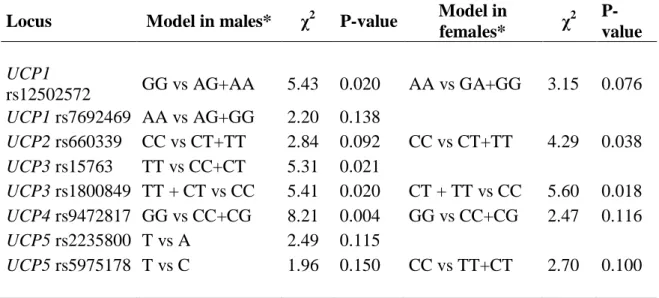
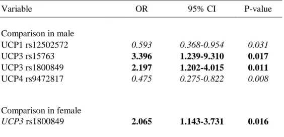
IV regressione logistica multivariata. Dall’analisi è emerso che le varianti genetiche UCP1 (rs12502572-GG), UCP3 (rs15763-TT e rs1800849-T/-), e UCP4 (rs9472817-GG) influenzano la possibilità dei maschi di sopravvivere fino ad età molto avanzata (valore di p: p = 0,031, p = 0,017, p = 0,011 ep = 0,008, rispettivamente), mentre nelle femmine solo UCP3 (rs1800849-T/-) ha mostrato tale associazione (p = 0,016).
Nel complesso, questi risultati suggeriscono che la variabilità genetica dei geni UCPs modula la sopravvivenza in età avanzata in maniera sesso-specifica. Dal momento che i geni UCPs presentano un’espressione tessuto specifica, i nostri risultati portano a ipotizzare un coinvolgimento del processo di disaccoppiamento nella modulazione della senescenza tessuto-specifica.
V
Summary
Aging is a natural and complex phenomenon characterized by a progressive decline in the functional capacity of various organs to maintain baseline tissue homeostasis and to respond adequately to physiological needs under stress. It is well known that the 20% - 30% of the variation in human lifespan can be attributed to genetic factors, which become more relevant ad advanced ages. Among several individual loci and pathways affecting this process, mitochondrial function is central to modulation of aging, being mitochondria the main cellular sites controlling energy metabolism and redox state. In recent years, the uncoupling activity of mitochondria, namely the uncoupling of respiration from energy production, has been considered as a process that can modulate the rate of aging and lifespan. Several experiments in model organisms have demonstrated a role for uncoupling mediated by UCP proteins in extending lifespan. In human, five uncoupling proteins (UCP1-5) have been described. These proteins seem to function as regulators of energy homeostasis and as antioxidants, and, even if their effect has not been yet established, a role of UCPs in human aging and longevity has been suggested.
In order to understand the role of UCPs in aging and longevity, during my PhD appointment I was involved in the study of the effects of the variability of UCP genes on survival at very old age. The analyses have been carried out on unrelated southern Italian individuals.
Firstly, we analyzed the variability of two polymorphisms (A-3826G and C-3737A) in strong linkage disequilibrium with each other and located at the 5’ of the gene UCP1 in a region which has been reported to mediate the transcriptional activation of the UCP1 gene. We found that the G-A haplotype showed a significant frequency variation with age (p=0.003) and that the diplotype A-C/A-C showed a highly significant increase with
VI age (p=0.005), while the diplotype A-C/G-A had a significant negative age related frequency variation (p<0.001). To test whether the transcriptional activity of that region was influenced by the haplotype variability, a functional study was also carried out both at basal condition and after stimulation. We found that the A-C constructs was upregulated after retinoic stimulation (p=0.027) and progesterone stimulation (p=0.014), while was down-regulated (p=0.028) when cells were treated with estradiol. The G-A constructs showed a low upregulation only after retinoic stimulation (p= 0.046). In conclusion, these results suggest that the A-3826G and C-3737A of the UCP1 gene may modulate survival, probably by affecting the levels of the protein.
Subsequently, we studied two variants of the UCP3 gene mainly expressed in skeletal muscle and we evaluated whether these variants were correlated to hand grip strength, one of the most important landmark of human aging. We found that the carriers of rs1800849 T allele has a significant impact on hand grip strength in our sample (p=0.010). Since this allele has been reported to promote a higher expression of the gene, we conclude that a more efficient uncoupling process has a beneficial effect on the aging muscle by slowing down its age related decay.
Finally, in order to analyze the role of all UCP genes in human aging ten SNPs of UCP1-5 genes were analyzed. To evaluate the effects of UCP genotypes on the probability to reach advanced ages we set up a multivariate logistic-regression model. We found that UCP1(rs12502572-GG), UCP3 (rs15763-TT and rs1800849-T/-), and UCP4 (rs9472817-GG) genetic variation affects male chance to survive to very old ages (p value: p=0.031, p=0.017, p=0.011 and p=0.008, respectively), while in females only the UCP3 (rs1800849-T/-) affects such chance (p=0.016).
On the whole, these results suggest that the UCPs gene variability modulates survival at old ages in a gender-specific way. As UCPs are differently expressed in various organs
VII and tissues, our results allowed some inferences on the involvement of the uncoupling process (and of energy storage and expenditure) in the elderly.
VIII
List of abbrevations
4HNE 4-hydroxy-2 ,3-transnonenale
ADP Adenosine DiPhosphate
ATP Adenosine TriPhosphate
BAT Brown Adipocite Tissue
BMI Body Mass Index
cDNA Complementary DeoxyriboNucleic Acid
CoQ Coenzyme Q
CoQH Ubisemiquinone
CoQH2 Ubiquinol
CR Calorie Restriction
CRE cAMP Response Elements
DNA DeoxyriboNucleic Acid
e- Electron
ETC Electron Transfer Chain
FADH2 Flavin Adenine DiNucleotide
GDP Guanosine DiPhosphate
Gly Glycine
GSH Glutathione
H+ Hydrogen ion
H2O Water
H2O2 Hydrogen Peroxide
HO2 Hydroperoxyl
KDa KiloDalton
Leu Leucine
Met Methionine
MREs MyoD responsive elements
mRNA messenger RNA
mtDNA mitochondrial DNA
NADH Nicotinamide Adenine DiNucleotide
NO Nitric Oxide
IX
OH Hydroxyl Radical
ONOO- Oxidant PeroxyNitrite
OXPHOS Oxidative Phosphorylation
PGC-1α Peroxisome Proliferator-activated receptor- α Coactivator
Pi Inorganic Phosphate
PolgA DNA Polymerase gamma A
PPARs (α and γ) Peroxisome Proliferator-Activated Receptors
PPRE Peroxisome Proliferator Response Element
RARE Retinoic Acid Response Elements
RNAi RNA interference
ROS Reactive Oxygen Species
SDH Succinate DeHydrogenase
Ser Serine
SNS Sympathetic Nervous System
SOD SuperOxide Dismutase
SREBPs Sterol Regulatory Element Binding Proteins
TRE Thyroid hormone Responsive Element
UCPs (UCP1,-5) UnCoupling Proteins (1, 2, 3, 4, 5)
1
1. Introduction
Aging is characterized by a progressive decline of the normal physiological functions. It is a complex process that characterizes every biological specie and leads to a dramatic reduction of the individual survival probability and, ultimately, to death. During the aging process continuous changes can be observed not only in the individual anatomy and physiology, but also at cellular and molecular levels. These changes may be characterized by gain, loss or maintenance of structure, function or capability to cope with endogenous and exogenous factors acting as stressors. Aging is a process that affects all organisms, but lifespan is species specific. In addition, a noticeable inter-individual variability exists with respect to the rate and the quality of aging.
Over the past 50 years, in the western society, there has been a gradual increase in the average lifespan of individuals that is mainly due to the improvement of living conditions, environmental hygiene, and to the development and application of new knowledge in medical and pharmaceutical fields (Kannisto, 1994). This has led to an increase in life expectancy which, as evidenced by the mortality curves, sharply increased the relative prevalence of elderly subjects, including nonagenarians and centenarians, in western societies. The increase of lifespan introduces new problems; in fact, the prevalence of age-related diseases such as Parkinson, Alzheimer and heart diseases, have also increased. Since aging-related diseases account for approximately 20% of healthcare costs, there has been a growing scientific interest regarding the study of the aging phenotype and the basis of individual variability in order to better understand which factors affect the quality of aging.
It is well known that environmental conditions (education, socio-economic status, and lifestyle choices such as diet, exercise, smoking habits, etc.) and genetic factors are
2 essential to modulate human aging and longevity. Several studies have been carried out to separate the genetic contribution from the environmental one. Studies on the variation of lifespan in twins reported that: i) the share of the variation in human life span which can be attributed to genetic differences among individuals ranges between 22% and 33% (McGue et al., 1993; Herskind et al., 1996; Ljungquist et al., 1998); ii) the heritability of longevity is estimated as 0.26 for males and 0.23 for females (Herskind et al., 1996). Subsequently, examples of familial clustering of longevity were reported by Perls and co-workers (Perls et al., 2000). By analyzing 444 centenarian pedigrees, and by comparing death rates and survival probabilities of siblings of centenarians with data from the same birth cohort, they found that relative survival probability for these siblings increased at old age and was significantly higher when compared with people born in the same birth cohort. What is more, siblings of centenarians had an one-half life-long reduction in risk of death, even up through very old age. These findings were also supported by data obtained from relatives of super-centenarians (age major or equal to 110 years), where a survival advantage was found for siblings and mothers of super-centenarians (Perls et al., 2002). The Leiden Longevity Study, carried out in the Dutch population, also confirmed the familial clustering of extreme longevity (Schoenmaker et al., 2006). However, these studies do not distinguish how much of the familial component is genetic or due to environmental factors shared by the members of the family. A study of Hjelmborg and co-workers (2006) showed that having a co-twin surviving to old ages significantly increases the chance of reaching the same old age much more in monozygotic than in dizygotic twins. This study clearly supports the existence of a genetic component affecting longevity in humans, especially at advanced ages.
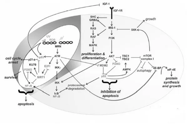
3 In the last years a large amount of studies aimed at identifying genes involved in aging and longevity in humans (for a review see Fontana et al., 2010). Studies on model organisms showed that the majority of the genetic variants having an influence on lifespan belong to a limited number of pathways, highly conserved during evolution. This suggests that a common ―core‖ of genes and pathways exists, responsible for modulating the lifespan of all animal species. The literature about this argument is huge and concordant to highlight a complex network of interactions linking metabolic pathways for nutrients metabolism and those that regulate response to external and / or internal stress factors. Figure.1 shows this complex network of interactions, whose efficient functioning is supposed to be critical for the modulation of lifespan.
Figure 1: Network of interaction between metabolism and stress response (from: Niedernhofer and Robbins, 2008).
4 In studies on human longevity the model of centenarians has emerged as essential because: i) it personifies the longevity phenotype naturally occurring in an outbreed species; ii) their environment continuously pushes the organism to cope with intrinsic and extrinsic antigenic loads; iii) several changes have been experimented due to the progress observed in the last century in all developed countries across the world; iv) they provide unique insights on the complex network of biological and non biological factors which guide individual survival at old age (De Benedictis and Franceschi, 2006). Taking into account the findings from model organisms, genetic studies in humans forwarded on specific genes belonging to the previously mentionated pathways. Many studies have found different genetic loci affecting longevity: 'cardiovascular genes' (APOE, APOC3, MTTP, ACE), 'immune system genes' (IL6), 'metabolism-related genes' (IGF1, GH1, HFE) and mitochondrial polymorphisms (Christensen et al., 2006 and references therein). As a consequence, due to their biological function, these ―longevity genes‖ may be useful in the study of human aging and longevity. It is important to highlight that human ―longevity genes‖ could function in several important ways: they may slow down the rate of age-related changes in cells and tissues, improve the effectiveness of repair mechanisms, and increase resistance to environmental stresses like infection and injury. Moreover, these genes could also affect a wide spectrum of debilitating age-related conditions.
1.1 Mitochondria: key components of the aging process
Over the last years, due to its crucial role in the energetic balance of the cell, mitochondrion emerged as a key factor in a number of complex traits, including ageing and numerous degenerative diseases. In fact, mitochondria are the organelles where Oxidative Phosphorylation (OXPHOS) takes place, and this implies that mitochondria
5 have a central position between energy uptake (that is food uptake and metabolism) and energy production. The direct consequence of this process is the mitochondria implication in several cellular processes such as heat production, apoptosis regulation, cellular differentiation, and especially in the production and the regulation of one of the most important by-products of cellular metabolism: the Reactive Oxygen Species (ROS). For both energy and ROS production, mitochondria play a central role in aging.
1.1.1 Mitochondria and energy production
Oxidative phosphorylation involves the coupling of electron transport, through the electron transfer chain (ETC), to the active pumping of protons across the inner mitochondrial membrane and ATP formation by the F1Fo-ATP synthase. The mitochondrial electron transport chain is made up of 80 component proteins that constitute five complexes designated for cellular energy production: complex I (NADH dehydrogenase), complex II (succinate dehydrogenase), complex III (ubiquinone cytochrome c oxidoreductase), complex IV (cytochrome oxidase), and complex V (F1Fo-ATP synthase).
The reducing equivalents extracted from the substrates are needed to start the transport of electrons through the mitochondrial ETC. The electron donors, nicotinamide adenine dinucleotide (NADH) and flavin adenine di nucleotide (FADH2), reduce equivalent transferring electrons to the ETC. This transferring is driven by a redox potential that is present across the chain. In particular, NADH is in turn oxidized by complex I (NADH-CoQ reductase), which transfers electrons to membrane-bound electron carrier, the ubiquinone (coenzyme Q, CoQ) to give ubisemiquinone (CoQH.) and then ubiquinol (CoQH2). FADH2 is an alternative substrate for the start of mitochondrial respiration. It is oxidized by complex II (succinate dehydrogenase, SDH), which then transfers
6 electrons to ubiquinone. The ubiquinol sends electrons to complex III (CoQ cytochrome c reductase), which, in turn, transfers them to cytochrome c oxidized. The reduced cytochrome c passes electrons to complex IV (cytochrome c oxidase), which, in the final step, reduces oxygen to water (4H+ + 4e- + O2 = 2H2O). This process of substrate oxidation and oxygen reduction, is also called "mitochondrial respiration" (Figure 2).
Figure 2: Mitochondrial oxidative phosphorylation and ROS production.
During respiration electron transfer along the redox potential gradient from NADH or FADH2 to oxygen is coupled to the active transport of hydrogen ions from the matrix to the cytosolic side of the inner membrane as described by the chemiosmotic theory of Mitchell (Mitchell, 1976). Complex I, III and IV of the respiratory chain pump protons from the mitochondrial matrix to the mitochondria intermembrane space, thereby establishing a gradient across the inner mitochondrial membrane. This electrochemical proton gradient, ΔP, has two components: a difference in electric potential (ΔΨ) and a difference in proton concentration (ΔpH) across the membrane (ΔP = ΔΨ + ΔpH). The
7 energy stored in the protons gradient across the inner mitochondrial membrane is used by ATP synthase (complex V) which, when protons are transported from the mitochondrial intermembrane space into the matrix, synthesizes ATP from ADP and inorganic phosphate (Pi) (Wallace DC, 2005). ATP synthase involves two protein complexes, F1 and F0.
Recently, Artal-Sanz and coworkers (2009) demonstrated that a component of the inner membrane of mitochondria (prohibitin) promotes longevity through modulation of mitochondrial proliferation. Moreover, genetic studies in both nematodes and rodents have reported that longevity may be promoted by moderate inactivation of genes important for mitochondrial electron transport chain (ETC) function. Hur and coworkers (2010) performed an RNAi screen in Drosophila melanogaster to test the role of ETC components in lifespan modulation. Five ETC genes turned out to be associated with increased longevity. However, only two of the knocked-down ETC genes decrease the abundance of fully assembled respiratory complexes. Moreover, none of the five silenced ETC genes affecting longevity was reported to cause a decrease in ATP levels.
1.1.2 Mitochondria and ROS generation
During oxidative phosphorylation, a small proportion of consumed oxygen, on average 0.4 - 4%, is converted to Reactive Oxigen Species (ROS). ROS includes a variety of molecules and free radicals (chemical species with one unpaired electron) derived from the metabolism of molecular oxygen. These molecules include: Superoxide anion (O2-), produced by an interaction between an oxygen and an electron escaped from the electron transport chain at other sites; Hydrogen peroxide (H2O2), derived from superoxide by a reaction catalized by SOD; Hydroxyl radical (OH-. ), one of the most toxic ROS that causes widespread oxidative damage. Moreover, O2- may
8 nonenzymatically react with nitric oxide (NO) to produce the powerful oxidant peroxynitrite (ONOO-) (Beckman et al., 1996; Radi et al., 2002).
Since the diffusion capability of most ROS is limited by their lipid solubility, their effect is mainly exerted on the molecules close to the mitochondrial transport chain where they are produced. These molecules include lipids, proteins and nucleic acids. Lipids can be damaged by free radicals directly by peroxidation or indirectly through the production of highly reactive aldehydes. The 4-hydroxy-2 ,3-transnonenale (4HNE) aldehyde is one of the main products of lipid peroxidation. It causes a variety of harmful effects on the molecules with which it comes into contact. For instance, the interaction between 4HNE and proteins induces structural and functional changes, while the interaction with membrane phospholipids decreases membrane fluidity and permeability. Moreover, such an interaction inhibits metabolic process and alterates ions transport (Nigam et al., 2000). The damage to mitochondria induced by lipid peroxidation can lead to further ROS generation (Green et al., 1998).
As lipids, also proteins are sensitive to ROS. The inner mitochondrial membrane contains a high proportion of protein physically associated with fats. Damage to these proteins, as the direct result of oxidative stress or as a consequence of lipid peroxidation, may occur in abnormal protein aggregation , in their degradation or in their loss of function.
Because of its proximity to the mitochondrial inner membrane and the lack of protective coating provided by histones, mitochondrial DNA (mtDNA) is the primary target of ROS. The 8-hydroxy-2-deoxyguanosine presence, which is the most abundant among the products of nucleotides oxidation, is used as an indicator of oxidative damage against DNA (Chomyn and Attardi, 2003). Several studies have shown that levels of 8-hydroxy-2-deoxyguanosine in mtDNA are higher than those observed in nuclear DNA
9 (Chung et al, 1992, Agarwal and Sohal, 1994). It has been suggested that oxidative damage is responsible for the accumulation of mutations in mitochondrial genome throughout life. This accumulation leads to synthesis of no functional subunits of the electron transport chain, the production of even more ROS and a consequent further increased mtDNA damage (Hiona et al, 2010).
In the aerobic cells ROS coexist in balance with biochemical antioxidants. Cells have two natural antioxidant systems to restore this balance: enzymes and low molecular weight antioxidants. Enzymatic antioxidant system includes the previously mentioned SOD, catalase, and peroxidase. The low molecular weight antioxidants include ascorbate, glutathione (GSH), phenolic compounds, and tocopherals. When the critical balance between ROS and antioxidants is disrupted (excess of ROS or antioxidants deplation) oxidative stress occurs.
1.1.3 Aging, ROS and the contribution of the uncoupling process
It is generally assumed that accumulate damages to a variety of cellular systems are the underlying cause of aging (Sinclair and Oberdoerffer, 2009). Harman was the first to propose the free radical theory of aging. According to this theory aging and age-associated degenerative diseases are attributed to the deleterious effects ROS (specifically hydroxyl, OH-, and hydroperoxyl, HO2-) on various cell components (Harman, 1956). In particular, mitochondria, a major site of ROS production, have a central role in this process (Harman, 1972). Several studies in model organisms confirmed that oxidative damage increases with age and that many forms of ROS may be the cause of accumulated oxidative damage (Bokov et al., 2004). Harman's original hypothesis has been developed and now is commonly known as the oxidative stress theory of aging (Sohal et al., 1996). It is also known that under normal physiological
10 conditions, a chronic state of oxidative stress exists and this is due to an imbalance between pro- and anti- oxidants (Sohal et al., 1996). This imbalance leads to the accumulation of damages to cellular macromolecules that contribute to a progressive decay of cells and tissues. In this frame, aging process may be directly influenced by the regulation of oxidative stress. Thus lifespan should be increased by a reduction of the oxidative stress, by an increase of the antioxidant defenses or by their combination. Remarkably, in Drosophila the manipulation of mitochondrial antioxidant systems is able to increase lifespan (Addabbo et al., 2009) while in mice this effect is not observed (Perez et al., 2009). To support the oxidative stress theory of aging several studies have analyzed whether long-lived animals have reduced oxidative damage or increased oxidative stress resistance.
In most mammalian models it has been demonstrated that life span can be extended by experimental intervention, such as calorie restriction (CR) or genetic manipulation. "Caloric restriction" means a diet in which calories are reduced by 30-40%, so it is characterized by a reduction of caloric intake without malnutrition. CR is the only non-genetic treatment that clearly increases mean and maximum lifespan in various animal models, including mice and rats (Weindruch and Walford, 1988; Jazwinsk, 2000, Rogina, et al, 2000; Bishop and Guarente, 2007; Sanz and Stefanatos, 2008). This effect on life span seems due to a reduction in oxidative damage/stress (Guarente and Kenyon, 2000). Rodents under CR show reduction in levels of oxidized protein, lipid, and DNA; reduced rates of mitochondrial ROS production, and increased resistance to oxidative stress compared to rodents fed ad libitum (Sohal et al, 1994; Li et al., 1998; Sun et al., 2001; Bokov et al., 2004; Richardson et al., 2004; Harper et al., 2006). However, it must be pointed out that CR alters more than free radical production (e.g. it decreases insulin signaling) and therefore the increase in life span cannot be exclusively attributed
11 to a decrease in mitochondrial ROS generation (Sanz and Stefanatos, 2008). Similarly, several genetic manipulations extend lifespan causing a reduction in oxidative damage/stress. For instance, a number of studies revealed in animal models where components of complex network of signaling pathways modulated by nutrients (IGF-1, TOR, sirtuins, AMP kinase, and PGC-1α) are up/down regulated phenotype characterized by the slowing of the aging process can be observed (Raffaello and Rizzuto, 2010).
Oxidative damage targeted to mitochondria and mtDNA is supposed to be one of the most important factors in determining age-related cellular decline. The link between mtDNA somatic mutations and aging phenotypes is generally accepted and supported by numerous studies (Kujoth et al. 2005). The ―mutator mice‖ (mice expressing a proofreading-deficient version of the catalytic subunit of mtDNA polymerase, PolgA) are characterized by high levels of mtDNA point mutations and deletions, and display many features of premature aging (Trifunovic et al. 2004; Edgar et al. 2009). However, the link between mtDNA mutations and increased ROS production during age is debated. For instance, in the ―mutator mice‖ the high mutation load in mtDNA was not associated with increased oxidative damage (Trifunovic et al. 2005); on the other hand, mice expressing peroxisomal catalase targeted to mitochondria showed increased lifespan which was associated with decreased damage to mtDNA and increased mitochondrial resistance to ROS damage (Schriner et al. 2005).
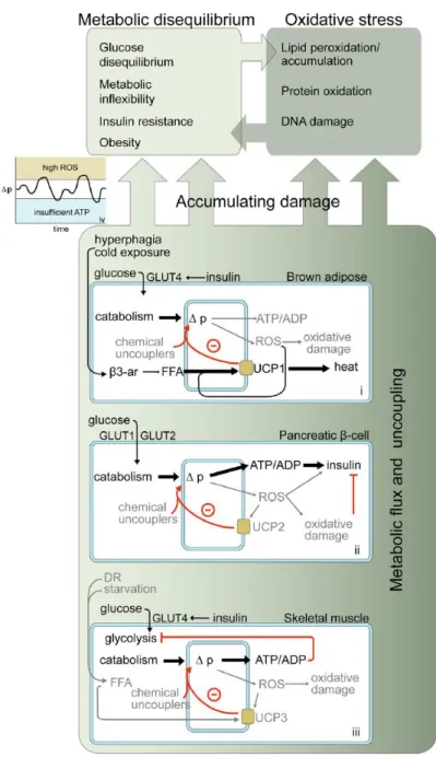
A mechanism that could be important for contrasting the accumulation of ROS, in minimizing oxidative damage to DNA, and in slowing aging, is the uncoupling of oxidative phosphorylation from ATP production. During oxidative phosphorylation not all the energy available in the electrochemical gradient is coupled to ATP synthesis.
12 Reducing the association between substrates oxidation and phosphorylation, decoupling allows to the electron transport chain to proceed without ATP synthesis. This uncoupling activity induces a "proton leak" that is a proton passage from the intermembrane space into the mitochondrial matrix not coupled to the ATP synthase activity. In this process the proton gradient becomes "relaxed", the membrane potential decreases and energy is released as heat (Stuart et al, 1999). This mitochondrial proton cycling, that accounts for up to 20-25% of basal metabolic rate, seems to be a general phenomenon, both in endotherm and ectotherm organisms. The high energetic cost of this futile proton cycle must be offset by high-benefit outcomes. Since it occurs in ectotherms, the heat production cannot be the most important function of the uncoupling process. A possibility is that it is a pathway of energy loss that might have a great importance from an ecological point of view. Interestingly, an important consequence of lowering of the mitochondrial membrane potential is a reduction in ROS generation. This process can justify the high energy costs imposed by the mitochondrial proton leak (Brand, 2000). In fact, even if cells have powerful antioxidant defences to protect themselves against ROS, prevention, rather than cure, would appear to be a more effective way to decrease oxidative damage.
Since ROS-induced damages and energy balance are among the major contributors to the aging process, the ―Uncoupling to survive theory" has been proposed. According to this theory, individuals with more coupled mitochondria are subject to less oxidative stress and age slow than animals with fewer uncoupling mitochondria (Brand, 2000; Van Voorhies, 2004; Wolkow and Iser, 2006). A confirmation of this hypothesis comes from a study by Speakman and coworkers (2004).They found that individual mice with a high metabolism lived 36% longer than those with low metabolism. Moreover, they exhibited higher resting oxygen consumption rate and they also possessed more
13 uncoupled mitochondria. In addition, tightly coupled cells showed greater deterioration with age than relatively uncoupled cells (Amara et al., 2007).
On the whole, these findings support uncoupling as a protective mechanism that minimizes ROS production and preserves mitochondrial function with age.
1.2. Human UnCoupling Proteins (UCPs)
The uncoupling of respiration from ATP production is a mitochondrial process by which stored energy is released as heat. This process is mediated by a group of five mitochondrial transporters present in the mitochondrial inner membrane well-known as UnCoupling Proteins (UCPs) (Krauss et al. 2005). These proteins constitute a subfamily of mitochondrial anion-carriers localized in the inner mitochondrial membrane. As uncouplers, UCPs uncouple ATP synthesis from the respiratory chain by transporting protons into the matrix causing proton motive force dissipation (Yu et al., 2000). UCPs have been identified in different species of invertebrates, including the nematode
Caenorhabditis elegans and the fruitfly Drosophila melanogaster (Hanak and Jezek,
2001; Sokolova and Sokolov, 2005), plants (Laloi et al, 1997), fungi and protozoa (Jarmuszkiewicz et al, 1999). However, the vast majority of UCPs is found in vertebrates including fish (Stuart et al, 1999), birds (Raimbault et al, 2001) and, especially, mammals. The human uncoupling proteins so far identified are five: UCP1, UCP2, UCP3, UCP4 and UCP5.
UCPs have a molecular mass of 31-34 KDa and share a common tripartite structure that consists of three repeat domains (of about 100 aminoacids) each with two hydrophobic regions forming a transmembrane α-helical spanning the mitochondrial inner membrane (Echtay, 2007). The amino- and carboxy-terminal ends protrude in the intermembrane space. In any repeat, the two helices are connected by a long hydrophilic loop that is
14 oriented on the matrix side of the membrane. It is believed that the functional unit of protein is a homodimer composed of two identical subunits. Site directed mutagenesis experiments suggest that all the α-helices constitute a hydrophilic channel in the UCP core, and that core access is controlled by "gates" formed by the loops (Arechaga et al, 2001).
Early studies have investigated mechanisms that modulate the activity of UCP1. It has been proposed that fatty acids play a role in UCPs positive regulation. Although the fatty acid-mediated activation is not fully understood, two plausible mechanisms have been proposed for the UCP1-mediated proton transport: the proton buffering model and the fatty acid-cycling model (Krauss et al., 2005; Echtay, 2007). The ―proton-buffering model‖ proposed that fatty acids function as cofactor/activator groups for UCP. In this model, fatty acids provide additional carboxyl moiety at the translocation channel through which protons enter the mitochondrial matrix with the help of proton-buffering amino acids. (Klingenberg and Huang, 1999). In the "fatty acid cycling‖ model, fatty acids, in the anionic form, are transported from the mitochondrial matrix to the intermembrane space through UCP1. Here, they accept protons and, in protonated form, are able to pass through the membrane and reach the matrix where the protons are released and a new cycle can begin (Garlid et al., 1998). The observation of uncoupling mediated by UCP1 in the absence of fatty acids suggests the existence of a proton pathway that operates when the cycle of fatty acids can not take place.
Purine nucleotides (especially ATP and GDP) are believed to regulate the activity of UCPs, but in a negative manner. It has been shown that the addition of purine nucleotides to mitochondria from Brown Adipocite Tissue (BAT) caused a reduction in uncoupled respiration mediated by UCP1 (Rafael et al., 1994). An inhibitory effect by purine nucleotides was also observed for UCP2 and UCP3 (Echtay et al., 2001). On the
15 basis of the proposed model, the binding of purine nucleotides to the nucleotide binding sites of UCP1 causes a conformational change which is believed to inhibit the transport activity of the protein (Modriansky et al., 1997). By aligning the sequences of the five UCPs, Ivanova et coworkers (2010) found that these binding sites are conserved in all five UCPs, thus reinforcing the idea that purine nucleotides act as UCPs inhibitors (Klingenberg et al., 1999).
By reducing ATP synthesis, and by the attenuation of ROS production, the uncoupling action of UCPs could directly or indirectly influence cellular metabolism. In fact, numerous data have been gathered on how UCP gene expression varies in different physiological and pathological processes of great importance (Li et al., 2008; Salopuro et al., 2009; Jia et al., 2010 and references therein). The relevance of the UCPs functions in several interconnected phenotypes is well depicted in Figure 3.
Figure 3: Proposed physiological roles and possible implication of uncoupling proteins in pathological events.(from: Nubel and Ricquier 2006).
16 The specific role of the five UCPs in the cellular physiology, and how each of them may affect aging and aging-related phenotypes will be discussed in the following sections.
1.2.1 Uncoupling Protein 1 (UCP1)
The Uncoupling Protein1 (UCP1), also known as "thermogenin", was observed for the first time in 1976 in the mitochondria of Brown Adipose Tissue (BAT) (Ricquier and Kader, 1976), and was isolated in 1980 (Lin and Klingenberg, 1980 ). The cDNA cloning of rat Ucp1 in 1985 (Bouillaud et al, 1985), led to the identification of the amino acid sequence (Bouillaud et al, 1986) of this protein which shows similarity to the ADP / ATP mitochondrial inner membrane carrier (Aquila et al, 1985).
The human UCP1 gene is located on the long arm of chromosome 4 (4q28.31) (Cassard-Doulcier et al, 1990). This gene is 13 kb long and has a structure highly conserved in rats and mice (where Ucp1 is located on chromosome 19 and 8, respectively). It contains six exons, each of which encodes for a transmembrane domain.
The regulation of UCP1 gene occurs mainly at the transcriptional level. It has been well described in murine model, although major features of the transcriptional regulation of the mouse and human UCP1 genes appear to be similar (Sears et al., 1996; del Mar Gonzalez-Barroso et al., 2000).
A complex enhancer region exists at -3,500 in humans (around -2,500 in rodents ) . This region is a multipartite response element with many response elements within a short sequence. cAMP response elements (CRE), retinoic acid response elements (RARE), containing three pairs of half-sites for RXR and RAR, are found which are functional.
17 PPAR response elements are also found (PPRE); both PPARα and PPARγ can bind these elements. Responsiveness to thyroid hormone (TRE) is also located here. These sites amplify the effect of norepinephrine on UCP1 transcription (del Mar Gonzalez-Barroso, et al, 2000; Cannon and Nedergaard, 2004 and references therein).
Moreover, upregulation of UCP1 expression in adipocytes is possible by chromatin remodeling. It has been proposed that gene silencing mechanisms involving DNA methylation of CRE motif, which contains a CpG dinucleotide, may be important in regulating UCP1 expression (Mancini et al., 1998; Kroft et al., 2001; Demura and Bulun, 2008). Shore and coworkers (Shore et al., 2010) demonstrated that in the murine
Ucp1 enhancer the methylation state of CpG dinucleotides occurs at specific position
and shows adipose tissue-specific patterns.
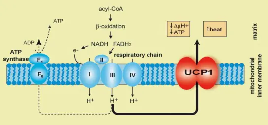
The role of UCP1 in adaptive thermogenesis is well established (Argyropoulos and Harper, 2002). Thermogenesis is a process that allows endotherm animals (mammals and birds) to produce heat. In some circumstances, such as exposure to cold, animals need to produce much more heat. As stated before, rather than using ATP to generate futile work as shivering, a way to achieve heat is to completely bypass the ATP generation system and to allow protons to turn back into the mitochondrial matrix where they can freely react with O2 forming water and releasing their energy directly as heat (Speakman, 2003) (Figure 4).
18
Figure 4: UCP1 location into inner mitochondrial membrane where is involved in heat production by dissipating the proton gradient.
Non-shivering thermogenesis, as opposed to shivering thermogenesis that occurs in skeletal muscle, occurs in BAT of mammals, which is specialized in this form of thermogenesis. BAT is located in the perirenal and interscapular areas of rodents, hibernating animals and, as will subsequently be discussed, in human (Mozo, et al, 2005). A high vascularization of BAT allows heat transfer to the tissues perfused by the blood that passes through the BAT (Smith, 1964). Cold exposure increases BAT vascularization by a mechanism involving stimulation of angiogenesis by SNS activation (Asano et al., 1997).
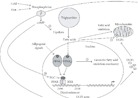
A rapid and full uncoupling of respiration from ATP synthesis leading to thermogenesis requires a large number of UCP1 molecules (Mattson et al., 2010). As described in figure 5, the expression of the UCP1 gene is directly regulated by peroxisome proliferator-activated receptors (PPARs) in association with adipogenic differentiation (via PPARγ) and in coordination with induction of gene expression required for active thermogenesis and fatty acid oxidation (via PPARα).
19
Figure 5: Schematic representation of the regulation of UCP1 gene expression (from: Villaroya et al., 2007)
Therefore, in response to certain stimuli such as cold or diet there is a norepinephrine release from the sympathetic nervous system innervating BAT. This norepinephrine release, by acting through β-adrenergic, cAMPdependent pathways and causing the activation of PPARα , PPARγ , and PGC-1α, contributes to the coordination of UCP1 gene transcription (Sell et al, 2004; Villaroya et al., 2007).
If the primary role of this protein in inducing thermogenesis is well recognized, recently it has been shown that an in vitro UCP1 overexpression adenovirus-mediated protects neurons from glucose induced degeneration by preventing mitochondrial hyperpolarization and ROS formation. That suggests a possible role of UCP1 in attenuation of ROS production (Echtay et al 2002; Vincent et al, 2004; Wolkow and Iser, 2006;).
Studies on model organisms have unequivocally demonstrated that BAT and UCP1 activity play an essential role in energy balance and in body weight control.
20 Experimental studies carried out on animals lacking of BAT or UCP1 showed that BAT thermogenesis protects against diet-induced obesity. For example, mice lacking UCP1 exhibit a marked attenuation of SNS-induced fatty acid utilization and nonshivering thermogenesis, which leads to obesity when the mice are fed a high fat diet (Kontani et al., 2005). Moreover, this study showed that, under a normal diet and at usual warm housing temperature, UCP1 deficient mice not develop obesity and appear to live a normal lifespan. In another study it has been shown that eliminating BAT noradrenergic input by disruption of the dopamine β-hydroxylase gene, mice were hyperphagic and more sensible to cold temperatures, but did not become obese because their basal metabolic rate was elevated (Thomas and Palmiter, 1997). Interestingly, in rodents the decline of the UCP1-mediated thermogenesis during aging contribute to weight gain and visceral adiposity, two phenomena that are involved in the development of age-related conditions (McDonald and Horwitz, 1999).
For a long time, it was believed that human thermogenin was expressed only in the newborn, however, recent studies have shown that UCP1 expression can be induced in adulthood. In fact, it has been found that human adults have several discrete areas of functional, UCP1-expressing brown cells and that in response to certain stimuli such as cold exposure and sympathetic stimulation, the white adipocytes can acquire features of brown cells (Tiraby et al., 2007; Nedergaard, 2007; Zingaretti et al., 2009). Moreover, although UCP1 is mainly expressed in BAT, recent works have shown that UCP1 is also expressed, although at low levels, in islet cells (Sale et al., 2007), and in thymocytes (Adams et al., 2008). Altogether, these findings strongly suggest that BAT likely plays a role in the regulation of body weight in human.
Population studies were carried out to elucidate the role of the UCP1gene variability in diabetes mellitus, obesity, and related metabolic disorders (Jia et al, 2009). The
21 polymorphisms A-1766G and A-112C at the 5’-flanking region and Ala64Thr polymorphism in exon 2 of UCP1 gene were studied in the Caucasian and Eastern Asian population (Japanese and Korean) where they are associated with body fat accumulation and body weight gain or body mass index (BMI) (Hamann et al., 1998; Herrmann et al., 2003; Kim et al., 2005; Kim et al., 2006). It has been shown that mutations, changing the activity or gene expression, change the uncoupling activity and impact pancreatic functions and insulin secretion. Two polymorphyisms in linkage disequilibrium, an A to C transition in exon 1 and a Met229Leu substitution in exon 5, were both associated with susceptibility to type-2 diabetes (Mori, 2001). The A-3826G transition is the UCP1 polymorphism most extensively studied. It is located at the 5’ of the gene in a region important for the gene regulation. Interestingly, this polymorphism was found to be associated with reduced mRNA expression indicating that the polymorphism has a functional significance (Sramkova et al, 2007). It is in strong linkage disequilibrium with a C to A variation at -3737, which affects a consensus site for the binding of members of the ATF/CREB (Activing Transcription Factors/cAMP Response Element Binding) family of transcription factors (Rousset et al., 2002). The A-3826G polymorphism has been extensively studied in relation to obesity phenotypes, diabetes mellitus and lipid/lipoprotein-related diseasebut the results were controversial (Jia et al, 2009 and reference therein).
Taken together, all of these data indicate that the UCP1 gene is an excellent candidate for these diseases even if further studies are required to investigate genetic polymorphisms of UCP1 in various populations to better elucidate the molecular and metabolic mechanism of association of these polymorphisms with obesity phenotypes, diabetes mellitus and lipid/lipoprotein-related disease.
22
1.2.2 Uncoupling Protein 2 (UCP2) and 3 (UCP3)
Human UCP2 and UCP3 genes form a cluster on chromosome 11 (11q13). UCP2 and
UCP3 are very similar to each other, about 70% of homology, and have more than 50%
of homology with UCP1 (59% and 57% respectively). Human UCP2 contains 8 exons and is 8 Kb long (Fleury et al, 1997). Exons 1 and 2 are not translated and the promoter region does not have a TATA box or CAAT box, although it contains a region rich in GC absent in UCP1. A particular feature of human and mouse Ucp2 gene is the presence of different ATGs in frame with an open reading frame for an unknown peptide of 36 amino acid in exon 2, while the UCP2 coding sequences starts in exon 3 (Pecqueur et al, 1999). UCP2 is ubiquitously expressed in different tissues including neurons (Fleury et al., 1997).
UCP3 human gene is located at 7 kb upstream of UCP2 (Pecqueur et al, 1999). It
contains seven exons and is 8.5 Kb long. UCP3 cDNA cloning revealed that the human gene is expressed as two variants generated by alternative splicing in the last intron. The amino acid sequences correspond to a protein of 312 amino acids, the long-form UCP-3L, and one of 275-amino acids, the short-form UCP-3S. UCP3S contains 5 putative transmembrane domains, while UCP3L contains an additional 37 amino acids at its C terminus that encodes a putative transmembrane domain and a putative purine nucleotide-binding domain. Human UCP3 is mainly expressed in the skeletal and heart muscle (Boss et al., 1997).
UCP2 and UCP3 discovery in mitochondria of various mammalian tissues, and evidence of their homology with UCP1 initially suggested that these two proteins were also involved in thermogenesis and regulation of energy expenditure (Boss et al, 1997). However, compared to UCP1, UCP2 and -3 are present in very low concentrations and they transport protons only when specifically activated (Esteves and Brand, 2005). This
23 observation, together with UCPs discovery in ectothermic fish and plants that do not require thermogenesis led to consider the possibility that uncoupling mediated by these proteins has a different and more general function. It has been observed that UCP2 and UCP3 activation causes a "mild uncoupling" which induces a limited increment in proton conductance, so that protonmotrice force is only slightly lowered, the respiration rate increased slightly, and the ATP synthesis can still occurs. This ―mild uncoupling― is sufficient to ensure a strong reduction of mitochondrial ROS production (Brand et al, 2004). Therefore, as UCP1, UCP2 and UCP3 are supposed to be involved in the control of reactive oxygen species production (Krauss et al., 2005). Numerous experimental evidences support this role. UCP-knockout mice have increased levels of ROS and showed signs of increased oxidative damage (Argyropoulos and Harper 2002; Rousset, et al., 2004; Echtay, 2007). For instance UCP3 knockout mice showed a higher ROS production (Brand et al, 2002), while UCP2 knockout mice were more resistant to parasitic infections, due to increased ROS production in their macrophages (Arsenijevic, et al, 2000). Furthermore, inhibition of UCP2 and 3 mediated by purine nucleotides increases the membrane potential and mitochondrial ROS production (Brand and Esteves, 2005).
It has been observed that UCP2 and UCP3 catalyze an inducible proton conductance in presence of specific activators, such as the aldehyde 4-hydroxynonenal (4HNE) and other aldehydes responsive. Proton conductance in the presence of these activators is inhibited, as previously reported, by ATP and GDP and is favored by fatty acids, which most likely act by removing inhibition induced by purine nucleotides (Rial et al, 2004). In addition, fatty acids increase UCP2 and UCP3 genes expression suggesting that proteins encoded by these genes are somehow involved in fatty acids metabolism. The mechanisms by which exposure to fatty acids increases UCP2 and UCP3 expression
24 have not been fully characterized. In addition to sterol regulatory element binding proteins (SREBPs), the G protein-coupled receptor GPR40, selectively expressed in β-cells and activated by fatty acids, seems to be implicated in UCP2 regulation (Villaroya et al., 2007 and references therein). Several studies have also evidenced that UCP2 and
UCP3 are under the control of PPARs, nuclear hormone receptors acting as sensor for
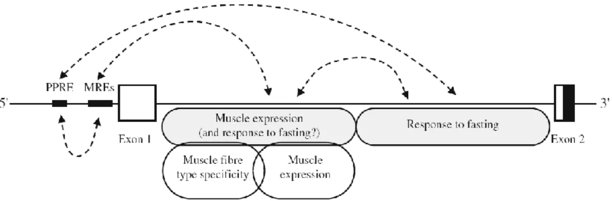
fatty acids and cholesterol-derived metabolites (Wang, 2010). It has been found that the proximal region of the UCP3 promoter contains PPAR responsive element and uncanonical MyoD responsive elements (MREs) which interact with critical elements in the 3′part of intron 1 to obtain a full response to fasting (Girousse et al., 2009) (Figure 6).
Figure 6: Potential interactions between cis-acting elements in the promoter and different regions of intron 1 of the human UCP3 gene. MREs, uncanonical MyoD responsive elements; PPRE, PPAR responsive element (from: Girousse et al, 2009).
Moreover, the regulation of these uncoupling proteins by dietary alterations, thyroid hormones and agonists of the β-3 adrenergic receptor, supports the hypothesis that UCP2 and UCP3 could play an important role in energy balance and body weight regulation (Bezaire et al., 2005; Echtay, 2007; Jia et al., 2009). In mice, high-fat feeding led to up-regulation of UCP2 in white adipose tissue, brown adipose tissue and skeletal
25 muscle. Likewise, UCP3 expression is elevated during states that are associated with increased fat metabolism in rodents and humans, for example, fasting, acute exercise, and high-fat feeding (Echtay, 2007).
Beside these roles, a tissue-specific function for UCP2 and UCP3 it has been also proposed. As for UCP2, implication in insulin secretion from β-cells and in the neuroprotection have been documented. Glucose uptake contributes to mitochondrial respiration for the production of ATP from ADP. The increase in ATP concentrations allows insulin to be released into the bloodstream. When the ATP/ADP ratios is high,
UCP2 may be activated. This activation cause an attenuation of ATP production
reducing the rate of insulin vesicle fusion, decreasing insulin release, and attenuating glucose uptake (Chan et al., 2001). Experiments in model systems, that either over- or under-express UCP2 gene, showed that UCP2 down-regulate the ability of beta cells to secret insulin. Moreover, induction of Ucp2 deficiency in ob/ob mice partially slow down the development of diabetes and insulin resistance (Zhang et al., 2001). This is of particular interest in metabolic control of complex phenotypes such as type 2 diabetes and aging.
It has been also showed that UCP2 affects several mechanisms involved in neuronal cell death, including excitotoxicity, mitochondria-mediated cell death and ROS. (Mattiasson and Sullivan, 2006). For these reasons UCP2 induction was proposed to have a potential therapeutic effect in the treatment of age-related neurodegenerative diseases such as Parkinson's disease, Alzheimer's disease, brain hypoxia and stroke.
As for UCP3, the high expression of UCP3 in skeletal muscle suggested that the gene could be important in the regulation of energy metabolism in this organ. Several observations support the proposed function of UCP3 as fatty acid anion transporter for increasing fatty acid oxidation capacity. Muscle UCP3 protein levels are increased
26 when rats are fed a diet high in long-chain triglycerides but not a diet high in medium-chain triglycerides which are oxidized via a different pathway (Schrauwen et al., 2003). Skeletal muscle mitochondria of mice overexpression Ucp3 show increased fatty acid oxidation rates and decreased intramuscular triglyceride stores (Wang et al., 2003). Nabben and coworkers (2008) showed that the physiological consequence of UCP3 overexpression in skeletal muscle might be the slowing down in the decline in muscle performance with ageing as a result of a decreased production of ROS, an increased protection of mitochondria from lipid peroxidation, and a better metabolic efficiency.
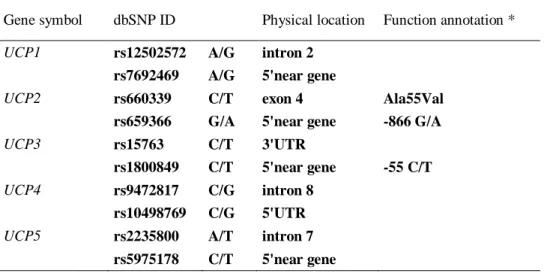
The variability of UCP2 and UCP3 genes has been analyzed in relation to different complex traits. Several polymorphisms in human UCP2 gene have been identified. In particular, the missense Ala55Val SNP (exon 5) and the G-866A polymorphism in the promoter region (Jia et al., 2009) are the more extensively studied. The Ala55Val polymorphism of the UCP2 gene it has been fully investigated. It has been shown that VV genotype causes a lower degree of uncoupling, lower energy expenditure (Astrup et al., 1999), higher exercise energy efficiency (Buemann et al 2001), and higher metabolic rate and risk in obesity, as well as a higher incidence of diabetes (Walder et al., 1998; Yu et al., 2005). Moreover, people with the VV genotype had greater weight loss and a higher BMI (Chen et al., 2007). Nevertheless, other studies indicated no association between this polymorphism and metabolic phenotypic features (Dalgaard et al., 2003; Wang et al., 2004; Hsu et al., 2008; Lee et al., 2008). Sesti and coworkers (2003) have noted that the G-866A polymorphism is associated with reduced insulin secretion with a "dose effect" of the A. The G-866A polymorphism was also identified as a risk factor in the development of multiple sclerosis, a chronic inflammatory disease caused by an autoimmune response directed against the myelin sheaths of the nerve
27 fibers. Also in this case, it has been hypothesized a "dose effect " for the A allele: carriers of homozygote genotype for the mutant allele are more protected against disease than heterozygote (Otaegui et al, 2007).
Genetic variants were also identified in the UCP3 gene. In particular, the best characterized polymorphism is the C-55T promoter region polymorphism. This polymorphism was associated with a significant increased gene expression in skeletal muscle (Schrauwen et al., 1999). It was also observed a positive correlation between resting metabolic rate and UCP3 expression. A low resting metabolic rate is a factor that predisposes to weight gain, therefore, higher UCP3 levels could cause a high resting metabolic rate and thus less prone to weight gain (Schrauwen et al, 1999).
To improve the statistical power in association studies and to better verify the role of A-866G and the 45 bp insertion of UCP2 gene and the C–55T variant of UCP3 gene (region expanding for a small region of 40 kb), a haplotype study was performed in relation to obesity and type 2 diabetes. It has been reported (Esterbauer et al., 2001) that haplotypes that included the 866A-allele and the 45 bp insertion were more frequent in lean compared with obese subjects and tend to be a protecting factor against T2D (Liu et al., 2005). Moreover, subjects carrying the two variants have been found to have a higher risk of type 2 diabetes (Wang et al., 2004).Even if in some studies (Ochoa et al., 2007) the individual polymorphisms are not found associated with obesity, the haplotype (–866G, De/45 bp, –55T) is associated significantly with obesity and causes a ninefold increase in the risk of the insulin resistance. Thus, the haplotype (–866A, Ins/45 bp, –55C) may protect against insulin resistance in obese populations.
28
1.2.3 Uncoupling Protein 4 (UCP4) and 5 (UCP5)
UCP4 (also called SLC25A27) and UCP5 (also termed brain-specific mitochondrial
carrier protein-1 (BMCP1) or SLC25A14) are located on chromosomes 6 (6p12.3) and X (Xp24), respectively. UCP4 and UCP5 have about 30% of homology with the other
members of UCP family (Adams et al., 2000; Graier et al., 2008). Human UCP4 gene contains 9 exons and 8 introns and is 24,3 Kb long, while UCP5 gene contains 6 exons and 5 introns and is 33,29 Kb long.
These two proteins, together with UCP2, are known as neuronal UCPs because are widespread in the brain. UCP4 is mainly expressed in central nervous system and at a lower level in other tissues (Yang et al., 2002; Krauss et al, 2005; Smorodchenko et al., 2009). UCP5 is particularly expressed in the brain and testis, and is also widely present at lower levels in other tissues such as kidney, uterus, heart, lung, stomach, liver, and skeletal muscle (Sanchis et al., 1998; Andrews et al., 2005; Echtay, 2007). UCP5 has three isoforms: a ―long‖ form of 325-amino acid (UCP5L), a ―short‖ form of 322-amino acid (UCP5S) that lacks amino acids Val–Ser–Gly (VSG) at position 23–25 of UCP5L, and a ―short insert‖ form of 353-amino acid (UCP5SI) that lacks the VSG amino acids but has a 31-amino acid insertion between transmembrane domains III and IV (Yu et al., 2000). The presence of multiple isoforms with a tissue-specific expression suggests a complexity in the UCP5 regulation.
Even if UCP4 and UCP5 are more widespread expressed in the brain than UCP2, their functions in neurons have been not completely established. Neurons are characterized by a very high metabolic rate and consequently by a high production of ROS, therefore, it has been presumed that by uncoupling activity, UCP4 and UCP5 may modulate oxidative metabolism in these cells. Andrews and co-workers observed that neuronal uncoupling activity leads to a decreased ROS levels, a decreased Ca2+
voltage-29 dependent influx and increased local temperature in neuronal microenvironment (Andrews et al, 2005). It has been shown that UCP4 activity can also induces an adaptative shift in energy metabolism, from mitochondrial respiration to glycolysis, that helps sustain neurons under conditions of metabolic and oxidative stress (Liu et al., 2006). Therefore, by decreasing free radical production and stabilizing cellular calcium homeostasis, UCPs expressed in neurons may positively influence neuronal function (synaptic transmission and plasticity) and retard cellular deterioration associated, for instance, with neurological disorders. It has been observed that increased UCP2, UCP4, and UCP5 levels (Sullivan et al., 2004), induced by dietary restriction and 2-deoxyglucose administration, improve behavioral outcomes and reduce dopaminergic neurodegeneration in models of Parkinson's disease (Duan and Mattson, 1999). Experimental evidences also showed that nutritional and temperature manipulations are able to modulated the mRNA abundance of UCP4 and UCP5 in a tissue-specific manner, suggesting their involvement in metabolic rate and adaptative thermoregulation (Yu et al., 2000; Yang et al., 2002; Andrews et al., 2005). For instance, their uncoupling activity may provide a basis for temperature as a neuromodulator. Moreover, temperature is critical for normal spermatogenesis (Steinberger, 1991), and it is conceivable that UCP5 may be involved in the regulation of local testicular temperature. The function of UCP4 in adipocytes is already unknown.
UCP4 and UCP5 are less studied with respect to UCP1, UCP2, and UCP3 and less is known about their regulation. A study carried out on bovine mammary epithelial cells (bMEC) showed that physiological concentrations of saturated fatty acids (stearate and palmitate), but not unsaturated fatty acids (oleate and linoleate), induced an elevated expression of UCP5, but not UCP4. Moreover, treatment with insulin induced down-regulation of UCP4 and UCP5. These results suggest that UCP4 and UCP5 are
30 regulated by insulin and/or fatty acids in mammary epithelial cells and lactating mammary glands, and thereby may play an important role in lipid and energy metabolism (Yonezawa et al., 2009).
Recently, Ho and coworkers (2010) characterized several cis-acting elements that might regulate UCP4 expression. They found that core promoter activity exists within 100 bp upstream of the transcription initiation site. In UCP4 transcription a CAAT box (-33/-27) and Sp1 (-62/-49) elements act synergistically. Moreover, a NF-kappaB putative binding site at -507/-495 exists, and it has been observed that mutation of this site decreases significantly the activity of the promoter. Activation of NF-kappaB by TNFalpha or cycloheximide increased, whereas its inhibition by 4-hydroxy-2-nonenal or transfection of pIkBαM suppressed, UCP4 promoter activity.
UCP4 and UCP5 variability has been analyzed in relation to several complex
phenotypes such as multiple sclerosis, leukoaraiosis, and schizophrenia. It was found that CC genotype for rs10807344 of UCP4 gene exerts a protective effect on occurrence of multiple sclerosis and of leukoaraiosis that is a vascular demyelinization of the white matter of the brain (Szolnoki et al, 2009 and 2010).
In a case-control study tag-SNPs for the neuronal UCPs were investigated in relation to schizophrenia. Modest associations was found for rs10807344 and rs2270450 in UCP4. Interestingly, a statistically significant synergistic interaction between UCP2 and UCP4 was found, suggesting that UCP2 and UCP4 have a modest but important involvement in the genetic etiology of schizophrenia (Yasuno et al., 2007).
Taken together, these data suggest that UCP4 and UCP5 play a role in antioxidant protection and preservation of neuronal dysfunction and for this reasons maybe
31 implicated in neuroprotection against mitochondrial dysfunction in various degenerative diseases.
1.3. The role of uncoupling proteins in lifespan
Based on the different functions of the uncoupling proteins described in previous sections, uncoupling proteins may provide a link between mitochondria, metabolism and lifespan. In fact, ROS production, metabolic rate and homeostasis are interconnected factors that are all likely to contribute to the accumulation of damage and dysfunction observed during life.
For this reason, starting from ―The uncoupling theory of aging‖ researchers try to highlight the connection between UCP-mediated uncoupling and lifespan (for reviews see: Harper et al., 2004; Wolkow and Iser, 2006; Dietrich and Horvath, 2010). The first evidence of the direct effect of UCPs in senescence came in 2005 by Fridell and coworkers (2005). In their work the authors overespressed the human UCP2 in adult fly neurons, and found an extantion of lispan. In these transgenic flies, they found an increase in proton leak, and a decrease in ROS production and oxidative damage. The same authors demonstrated that transgenic Drosophila lines which targeted UCP expression in insulin producing cells showed an attenuated systemic insulin signaling and a significant lifespan extantion (Fridell et al., 2009). In another study, Conti and coworkers (2006) generated transgenic mouse over-expressing UCP2 in neurons. They found that these mice have increased lifespan. Additional data on the effect of UCP2 regard to mammalian longevity came from the study of Andrews and Horvath (2009) who showed that knockout mice had a significantly shorter survival age compared to their wild-type littermates. However these results show some contradictions with respect to the study by McDonald and coworkers (2008). Interestingly recent