Università degli Studi
"ROMA TRE”
Scuola Dottorale in Biologia (XXI ciclo)
Sezione “Scienze biomolecolari e cellulari”
A Study on Flavin-containing Amine
oxidases in Arabidopsis thaliana
Studio di Ammino Ossidasi Flaviniche
in Arabidopsis thaliana
Candidata: Dott.ssa Valentina Spedaletti
TABLE OF CONTENTS
ABSTRACT 1
1. INTRODUCTION 6
1.1 Polyamines and polyamine oxidases 6
1.1.1 General characteristics of polyamines 6
1.1.2 Physiological roles of polyamines 7
1.1.3 Polyamine biosynthesis 9
1.1.4 Polyamine catabolism 11
Plant copper-containing amine oxidases 13
Plant polyamine oxidases 13
Physiological roles of CuAOs and PAOs in plants 15
1.2 Histones and histone demethylases 18
1.2.1Epigenetic modifications of histones 18
1.2.2Histone methylation 21
Lysine and arginine methylation 21
Lysine-specific histone demethylases 21
Peptidyl-arginine deiminases 24
Jumonji C domain-containing histone demethylases 25
1.3 Arabidopsis thaliana as a model plant 26
1.4 Aims of this thesis 27
2. RESULTS 28
2.1 Polyamine oxidases of A. thaliana 28
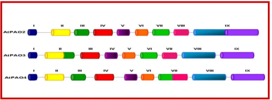
2.1.1Description of the PAO gene family in A. thaliana 28
2.1.2Heterologous expression of AtPAO2, AtPAO4 and AtPAO5 in Escherichia coli 29
2.1.3Purification and biochemical characterization of recombinant AtPAO2 and AtPAO4 31
2.1.4Characterization of AtPAO2 and AtPAO4 reaction products 33
2.1.5Determination of the polyamine back-conversion pathway in vivo in A. thaliana 34
2.2: Histone demethylases of A. thaliana 37
2.2.1Description of the lysine-specific histone demethylase gene
family in A. thaliana 37
2.2.2Expression pattern of the AtLSD gene family in
different Arabidopsis organs 38
2.2.3Heterologous expression and biochemical characterization
of AtLSD1 in E. coli 38
2.2.4Characterization of the reaction products of AtLSD1 by
mass spectrometry analysis 42
2.2.5Analysis of the ability of AtLSD1 to interpret the histone code 42
2.2.6Comparative analysis of the three-dimensional structure
of AtLSD1 44
2.2.7Characterization of T-DNA insertional mutants for AtLSD1, AtLSD2, AtLSD3 and AtLSD4 46
2.2.8Preparation of trangenic Arabidopsis plants transformed with an AtLSD1prom::GFP-GUS construct and with constructs for AtLSD1 overexpression 49
3. DISCUSSION 52
ABSTRACT
Le poliammino ossidasi (PAO) sono enzimi FAD-dipendenti che ossidano le poliammine spermina (Spm) e spermidina (Spd) e/o i loro derivati acetilati a livello del gruppo amminico secondario. L’identità chimica dei prodotti di reazione delle PAO dipende dall’origine dell’enzima e riflette la modalità di ossidazione del substrato. In particolare, le PAO presenti nelle piante monocotiledoni, come la PAO di mais (ZmPAO), ossidano l’atomo di carbonio interno adiacente al gruppo amminico secondario della Spm e della Spd, con produzione di 1,3-diamminopropano (Dap), perossido di idrogeno e di un’amminoaldeide ed in tale maniera partecipano ad una via catabolica terminale delle poliammine. Le PAO animali e le spermina ossidasi (SMO) ossidano, invece, l’atomo di carbonio esterno adiacente al gruppo amminico secondario della Spd o della Spm (o dei loro derivati acetilati) producendo rispettivamente putrescina (Put) o Spd, un’amminoaldeide e perossido di idrogeno ed in tale maniera sono coinvolte in una via di interconversione delle poliammine.
In Arabidopsis thaliana, sono stati identificati cinque geni codificanti per PAO putative: l’At5g13700 (AtPAO1), l’At2g43020 (AtPAO2), l’At3g59050 (AtPAO3), l’At1g65840 (AtPAO4), e l’At4g29720 (AtPAO5). L’AtPAO1 e l’AtPAO5 presentano un’omologia di sequenza con la ZmPAO (la PAO vegetale maggiormente caratterizzata ed avente una localizzazione apoplastica) rispettivamente del 23% e del 25% ed una probabile localizzazione citosolica. L’AtPAO2, l’AtPAO3 e l’AtPAO4 presentano un’omologia con la ZmPAO del 23%, un’omologia fra loro che varia tra il 58% e l’85% ed una localizzazione perossisomale. Recentemente, è stato dimostrato che l’AtPAO1 è in grado di ossidare la Spm, ma non la Spd e che è coinvolta in una via di interconversione delle poliammine in maniera simile a alle PAO animali e alle SMO.
Nel presente lavoro di tesi, è stato effettuato uno studio sulle proprietà biochimiche delle proteine ricombinanti AtPAO2 e AtPAO4 in seguito alla loro espressione eterologa in Escherichia coli. Tale studio ha dimostrato che le proteine ricombinanti AtPAO2 e AtPAO4 sono attive nei confronti della Spm e della Spd e che producono Spd dall’ossidazione della Spm e Put dall’ossidazione della Spd. Questi dati indicano quindi che queste due AtPAO hanno una modalità di ossidazione del substrato simile a quella dell’AtPAO1 e delle PAO e SMO animali e che sono coinvolte in una via di interconversione delle poliammine. L’esistenza di una via di interconversione delle poliammine in A. thaliana è stata dimostrata anche in vivo. Infatti, durante l’incubazione di protoplasti ottenuti da foglie di A. thaliana con Spd o Spm radioattiva è stato osservato un aumento della quantità di Put o Spd radioattiva. Tale aumento risulta fortemente inibito in presenza di guazatina, un inibitore specifico delle PAO.
Nel presente lavoro, è stato dimostrato anche che le proteine ricombinanti AtPAO1, AtPAO2 e AtPAO4 sono in grado di ossidare le poliammine non comuni termospermina (Termo-Spm) e norspermina (Nor-Spm), che sono state associate alla tolleranza agli stress. In particolare, è stato dimostrato queste poliammine non comuni sono per l’AtPAO1 dei substrati migliori rispetto alla Spm facendo ipotizzare che potrebbero essere i suoi substrati fisiologici. Questo dato è di fondamentale importanza se si considera che recentemente è stato identificato un gene (ACAULIS5) codificante per una proteina capace di sintetizzare la Termo-Spm dalla Spd e che piante di A. thaliana che presentano una mutazione in questo gene (acaulis5) mostrano dei difetti nell’allungamento dello stelo.
In A. thaliana sono presenti altri quattro geni: l’At1g62830 (AtLSD1), l’At3g13682 (AtLSD2), l’At3g10390 (AtLSD3) e l’At4g16310 (AtLSD4) che codificano per proteine aventi un dominio ammino ossidasico. Queste proteine presentano anche un dominio SWIRM, che è generalmente presente nei complessi coinvolti nelle modificazioni della cromatina, ed hanno un’omologia di sequenza con la proteina umana HsLSD1 (KIAA0601) che varia dal 26 al 30%. È stato dimostrato che l’HsLSD1, che ha gli stessi domini funzionali delle AtLSD, catalizza la demetilazione ossidativa dell’istone H3 mono o dimetilato sulla lisina 4 e fa parte di complessi multiproteici importanti nella regolazione dell’espressione genica.
Nel presente lavoro, in seguito ad espressione eterologa in E. coli è stata effettuata una parziale caratterizzazione biochimica della proteina AtLSD1, scelta come membro rappresentativo di questa famiglia genica, ed è stato dimostrato che questo enzima vegetale ha un’attività iston-demetilasica e presenta la stessa specificità di substrato della corrispondente proteina umana. Inoltre, dall’analisi del modello della struttura tridimensionale dell’AtLSD1, eseguito sulla base del cristallo dell’HsLSD1, è risultato un’elevato grado di conservazione delle strutture secondarie dell’HsLSD1 e dei residui facenti parte del sito catalitico e sono emerse alcune importanti differenze che suggeriscono che i partners molecolari dell’AtLSD1 possano essere differenti da quelli della proteina ortologa umana. Per effettuare un’analisi del profilo di espressione dei geni AtLSD, sono stati condotti degli esperimenti di RT-PCR dai quali è emerso che i livelli di espressione di tali geni risultano simili nei vari organi testati. Inoltre, allo scopo di approfondire l’analisi del profilo di espressione dell’AtLSD1, il promotore di tale gene è stato amplificato tramite PCR dal DNA totale estratto da foglie di A. thaliana ed è stato clonato in un vettore per l’espressione in pianta, mediata da Agrobacterium tumefaciens, a monte della sequenza codificante per la green-fluorescent protein (GFP) in fusione con la
β-glucuronidasi (GUS). In questo modo è stato ottenuto un costrutto AtLSD1
prom::GFP-GUS. Per individuare i geni regolati dall’AtLSD1 tramite analisi microarray ed esperimenti di immunoprecipitazione della cromatina è stato preparato un costrutto per la sovraespressione dell’AtLSD1 in A. thaliana. In
particolare, la regione codificante per l’AtLSD1 è stata amplificata tramite PCR utilizzando primers sequenza-specifici disegnati in modo tale da permetterne il clonaggio in un vettore che guidi la sovraespressione delle proteine in pianta e per aggiungere all’estremità 3’ una coda di 6 istidine che faciliti l’individuazione della proteina. Per isolare i complessi nei quali la proteina AtLSD1 è eventualmente coinvolta attraverso cromatografia di affinità, è stato preparato anche un costrutto per la sovraespressione della proteina in fusione con una coda FLAG-HA. Tutti i plasmidi ricombinanti sono stati utilizzati per trasformare il ceppo GV301 di A. tumefaciens ed i batteri trasformati sono stati al loro volta utilizzati per trasformare piante di A. thaliana. Per determinare i ruoli fisiologici svolti dalle AtLSD, mutanti inserzionali per ognuno dei quattro geni (Atlsd1, Atlsd2, Atlsd3 e Atlsd4) sono stati ottenuti da banche di semi di A. thaliana e sono stati analizzati. In particolare, per confermare la presenza dell’inserzione del T-DNA e per identificare le piante mutanti omozigoti per l’inserzione è stata effettuata un’analisi tramite PCR del DNA totale estratto dalle piante mutanti. In seguito, è stata eseguita un’analisi dettagliata del fenotipo dei mutanti Atlsd che ha evidenziato un fenotipo nel mutante Atlsd3 caratterizzato da un ritardo nella fioritura.
Polyamine oxidases (PAOs) are FAD-dependent enzymes which oxidize the polyamines spermine (Spm) and spermidine (Spd) and/or their acetylated derivatives at the secondary amino group. The chemical identity of PAO reaction products depends on the enzyme source and reflects the mode of substrate oxidation. In particular, PAOs from monocotyledonous plants, such as maize PAO (ZmPAO), oxidize the carbon on the endo-side of the secondary amino group of Spm and Spd producing 1,3-diaminopropane (Dap), H2O2 and
an aminoaldehyde, and are considered involved in a terminal catabolic pathway of polyamines. Conversely, animal PAOs and spermine oxidases (SMOs) oxidize the carbon on the exo-side of the secondary amino group of Spd or Spm (or their acetylated derivatives) producing putrescine (Put) or Spd, respectively, in addition to an aminoaldehyde and H2O2, and are considered involved in a
polyamine back-conversion pathway.
In Arabidopsis thaliana, five putative PAO genes have been identified: At5g13700 (AtPAO1), At2g43020 (AtPAO2), At3g59050 (AtPAO3), At1g65840 (AtPAO4), At4g29720 (AtPAO5). AtPAO1 and AtPAO5 have a sequence homology of 45% and 25%, respectively, with ZmPAO (the so far best characterized plant PAO which has an apoplastic localization) and a predicted cytosolic localization. AtPAO2, AtPAO3 and AtPAO4 display an homology of about 23% with ZmPAO, an homology of 58-85% to each other and a peroxisomal localization. Recently, AtPAO1 has been shown to oxidize only
Spm and not Spd and to be involved in a polyamine back-conversion pathway similarly to the animal PAOs/SMOs.
In the present work, a study on the biochemical properties of recombinant AtPAO2 and AtPAO4 expressed in Escherichia coli was performed. This study demonstrated that recombinant AtPAO2 and AtPAO4 are active towards both Spd and Spm and that produce Spd from Spm and Put from Spd. These data indicate that these two AtPAOs have a mode of substrate oxidation similar to that of AtPAO1 and animal PAOs/SMOs and thus that they are also involved in a polyamine conversion pathway. The existence of a polyamine back-conversion pathway in A. thaliana has been demonstrated also in vivo. Indeed, incubation of A. thaliana protoplasts with radiolabelled Spd or Spm resulted in the accumulation of radiolabelled Put or Spd, respectively, which was strongly reduced in the presence of the PAO-specific inhibitor guazatine.
In the present work, it was also shown that recombinant AtPAO1, AtPAO2 and AtPAO4 are able to oxidize the stress related uncommon polyamines thermospermine (Thermo-Spm) and norspermine (Nor-Spm). In particular, it was shown that these uncommon polyamines are better substrates than Spm for AtPAO1, suggesting that these polyamines may be the physiological substrates of this enzyme. This is of great importance considering that a gene (ACAULIS5) encoding for a protein able to synthesize Thermo-Spm from Spd has been recently characterized in A. thaliana and the acaulis5 Arabidopsis mutant presents defects in stem elongation.
In A. thaliana, four more genes have been also identified: At1g62830 (AtLSD1), At3g13682 (AtLSD2), At3g10390 (AtLSD3), At4g16310 (AtLSD4) encoding for proteinswith an amine oxidase domain. These proteins bear also a SWIRM domain, which is usually present in chromatin-modifying complexes, and display a 26-30% sequence homology with human HsLSD1 (KIAA0601). HsLSD1, which has the same functional domains as the four AtLSDs, has been shown to catalyse the oxidative demethylation of mono- or dimethylated lysine 4 of histone H3 and to participate in multiprotein complexes important in the regulation of gene expression.
In this work, partial biochemical characterization of AtLSD1, chosen as a representative member of this gene family, following expression in E. coli demonstrated that this plant enzyme has a demethylase activity with the same substrate specificity as the corresponding human protein. Modeling of the AtLSD1 three-dimensional structure, using the HsLSD1 crystal structure, evidenced a high degree of conservation of the HsLSD1 secondary structures and of the residues building up the catalytic site, but also some important differences which suggest that the AtLSD1 molecular partners are probably different from those of the human orthologue. To analyse the expression pattern of AtLSDs, RT-PCR experiments were performed which showed that the AtLSD1, AtLSD2, AtLSD3 and AtLSD4 transcripts are present at similar levels in all organs tested. With the aim to go deeper into the AtLSD1
expression pattern, the AtLSD1 promoter was amplified by PCR from Arabidopsis total DNA and cloned in an Agrobacterium tumefaciens-based plant expression vector upstream of the sequence encoding green-fluorescent protein (GFP) in fusion with β-glucuronidase (GUS). In this way, an AtLSD1 prom::GFP-GUS construct was obtained. Furthermore, to identify the genes regulated by AtLSD1 through microarray analysis and chromatin immunoprecipitation experiments, a construct for AtLSD1 overexpression in A. thaliana was prepared. Indeed, the coding region of AtLSD1 was amplified by PCR using sequence specific primers designed in a way to allow AtLSD1 cDNA cloning through the Gateway technology in a vector which guides overexpression of proteins in plant and to add at the 3′ terminus of cDNA a sequence encoding for a 6-His tag to facilitate detection. To isolate the complexes in which AtLSD1 is eventually involved through a two-step affinity chromatography, a construct for AtLSD1 overexpression in A. thaliana in fusion with a FLAG-HA tag was also prepared. All the recombinant plasmids were used to transform the A. tumefaciens GV301 strain and the transformed bacteria were in turn used to transform A. thaliana plants. To determine the physiological roles of AtLSDs, insertional knock-out mutants for each one of the four AtLSD genes (Atlsd1, Atlsd2, Atlsd3 and Atlsd4 mutants) were obtained from A. thaliana seed banks and analyzed. In particular, PCR analysis of total DNA from mutant seedlings was performed to confirm the presence of T-DNA insertion and to identify the homozygous mutant plants for this insertion. Detailed phenotypic analysis of the Atlsd mutants was also performed which evidenced a delayed flowering phenotype for Atlsd3 mutant.
1. INTRODUCTION
1.1 Polyamines and polyamine oxidases 1.1.1General characteristics of polyamines
Polyamines are aliphatic polycations of low molecular mass that are found ubiquitously in all living organisms. The most common polyamines are the diamine putrescine (Put), the tri-amine spermidine (Spd), and the tetra-amine spermine (Spm) (Galston and Sawhney, 1990) (Fig. 1). In nature, there are also uncommon polyamines, such as 1,3-diaminopropane (Dap), cadaverine (Cad), norspermidine (Nor-Spd), norspermine (Nor-Spm) and thermospermine (Thermo-Spm) (Fig. 1). Nor-Spd, Nor-Spm and Thermo-Spm, which are abundant in the extreme thermophilic bacterium Thermus thermophilus, have also been detected in other bacteria, algae, fungi, animals and higher plants (Cohen, 1998). In particular, in the latter, Nor-Spd and Nor-Spm have been found in drought-tolerant cultivars of Medicago sativa L. and in heat-tolerant cotton species (Rodriguez-Garay et al., 1989; Kuehn et al., 1990).
Plant polyamines often occur in free soluble form. However, they can be also conjugated to low molecular mass molecules, such as hydroxycinnamic acid, by the formation of an amide linkage or bound to macromolecules such as proteins (Martin-Tanguy, 1997).
Fig. 1. Structures of common and uncommon polyamines
Common polyamines NH2-(CH2)4-NH2 Putrescine NH2-(CH2)3-NH-(CH2)4-NH2 Spermidine NH2-(CH2)3-NH-(CH2)4-NH-(CH2)3-NH2 Spermine Uncommon polyamines NH2-(CH2)3-NH2 1,3-diaminopropane NH2-(CH2)5-NH2 Cadaverine NH2-(CH2)3-NH-(CH2)3-NH2 Norspermidine NH2-(CH2)3-NH-(CH2)3-NH-(CH2)3-NH2 Norspermine NH2-(CH2)4-NH-(CH2)3-NH-(CH2)3-NH2 Thermospermine
1.1.2Physiological roles of polyamines
Polyamines are positively charged at physiological pH and this property allows them to interact with negatively charged macromolecules, such as DNA, RNA, proteins and phospholipids. In this way, polyamines are involved in the regulation of physical and chemical properties of membranes, stabilization of nucleic acid structure and modulation of enzyme activities (Galston and Sawhney, 1990). They are also known to protect DNA from damage caused by alkylating reagents, reactive oxygen species (ROS) and γ-rays (Ha et al., 1998; Mackintosh and Pegg, 2000).
Polyamines are also involved in cell cycle progression. Indeed, polyamine concentration vary during cell cycle. In particular, Put level increases during S e G2 phases, that of Spd during the entire cycle and that of Spm mainly during G1 and S phases (Thomas and Thomas, 2001). It has been suggested that Put is essential for the cell to enter S-phase, possibly driving the cell through the G1 restriction point prior to DNA synthesis and by the regulation of cyclin D1 (Wallace et al., 2003).
Polyamines are also considered to be essential for cell growth and differentiation (Cohen, 1998; Igarashi and Kashiwagi, 2000; Thomas and Thomas, 2001; Hanfrey et al., 2001). Inhibition of Spd synthesis, through deletion of enzymes involved in polyamine biosynthesis, is lethal at very early embryonic stages in mice (Wang et al., 2004a) and leads to lethal defect in embryo development in Arabidopsis thaliana plants (Imai et al., 2004; Urano et al., 2005). These data suggest an important role of Spd for viability of eukaryotic cells probably because it is a precursor of the unusual amino acid hypusine which in turn is involved in posttranslational modification of the eukaryotic translational initiation factor 5A (eIF5A) (Park, 2006).
On the other hand, polyamines are also involved in apoptotic cell death. Indeed, several studies suggest that higher or lower polyamine levels with respect to the physiological levels are implicated in apoptosis. Infact, it has been shown that an excessive production of polyamines can cause apoptosis, at least in some cell systems (Schipper et al., 2000). On the contrary, inhibitors of Spm biosynthetic enzymes have been shown to induce apoptosis in different cell lines. Data also exist suggesting a protective role of Spm against apoptosis (Kaneko et al., 1998; Hashimoto et al., 1999; Schipper et al., 2000; Seiler and Raul, 2005). In particular, it has been demonstrated that Spm prevents an increase in caspase 3 activity which generally precedes apoptosis. Other mechanisms that could be on the basis of the protective effect of Spm are inhibition of endonucleases, stabilization of DNA and protection of DNA against oxidative stresses (Schipper et al., 2000; Seiler and Raul, 2005).
In plants, polyamines have been suggested to play important roles in morphogenesis, growth, embryogenesis, organ development and leaf senescence (Kumar et al., 1997; Walden et al., 1997; Malmberg et al., 1998;
Liu et al., 2000; Kusano et al., 2007a). In general, cells undergoing division contain high levels of free polyamines while cells undergoing expansion and elongation contain low levels of free polyamines (Galston and Sawhney, 1995). In higher plants, polyamines are also implicated in responses to various types of abiotic stress (Bouchereau et al., 1999; Alcázar et al., 2006; Kusano et al., 2007b; Groppa and Benavides, 2008). In Arabidopsis plants, K+ deficiency induces an increase in Put accumulation and several studies have established a specific role of Put in maintaining a cation-anion balance in plant tissues (Boucherau et al., 1999). Furthermore, in A. thaliana, the expression levels of various enzymes involved in polyamine biosynthesis are increased following dehydration and cold stress (Pérez-Amador et al., 2002; Urano et al., 2003; Alcázar et al., 2006; Hummel et al., 2004; Vergnolle et al., 2005). In addition, different studies reported increase in Spd level in water-stressed and cold-tolerant tissues, indicating the stress-specific role of this polyamine (He et al., 2002; Nayyar et al., 2005). Salinity causes a significant increase in Spd and Spm levels and a decrease in Put level almost in all plant species studied. Furthermore, cellular alterations induced by sodium chloride treatment in wheat roots can be alleviated by exogenous Spd or Spm (Mansour and Al-Mutawa, 1999). These results would indicate a role for Spd and Spm in protecting plasma membrane under salinity and, thus, enhancing salt tolerance. The protective role of Spd and Spm in plant responses to abiotic stresses was also demonstrated using transgenic plants overexpressing polyamine biosynthetic genes (Kasukabe et al., 2004; Kasukabe et al., 2006) and loss-of-function mutant plants. In particular, enhancement of Spd synthesis in A. thaliana via a transgenic approach confers multi-stress tolerance and Arabidopsis acl5/spms mutant plants, which are unable to produce Spm, are hypersensitive to salt and drought stresses. Arabidopsis acl5/spms mutant plants are also Ca2+ deficient and lose more water compared to control plants, due to a failure of stomatal closure upon onset of drought (Yamaguchi et al., 2006; Yamaguchi et al., 2007). These results suggest that the absence of Spm may cause deregulation of Ca2+ trafficking, resulting in a lack of proper adaptation to high sodium chloride or drought stresses (Kusano et al. 2007a; Kusano et al., 2007b). It is plausible that one of the tasks of stress-induced polyamines is to modulate the activity of a certain set of ion channels to adapt ionic fluxes in response to environmental changes (Shabala et al., 2007; Zhao et al., 2007).
In plants, polyamines are also involved in plant-pathogen interactions, both incompatible and compatible ones (Walters, 2003). During the hypersensitive response (HR), which follows an incompatible plant-pathogen interaction and which consists in rapid H2O2 production and cell death at the site of pathogen
entry, an enhanced polyamine synthesis and an apoplastic accumulation of Spm were reported (Torrigiani et al., 1997; Marini et al., 2001). Apolastic Spm may directly affect cation channel(s) and/or be catabolized to produce H2O2.
mithocondrial malfunction and cell death (Takahashi et al., 2003; Amirsadeghi et al., 2007; Kusano et al., 2007a).
Changes in polyamine metabolism were reported also during compatible plant-pathogen interactions. In particular, increased polyamine concentrations were found in green islands which are formed on cereal or barley leaves infected by biotrophic fungal pathogens, like rust and powdery mildew (Walters, 2000). Green islands surround the infection sites and are thought to represent regions in which a juvenile condition is maintained to ensure absorption of nutrients by the pathogen (Walters, 1989). It was suggested that the increased polyamine concentration in infected cereal or barley leaves might be related to green island formation (Walters, 2003).
1.1.3Polyamine biosynthesis
Intracellular polyamines pools appear to be sensitively regulated by various homeostatic processes that include pathways for polyamine biosynthesis, catabolism, and transport across the cell membrane (Wallace et al., 2003).
In plants, two alternative polyamine biosynthetic pathways are present (Bagni and Tassoni, 2001; Liu et al., 2007). In particular, the diamine Put can be synthesized, starting either from arginine (Arg) through the arginine decarboxylase (ADC) pathway or from ornithine through the ornithine decarboxylase (ODC) pathway (Fig. 2). In the ADC pathway, the Arg is first decarboxylated by ADC to form agmatine which is subsequently converted to Put by the combined action of agmatine iminohydrolase and N-carbamoylputrescine amidohydrolase. In the ODC pathway, Put is the direct product of ornithine decarboxylation by the action of ODC. Spd and Spm are formed by the subsequent addition of an aminopropyl group onto Put and Spd, respectively, in reactions catalysed by the enzymes spermidine synthase (SPDS) and spermine synthase (SPMS). The aminopropyl group is transferred from the decarboxylated S-adenosylmethionine (dcSAM), which in turn is synthesized from methionine in two sequential reactions catalyzed by methionine adenosyltransferase and S-adenosylmethionine decarboxylase (SAMDC), respectively (Fig. 2).
In A. thaliana, the ODC pathway seems to be absent since none ODC gene has been identified in the sequenced genome of this plant (Hanfrey et al., 2001), whereas all other polyamine biosynthetic genes have been assigned in A. thaliana genome. In detail, there are two ADC genes (ADC1 and ADC2) (Watson and Malmberg, 1996; Watson et al., 1997), two SPDS genes (SPDS1 and SPDS2) (Hanzawa et al., 2002), one SPMS gene (Panicot et al., 2002; Clay and Nelson, 2005) and at least four SAMDC genes (SAMDC1, SAMDC2, SAMDC3 and SAMDC4) (Urano et al., 2003; Ge et al., 2006). Recently, a gene (ACAULIS5 or ACL5) encoding for an enzyme able to produce Thermo-Spm from Spd has been reported (Knott et al., 2007). Furthermore, a
loss-of-function mutation on ACL5 leads to defects in stem elongation (Hanzawa et al., 1997) and in vascular development (Clay and Nelson, 2005). Daily application of Thermo-Spm onto the shoot apex partially rescued the dwarf phenotype of acl5 mutant (Kakehi et al., 2008).
In bacteria, both the ADC and the ODC pathways are present. However, in this case Put can be also synthesized directly from agmatine by agmatinase. Furthermore, in bacteria no SPMS gene is present thus Spm is not synthesized (Wortham et al., 2007) (Fig. 2). In animals, Put is prevalently synthesized by the ODC pathway, the ADC pathway being just a minor pathway in specific mammalian tissues (Gilad et al., 1996) (Fig. 2).
Fig. 2. Polyamine biosynthetic pathways. Plant pathway is indicated by green bold arrows. Blue
and red arrows indicate bacterial and animal pathways, respectively. Figure taken from Kusano et
1.1.4Polyamine catabolism
Polyamines are catabolized by the action of two classes of amine oxidases, the copper-containing amine oxidases (CuAOs) and the FAD-dependent amine oxidases (PAOs) (Cona et al., 2006).
CuAOs oxidize Put at the primary amino group producing 4-aminobutanal, H2O2 and ammonia (Cohen, 1998) (Fig. 3). 4-Aminobutanal cyclizes
spontaneously to yield ∆1-pyrroline that can be further oxidized to form γ-aminobutyric acid (GABA). GABA is subsequently transaminated and oxidized to succinic acid, which is incorporated into the Kreb’s cycle, thus ensuring the recycling of carbon and nitrogen from Put (Cona et al., 2006) (Fig. 3).
PAOs catalyze the oxidation of Spm, Spd, and/or their acetylated derivatives at the secondary amino group (Federico and Angelini, 1991; Wang et al., 2001; Wu et al., 2003; Cona et al., 2006). The chemical identity of PAO reaction products depends on the enzyme source and reflects the mode of substrate oxidation. The until now characterized PAOs from monocotyledonous plants oxidize the carbon on the endo-side of the N4-nitrogen of Spd and Spm, producing 4-aminobutanal and N-(3-aminopropyl)-4-aminobutanal, respectively, in addition to Dap and H2O2 (Cona et al., 2006) (Fig. 3). Animal
PAOs and yeast (Saccharomyces cerevisiae) spermine oxidase (Fms1) oxidize the carbon on the exo-side of N4-nitrogen of N1-acetyl-Spm and N1-acetyl-Spd to produce Spd and Put, respectively, in addition to 3-acetamidopropanal and H2O2 (Landry and Sternglanz, 2003; Vujcic et al., 2003; Wu et al., 2003) (Fig.
3). In this catabolic pathway, polyamine acetylation is catalyzed by the tightly regulated Spd/Spm N1-acetyltransferase (SSAT), which is the rate-limiting factor (Wallace et al., 2003). Animal spermine oxidases (SMOs) and Fms1 also oxidize the carbon on the exo-side of N4-nitrogen of Spm to produce Spd, 3-aminopropanal, and H2O2 (Wang et al., 2001; Vujcic et al., 2002; Cervelli et
al., 2003; Landry and Sternglanz, 2003) (Fig. 3). Aminopropanal and 3-acetamidopropanal, produced by polyamine catabolism, can be metabolized by an aldehyde dehydrogenase (ADH) to form β-alanine and N-acetyl-β-alanine, respectively, and N-acetyl-β-alanine in turn can be converted to β-alanine by the action of a selective hydrolase (HDL) (Fig. 3).
The animal PAOs and SMOs, as well as the yeast Fms1, are considered involved in a polyamine back-conversion pathway since they produce a common polyamine by the oxidation of another common polyamine (Seiler, 2004). On the contrary, the until now characterized PAOs from monocotyledonous plants are involved in the terminal catabolism of polyamines (Fig. 3). Only recently, a PAO from A. thaliana (AtPAO1) has been shown to be involved in a polyamine back-conversion pathway similarly to animal PAOs and SMOs (Tavladoraki et al., 2006).
Fig. 3. Schematic representation of the polyamine catabolic pathways in animals, yeasts and
plants. Catabolic reactions common to all organisms are indicated by green arrows, those specific
to animal and yeast are indicated by blue arrows and those specific to plants are indicated by red arrows. The same colours are used for the corresponding catabolic enzymes. Broken arrows indicate metabolic pathways that involve polyamine oxidation products. Biosynthetic pathways are shown in black. In the insert, blue and red arrows indicate the Spd carbon atoms that are oxidized by animal and the until now characterized PAOs from monocotyledonous plants, respectively. Abbreviations: CuAO, copper-containing amine oxidase; PAO, polyamine oxidase; SMO, animal spermine oxidase; Fms1, yeast spermine oxidase; SSAT, spermidine-spermine N1-acetyltransferase;
ADH, aldehyde dehydrogenase; HDL, hydrolase; GDC, Glutamate decarboxylase; SPMS, Spm synthase; SPDS, Spd synthase; SRD, Schiff-base reductase/decarboxylase; AMT, β-alanine N-methyltransferase ; GABA, γ-aminobutyric acid. Figure modified from Cona et al., 2006.
Plant copper-containing amine oxidases
CuAOs are homodimers, each subunit of which contains a copper ion and a 2,4,5-trihydroxyphenylalanine quinone cofactor (TPQ) generated by a post-translational autocatalytic modification of a tyrosine residue in the active site (Medda et al., 1995). In plants, and in particular in Pisum sativum, Lens culinaris and Cicer arietinum seedlings, CuAOs represent the most abundant proteins of the extracellular fluids (Federico and Angelini, 1991).
The crystal structure of Pisum sativum CuAO (PSAO) (Kumar et al., 1996) showed that the copper ion, which was found close to the TPQ cofactor, is coordinated by three histidine residues and two water molecules present in the active site. An important characteristic of PSAO three-dimensional structure is the conformational flexibility of both the TPQ side chain and the enzyme surface, which seems to be essential for the catalytic reaction (Kumar et al., 1996).
In A. thaliana, 12 putative CuAO genes are present (ATAOs) (Alcázar et al. 2006). ATAO1, which shows 48% identity to PSAO, is the only until now biochemically characterized (Møller and McPherson, 1998). In particular, recombinant ATAO1 was expressed in insect cells and was shown to oxidize Put and, with a lower activity, Spd. Furthermore, the analysis of the ATAO1 expression pattern in A. thaliana revealed that the highest expression level occurs in lateral root cap cells and in root differentiating vascular tissue.
Plant polyamine oxidases
The best characterized plant PAO is from Zea mays (ZmPAO). ZmPAO is a monomeric glycoprotein of 53 kDa, containing one molecule of FAD and having an apoplastic localization (Federico and Angelini, 1991). There are three genes encoding for ZmPAO (ZmPAO1, ZmPAO2, ZmPAO3), which show a conserved gene organization and identical amino acid sequence (Tavladoraki et al., 1998; Cervelli et al., 2000). Its crystal structure has been solved to a resolution of 0.19 nm and contains two domains, the substrate-binding domain and the FAD-binding domain, which define a remarkable 30 Å long U-shaped catalytic tunnel at their interface (Fig. 4). The innermost part of the tunnel is positioned in front of the flavin isoalloxazine ring and forms the catalytic center (Binda et al., 1999; Binda et al., 2001).
ZmPAO shows a typical absorption spectrum of oxidised flavoprotein with absorption maxima at 280, 380 and 460 nm (Federico and Angelini, 1991). It catalyses the oxidation of Spm (Km = 1.6 µM; kcat = 32.9 s-1) and Spd (Km = 2.1
µM; kcat = 50.2 s-1) with an optimal pH of 6.5 (Polticelli et al., 2005). Another
important biochemical characteristic of ZmPAO is the high value of Km for
oxygen (0.2 mM), which suggests that the oxygen concentration may be a relevant rate-limiting factor in vivo (Bellelli et al., 1997).
Fig. 4. Crystal Structure of ZmPAO. The FAD-binding domain is indicated in red, the substrate
domain is indicated in green and the prosthetic group is shown in yellow ball-and-stick representation. Figure taken from Binda et al., 1999.
Furthermore, the aminoaldehydes, 4-aminobutanal and N-(3-aminopropyl)-4-aminobutanal, produced during the reaction for Spd and Spm respectively, are competitive inhibitor of the enzyme itself (with Ki values being 400 µM and
100 µM, respectively) (Federico et al., 1990).
In barley (Hordeum vulgare), two PAO genes (HvPAO1 and HvPAO2) were isolated which encode for two proteins that display a high overall sequence homology with each other (73%) and with ZmPAO (84% and 73%, respectively) (Cervelli et al., 2001). HvPAO1 enzyme was purified from immature caryopsis and was also in vitro synthesized. It oxidizes both Spm and Spd with Km values of 8.9 µM and of 5.0 µM respectively and a pH of optimum
activity of 7.0 for both substrates similarly to ZmPAO (Cervelli et al., 2006). The similar catalytic properties shown for HvPAO1 and ZmPAO could be ascribed to the close phylogenetic relationship existing between them. On the contrary, HvPAO2 enzymatic features differ from the ones of HvPAO1. Indeed, despite the elevated sequence homology between HvPAO1 and HvPAO2, HvPAO2 purified from barley seedlings has a pH of optimum activity (5.5 for Spm and 8.0 for Spd) which is different from that of HvPAO1. Furthermore, HvPAO2 has a higher affinity for Spm (Km = 4.8 µM) than for
Spd (Km = 560 µM) (Cervelli et al., 2001). Recently, it has been demonstrated a
symplastic localization for HvPAO2. In particular, it has been shown that a C-terminal extension of eight amino acid residues present in the HvPAO2 sequence is a signal for protein targeting into the plant vacuoles (Cervelli et al.,
2004). On the contrary, the presence in HvPAO1 of an N-terminal signal peptide specific for the secretory pathway and the lack of the C-terminal extension present in HvPAO2 could suggest an apoplastic localization for HvPAO1 similarly to ZmPAO. The differences between HvPAO1 and HvPAO2 in catalytic properties and localization suggest that, in barley, the two PAO genes evolved separately, after a duplication event, to encode for two distinct enzymes, and they are likely to play different physiological roles (Cervelli et al., 2006).
In A. thaliana, five putative PAO genes are present: At5g13700 (AtPAO1), At2g43020 (AtPAO2), At3g59050 (AtPAO3), At1g65840 (AtPAO4) and At4g29720 (AtPAO5) (Tavladoraki et al., 2006). AtPAO1 and AtPAO5 have an amino acid sequence homology of 45% and 25%, respectively, with ZmPAO and a predicted cytosolic localization. AtPAO2, AtPAO3 and AtPAO4 display an homology of about 23% with ZmPAO, an homology of 58-85% to each other and a peroxisomal localization (Kamada-Nobusada et al., 2008; Moschou et al., 2008). Recently, AtPAO1 has been shown to oxidize only Spm (Km =
110 µM; kcat = 2.7 s-1) and not Spd with a pH value of optimum activity of 8.0
(Tavladoraki et al., 2006). It has been also demonstrated that AtPAO1 has a mode of substrate oxidation similar to that of animal PAOs producing Spd from Spm and thus that it is involved in a polyamine back-conversion pathway. In this way, AtPAO1 represents the first plant PAO shown to be involved in a polyamine back-conversion pathway (Tavladoraki et al., 2006).
Physiological roles of CuAOs and PAOs in plants
CuAOs and PAOs contribute to important physiological processes not only through regulation of cellular polyamine levels but also through their reaction products: aminoaldehydes, Dap and mainly H2O2 (Boucherau et al., 1999;
Sebela et al., 2001; Walters, 2003; Cona et al., 2006). In particular, 4-aminobutanal can be further metabolized to GABA (Fig. 3), which is an important metabolite associated with various physiological processes (including the regulation of cytosolic pH, carbon fluxes into the citric acid cycle, insect deterrence, protection against oxidative stress and signalling) and is largely and rapidly produced in response to biotic and abiotic stresses (Bouchè and Fromm, 2004). Dap is a precursor of β-alanine and uncommon polyamines (Terano and Suzuki, 1978; Koc et al., 1998) (Fig. 3), which in plants are associated with stress tolerance. H2O2 has a key role in both development and defence and is
produced in the apoplast and intracellular compartments by several enzymatic systems (Apel and Hirt, 2004; Mittler et al., 2004; Yesbergenova et al., 2005).
The abundant localization of plant CuAOs and ZmPAO in tissues undergoing lignification or extensive cell wall-stiffening events suggests that these enzymes could influence, through H2O2 production, plant growth and
Angelini et al., 1990; Laurenzi et al., 2001; Cona et al., 2005; Paschalidis and Roubelakis-Angelakis, 2005). In this regard, a positive spatial correlation between lignin, POD and CuAO levels has been found in chick-pea and tobacco, supporting a functional correlation between the two enzymes (Angelini et al., 1990; Paschalidis and Roubelakis-Angelakis, 2005).
A lot of biochemical, histochemical and immunocytochemical evidence also indicates the involvement of PAO in light-induced inhibition of maize mesocotyl growth (Cona et al., 2005; Laurenzi et al., 1999; Cona et al., 2003). Interestingly, the time course of the light-induced increase in PAO level is strongly correlated to the inhibition of extension growth in the mesocotyl apex (Laurenzi et al., 1999). Exogenously supplied auxin, which is involved in mesocotyl growth, inhibited the light-induced increase in PAO expression level, whereas auxin polar transport inhibitors caused an increase in PAO expression level under the same stimulus (Cona et al., 2003).
Recently, a new role in programmed cell death (PCD) associated with developmental differentiation has been proposed for CuAOs and PAOs (Møller and McPherson, 1998). Indeed, the considerable presence of ZmPAO and ATAO1 proteins in developing tracheary elements and root cap cells suggests the possibility of their specific involvement in the PCD of both cell types (Møller and McPherson, 1998; Cona et al., 2005).
Several studies have shown that in plants, CuAOs and PAOs contribute to the preformed and inducible defence responses that occur in the apoplast following biotic stress, mainly through H2O2 production. In particular, studies
of the interaction between Cicer arietinum and the necrotrophic fungus Ascochyta rabiei have identified different distribution patterns and expression level of CuAO between susceptible (cv Calia) and resistant (cv Sultano) cultivars, the expression level being higher in the resistant cultivar compared to the susceptible one (Angelini et al., 1993). Furthermore, it has been demonstrated that the defence capacity of cv Sultano during interaction with A. rabiei is strongly impaired by 2-bromoethylamine, a potent, selective, mechanism-based inhibitor of CuAOs (Rea et al., 2002). The involvement of CuAOs in the extra-cellular cross-linking of structural proteins or lignin precursors has also been shown in A. thaliana during interactions with nematode parasites (Møller et al., 1998).
H2O2 derived from polyamine catabolism has been also shown to contribute
to HR cell death (Yoda et al., 2003). Indeed, it has been reported that in Nicotiana tabacum plants resistant to tobacco mosaic virus (TMV), PAO expression level increases in tissues exhibiting TMV-induced HR and that PAO inhibition by the specific inhibitor guazatine markedly reduces HR (Yoda et al., 2003).
Further evidence regarding the participation of polyamine catabolism in defence mechanisms came from the observation that CuAOs are part of the complex network leading to wound- or herbivore-induced systemic protection.
Indeed, in chickpea tissue damage elicits a local and systemic increase in CuAO expression level. Jasmonic acid, which is a mobile wound signal, induces a further increase in CuAO expression level, whereas salicylic acid, a crucial component of systemic acquired resistance, exerts an opposite effect. Furthermore, inhibition in vivo of CuAOs by 2-bromoethylamine strikingly reduces local and systemic wound-induced H2O2 accumulation (Rea et al.,
2002).
In several cases, the plant response to abiotic stress, such as drought, salinity, osmotic stress and heat stress, is associated with a stimulation of polyamine oxidation. In tomato leaf discs treated with sodium chloride, polyamine catabolism has been shown to be closely related to proline accumulation, which is one of the most commonly induced metabolic responses to water stress or salinity in higher plants. Furthermore, treatment with aminoguanidine, an inhibitor of CuAOs, strongly inhibited the parallel accumulation of proline (Aziz et al., 1998).
In rape leaf discs, osmotic stress caused by polyethylene glycol has been associated with increases in Put, Cad and Dap levels as well as a decrease in Spd level. Inhibitor studies have indicated that the stress-induced reduction of Spd level is due to stimulation of Spd oxidation and not to a block in Spd biosynthesis (Aziz et al., 1997). These results support the idea that under osmotic stress, rape leaf cells are induced to produce Dap through activation of PAO. The specific effect of this end product of polyamine catabolism is not well understood, but it can involve biosynthesis of uncommon polyamines and/or β-alanine. Indeed, Dap can be converted to stress associated uncommon polyamines, such as Nor-Spm and Nor-Spd, by the action of a Schiff-base reductase/decarboxylase (Fig. 3). In addition, Dap can be also converted in β-alanine through the concerted action of both a Dap-aminotransferase, reported in bacteria but not yet characterized in plants, and an aldehyde dehydrogenase. The role of β-alanine in stress responses may be due to its involvement, through the action of a alanine N-methyltransferase, in the production of β-alanine betaine (Fig. 3), an osmoprotectant found in species of Plumbaginaceae adapted to a wide range of adverse stress environments including saline and hypoxic conditions (Hanson et al., 1994; Raman and Rathinasabapathi, 2003).
Polyamine catabolism in plants is also associated with heat stress. For example, heat stress caused an increase in the levels of PAO and ADC in rice callus (Roy and Ghosh, 1996). This increase was higher in calli raised from a heat-tolerant cultivar than in those from a heat-sensitive one and correlated well with the increased levels of free and conjugated polyamines. Interestingly, PAO and ADC levels were also well correlated with the accumulation of uncommon polyamines under non-stress and stress conditions (Roy and Ghosh, 1996).
1.2 Histones and histone demethylases 1.2.1Epigenetic modifications of histones
In eukaryotic cells, the basic unit of chromatin is the nucleosome core particle, a repeating element consisting of an histone octamer with 146 bp of DNA wrapped around it. Two copies of each of four histones H3, H4, H2A and H2B form the core octamer by protein–protein interactions of their globular domains and crystallographic analysis has revealed that the N-terminal tails of these histones protrude from the octamer (Luger et al., 1997).
For many years it was believed that the role of histones is constraint to their packaging function and that non-histone proteins carry the instructions for the chromatin activity and regulate gene expression. However, in recent years it became clear that the nucleosome core particle contributes to the dynamic remodeling of chromatin during gene activation/repression and carries important epigenetic information. This information resides primarily in the histone tails, which are subject to various covalent modifications, including acetylation, methylation, phosphorylation, ubiquitination, sumoylation and ADP-ribosylation (Jenuwein and Allis, 2001; Berger, 2002). In particular, it has been suggested that the specific histone tail modifications and their combinations constitute a “histone code” which provides signals for recruitment of specific chromatin-associated proteins, which in turn alter chromatin state and affect transcriptional regulation (Junewein and Allis, 2001; Zhang and Reinberg, 2001).
Acetylation of the ε-amino group of Lys residues is one of the first histone modifications described to be correlated with transcriptional activity (Allfrey et al., 1964; Hebbes et al., 1988). Acetylation influences transcription by neutralizing the positive charge of the histone tails and thus decreasing their affinity for DNA. Histones residues found in acetylated form are Lys 9 of histone H3 (H3K9), H3K14, H3K18, H3K23, H4K5, H4K8, H4K12, H4K16 and H4K20 (Fig. 5). Histone acetylation is regulated by the opposing activities of histone acetyltransferase (HATs) and histone deacetylase (HDACs) (Brownell and Allis, 1996; Kuo and Allis, 1998; Roth et al., 2001). HATs are classified into two categories (type A and type B) based on their subcellular distribution. The type A HATs are nuclear proteins, whereas the type B HATs are cytoplasmic proteins. In A. thaliana, AtGCN5, a type A HAT, appears to interact with the Arabidopsis homologs of the yeast transcriptional adaptor proteins ADA2a and ADA2b. Mutations disrupting ADA2b and AtGCN5 induce various pleiotropic defects in A. thaliana, including dwarfism, aberrant root development, short petals and stamens, and reduced expression of cold regulated genes in cold acclimation (Vlachonasios et al., 2003).
Fig. 5. Epigenetic modifications of H3 and H4 histones tails. Sites of histone acetylation (Ac,
red), methylation (Me, blue) and phosphorylation (P in green circle) are indicated. Asterisks indicate plant-specific acetylation of H4K20 and plant-specific methylation of H3K14, H3K18, and H3K23. Figure modified from Chen and Tian, 2007.
Phylogenetic analysis subdivided HDACs into four classes (I - IV). Classes I, II, and IV enzymes utilize an active-site metal dependent catalytic mechanism, whereas class III HDACs utilize a NAD+ dependent catalytic mechanism (Frye, 2000; Imai et al., 2000; Gregoretti et al., 2004). In A. thaliana, class I HDACs, such as AtHD1 and AtHDA6, are the best characterized HDACs. Down-regulation of AtHD1 induces various developmental defects, including early senescence, serrated leaves, aerial rosettes, defects in floral organ identity and late flowering (Wu et al., 2000; Tian and Chen, 2001, Tian et al., 2005). AtHDA6 is mainly responsible for repression of repetitive transgenes and endogenous genes as well as maintenance of NORs (Nucleolus Organization Regions) (Probst et al., 2004; Chen and Tian, 2007). AtHD1 and AtHDA6 are also involved in plant responses to pathogens and environmental stresses (Zhou et al., 2005; Chen and Tian, 2007).
Specific histone residues can be also phosphorylated, such as H3S10 and H3S28 (Fig. 5). Recently, important progresses have been made toward understanding the role of histone phosphorylation in important cellular processes. Phosphorylation of H3S10 has been shown to correlate with chromosome condensation and segregation and with the activation of early response genes (including c-fos and c-jun) in mammalian cells (Nowak and Corces, 2004) as well as with the induction of transcription during heat-shock response in Drosophila melanogaster (Ivaldi et al., 2007; Hartzog and Tamkun, 2007). Furthermore, H3S10 phosphorylation modulates transcription by influencing other covalent modifications of the histone H3 tail. For example, the phosphorylation of H3S10 can activate transcription by promoting the H3K14 acetylation by specific type A HATs (Lo et al., 2001; Hartzog and Tamkun, 2007). In recent studies, an important role of H3S10 and H3S28 phosphorylation in DNA-damage response processes has been also suggested (Ozawa, 2008). Furthermore, it has been recently shown that Thr 11 of histone H3 (H3T11) is phosphorylated by protein-kinase-C-related kinase 1 (PRK1)
during androgen-receptor dependent transcription. Thus, phosphorylation of H3T11 can be considered as a novel chromatin mark for transcriptional regulation (Metzger et al., 2008).
Histone methylation involves Lys and Arg residues such as H3K4, H3K9, H3K27, H3K36, H4K20, H3R2, H3R17, H3R26 and H4R3 (Fig. 5). Lys residues can be mono-, di-, or trimethylated, whereas Arg residues can be either mono- or dimethylated (Zhang and Reinberg, 2001). Methylation of H3K9 and H3K27 have been linked to epigenetic gene silencing. Indeed, methylated H3K9 is enriched in heterochromatin and has the potential to initiate chromatin condensation and silencing in animals (Peters et al., 2001). In A. thaliana, mono- and di-methylated H3K9 (H3K9me1 and H3K9me2) are enriched in centromeric and pericentromeric repeats (Gendrel et al., 2002; Jackson et al., 2004). Furthermore, in A. thaliana it has been demonstrated that, during vernalization, H3K9 and H3K27 dimethylation is increased in discrete domains within a negative regulator of flowering, the FLOWERING LOCUS C (FLC) (Bastow et al., 2004; Yan et al., 2004). In contrast to H3K9 and H3K27 methylation, which represses transcription, H3K4 methylation is linked to transcriptional activity. In particular, in A. thaliana H3K4 methylation is exclusively localized to euchromatin and is required for activation of FLC expression. Like H3K4 methylation, Arg methylation of H3 and H4 correlates with transcriptional activation; for instance, H4R3 methylation promotes gene expression facilitating acetylation of H4 (Zhang and Reinberg, 2001).
Ubiquitination is another posttranslational modification which can also involve histones residues. This process consists in the attachment of ubiquitin, a small globular protein of 76 amino acids, to a target substrate through a series of steps referred to as activation (E1), conjugation (E2) and ligation (E3) (Jason et al., 2002). The major role identified thus far for ubiquitination of cellular proteins is their targeting to proteasome for degradation (Hochstrasser, 1996). However, histone ubiquitination has been recently associated to several processes other than degradation. For instance, H2A and H2B ubiquitination, which involves Lys residues (H2AK119 and H2BK120), varies during the cell cycle and its absence during G2/M transition is believed to be essential for cell cycle progression (Jason et al., 2002). Furthermore, H3 ubiquitination, which has been recently reported in vivo in elongating spermatid of rat testes, could play a role in histone desplacement (Chen et al., 1998; Jason et al., 2002; He and Lehming, 2003).
An additional histone modification is sumoylation, a process that conjugates small ubiquitin modifier peptides (SUMO) to Lys residues of histones and is catalyzed by an enzyme cascade similar to that for ubiquitination. It was reported that histone sumoylation mediates transcriptional repression and in plants trascriptional repression by histone sumoylation is involved in the regulation of flowering and development (Shiio and Eisenman, 2003; Jin et al., 2008).
Furthermore, histone residues can be also ADP-ribosylated. In particular, several studies have indicated that histones can be covalently modified by mono-ADP-ribose in response to DNA damage. In addition, it has been shown the existence of a cross-talk between mono-ADP-ribosylation and other posttranslational modifications of histones, such as acetylation or phosphorylation. Thus, it is possible that mono-ADP-ribosylation, along with other modifications of histone tails, may regulate subsequent steps in DNA damage response pathways (Hassa et al., 2006).
1.2.2Histone methylation Lysine and arginine methylation
Histone methylation is catalyzed by Lys-specific methyltransferases (PKMTs) and Arg-specific methyltransferases (PRMTs). PKMTs catalyze mono-, di-, and trimethylation of the Lys ε-amino group in a S-adenosylmethionine (SAM)-dependent manner (Zhang and Reinberg, 2001). PKMTs consist of two main classes, the SET domain containing family and the DOT1 family. The SET domain is a sequence comprising approximately 130 amino acids and its name is related to the three D. melanogaster genes involved in epigenetic processes in which it was originally identified: Su(var)3–9, En(zeste) and Trithorax (Jenuwein et al, 1998). In A. thaliana several proteins with homology to Su(var)3–9 have been identified, with KRYPTONITE (KYP) being the predominant PKMT (Jackson et al., 2002). KYP methylates H3K9 and the kyp mutant was isolated as a suppressor of gene silencing at the Arabidopsis SUPERMAN (SUP) locus, which is a regulator of floral homeotic genes (Jackson et al., 2002). DOT1-PKMTs do not contain a SET domain, methylate the H3K79 residue within the core domain and are involved in heterochromatin-mediated silencing (Feng et al., 2002; Ng et al., 2002).
PRMTs are separated into two main types. Whereas both types catalyze the formation of monomethyl-Arg, type I PRMTs continue to form asymmetric N,N′-dimethyl-Arg and type II PRMTs continue to form symmetric N,N-dimethyl-Arg (Zhang and Reinberg, 2001). Recently, it has been shown that AtPRMT5, a type II PRMT of A. thaliana, is involved in the vernalization-induced epigenetic silencing of FLC (Schmitz et al., 2008).
Lysine-specific histone demethylases
Although some histone modifications are highly dynamic, histone methylation had been initially regarded as irreversible. However, the recent discovery of several histone demethylases which can reverse methylation of Lys and Arg residues (Bannister et al., 2002; Shi et al., 2004) has changed this point of view.
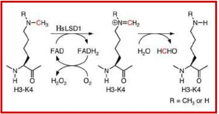
Human Lys-specific histone demethylase 1 (HsLSD1 alias KIAA0601 and BHC110) is the first discovered histone demethylase. HsLSD1 acts on H3K4me1 and H3K4me2, respectively, through a FAD-dependent mechanism (Shi et al., 2004). The reaction involves two steps. First, the histone substrate is bound and its methylated K4 side chain is oxidized by the FAD with resultant reduction of oxygen. Then, the resulting imine intermediate is hydrolyzed to generate the demethylated H3 tail and formaldehyde (Forneris et al., 2006) (Fig. 6; Supplementary Fig. 1).
HsLSD1 was originally identified as a component of transcriptional repressor complexes. Indeed, HsLSD1 is typically associated with the transcriptional corepressor protein CoREST and with HDAC1 or HDAC2, to form a stable core subcomplex recruited by several chromatin-remodeling multiprotein complexes (Ballas et al., 2001, Shi et al., 2003). For instance, HsLSD1-CoREST–HDAC core was found in complex with the repressor element 1-silencing transcription factor (REST) to mediate long-term repression of neuronal genes in non-neuronal cells (Ballas et al., 2005; Ooi and Wood, 2007).
HsLSD1 is also involved in gene activation processes, thus highlighting its multifaceted function in chromatin regulation. In particular, activation of androgen receptor target genes requires HsLSD1-dependent histone H3K9 demethylation (Metzger et al., 2005).
The dual role of HsLSD1 in gene repression/activation is also demonstrated by its role in the fine regulation of growth hormone (Gh) production during pituitary development (Wang et al., 2007).
Fig. 6. Mechanism of HsLSD1-catalyzed demethylation of H3K4. The carbon atom that is
Activation of the Gh gene is regulated by the transcriptional activator pituitary transcription factor 1 (Pit1) during the early phases of development through recruitment of the HsLSD1-containing MLL1 (mixed lineage leukemia 1) coactivator complex. Pit1 and its associated complex is later replaced by the ZEB1 (zinc finger E-box binding homeobox 1) transcriptional repressor which recruits a corepressor complex containing CtBP (C-terminal Binding Protein), CoREST and HsLSD1, in this way switching off Gh gene expression. Thus, HsLSD1 is the key component of two opposing coactivator and corepressor complexes that fine-tune the temporal expression of a single target.
An important feature in the HsLSD1-mediated demethylation process is that, though HsLSD1 alone can demethylate H3K4 in peptides or bulk histones, the binding of HsLSD1 to the C-terminal SANT domain of CoREST renders HsLSD1 able to catalyze H3K4 demethylation on intact nucleosomal particles and also less prone to proteasomal degradation (Shi et al., 2005). Another important characteristic of HsLSD1 is the mechanism for substrate recognition. It has been demonstrated that HsLSD1 requires the first 20 N-terminal amino acids of the histone tail for productive binding (Forneris et al., 2005a). Such a specific recognition mechanism enables HsLSD1 to sense the epigenetic message encoded by the histone tail, as evidenced by the finding that the presence on H3 of other epigenetic markers affects HsLSD1 catalytic activity, decreasing or even completely hampering it (Shi et al., 2004; Shi et al., 2005; Forneris et al., 2005a; Forneris et al., 2006).
The crystal structures of HsLSD1 in free form and in complex with CoREST was recently solved (Stavropoulos et al., 2006; Chen et al., 2006; Yang et al., 2006). HsLSD1 is an asymmetric molecule consisting of three distinct structural domains. Two of them, the N-terminal SWIRM domain (named for its presence in the proteins Swi3, Rsc8, and Moira) and the C-terminal FAD-binding amine oxidase domain, closely pack against each other, forming a globular core structure from which the third domain, named Tower domain, protrudes as an elongated helix-turn-helix motif (Fig. 7).
Insight into substrate binding was obtained from recent crystallographic analyses of HsLSD1-CoREST in complex with an histone H3 peptide modified by the addition of a reactive chemical group (a propargyl unit) on the K4 side chain (Culhane et al., 2006; Yang et al., 2007). In this way, the peptide functions as an inhibitor through formation of a covalent adduct between its modified K4 and the flavin. The crystal structure of this complex revealed that the peptide binds to the amine oxidase domain, adopting a folded conformation that enables the substrate-binding site to accommodate the relatively long stretch of the N-terminal H3 tail. This binding mode positions the reactive H3K4 side chain in proximity to FAD. In general, the architecture of the substrate-binding site is characterized by the presence of various niches that accommodate the side chains of the histone peptide through formation of specific interactions (Yang et al., 2007). The addition of more epigenetic
Fig. 7. Structural biology of HsLSD1 in complex with CoREST and a peptide substrate.
HsLSD1 (blue) consists of three domains: the amine oxidase domain, the SWIRM domain and the helical tower domain. HsLSD1 tightly associates with the CoREST C-terminal SANT domain (red). The histone H3 N-terminal peptide (green) binds deeply in the HsLSD1 amine oxidase domain in proximity to the flavin cofactor (yellow). Figure modified from Forneris et al., 2008. markers on the H3 N-terminal tail introduces steric and electrostatic perturbations which alter this network of interactions predictably, thus explaining the negative effect that nearly all epigenetic modifications have on HsLSD1–H3 binding (Forneris et al., 2007; Forneris et al., 2008).
Recently, it has been reported that HsLSD1 is also active toward a non-histone protein such as the tumor suppressor p53 methylated at Lys 370 residue. Thus blocking its pro-apoptotic activity. This finding leaves open the question of which are the in vivo substrates of the enzyme (Huang et al., 2007).
In A. thaliana, four cDNAs: At1g62830, At3g13682, At3g10390 (FLOWERING LOCUS D o FLD), At4g16310 were identified encoding for proteins displaying 26-30% sequence homology with HsLSD1 and bearing, similarly to HsLSD1, both a flavin amine oxidase domain and a SWIRM domain (Shi et al., 2004). Recently, FLD has been shown to interact with a plant-specific C2H2 zinc finger-SET domain protein and to repress FLC expression (He et al., 2003; Krichevsky et al., 2007).
Peptidyl-arginine deiminases
Arg methylation is prone to enzymatic conversion through a deimination reaction (Cuthbert et al., 2004; Kubicek and Jenuwein, 2004). The responsible enzymes, termed peptidyl-Arg deiminases or demethylases (PADs), are not very prominent in mammals and comprise only five members (PAD1–PAD4 and PAD6). The described reaction of monomethyl-Arg deimination is not a true reversion of the methyl mark since it generates an altered amino acid
(citrulline) and methyl-ammonium (Supplementary Fig. 1). PADs can deiminate unmodified Arg and monomethylated Arg residues of H3 and H4 histone tails (Cuthbert et al., 2004; Wang et al., 2004b). PAD4, the most characterized PAD, was reported to suppress the transcription of the estrogen-regulated genes by citrullination of methylated H3R17 and H4R3 (Cuthbert et al., 2004).
Jumonji C domain-containing histone demethylases
Recently, a new family of histone demethylases has been identified. Although the various members of this family contain numerous domains, such as PHD (Plant Homeodomain) and Tudor domains, they each feature a jumonjiC (JmjC, Japanese for ‘cruciform’) domain responsible for their demethylase activity, and for this reason they have been designated as jumonji C histone demethylases (JHDMs) (Tsukada et al., 2005).
JHDMs operate on methylated Lys and Arg residues via Fe(II)- and 2-oxoglutarate–dependent dioxygenation and proceed through a radical mechanism involving an iron-oxo intermediate (Supplementary Fig. 1). Overall, this reaction results in the demethylation of the Lys or methyl-Arg moiety to produce succinate and formaldehyde as the resulting byproducts (Anand and Marmorstein, 2007) (Supplementary Fig. 1). One important aspect of the JHDMs catalytic mechanism, in contrast to the mechanism employed by HsLSD1, is that it does not require a protonated nitrogen for activity and hence permits demethylation of trimethylated Lys residues.
There are 27 different JHDMs with varying substrate specificity within the human genome, whereas in A. thaliana 21 putative JHDMs were identified (Agger et al., 2008; Lu et al., 2008). Human JHDM1A, which is the first described JmjC domain-containing demethylase, specifically demethylates mono- and dimethylated H3K36 (H3K36me1 and H3K36me2; Tsukada et al., 2005). Interestingly, JHDM1B, a close homologue of JHDM1A involved in transcriptional repression of ribosomal RNA genes and c-jun, was recently reported to bear demethylase activity toward the H3K4me3 (Frescas et al., 2007; Koyama-Nasu et al., 2007).
In recent studies it has been shown that the various members (JMJD2A, JMJD2B, JMJD2C and JMJD2D) of JMJD2 family, a subfamily of JmjC domain containing proteins, prevalently catalyze the demethylation of H3K9me3 or H3K9me2 and H3K36me3 or H3K36me2 and are associated to prostate cancer (Cloos et al., 2006; Klose et al., 2006; Wissmann et al., 2007). In addition to the various members of the JMJD2 family, also JMJD1A can demethylate methylated H3K9. However, JMJD1A is specific for H3K9me1 and H3K9me2, in particular at androgen receptor target genes (Yamane et al., 2006). The jmjc-containing members of JARID1 family (JARID1A, JARID1B, JARID1C, JARID1D) specifically demethylate H3K4me3. Despite, the