Abstract. – OBJECTIVE: This review is aimed at demonstrating the progesterone-like activity exerted by the active form of vitamin D, or cal-citriol (1,25(OH)2D). To achieve this outcome, we compared the effects in vivo and in vitro exert-ed by progesterone and vitamin D, with a spe-cial focus on the female reproductive system and pregnancy.
MATERIALS AND METHODS: This is a litera-ture review of the most important articles pub-lished in English on vitamin D as a hormone, mainly found by MEDLINE. Furthermore, a sec-tion of our review contains some unpublished data, concerning the analysis in silico of the sim-ilarities between the steric structure of proges-terone and calcitriol, based on the availability of the experimental structures of progesterone and vitamin D3 receptors in complex with their phys-iological ligands in the RCSB Protein Data Bank. RESULTS: Vitamin D was shown to exert ma-ny physiological activities during the very ear-ly stages of gestation in perfect synchrony with progesterone. Both the molecules mutually help and reinforce the activity exerted by each one. A little bit later than progesterone is released, vita-min D secretion rises, but only if pregnancy oc-curs. Calcitriol contributes to prepare the endo-metrium to be receptive. Moreover, it supports the implantation process and the course of preg-nancy through different but similar pathways to those used by progesterone, giving rise to a sig-nificant synergy of action. It is increasingly ev-ident that vitamin D gives an essential support from the luteal phase onwards.
CONCLUSIONS: Based on the evidence dis-played in this review we may define appropriate-ly vitamin D as a steroid hormone with proges-terone-like activity.
Key Words:
Calcitriol, CYP24A1, CYP27A1, CYP27B1, Embryo implantation, Inflammation, Osteopontin, Pregnancy, Progesterone, Progesterone receptor, Vitamin D, Vita-min D receptor.
Abbreviations
1,25(OH)2D = 1α,25-dihydroxyvitamin D; 1α-OHase: 1α-hydroxylase; CL = corpus luteum; CRF = corticotro-pin-releasing factor; CSF = colony-stimulating factor; CYP24A1 = Cytochrome P450 family 24 subfamily A member 1; CYP27A1 = Cytochrome P450 Family 27 Subfamily A Member 1; CYP27B1 = Cytochrome P450 Family 27 Subfamily B Member 1; DBP = vitamin D Binding Protein; FGF = Fibroblast growth factor; IL = interleukin; TNF-α = tumour necrosis factor alpha; Treg cells = T regulatory cells; VDR = vitamin D receptor; VEGF = Vascular endothelial growth factor.
Introduction
A Story Coming from the Thirties
Between the end of sixties and the beginning of seventies some advanced researches and stu-dies brought out to understand much better the activity of calcitriol or 1,25(OH)2D, two names referred to the active form of vitamin D (in our paper “vitamin D” will be used with the same meaning of them). They offered a wider picture of this very intriguing molecule, until then consi-dered only an essential vitamin for bone growth and for fighting rickets. In that period vitamin D was defined, for the first time, as a new steroid hormone and its chemical structure was characte-rized1,2. In 1971 Norman et al3 published a
semi-nal paper where they stated that “It is possible that 1,25- dihydroxycholecalciferol should be reclassi-fied as a steroid hormone. Certainly, its secretion by the kidney followed by its selective accumula-tion in the target intestinal mucosa where it exerts its characteristic physiological effect on calcium metabolism satisfies the minimal definitions of a hormone”. Furthermore, in the same years, its nuclear receptor was identified4,5. Vitamin D
G. MONASTRA
1, S. DE GRAZIA
2, L. DE LUCA
3, S. VITTORIO
3, V. UNFER
41Department of Experimental Medicine, Sapienza University of Rome, Rome, Italy 2Department of Research and Development, Lo.Li. Pharma, Rome, Italy
3Department of Chemical, Biological, Pharmaceutical and Environmental Sciences, Polo Universitario SS. Annunziata, University of Messina, Messina, Italy
4Faculty of Medicine and Psychology, Department of Developmental and Social Psychology, Sapienza University of Rome, Rome, Italy
Vitamin D: a steroid hormone with
progesterone-like activity
started to be considered a hormone, even thou-gh many years had to pass before this concept became established in the scientific community, as stated in 1997 by the Nutritional Committee Report for both North America and Europe6.
De-spite this and other authoritative opinions and de-clarations7, some scholars still do not accept this
classification8, but they are a minority. It is worth
of note that a couple of decades before the six-ties, some studies (1934) demonstrated the hor-monal properties exerted by a group of molecules having the cyclopentanoperhydrophenanthrene ring, with seventeen-carbon tetracyclic hydro-carbon. Ovariectomized rats (Wistar strain) were injected with 100 mg of each compound dissolved in sesame oil and the treatment was able to induce estrus in these animals, showing that such effect is due to the presence of the classic steroid skele-ton9. A marked activity was shown by ergosterol
and its irradiation product calciferol (crystalline vitamin D), and still greater activity by neo-ergo-sterol9. Later, Feyel-Cabanes (1949) and Chambon
(1951) highlighted that ergosterol and 7-dehydro-cholesterol, administered at non-estrogenic nor progestogenic doses, act synergically with proge-sterone in sustaining the pregnancy10 or obtaining
implantation of ovariectomized rabbits11. These
interesting results were then forgotten. The times were not yet ripe to go ahead in that direction of research.
Vitamin D and Steroid Hormones
In fairly recent years, the studies on calci-triol had a revival, even under the impulse of the discovery of areas, in developed countries, with deficiency and widespread rickets. Another cause for this renewed interest in the field came from the finding that many vitamin D receptors (VDR) in tissues and cells are not connected with its effects on bone, being linked to other, before unknown, benefits for health. Nowa-days, vitamin D and its metabolites, from the structural point of view, are called secosteroids, i.e., compounds whose structure shows the abo-ve-cited steroid skeleton with seventeen-carbon tetracyclic hydrocarbon, that is not joined at the level of the bond located in position 9-10 of the ring B. Therefore, chemically they are like the mineralocorticoids, glucocorticoids, testoste-rone, progestetestoste-rone, estrogens, chorionic gona-dotropin. By now, both calcidiol and calcitriol are considered respectively pre-hormone and hormone. Indeed, they are synthetized endoge-nously in the skin by means of UVB stimulated
photoconversion of 7-dehydrocholesterol and subsequent thermosensitive but noncatalytic iso-merization to cholecalciferol. Then, cholecalci-ferol is hydroxylated by liver mitochondrial and microsomal 25-OHase CYP27A1. The resultant 25-hydroxycholecalciferol (calcidiol) is 1α-hyd-roxylated in the kidney by CYP27B1 (a mito-chondrial 1α-OHase). This reaction yields the secosteroid 1α,25(OH)2D (calcitriol). Therefore, CYP27A1 and CYP27B1 are key enzymes invol-ved in the synthesis of vitamin D, whereas the inactivating enzyme CYP24A1 determines the rate-limiting step for calcitriol catabolism12. In
physiologic conditions at least the 80% of this vitamin is produced by the skin, whereas the re-maining 20% or less is taken up from dietary sources. In areas rich in sunlight, this endoge-nous production provides enough quantity of vitamin D, at least in healthy young subjects. However, among people with darker skin, a se-vere and widespread deficiency may be found as seen in the US especially among blacks and Hispanics13. Moreover, subjects who are obese,
have hypertension, low high-density lipoprotein cholesterol level, do not drink milk daily, are all significantly, independently associated with vi-tamin D deficiency13.
Besides the chemical structure, there are other very convincing connections between vitamin D and the class of (steroid) hormones. These last ones are bound to transport proteins in plasma, howe-ver only the free molecule can spread into its target cells. These hormones are chemical messengers in a wide number of species and tissues to transmit signals. Obviously, the ability of steroid hormones to give rise to biological message depends on the presence of their receptors on their specific targets. Unlike the growth factors and peptide hormones which bind only to receptors on cell membrane, the fat-soluble steroid hormones can pass through the lipid bilayer surrounding the cell and interact also with their related receptors on the cell nucleus, inducing a genomic response. The receptor-hor-mone complex binds to DNA and triggers or re-presses the transcription of one or more genes14.
Furthermore, steroid hormones bind to a variety of receptor types located near or associated with the plasma membrane15-17. In this way, they activate
sy-stems of second messengers, with rapid cellular re-sponses that may range from seconds (e.g., opening of ion channels) to 10-60 min (e.g., activation of phosphatidylinositol-3’-kinase or endothelial nitric oxide synthase). Instead, the genomic responses commonly need from some hours to days before
becoming entirely manifest; they may be stopped by transcription or translation inhibitors16.
There-fore, many steroid hormone receptors are in the cytoplasm and/or nucleus.
The parallelism between the above picture and the behaviour of calcitriol, its receptor and the re-sulting downstream activity, is striking. In fact, as for steroid hormones, vitamin D and its meta-bolites circulate in plasma linked to a transport protein produced by the liver: vitamin D Binding Protein (DBP). Additionally, as for the classic ste-roid hormones, VDR is a transcription factor lo-cated both in the nucleus of cells and on the cell membrane. The complex between vitamin D and its nuclear receptor, once activated, binds to DNA and, in this way, modifies the expression of nu-merous genes (genomic response). On the other side, the interaction with the VDR located on the cell membrane induces the formation of second cellular messengers (such as cAMP, diacylglyce-rol, inositol triphosphate, arachidonic acid) or the phosphorylation of some cellular proteins. The-refore, calcitriol, as progesterone, estrogens, etc., exerts genomic and nongenomic actions. On the whole, this picture further confirms the hormonal nature of vitamin D16.
Vitamin D deficiency causes osteopenia/oste-oporosis, immune system disorders, metabolic syndrome (obesity and diabetes), cancer, kidney disease, neurological and cardiovascular diseases, pregnancy complications, and its administration proved to be useful in these pathologies. As well known, only cells having the specific receptor for a given hormone, respond to its stimuli; therefo-re, it is important to highlight that the distribution of VDR is almost ubiquitous, involving various organs and tissues. VDR was found in hema-topoietic tissues, immune system, monocyte/ macrophages, lymphocytes, skin, muscle, smo-oth muscle cells, myoblasts, heart cardiac mu-scle cells and atrial myocytes, pancreas β cells, mammary gland, adrenal gland medullary cells, prostate, brain hippocampus/selected neurons, cartilage chondrocytes, liver parenchymal cells, lung, Sertoli/seminiferus tubule, pituitary gland, thyroid, parathyroid, ovarian, myometrial and en-dometrial cells, placenta18-22.
Progesterone and Vitamin D Structural Similarities
It was discovered that all the nuclear receptors of steroid hormones show common structure/fun-ction domains and are named as “nuclear receptors superfamily” including four classes with a shared
evolutionary process. Progesterone and vitamin D receptors belong to two different classes of the abo-ve-mentioned superfamily23. This relationship has
to be keep in mind, being in perfect agreement with the core of our review. In this context, we aimed at determining which are the similarities between the steric structure of progesterone and vitamin D, in consideration of their common phylogenesis, by using in silico methods.
The crystal structures of progesterone in complex with the ligand-binding domain (LBD) of the human progesterone receptor (PDB code 1A28)24 and of
vi-tamin D in complex with the LBD of the vivi-tamin D nuclear receptor (VDR) (PDB code 1DB1)25 have
been reported in the RCSB Protein Data Bank. By structure-based pharmacophore model tool of Li-gandScout software26, we highlighted the binding
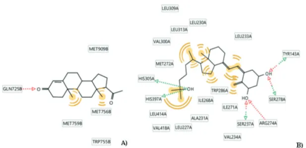
requirements for these two ligands that are schema-tically represented in Figure 1.
Progesterone showed the ability to bind its re-ceptor establishing i) hydrogen bond interaction with Gln725 and ii) hydrophobic contacts through the methyl groups with Met909, Met759, Met756 and Trp755 (Figure 1A).
On the other hand, vitamin D interacts with its receptor establishing three different hydrogen bonds interactions: i) with Ser237 and Arg274 through the hydroxyl group in position 1, ii) with Tyr143 and Ser278 through the hydroxyl group in position 3, and iii) with His305 e His397 through the hydroxyl group in position 25. Furthermore, there are some hydrophobic contacts with the dif-ferent hydrophobic residues present in the binding site (Figure 1B).
To obtain more insight on the structural simila-rity between progesterone and vitamin D, the two ligands have been aligned in their binding confor-mation creating a shared feature pharmacophore model by LigandScout software (Figure 2).
The alignment shows an interesting overlap-ping of vitamin D (stick grey) and progesterone (stick green) highlighting three convergent points that have been translated in three pharmacophore features describing chemical functionalities: – one hydrophobic sphere (yellow), mapped by
the methylene group in position 19 of vitamin D and the methyl group in position 19 of proge-sterone;
– another hydrophobic sphere overlapped by the methyl group in position 18 of vitamin D and the methyl group in position 18 of progesterone – one hydrogen bond acceptor (red sphere) occu-pied by hydroxyl group in position 3 of vitamin D and the carbonyl group of progesterone.
These information reveal that vitamin D con-tains some chemical functionalities useful for the binding to progesterone receptor.
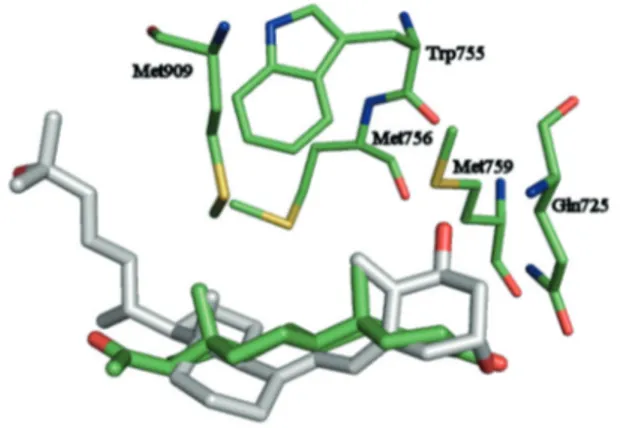
In Figure 3 is represented a possible interaction of vitamin D with progesterone receptor obtained by superimposition of the two proteins 1A28 and 1DB1. The results (Figure 3) showed that vitamin D could hypothetically interact with the proges-terone receptor by establishing a H bond with Gln725 and hydrophobic interactions with the same hydrophobic amino acid residues involved in the interaction with progesterone. Clearly, this is only a hypothesis that has to be carefully tested in depth. However, this interaction, even if it is real, does not necessarily imply the induction of a physiological effect.
Progesterone Release in the Luteal Phase for Preparing the Scenario
The female reproductive system plays very com-plex roles, with the participation of a huge number of actors whose activity is finely regulated. In this pro-cess, progesterone and calcitriol play pivotal func-tions, and without their appropriate levels, reached in well-determined periods, gestation cannot occur. The menstrual cycle determines an essential tim-ing for reproduction and it is divided into two main phases, the follicular and luteal phases, that must work in series, in an integrated manner, to allow pregnancy. During the follicular phase, also called proliferative phase, the follicles inside the ovaries
start their development to prepare ovulation. This process occurs from the 1st to the 14th day of the
menstrual cycle. The next luteal phase begins after ovulation and lasts normally from the 15th to the 28th
day. It needs, on one hand, satisfactory secretions of progesterone by the corpus luteum (for this reason it is also termed secretory phase), and, on the other hand, a prompt endometrial response to its stimu-lus. In the luteal phase, the corpus luteum (CL), a transient endocrine gland which develops in this specific period, undergoes a prompt neovasculariza-tion. Such process is principally under the control of Vascular endothelial growth factor (VEGF) and Fibroblast growth factor (FGF), both upregulated in the luteinized granulosa cells and then
absolute-Figure 1. Interaction patterns of progesterone-LBD of progesterone receptor (A) and vitamin D-LBD of VDR (B). The labels refer to the amino acid residues involved in the interaction with the receptors: green and red arrows represent H-bond inte-ractions, yellow areas highlight the groups involved in the hydrophobic contacts.
Figure 2. Shared-feature pharmacophore model. Progeste-rone and vitamin D are represented in green and grey stick respectively. Yellow spheres represent hydrophobic contacts while red sphere represents hydrogen bond acceptor.
ly necessary for the embryo implantation27,28. CL
produces several different hormones; among them, progesterone is the most important since its activity is necessary and sufficient to hinder the proliferation of endometrium and promote its receptive state for blastocyst implantation to maintain early pregnancy. Progesterone is produced by the luteal cells in a pul-satile way with levels that may vary up to sevenfold in a range of a few hours. Its synthesis depends on the availability of appropriate levels of circulating cholesterol as substrate and on low stimulation by luteinizing hormone (LH), produced by gonado-tropic cells of the anterior pituitary gland. Therefore, progesterone prepares the environment conducive to pregnancy success and it is categorically necessary, during the secretory phase, to induce the endometri-um receptivity to the embryo implantation, even if moderate concentrations are sufficient29. In this
sce-nario, vitamin D plays the role of second main actor and its function comes to light from several exam-ples that we present below. Overall, it modulates the innate and adaptive immune system, controls cell proliferation and functions as a regulator of various and important metabolic processes, with effects on the implant, the production of cytokines and the im-mune response to infections30-33.
Activities in Common Between Vitamin D and Progesterone in the inception of Pregnancy
Vitamin D
The metabolism of 1,25(OH)2D in pregnancy differs drastically from what normally occurs in the non-pregnant state. Increased circulating
lev-els of vitamin D were detected through human pregnancy, independently of calcium homeosta-sis34-36. In addition, the above-cited presence of
VDR on ovaries, uterus, endometrium and pla-centa should be emphasized. It is noteworthy that the number of VDR increases continuously during the first and second trimester of pregnancy, mean-ing that this vitamin plays a key physiological role in pregnancy. Especially in the first months of gestation adequate levels of vitamin D are even more essential, affecting not only the well-being of the mother but also that one of the fetus. VDR was detected in the endometrium throughout all the phases of the menstrual cycle and also later, namely in proliferative phase endometrial stromal cells, in secretory phase endometrial stromal cells and in early pregnant decidual cells37. The levels
of 1α-OHase mRNA were comparable in endo-metrial stromal cells without any influence due to the phase of the cycle. Instead, such concentra-tions were found significantly higher in decidual cells37. As a side note, we observe that vitamin D,
detected in the seminal fluid38, could also
func-tion as a trigger molecule to provide addifunc-tional levels to further support certain processes under-lying a successful onset of pregnancy. It was ob-served that, in response to IL-1β secreted by the blastocyst, the dendritic cells of the endometrium and the macrophages produce 1α-OHase (the en-zyme responsible for the last step of the synthesis process) and, consequently, calcitriol37. On the
other hand, the stimulation of the endometrial 1α-OHase expression due to TNF is not as potent as that one achieved using very low concentra-tions of IL-1 β37. This evidence means that
be-tween the two main pro-inflammatory cytokines, at least in this case, the central role is played by IL-1 β, whereas TNF exerts a secondary function, despite its strong effects in many contexts. Thus, there is not only an increased calcitriol production in the first stages of pregnancy, but also a wide array of new synthetized receptors in the endome-trium, ready for binding.
Using blood cells of women with a history of recurrent pregnancy losses (RPL) Vitamin D was reported to hinder in vitro the proliferation of T helper 1 (Th1) cells and to decrease the produc-tion of their cytokines, such as IFN-γ, IL-2 and TNF-α. On the other hand, vitamin D stimu-lated the secretion of T helper 2 (Th2) cytokines; among them, IL-10 is the most important anti-in-flammatory cytokine released under the effect of calcitriol39. Furthermore, in vitro vitamin D
sig-nificantly decreased in a dose-dependent manner
Figure 3. Superimposition of the crystal structures 1A28 and 1DB1. Amino acid residues present in the progestero-ne receptor, involved in the interaction with the ligand, are represented by green stick. Progesterone is represented by green stick while vitamin D is represented by grey stick.
the cytotoxicity of natural killer (NK) cells39 and
it was able to suppress in the same cells the secre-tion of proinflammatory cytokines, such as IFN-γ and TNF-α, and to increase IL-10 release39, as
well as CSF2, IL-1, IL-6 too, thus modulating the inflammation that normally develops in the initial phase of pregnancy40.
Accordingly to that, vitamin D significantly lowers the frequency of T helper 17 (Th17) cells in addition to reducing the Th17/Treg ratio in peripheral blood of patients with recurrent mis-carriage compared with the control group41. The
same modulatory effect of inflammation was seen in the induction and upregulation of the sup-pressive activity of T regulatory (Treg) cells in skin-draining lymph nodes42.
Vitamin D by binding to VDR on the endo-metrium, upregulates the target genes, such as calbindin and osteopontin, also called secreted phosphoprotein one (SPP1)37. Development of
re-ceptivity requires changes in endometrial gene expression; endometrial expression of calbindin mRNA was shown to be highly regulated during implantation. Indeed, calbindin participates to the regulation of endometrial receptivity acting as calcium binding protein: it is well known that the function of reproductive organs and its mus-culature is depending on the influx of extracellu-lar calcium or the release of intracelluextracellu-lar stores43.
Osteopontin is an adhesion protein involved in implantation and decidualization44. Its action has
a direct influence as signal transduction at the uterine-placental interface throughout pregnancy, including the regulation of immune cells behavior and cytokine production. Both calbindin and oste-opontin are essential for the embryo implantation and for the development of the placenta. Their effects on the endometrium favor the success of pregnancy37,45-47. Therefore, 1,25(OH)2D is
reput-ed to exert an autocrine/paracrine action aimreput-ed at regulating both acquired and innate immune responses at the fetal-maternal interface, as con-firmed by the presence of the VDR in the endo-metrium. This hypothesis, in general terms, was born about twenty years ago and is based on the widespread co-expression of VDR and 1α-OHase in many tissues where vitamin D plays various physiological functions48.
Notably, vitamin D in human cumulus granu-losa cells significantly increases the expression of 3β-HSD, necessary for conversion from pregnen-olone to progesterone49 and in this way it
signifi-cantly increases the production of such hormone by granulosa cells. Moreover, vitamin D directly
stimulates the production of progesterone in cul-tures of human ovarian cells50. It also provokes the
synthesis or blocks the release of key molecules that have, respectively, beneficial (such as activin A, CRF, VEGF)51-53 or harmful effects (such as
estrogens, besides the already cited TNFα and other inflammatory cytokines)53-56 in the period
between the luteal phase and the 12th week of
pregnancy (decidualization and so on).
Vitamin D production and VDR expression pro-mote an increase in the level of two target genes in both cycling and early pregnant endometrium, CYP24 and the already cited osteopontin. CYP24, the rate-limiting step for calcitriol catabolism, is one of the most potent 1,25(OH)2D-responding genes and its protein product is responsible for the hydroxylation reaction that deactivates the vi-tamin D, by a negative feedback mechanism57,58.
Progesterone
Progesterone receptor, as VDR, is expressed in the glandular epithelium and stroma of the hu-man endometrium during the menstrual cycle59.
In experiments with human and mouse T cells progesterone was seen to induce the expression of the VDR gene, which is one of the major genes responding to progesterone. It was able to elicit the VDR mRNA expression in human Treg, Th1, and Th17 cells with various cytokine conditions, and in spleen CD4+ T cells and uterus from pregnant or progestin-injected mice60. The specificity of this
effect is demonstrated by its suppression due to the receptor antagonist of progesterone, RU48660.
Therefore, progesterone promotes VDR gene ex-pression in T cells in heterogeneous conditions and different species. The expression of VDR due to progesterone rises the sensitivity of T cells to cal-citriol and facilitates a more efficient regulation of T cells by calcitriol with an increase of Tregs and suppression of effector T cells which exert a poten-tial pro-inflammatory activity60.
Progesterone-in-duced VDR in T cells allows to prevent adverse immune responses, causing complications during gestation60. Of note, if VDR gene is knocked down
with siRNA, the inhibition of T cell differentiation into effector T cells caused by progesterone and calcitriol significantly decreases60.
Furthermore, progesterone promoted Th2 cells formation and the release of their anti-inflamma-tory cytokines61 and regulated the differentiation
of human naive cord blood fetal T cells into Treg cells62. It also suppressed Th1 and
Th17-relat-ed genes and enhancTh17-relat-ed Th2-relatTh17-relat-ed genes in the lymphocytes obtained from pregnant cows63. It is
remarkable that progesterone inhibited IFN-γ and IL-17 release in dose-dependent manner in both pregnant and non-pregnant cows63. In addition,
progesterone neutralized the cytotoxicity of NK cells by hindering their degranulation and perfo-rin release64. Moreover, in mononuclear cells
iso-lated from human placental blood progesterone was able to antagonize the effects of lipopolysac-charide (LPS) significantly reducing TNF-α, IL-1β, IL-6, IL-8, and MIP-1α release. Instead, the levels of IL-10 grew in a remarkable way when LPS was added in vitro with progesterone65. As
calcitriol, progesterone stimulates the synthesis or suppresses the release of pivotal molecules exert-ing, respectively, helpful (such as activin A, CRF, VEGF) or detrimental effects (such as estrogens or TNF)64,66-69 in the period between the luteal
phase and the 12th week of pregnancy. Finally,
progesterone induced the release of osteopontin in human decidua70 and calbindin in the uterus of
Egyptian buffalo46. These effects are briefly
de-picted in Figure 4.
Vitamin D effect to promote Treg activity in presence of progesterone appears strongly redu-ced when VDR gene is knocked down60. Of note,
progesterone induced a greater sensitivity in T cell toward calcitriol to up-regulate the Treg-associated molecules (FoxP3, CD8, LAP-TGF-β1).
The efficacy of Treg cells in immune suppres-sion is higher when they are produced under the combined effect of calcitriol and progesterone than when there is only one of these effector mo-lecules60. All these data demonstrate that
proge-sterone potentiates the anti-inflammatory effects of vitamin D, and vice versa.
Studies on Vitamin D in Pregnancy and Assisted reproductive technology (ART)
The relevance of vitamin D activity for pre-gnancy, from the luteal phases onwards, is con-firmed by the findings of several studies. Insuffi-cient levels of vitamin D have been related to the pathogenesis and the abnormal development of luteal phase. Vitamin D deficiency is prevalent in pregnant women with a major risk of miscarria-ge and recurrent pregnancy losses. Furthermore, the treatment with vitamin D or its precursor was found effective in women looking for gestation and in expectant mothers71. Several investigations
confirm this picture. A study analyzed 1072
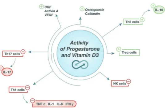
wo-Figure 4. Progesterone and vitamin D share several biological activities, as an inducer of anti-inflammatory pathways (Th2 and Treg cells). In addition, they stimulate the release of corticotropin-releasing factor (CRF), activin A, vascular endothe-lial growth factor (VEGF), osteopontin, calbindin. Moreover, both progesterone and vitamin D inhibit the proinflammatory pathways (NK, Th1 and Th17 cells). All these effects contribute in supporting pregnancy since its inception. It is noteworthy that their action is synergic.
men attending an academic infertility centre: a high rate of hypovitaminosis D among childbea-ring women was demonstrated to be related with infertility72. In another work, the estimation of
25-hydroxy cholecalciferol (25-OHD) in patients was made before the treatment protocol for ICSI. The mean value of 25-OHD, number of oocytes, fertilized oocytes and endometrial thickness were significantly higher in pregnant women. A significant positive association of 25-OHD with clinical pregnancy and thickness of endometrium was observed. Deficiency of 25-OHD in females hinders the accomplishment of optimal endome-trial thickness required for implantation of the embryo after ICSI. The improvement in vitamin D status positively affected the success results in ART73. Also, Ozkan et al74 found that women with
higher vitamin D level in their serum are signifi-cantly more likely to achieve clinical pregnancy following IVF-embryo transfer. The same group highlighted that another favourable parameter for ART outcome is the higher content of vitamin D in follicular fluids. As stated by Hollis and Wa-gner75, there is growing evidence of the important
role exerted by vitamin D supplementation during pregnancy to achieve satisfying circulating levels, associated to a lower risk of comorbidities of pre-gnancy and better outcomes. Concerning the sa-fety in pregnant women, vitamin D supplementa-tion was demonstrated to be completely safe at the dose of 4,000 IU/day for 24-28 weeks76,77. Finally,
a recent systematic review78 showed clearly an
association between vitamin D levels in women undergoing ART and their reproductive outco-mes. Vitamin D deficiency and insufficiency re-present a crucial key point to consider in women following ART procedures.
Conclusions
Vitamin D and progesterone modulate nume-rous components of the immune system in the whole organism and at fetoplacental level too. Their action involves both the immune cells (macrophagic, dendritic, uterine natural killer, and lymphocytic cells) and non-immune cel-ls (trophoblast celcel-ls), shifting the equilibrium toward a Th2 profile. Vitamin D shows several physiological activities during gestation in per-fect synchrony with progesterone or also acting, in some cases, as a substitute. The molecules mutually help and reinforce the activity exerted by each one. Overall, the data so far known
sug-gest that vitamin D, whose metabolism is stri-ctly regulated, contributes to prepare the endo-metrium to be receptive. Moreover, it supports the implantation process and the course of pre-gnancy through different, but similar pathways to those used by progesterone, giving rise to a significant synergy of action79. A little bit later
than progesterone is released, vitamin D enters the scene, and at endometrium level, its con-centration strongly increases only if pregnancy occurs (i.e., after fertilization). It is increasingly evident that calcitriol gives an essential support from the luteal phase onwards, as glimpsed for the first time by the pioneering studies carried out by Feyel-Cabanes10 and Chambon11 between
the thirties and the fifties of the past century. In conclusion, based on the evidence displayed in this review we may define appropriately vitamin D as a steroid hormone with progesterone-like activity.
Conflict of Interest
Vittorio Unfer and Sara De Grazia are employees at Lo.Li. Pharma, Rome, Italy. The other authors declare that they have no conflict of interests.
References
1) Haussler Mr MJ, NorMaN aW. The association of a metabolite of vitamin D3 with intestinal mucosa chro-matin in vivo. J Biol Chem 1968; 243: 4055-4064. 2) Myrtle JF HM, NorMaN aW. Evidence for the
biolo-gically active form of cholecalciferol in the intesti-ne. J Biol Chem 1970; 245: 1190-1196.
3) NorMaN aW MJ, Midgett rJ, NoWicki Hg, WilliaMs V, PoPJak g. 1,25-dihydroxycholecalciferol: identi-fication of the proposed active form of vitamin D3 in the intestine. Science 1971; 173: 51-54.
4) Haussler Mr, NorMaN aW. Chromosomal recep-tor for a vitamin D metabolite. Proc Natl Acad Sci USA 1969; 62: 155-162.
5) tsai Hc, NorMaN aW. Studies on the mode of action of calciferol. VI. Effect of 1,25-dihydroxy-vi-tamin D3 on RNA synthesis in the intestinal mu-cosa. Biochem Biophys Res Commun 1973; 54 622-627.
6) iNstitute oF MediciNe (us) staNdiNg coMMittee. Die-tary reference intakes: calcium, phosphorus, ma-gnesium, Vitamin D, and fluoride. 1997, Washin-gton (DC): National Academy Press.
7) NorMaN aW. From vitamin D to hormone D: fun-damentals of the vitamin D endocrine system es-sential for good health. Am J Clin Nutr 2008; 88: 491S-499S.
8) VietH r. Why “Vitamin D” is not a hormone, and not a synonym for 1,25-dihydroxy-vitamin D, its analogs or deltanoids. J Steroid Biochem Mol Biol 2004; 89-90: 571-573.
9) cook J. W. dec, HeWett c. l., laWsoN W. The oe-strogenic activity of some condensed-ring com-pounds in relation to their other biological activi-ties. Proc Roy Soc London Series B 1934; 114: 272-286.
10) Feyel-cabaNes t. Maintien de la grossesse chez le lapine gestante castrée par une association hor-mone-stérol. C R Seances Soc Biol Fil 1949; 143: 1454-1458.
11) cHaMboN y. [Synergic action of vitamin D and of progesterone in obtaining ovum implantation in the castrated pregnant rabbit]. C R Seances Soc Biol Fil 1951; 145: 955-959.
12) karlic H, Varga F. Impact of vitamin D metabolism on clinical epigenetics. Clin Epigenetics 2011; 2: 55-61.
13) Forrest ky, stuHldreHer Wl. Prevalence and cor-relates of vitamin D deficiency in US adults. Nutr Res 2011; 31: 48-54.
14) o’Malley bW. Mechanisms of action of steroid hormones. N Engl J Med 1971; 284: 370-377. 15) NorMaN aW, MizWicki Mt, NorMaN dP.
Steroid-hor-mone rapid actions, membrane receptors and a conformational ensemble model. Nat Rev Drug Discov 2004; 3: 27-41.
16) NorMaN aW. Minireview: vitamin D receptor: new assignments for an already busy receptor. Endo-crinology 2006; 147: 5542-5548.
17) leViN er. Extranuclear steroid receptors are es-sential for steroid hormone actions. Annu Rev Med 2015; 66: 271-280.
18) ValdiVielso JM. The physiology of vitamin D recep-tor activation. Contrib Nephrol 2009; 163: 206-212. 19) cHristakos s, HeWisoN M, gardNer dg, WagNer cl,
sergeeV iN, rutteN e, Pittas ag, bolaNd r, Ferruc -ci l, bikle dd. Vitamin D: beyond bone. Ann N Y Acad Sci 2013; 1287: 45-58.
20) acker gM, garabediaN M, guillozo H, PecquiNot Ma, balsaN s. In vitro formation of 25-hydroxyvita-min D3 metabolites in endometrium: dependence on the hormonal status of the rat. Endocrinology 1982; 111: 2103-2109.
21) delViN ee, arabiaN a, glorieux FH, MaMer oa. In vitro metabolism of 25-hydroxycholecalciferol by isolated cells from human decidua. J Clin Endo-crinol Metab 1985; 60: 880-885.
22) kacHkacHe M, rebut-boNNetoN c, deMigNoN J, cy -Nober e, garabediaN M. Uterine cells other than stromal decidual cells are required for 1,25-dihy-droxyvitamin D3 production during early human pregnancy. FEBS Lett 1993; 333: 83-88.
23) MaNgelsdorF dJ, tHuMMel c, beato M, HerrlicH P, scHutz g, uMesoNo k, bluMberg b, kastNer P, Mark M, cHaMboN P, eVaNs rM. The nuclear receptor superfamily: the second decade. Cell 1995; 83: 835-839.
24) WilliaMs sP, sigler Pb. Atomic structure of proge-sterone complexed with its receptor. Nature 1998; 393: 392-396.
25) rocHel N, Wurtz JM, MitscHler a, klaHolz b, Moras d. The crystal structure of the nuclear receptor for vitamin D bound to its natural ligand. Mol Cell 2000; 5: 173-179.
26) Wolber g, laNger t. LigandScout: 3-D pharma-cophores derived from protein-bound ligands and their use as virtual screening filters. J Chem Inf Model 2005; 45: 160-169.
27) cHakraborty i, das sk, dey sk. Differential expression of vascular endothelial growth factor and its recep-tor mRNAs in the mouse uterus around the time of implantation. J Endocrinol 1995; 147: 339-352. 28) sak Me, gul t, eVseN Ms, soydiNc He, sak s, ozler
a, alabalik u. Fibroblast growth factor-1 expres-sion in the endometrium of patients with repeated implantation failure after in vitro fertilization. Eur Rev Med Pharmacol Sci 2013; 17: 398-402. 29) youNg sl. Oestrogen and progesterone action on
endometrium: a translational approach to under-standing endometrial receptivity. Reprod Biomed Online 2013; 27: 497-505.
30) NisWeNder gd, JueNgel Jl, silVa PJ, rollysoN Mk, MciNtusH eW. Mechanisms controlling the function and life span of the corpus luteum. Physiol Rev 2000; 80: 1-29.
31) MeseN tb, youNg sl. Progesterone and the luteal phase: a requisite to reproduction. Obstet Gyne-col Clin North Am 2015; 42: 135-151.
32) Herr d, bekes i, WulFF c. Regulation of endothe-lial permeability in the corpus luteum: a review of the literature. Geburtshilfe Frauenheilkd 2013; 73: 1107-1111.
33) daVis Js, rueda br. The corpus luteum: an ovarian structure with maternal instincts and suicidal ten-dencies. Front Biosci 2002; 7: d1949-1978. 34) kuMar r. The metabolism of
1,25-dihydroxyvita-min D3. Endocr Rev 1980; 1: 258-267.
35) HartWell d, riis bJ, cHristiaNseN c. Changes in vi-tamin D metabolism during natural and medical menopause. J Clin Endocrinol Metab 1990; 71: 127-132.
36) salle bl, delViN ee, laPilloNNe a, bisHoP NJ, glo -rieux FH. Perinatal metabolism of vitamin D. Am J Clin Nutr 2000; 71: 1317S-1324S.
37) VigaNo P, lattuada d, MaNgioNi s, erMelliNo l, VigNa -li M, caPorizzo e, PaNiNa-bordigNoN P, besozzi M, di blasio aM. Cycling and early pregnant endome-trium as a site of regulated expression of the vita-min D system. J Mol Endocrinol 2006; 36: 415-424. 38) bloMberg JeNseN M. Vitamin D and male
repro-duction. Nat Rev Endocrinol 2014; 10: 175-186. 39) ota k, daMbaeVa s, kiM MW, HaN ar, Fukui a,
gilMaN-sacHs a, beaMaN k, kWak-kiM J. 1,25-Dihy-droxy-vitamin D3 regulates NK-cell cytotoxicity, cytokine secretion, and degranulation in women with recurrent pregnancy losses. Eur J Immunol 2015; 45: 3188-3199.
40) eVaNs kN, NguyeN l, cHaN J, iNNes ba, bulMer JN, kilby Md, HeWisoN M. Effects of 25-hydroxyvita-min D3 and 1,25-dihydroxyvita25-hydroxyvita-min D3 on cytokine production by human decidual cells. Biol Reprod 2006; 75: 816-822.
41) raFiee M, gHaragozloo M, gHaHiri a, MeHrabiaN F, Maracy Mr, kouHPayeH s, PiePer il, rezaei a. Alte-red Th17/Treg ratio in recurrent miscarriage after treatment with paternal lymphocytes and vitamin D3: a double-blind placebo-controlled study. Iran J Immunol 2015; 12: 252-262.
42) gorMaN s, geldeNHuys s, Judge M, WeedeN ce, Wai -tHMaN J, Hart PH. Dietary vitamin D increases percentages and function of regulatory T cells in the skin-draining lymph nodes and suppresses dermal inflammation. J Immunol Res 2016; 2016: 1426503.
43) luu kc, Nie gy, salaMoNseN la. Endometrial cal-bindins are critical for embryo implantation: evi-dence from in vivo use of morpholino antisense oligonucleotides. Proc Natl Acad Sci U S A 2004; 101: 8028-8033.
44) JoHNsoN ga, burgHardt rc, bazer FW, sPeNcer te. Osteopontin: roles in implantation and placenta-tion. Biol Reprod 2003; 69: 1458-1471.
45) cHoi kc, leuNg Pc, JeuNg eb. Biology and phy-siology of Calbindin-D9k in female reproductive tissues: involvement of steroids and endocrine disruptors. Reprod Biol Endocrinol 2005; 3: 66. 46) eMaM Ma, abouelroos Me, gad Fa. Expression of
calbindin-D9k and vitamin D receptor in the ute-rus of Egyptian buffalo during follicular and luteal phases. Acta Histochem 2016; 118: 471-477. 47) labudzyNskyi d, sHyMaNskyy i, Veliky M. Role of
vi-tamin D3 in regulation of interleukin-6 and oste-opontin expression in liver of diabetic mice. Eur Rev Med Pharmacol Sci 2016; 20: 2916-2919. 48) HeWisoN M, zeHNder d, blaNd r, steWart PM.
1al-pha-Hydroxylase and the action of vitamin D. J Mol Endocrinol 2000; 25: 141-148.
49) MerHi z, dosWell a, krebs k, ciPolla M. Vitamin D alters genes involved in follicular development and steroidogenesis in human cumulus granulosa cel-ls. J Clin Endocrinol Metab 2014; 99: E1137-1145. 50) ParikH g, VaradiNoVa M, suWaNdHi P, araki t, ro
-seNWaks z, Poretsky l, seto-youNg d. Vitamin D re-gulates steroidogenesis and insulin-like growth factor binding protein-1 (IGFBP-1) production in human ovarian cells. Horm Metab Res 2010; 42: 754-757.
51) Woeckel VJ, VaN der eerdeN bc, scHreuders-koedaM M, eiJkeN M, VaN leeuWeN JP. 1alpha,25-dihydroxy-vitamin D3 stimulates activin A production to fi-ne-tune osteoblast-induced mineralization. J Cell Physiol 2013; 228: 2167-2174.
52) Wierzbicka JM, zMiJeWski Ma, PiotroWska a, Ne -doszytko b, laNge M, tuckey rc, sloMiNski at. Bio-active forms of vitamin D selectively stimulate the skin analog of the hypothalamus-pituitary-adrenal axis in human epidermal keratinocytes. Mol Cell Endocrinol 2016; 437: 312-322.
53) soNg J, li y, aN r. Vitamin D restores angiogenic balance and decreases tumor necrosis factor-al-pha in a rat model of pre-eclampsia. J Obstet Gy-naecol Res 2017; 43: 42-49.
54) Halder sk gs, al-HeNdy a. Vitamin D exhibits an-tiestrogenic effects in human uterine leiomyoma cells. Fertil Steril 2010; 94: S219-S220.
55) krisHNaN aV, sWaMi s, PeNg l, WaNg J, MoreNo J, FeldMaN d. Tissue-selective regulation of aromata-se expression by calcitriol: implications for breast cancer therapy. Endocrinology 2010; 151: 32-42. 56) barrera d, diaz l, Noyola-MartiNez N, HalHali a.
Vitamin D and inflammatory cytokines in healthy and preeclamptic pregnancies. Nutrients 2015; 7: 6465-6490.
57) iNouye k, sakaki t. Enzymatic studies on the key enzymes of vitamin D metabolism; 1 alpha-hy-droxylase (CYP27B1) and 24-hyalpha-hy-droxylase (CYP24). Biotechnol Annu Rev 2001; 7: 179-194. 58) Parise ra, egoriN MJ, kaNtereWicz b, taiMi M, Petko
-VicH M, leW aM, cHuaNg ss, NicHols M, el-HeFNaWy t, HersHberger Pa. CYP24, the enzyme that cata-bolizes the antiproliferative agent vitamin D, is in-creased in lung cancer. Int J Cancer 2006; 119: 1819-1828.
59) Mote Pa, balleiNe rl, McgoWaN eM, clarke cl. Co-localization of progesterone receptors A and B by dual immunofluorescent histochemistry in human endometrium during the menstrual cycle. J Clin Endocrinol Metab 1999; 84: 2963-2971.
60) tHaNgaMaNi s, kiM M, soN y, HuaNg x, kiM H, lee JH, cHo J, ulricH b, broxMeyer He, kiM cH. Cutting edge: progesterone directly upregulates vitamin d receptor gene expression for efficient regulation of T cells by calcitriol. J Immunol 2015; 194: 883-886. 61) PicciNNi MP, giudizi Mg, biagiotti r, beloNi l, giaNNa -riNi l, saMPogNaro s, ParroNcHi P, MaNetti r, aNNuNzia -to F, liVi c, etal. Progesterone favors the develop-ment of human T helper cells producing Th2-type cytokines and promotes both IL-4 production and membrane CD30 expression in established Th1 cell clones. J Immunol 1995; 155: 128-133.
62) lee JH, ulricH b, cHo J, Park J, kiM cH. Progestero-ne promotes differentiation of human cord blood fetal T cells into T regulatory cells but suppres-ses their differentiation into Th17 cells. J Immunol 2011; 187: 1778-1787.
63) Maeda y, oHtsuka H, toMioka M, oikaWa M. Effect of progesterone on Th1/Th2/Th17 and regulatory T cell-related genes in peripheral blood mononucle-ar cells during pregnancy in cows. Vet Res Com-mun 2013; 37: 43-49.
64) druckMaNN r, druckMaNN Ma. Progesterone and the immunology of pregnancy. J Steroid Biochem Mol Biol 2005; 97: 389-396.
65) Preciado-MartiNez e, garcia-ruiz g, Flores-esPiNo -sa P, berMeJo-MartiNez l, esPeJel-NuNez a, estra -da-gutierrez g, razo-aguilera g, graNados-cePeda M, Helguera-rePetto ac, irles c, zaga-claVelliNa V. Progesterone suppresses the lipopolysacchari-de-induced pro-inflammatory response in primary
mononuclear cells isolated from human placental blood. Immunol Invest 2018; 47: 181-195.
66) Florio P, rossi M, sigurdardottir M, ciarMela P, luisi s, VigaNo P, grasso d, Fiore g, cobellis l, di blasio aM, Petraglia F. Paracrine regulation of endome-trial function: interaction between progesterone and corticotropin-releasing factor (CRF) and acti-vin A. Steroids 2003; 68: 801-807.
67) MakrigiaNNakis a, Margioris aN, cHatzaki e, zouMa -kis e, cHrousos gP, graVaNis a. The decidualizing effect of progesterone may involve direct tran-scriptional activation of corticotrophin-releasing hormone from human endometrial stromal cells. Mol Hum Reprod 1999; 5: 789-796.
68) kiM M, Park HJ, seol JW, JaNg Jy, cHo ys, kiM kr, cHoi y, lydoN JP, deMayo FJ, sHibuya M, Ferrara N, suNg Hk, Nagy a, alitalo k, koH gy. VEGF-A regu-lated by progesterone governs uterine angioge-nesis and vascular remodelling during pregnancy. EMBO Mol Med 2013; 5: 1415-1430.
69) kuMar P, MagoN N. Hormones in pregnancy. Niger Med J 2012; 53: 179-183.
70) qu x, yaNg M, zHaNg W, liaNg l, yaNg y, zHaNg y, deNg b, gao W, liu J, yaNg q, koNg b, goNg F. Oste-opontin expression in human decidua is associated with decidual natural killer cells recruitment and re-gulated by progesterone. In Vivo 2008; 22: 55-61. 71) kWak-kiM J, skariaH a, Wu l, salazar d, suNg N, ota
k. Humoral and cellular autoimmunity in women with recurrent pregnancy losses and repeated im-plantation failures: a possible role of vitamin D. Autoimmun Rev 2016; 15: 943-947.
72) PagliardiNi l, VigaNo P, Molgora M, Persico P, salo -Nia a, Vailati sH, PaFFoNi a, soMigliaNa e, PaPaleo
e, caNdiaNi M. High prevalence of vitamin D de-ficiency in infertile women referring for assisted reproduction. Nutrients 2015; 7: 9972-9984. 73) abdullaH uH, lalaNi s, syed F, ariF s, reHMaN r.
Association of vitamin D with outcome after intra cytoplasmic sperm injection. J Matern Fetal Neo-natal Med 2017; 30: 117-120.
74) ozkaN s, JiNdal s, greeNseid k, sHu J, zeitliaN g, Hi -ckMoN c, Pal l. Replete vitamin D stores predict reproductive success following in vitro fertiliza-tion. Fertil Steril 2010; 94: 1314-1319.
75) Hollis bW, WagNer cl. Vitamin D supplementa-tion during pregnancy: Improvements in birth outcomes and complications through direct ge-nomic alteration. Mol Cell Endocrinol 2017; 453: 113-130.
76) Hollis bW, JoHNsoN d, Hulsey tc, ebeliNg M, Wa -gNer cl. Vitamin D supplementation during pre-gnancy: double-blind, randomized clinical trial of safety and effectiveness. J Bone Miner Res 2011; 26: 2341-2357.
77) daWodu a, saadi HF, bekdacHe g, JaVed y, altaye M, Hollis bW. Randomized controlled trial (RCT) of vitamin D supplementation in pregnancy in a po-pulation with endemic vitamin D deficiency. J Clin Endocrinol Metab 2013; 98: 2337-2346.
78) cHu J, gallos i, tobias a, taN b, eaPeN a, cooMara -saMy a. Vitamin D and assisted reproductive treat-ment outcome: a systematic review and meta-a-nalysis. Hum Reprod 2018; 33: 65-80.
79) kiM cH. A functional relay from progesterone to vitamin D in the immune system. DNA Cell Biol 2015; 34: 379-382.