www.aging‐us.com
AGING 2017, Vol. 9, No. 6
Mega-Review
Effects of resveratrol, curcumin, berberine and other nutraceuticals
on aging, cancer development, cancer stem cells and microRNAs
James A. McCubrey
1, Kvin Lertpiriyapong
2, Linda S. Steelman
1, Steve L. Abrams
1, Li V. Yang
3,
Ramiro M. Murata
1,4, Pedro L. Rosalen
5, Aurora Scalisi
6, Luca M. Neri
7, Lucio Cocco
8, Stefano
Ratti
8, Alberto M. Martelli
8, Piotr Laidler
9, Joanna Dulińska‐Litewka
9, Dariusz Rakus
10, Agnieszka
Gizak
10, Paolo Lombardi
11, Ferdinando Nicoletti
12, Saverio Candido
12, Massimo Libra
12, Giuseppe
Montalto
13,14, Melchiorre Cervello
14
1Department of Microbiology and Immunology, Brody School of Medicine at East Carolina University, Greenville, NC 27858, USA
2Department of Comparative Medicine, Brody School of Medicine at East Carolina University, Greenville, NC 27858, USA
3Department of Internal Medicine, Hematology/Oncology Section, Brody School of Medicine at East Carolina University, Greenville, NC 27858, USA 4Department of Foundational Sciences, School of Dental Medicine, East Carolina University, Greenville, NC 27834, USA 5Department of Physiological Sciences, Piracicaba Dental School, State University of Campinas, Piracicaba, Brazil 6Unit of Oncologic Diseases, ASP‐Catania, Catania 95100, Italy 7Department of Morphology, Surgery and Experimental Medicine, University of Ferrara, Ferrara, Italy 8Dipartimento di Scienze Biomediche e Neuromotorie, Università di Bologna, Bologna, Italy 9Chair of Medical Biochemistry, Jagiellonian University Medical College, Kraków, Poland 10Department of Molecular Physiology and Neurobiology, Wroclaw University, Wroclaw, Poland 11Naxospharma, Novate Milanese 20026, Italy
12Department of Biomedical and Biotechnological Sciences, Oncological, Clinical and General Pathology Section, University of Catania, Catania, Italy
13Biomedical Department of Internal Medicine and Specialties, University of Palermo, Palermo, Italy
14Consiglio Nazionale delle Ricerche, Istituto di Biomedicina e Immunologia Molecolare “Alberto Monroy”, Palermo, Italy
Correspondence to: James A. McCubrey, Melchiorre Cervello; email: [email protected]; [email protected]
Keywords: miRs, SIRT, gene methylation, CSCs, natural products, resveratrol, curcumin Received: May 19, 2017 Accepted: June 4, 2017 Published: June 12, 2017
Copyright: McCubrey et al. This is an open‐access article distributed under the terms of the Creative Commons Attribution License (CC‐BY), which permits unrestricted use, distribution, and reproduction in any medium, provided the original author and source are credited.
ABSTRACT
Natural products or nutraceuticals have been shown to elicit anti‐aging, anti‐cancer and other health‐enhancing effects. A key target of the effects of natural products may be the regulation of microRNA (miR) expression which results in cell death or prevents aging, diabetes, cardiovascular and other diseases. This review will focus on a few natural products, especially on resveratrol (RES), curcumin (CUR) and berberine (BBR). RES is obtained from the skins of grapes and other fruits and berries. RES may extend human lifespan by activating the sirtuins and SIRT1 molecules. CUR is isolated from the root of turmeric (Curcuma longa). CUR is currently used in theINTRODUCTION
Hippocrates is reported to have stated, “Let food be thy medicine and medicine be thy food”. Numerous natural products/nutraceuticals derived from food, plants and other organisms have been evaluated for their effects on various medical conditions including: inflammation, bacterial and viral infections, obesity, diabetes, mental disorders, cardiovascular problems and more recently cancer.
RES is being examined in at least 110 clinical trials on: age-related macular degeneration, aging, Alzheimer's disease (AD), cancer including, (colon, follicular lymphoma, liver cancer, multiple myeloma (MM) and neuroendocrine tumors), cardiovascular problems including, (diastolic heart failure, hypertensive heart disease, cerebral blood flow, heart failure, heart failure with preserved ejection fraction), chronic renal insufficiency, cellulite, cognitive disorders, dyslipidemia, diabetes, diabetic nephropathy, endometriosis, eye diseases, Friedreich Ataxia, Huntington disease, memory, metabolic disorders, mood disorders, polycystic ovary syndrome (PCOS), seasonal allergic rhinitis, nonalcoholic fatty liver disease, peripheral arterial disease, obesity, schizophrenia, sedentary lifestyle, sports concussion, and other disorders.
CUR is being evaluated in at least 129 clinical trials for various conditions including: acute kidney injury, Alzheimer's disease, atopic asthma, cancer (breast, head and neck cancer, osteosarcoma, chronic lymphocytic leukemia, glioblastoma, cutaneous T-cell lymphoma, colo-rectal and rectal cancer (CRC), non-small cell lung cancer, prostate, pancreatic neoplasms, multiple myeloma, myelodysplastic syndrome, cervical intraepithelial neoplasia), schizophrenia, cardiovascular abnormalities, chemotherapy induced oral mucositis, Crohn's disease, chronic kidney disease, chronic obstructive pulmonary disease, chronic periodontitis, cystic fibrosis, diabetes (glucose tolerance and insulin resistance), end stage renal failure, erectile dysfunction,
Helicobacter Pylori infection, hyperprolactinoma,
inflammation in cancer patients, irritable bowel syndrome, knee osteoarthritis, lichen planus, major depression, metabolic syndrome, migraine, multiple sclerosis, optic atrophy, oral submucous fibrosis, proteinuria, psoriasis, radiation dermatitis, rheumatoid
arthritis, ulcerative colitis, vascular stiffness and other health problems.
BBR is being examined in at least 35 clinical trials for treatment of: cardiovascular diseases, colorectal adenoma reoccurrence, diabetes, defective endothelial function, glucose metabolism/metabolic syndrome,
Helicobacter pylori infection, hyperglycemia/glycemic
control, hyper-lipemia, insulin sensitivity and insulin secretion, inflammation, non-alcoholic fatty liver disease, platelet aggregation, polycystic ovary syndrome and schizo-phrenia. Clearly these nutraceuticals, as well as many others, are being evaluated in the treatment of many health problems.
Nutraceuticals effect on miR expression
Nutraceuticals (natural products) have been proposed to exert their effects on CSCs by interacting with the expression of miRs. miRs such as: miR-21, miR-22, miR-34, miR-101, miR-146a miR-200 and let-7 have been associated with the CSC phenotype. miRs may be involved in drug resistance, invasion, metastasis and angiogenesis, key events in the biological charac-teristics of CSCs [1,2].
Natural products such as CUR, 3,3'-diindolylmethane, (-)-epigallocatechin-3-gallate, indole-3-carbinol, isofla-vone, RES and others may alter the expression of miRs. These changes may result in suppression of cell growth and induction of apoptosis. The natural products may also alter epithelial to mesenchymal transition (EMT) and the sensitivity to chemotherapy [3].
Resveratrol-an anti-aging, anti-cancer nutraceutical
RES has been evaluated in over 110 clinical trials, frequently consisting of diabetes and metabolic syndrome patients and certain types of cancer patients. In 2012, the global market for RES was estimated at $50 million (http://www.nutraingredients.com/Markets-and-Trends/US-dominates-global - resveratrol - market). Many different effects on cellular physiology have been attributed/proposed for RES. It has been reported that RES affects NF-kappaB activity and inhibits cyto-chrome P450 isoenzyme (CYP A1) drug metabolism and cyclooxygenase activity. Moreover, RES may influence TP53, FAS/FAS-ligand (FAS-L = CD95, tu- treatment of many disorders, especially in those involving an inflammatory process. CUR and modified derivatives have been shown to have potent anti‐cancer effects, especially on cancer stem cells (CSC). BBR is also isolated from various plants (e.g., Coptis chinensis) and has been used for centuries in traditional medicine to treat diseases such as adult‐ onset diabetes. Understanding the benefits of these and other nutraceuticals may result in approaches to improve human health.
mor necrosis factor receptor superfamily member 6 [TNFRSF6]) induced apoptosis and mammalian target of rapamycin/mechanistic target of rapamycin (mTOR) activity and other biochemical pathways related to metabolism, oxidation of fatty acids, mitochondrial biogenesis and respiration and gluconeogenesis. RES may also induce apoptosis of activated T cells and suppress tumor necrosis factor-alpha (TNF-alpha), interleukin 17 (IL-17) and additional pro-inflammatory cytokines. Hence, it has been proposed that RES may be useful in auto-immune diseases. Importantly, RES may inhibit also the expression of hypoxia-inducible factor-1alpha (HIF-factor-1alpha) and vascular endothelial growth factor (VEGF) and thus may have anti-cancer properties. RES may also affect brain-derived neuro-tropic factor expression which is important in obesity, diabetes, metabolic syndrome, depression, schizo-phrenia, bipolar disorder, and autism. Thus, RES may be an important natural product which could be used to treat many medical disorders [4].
RES is also present in Polygonum cuspidatum, which is also called bamboo in certain parts of the world and is considered an invasive weed. Thus, it could be inexpensive to produce large quantities of RES [5]. There are other compounds that are structurally related to RES that inhibit CSCs. Bedaquiline is an anti-microbiological agent that is used to treat multi-drug resistant tuberculosis. Recently it has been shown that bedaquiline inhibits the growth of mammary CSCs. Bedaquiline and RES inhibit the mitochondrial ATP-synthase. Both bedaquiline and RES also have anti-aging properties [6].
RES has been shown to inhibit pancreatic beta cell dysfunction, arterial stiffening and metabolic decline that can occur after administration of a high-fat/high-sugar (HFS) diet to nonhuman primates. In additional studies with this model, the effects on neuroprotection in the cortical brain tissue of these animals were investigated. These studies indicated that the non-human primates which had been fed RES and the HFS diet had neuroprotection against cerebral vascular dysfunction. Decreased mitochondrial aldehyde de-hydrogenase 2 levels, dysregulation in endothelial nitric oxide synthase, and reduced capillary density were observed in the non-human primates fed RES and HFS diet in comparison to monkeys fed only the HFS diet [7].
Effects of resveratrol on sirtuins in cancer and aging
RES induces sirtuins, a class of proteins involved in regulation of gene expression. Most sirtuins are histone deacetylates and in some cases, they are ADP-ribosyl
transferases. The induction of sirtuins by RES may be responsible in part for the beneficial effects of the Mediterranean diet which is rich in RES [8].
The inherent defense mechanisms in plants results in the production of the phytoalexins RES and pterostilbene. These compounds can affect gene methylation, however, the concentrations required to elicit these changes are higher than normally obtained by casual consumption of food and beverage products containing these compounds. The effects of these compounds have been examined on the hepatocellular carcinoma HCC1806 cells and the triple negative breast cancer (TNBC) MDA-MB-157 cell line and as a control the non-malignant breast epithelial MCF-10A cells. Combination of RES and pterostilbene at physiologically relevant concentrations resulted in a synergistic inhibition of cell proliferation. The expression of the SIRT1, gamma-H2A histone family-member X (gamma-H2AX) and telomerase was detected at decreased levels in response to the combined treatment in HCC1806 and MDA-MB-157 cells but not in breast epithelial MCF-10A cells. Knockdown of SIRT1 mimicked the effects of resveratrol and pterostilbene on inhibition of SIRT1 in terms of the effects on telomerase and gamma-H2AX expression in HCC1806 breast cancer cells. The effects of these compounds on the ability of SIRT1 to recruit the DNA methyltransferases (DNMTs) were determined. These compounds resulted in down-regulation of the DNMT enzymes in the breast cancer cells but not in the MCF-10A cells, thus RES appeared to target SIRT1 specifically in breast cancer but not in normal cells [9]. RES is also considered to be a SIRT1-activating compound (STACs). STACs may have potential in cancer treatment. However, the effects of RES remain controversial as it has been reported to increase as well as decrease the effects of chemotherapy. The effects of RES and the synthetic STAC SRT1720 on etoposide and vincristine-induced cell death of Ewing’s sarcoma cells were examined. The effects of STACs can depend on the TP53 gene status. In this study, the effects of STACs were examined in cells with different TP53 genotypes, Ewing’s sarcoma cells with WT (WE-68), mutant TP53 (SK-ES-1) and TP53 null (SK-N-MC) gene configurations. Interestingly, when used as a single agent, the effects of the STACs were independent of TP53 gene status. However, when either SRT1720 or RES were added in combination with a chemo-therapeutic drug, SRT1720 enhanced the effects of chemotherapeutic drugs on cell death while RES suppressed it [10].
The Klotho gene is a tumor suppressor gene that may be important in age-associated diseases. It is activated by
RES in kidney cells. RES activates the activating transcription factor 3 (ATF-3)/c-Jun complex that results in Klotho expression. Dominant negative (DN) ATF-3 or c-Jun complexes suppressed the induction of Klotho in response to RES treatment [11].
In the Invecchiare in Chianti (InChianti) study, RES levels (metabolites in 24-hour urine samples) were not associated with improvement in the longevity of the patient populations (783 men and women, 65 years or older) or inflammation, cancer or cardiovascular disease. The authors in this study concluded that RES levels were not associated with the levels of serum C-reactive protein (CRP), interleukin-6 (IL-6), IL-1beta, TNF-alpha, cardiovascular disease, or cancer [12]. Thus, the roles of RES in anti-aging remain controversial.
Effects of resveratrol on 5' adenosine mono-phosphate-activated protein kinase (AMPK)
RES may target nutrient sensing pathways which are implicated in aging and cancer. These pathways include, among others, insulin/insulin like growth factor-1 (IGF-1), mTORC, AMPK and sirtuins. When these pathways are active, they affect the conversion of normal cells into senescent cells and can result in abnormal aging, AD and growth (cancer) [13,14].
Caloric restriction (CR) will function as a neuroprotector in Apolipoprotein E (ApoE)-deficient mice. This has recently been shown to be due to the upregulation of fibroblast growth factor 21 (FGF21) expression which phosphorylates AMPK that leads to decreased mTOR signaling which is important in AD [15]. AMPK has also been shown to be important in the life span of Caenorhabditis elgans. Induction of AMPK was determined to increase life span [16].
The Sirtuin Sirt4 has been shown to regulate ATP homeostasis. Part of the effects of Sirt4 are due to a feedback loop involving AMPK [17].
RES has been shown to regulate TP53 expression. TP53 can regulate glucose metabolism. Treatment of serum-deprived or RES-treated cells resulted in increased levels of the tumor suppressor TP53 protein. RES was shown to increase the TP53 promoter activity in HeLa S3 cells. This has been suggested to be in part responsible for the anti-aging effects that are elicited by RES [18].
Resveratrol and senescence
The induction of premature senescence after DNA damage in human primary dermal fibroblasts (BJ) has
been associated with down-regulation of SIRT1 and SIRT2. RES was shown to decrease BJ proliferation and induce premature senescence. The phosphorylation of gamma-H2AX, and increased levels of TP53, p21Cip-1 and p16INK4A were detected after RES treatment of BJ cells. In contrast, decreased levels of SIRT1 and SIRT2 were observed after treatment of BJ cells with concentrations of RES which induced premature cellular senescence. Silencing SIRT1 or SIRT2 or treatment with sirtinol induced premature cellular senescence. Doxorubicin also suppressed SIRT1 and SIRT2 levels and induced premature senescence [19]. Decreased levels of miR-15b were associated with increased expression of SIRT4 in cellular senescence and in photoaged skin. SIRT4 is linked with regulation of life span, metabolism and mitochondrial dysfunction. Senescence triggered by UVB or gamma irradiation of human dermal fibroblasts resulted in increased SIRT4 expression. In contrast, decreased levels of miR-15b were detected. miR-15b can target the SIRT4 gene. These studies point to the importance of miR-15b in regulation of SIRT4 which is involved in cellular senescence, mitochondrial dysfunction and photoaging of human skin [20].
Resveratrol and suppression of drug-induced cardiotoxicity
RES has been shown to suppress doxorubicin-induced cardiotoxicity. The effect of RES on doxorubicin-induced endoplasmic reticulum (ER) stress and cardiomyocyte apoptosis in H9c2 rat heart tissue cells was examined by determining the extent of activation of the SIRT1 pathway. Treatment of H9c2 cells with doxorubicin was associated with increased expression of glucose‑regulated protein 78 (GRP78) and C/EBP homologous protein (CHOP). RES could maintain viability of the cells and decrease the expression of the ER stress proteins in the presence of doxorubicin. Combined treatment of RES and doxorubicin resulted in a significant increase in SIRT1 expression, whereas single treatment with either RES or doxorubicin only led to a slight increase in SIRT1 expression. Nicotina-mide, a SIRT1 inhibitor, prevented the effects of RES on SIRT1 expression in doxorubicin-treated cells and suppressed the effects on GRP78 and CHOP. These results indicate that RES has protective effects on H9c2 cells on doxorubicin-induced ER stress through activation of the SIRT1 pathway which may be important in the survival of cardiac and other cell types [21].
In addition, RES has been shown to protect against doxorubicin-induced cardiotoxicity via restoration of SIRT1 and suppression of catabolic/apoptotic pathways
controlled by ubiquitin specific peptidase 7 (USP7), a TP53-deubiquitinating protein. 2-month-old (young) and 8-month-old (old) senescence-accelerated mice prone 8 (SAMP8) were treated with doxorubicin, and doxorubicin and RES, in the presence or absence of SIRT1 inhibitors, sirtinol or EX527. RES suppressed the induction of cardiotoxicity induced by doxorubicin. In these mice, the SIRT1 inhibitors prevented these effects of RES on doxorubicin-induced damage [22].
Resveratrol and exercise
Exercise can prevent some of the frailty which results from aging. The effects of RES supplementation to aged and young mice (C57BL/6J, 16 month-old and 10 week-old, respectively) that were involved in an exercise regime were analyzed. The performance of the mice was evaluated using forelimb grip strength and exhaustive swimming exercise. The plasma levels of lactate, ammonia, glucose, and creatine kinase were evaluated after exercise. The addition of RES improved the performance of the old but not young mice. The combination of RES and exercise training together was necessary to achieve enhanced performance and it may limit the extent of deterioration that normally occurs with aging [23].
The heart function in old rats has been shown to improve upon exercise or treatment with RES. While the RES and exercise treatments each resulted in activation of the PI3K/PTEN/Akt pathway, only the RES-treated rats displayed elevated SIRT1. The combination of RES and exercise treatment was determined to enhance FOXO3 phosphorylation by activation of PI3K/PTEN/Akt and SIRT1 pathways in the swimming exercise model of aged rats. Increased levels of SIRT1 and PI3K/PTEN/Akt were detected in the groups of rats that underwent treatment with RES and exercise treatment as well as increased levels of phosphorylated FOXO3 which results in its inactivation [24].
The effects of exercise training and RES on vascular health in aging is influenced by various factors. Impaired vascular function is often due to endothelial dysfunction. This may be mediated by an altered redox balance which may result from increased reactive oxygen species (ROS) and decreased antioxidant ability. These changes result in decreased amounts of nitric oxide (NO). Physical inactivity and aging have negative effects on the vascular system. The NO bioavailability, redox balance and plasma lipids are improved by exercise. Some of these effects are mediated by SIRT1, AMPK and the estrogen receptor (ER). It turns out that RES can also activate these pathways, however, in some cases, RES can
counteractive the positive effects of regular exercise [25].
Resveratrol enriched rice (DJ526) has been generated by genetic modifications that enables the rice plant to synthesize RES. This rice has been examined for its effects on physical strength and motor coordination. This DJ526 rice improved changes associated with aging [26].
Resveratrol and diabetes
RES induces SIRT1, that may modulate Foxo1 signaling which is important in insulin signaling. Foxo1 inhibits glucose uptake and utilization in skeletal muscles. SIRT1 may deacetylase and suppress Foxo-1 which has effects on the transactivation of pyruvate dehydrogenase lipoamide kinase 4 (PDK4). PDK4 is a negative regulator of the glycolytic enzyme pyruvate dehydrogenase (PDH). Thus, RES has effects on glycolysis in aging skeletal muscle and insulin sensitivity. RES may have effects on prevention of reduction of skeletal muscle and insulin sensitivity, two important components in aging and diabetes [27]. Many of the beneficial effect of RES, CUR and BBR have been attributed to their anti-inflammatory properties. They have beneficial effects in pancreatic beta-cell function by suppressing phosphodiesterase activity which plays critical roles in glucose- and incretin-stimulated insulin secretion that is important in type 2 diabetes. The phosphodiesterases, which normally degrade cAMP, are important targets in type 2 diabetes. Recently, it was determined that both RES and CUR inhibit phosphodiesterase activity. RES and CUR were observed to suppress the expression of mRNAs encoding many of the phosphodiesterase isoforms such as: PDE3B, PDE8A, and PDE10A. These phospho-diesterase isoforms are important in the regulation of insulin secretion in the islets. These results suggest that RES and CUR could be important in the treatment of certain diabetics as they may enhance pancreatic beta-cell function [28].
Both RES and melatonin (MEL) will activate SIRT1 expression. Endogenously produced melatonin decreases with aging and is responsible for some of the deleterious effects associated with aging. MEL and RES are both found in some foods and may have anti-aging effects. CR will also activate sirtuin expression [29].
Resveratrol and memory loss
RES has been shown to inhibit memory loss and mood dysfunction which can occur during aging. This occurs via increased hippocampal neurogenesis and
micro-vasculature and inhibition of glial activation in small rats. RES supplementation resulted in improved learning in the rats. This has been associated with increased angiogenesis and decreased astrocytic hypertrophy and decreased microglial activation in the hippocampus. The beneficial effects of RES may be due to effects on the hippocampus plasticity and suppression of chronic low-level inflammation. This study suggests that addition of RES to the diets of adults could have promise in preventing some of the deleterious effects that aging has on memory and other related functions [30].
Importantly, the effects of RES have been examined on human subjects. RES was shown to enhance memory performance and glucose metabolism in older heathy adults. 23 patients (50-75 years old) consumed RES, while 23 received placebo for 26 weeks. Patients treated with RES also exhibited decreased glycated hemoglobin (HbA1c) and body fat, and increases in leptin in comparison with control patients treated with placebo [31].
Resveratrol and Alzheimer’s disease-enhancing cerebral circulating function
RES may have neuroprotective roles in AD and may improve memory function in dementia. In studies with rats in a water maze model, RES inhibited the expression of inflammatory cytokines (TNF-alpha and IL-1beta) in the older rats. The studies point to potential benefits of RES in terms of memory decline which occurs during aging [32]. In a rat model of ibotenic acid induced AD, RES was shown to ameliorate memory deficiency associated symptoms, alleviating cholinergic pathways, and reduce oxidative stress [33]. In a mouse model with beta amyloid (Abeta1)-42-induced cognitive impairment, RES was shown to inhibit phosphodies-terase-4 related signaling which is important in beta amyloid-induced memory impairment [34]. miR-603 has been associated with AD risk and pathogenesis. miR-603 binds the 3’untranslated region of low density lipoprotein-related protein-associated protein 1 (LRPAP1) mRNA and downregulates the mRNA and protein [35]. LRPAP1 acts as a chaperone to aid in the trafficking of the LDL receptor family members, LRP1 and LRP2. LRPAP1 and LRP1 have opposite effects. miR-603 decreases LRPAP1 levels while increasing LRP1 levels. miR-603 and the associated rs11014002 single nucleotide polymorphism (SNP) may serve as a protective factor against AD. The SNP promotes the biogenesis of mature miR-603 and has a protective effect with regards to AD risk. The expression of the LRPAP gene is down regulated by miR0603 binding the 3’UTR. In contrast, miR-603 increases LRP1
expression. LRP1 and LRPAP1 are involved in Abeta protein clearance and AD pathogenesis.
Resveratrol and aging in the eyes
The roles of RES and SIRT1 in controlling the resistance to oxidative stress in lens epithelial cells (LECs) have been examined. RES is an activator of SIRT1. Cells were treated with RES and an inhibitor of SIRT1, nicotinamide, and incubated with H2O2 which induces oxidative stress. The effects of this treatment on SIRT1, TP53 and acetylated TP53 were examined. Upon H2O2 treatment, SIRT1 was increased and increased further upon RES treatment. RES prevented the morphological changes and apoptosis induced by H2O2 treatment and promoted proliferation and a decrease in acetylated TP53. Nicotinamide treatment, on the other hand, enhanced apoptosis under oxidative stress conditions, decreased proliferation and resulted in increased acetylated TP53. The TP53 inhibitor pifithrin-alpha (PFT-pifithrin-alpha) prevented the effects of nicotinamide [36].
The ability of SIRT1 to serve as a regulator of aging in the retina has been examined after 1 month and 19 months after oral administration of RES to rats. The expression of SIRT1, brain derived neurotropic factor (BDNF) and tropomyosin receptor kinase B (TRKB) was examined in these studies. SIRT1 was determined to decrease in aged retinas in comparison to young retinas. The expression of BDNF, TRKB and SIRT1 and b-wave amplitude was increased in the retinas upon RES treatment. More apoptosis was detected in the aged rat retinas which did not receive RES than in the retinas of the rats which did receive RES. These studies suggest that RES treatment increased SIRT1 levels may prevent some of the deleterious effects associated with retina aging [37]. The effects of RES on macular degene-ration were examined. RES had effects on TGF-beta and hypoxia-induced VEGF secretion by human retinal pigment epithelial cells (HRPE) that were derived from elderly patient’s eyes. Treatment with cytokines such as: interferon-gamma (IFN-gamma), TNF-alpha and IL-1beta resulted in increased expression of vascular endothelial growth factor-A (VEGF-A) and VEGF-C in the eye cultures. RES suppressed the secretion of the VEGF-A and VEGF-C in the eye cultures. RES was also demonstrated to suppress the mobility of the eye cells in scratch wound closure assays. Overexpression of VEGF is often associated with macular degeneration and one the treatments is by ocular injection of a monoclonal antibody such as bevacizumab which is directed to VEGF. RES may be beneficial in the treatment of macular degeneration and diabetic retinopathy [38].
miR-320a has been shown to target the VEGF signaling pathway. Elevated miR-320a expression was determined to reduce cardiac microvessel density. This resulted in decreased cardiac function in mice treated with doxorubicin. VEGF-A is a target of miR-320a. These results indicate that miR-320a and VEGF-A play key roles in cardiotoxicity induced by doxorubicin. Targeting miR-320a may alienate some of the cardiotoxicity associated with doxorubicin treatment by suppressing miR-320c [39].
Resveratrol and hearing loss
The expression of TP53/miR-34a/SIRT-1 axis was examined during aging in cochlear hair cells which are involved in hearing and deafness. Increased levels of miR-34a, acetylated TP53 and apoptosis were observed
in cochlear hair cells from aged C57BL/6 mice while the levels of SIRT-1 decreased. Overexpression of miR-34a was determined to inhibit SIRT1 expression in the inner ear HEI-OC1 cell line. This resulted in increased TP53 acetylation and apoptosis. In contrast, suppression of miR-34a increased SIRT1 expression and decreased TP53 acetylation and apoptosis. RES was shown to rescue the detrimental effects of miR-34a over-expression on HEI-OC1 cells and had positive effects with regards to protection from hearing loss and hair cell loss in C57BL6/J mice [40].
Potential of resveratrol in cancer and CSCs
The abilities of RES to inhibit various types of cancer have been examined. RES has been shown to stimulate the differentiation of CSCs as well as induce apoptosis
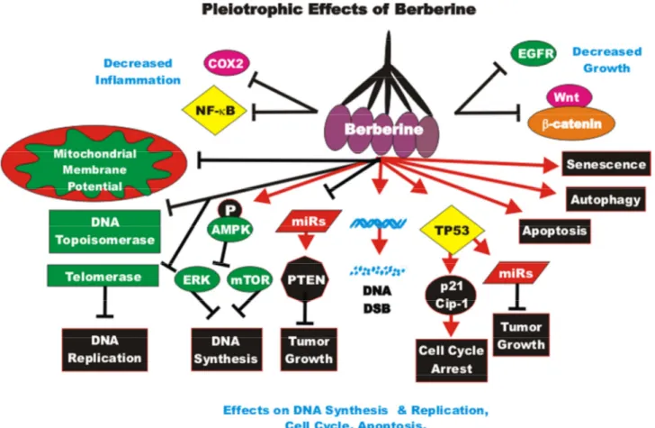
Figure 1. Pleiotropic effects of resveratrol on signaling pathways involved in cell growth.Some of the pathways affected by RES are indicated. This diagram focusses on signaling pathways predominately involved in aging and cancer. Red arrows indicate induction of an event; black closed arrows indicate suppression of an event.
and autophagy. In some cases, RES has been shown to synergize with chemotherapeutic drugs and inhibit proliferation. We will briefly summarize some of the different types of cancer that RES has been examined on. We will focus primarily on RES and CSCs. An overview of the pleiotropic effects of RES on cancer pathways is presented in Figure 1.
Effects of resveratrol on brain cancer
The effects of RES on the induction of autophagy and apoptosis were examined in glioblastoma (GBM) CSCs. RES induced autophagosome formation in three GBM cell lines. The expression of proteins associated with autophagy such as: autophagy protein 5 (Atg5), beclin-1 and Microtubule-associated proteins 1A/1B light chain 3B (LC3-II) were increased after RES treatment. Suppression of the autophagy induced by RES resulted in apoptosis. Suppression of both autophagy and apoptosis was required to block the toxicity of RES. RES suppressed the sphere-forming capacity of the GMB cells and the percentage of CD133 and octamer-binding transcription factor-4 (OCT4) positive cells [41].
Increased levels of signal transducer and activator of transcription 3 (STAT3) have been detected in CD133+, GMB CSCs. Suppression of STAT3 by either pharmacological or genetic approaches inhibited the CSC properties of GMB CD133+ cells. RES was determined to inhibit STAT3, induce apoptosis, suppress the stemness gene signature and induced differentiation. RES or sh-STAT3 treatment was determined to suppress GBM CSC survival in mice and their combined treatment synergistically increased radiosensitivity [42].
The effects of RES on human U87MG cells and primary human glioblastoma cultures were examined. RES suppressed the invasive growth and stimulated differentiation. RES decreased NESTIN and stimulated glial acidic fibrillary protein and betaIII-tubulin and senescence [43].
RES inhibited the self-renewal and tumor-initiating capacity of patient-derived glioblastoma stem cells (GSC). RES was determined to suppress the expression of NANOG through proteasomal degradation which could be inhibited by the proteasome inhibitor MG132. TP53 activation is important in the suppression of NANOG after RES treatment. RES treatment activated TP53 and p21Cip1. Suppression of NANOG inhibited self-renewal and tumor-forming capacity of the GSCs. These studies indicated that RES could induce GSC differentiation and NANOG was important in stemness [44].
The effects of RES have been compared on GSCs and normal neural stem cells (NSCs). RES was determined to block GSC proliferation, while it did not appear to have any effects on NSCs. SIRT1 and SIRT3 were expressed in both GSCs and NSCs, however, SIRT2 was only expressed in GSCs. Inhibition of SIRT2 suppressed the anti-proliferative effects of RES [45]. The effects of combining RES with radiation have been examined in the radioresistant GBM SU-2 cell line. RES was demonstrated to inhibit proliferation, enhance radiosensitivity, suppress neural stem cell markers and induce differentiation in SU-2 cells. The combination of RES and radiation resulted in increased autophagy, apoptosis in in vitro and in vivo models. In addition, RES treatment suppressed repair of radiation-induced DNA damage [46].
Effects of resveratrol on breast cancer
RES inhibited the proliferation of CSCs isolated from MCF-7 and SUM159 breast cancer cells. RES also reduced the presence of mammospheres in these two cell lines and inhibited tumor formation of the mammospheres in NOD/SCID mice. RES also induced autophagy in the breast mammospheres as determined by analysis of GFP-LC3-II puncta formation assay and the levels of LC3-II, Beclin1 and Atg 7 proteins. RES suppressed WNT/beta-catenin pathway expression [47]. The abilities of RES and the chemically-related pterostilbene to combine and restore the ERalpha expression in ERalpha-negative breast cancer have been examined. After combined treatment with RES and pterostilbene, increased levels of active acetyl-H3, acetyl-H3lysine9 (H3K9) and acetyl-H4 binding to the ERalpha promoter analysis were observed by chromatin immunoprecipitation analysis (ChIP). The combined treatment also resulted in changes in HDAC and histone acetyl transferase activity (HAT) as reduction in DNMT enzyme activity and 5-methylcytosine levels were observed in the normally ER- MDA-MB-157 breast cancer cells. The combined treatment also rendered the cells sensitive to the ER antagonist tamoxifen. This combination approach may be a safer alternative to restore ER expression and sensitivity to anti-ER based therapeutics in certain breast cancer patients [48]. The effects of the RES analog 4,4'-dihydroxy-trans-stilbene (DHS) were examined on normal mouse and human fibroblasts and human breast cancer cells. DHS was determined to inhibit the growth of human fibroblast cells more than normal RES. DHS was determined to suppress chemically-induced (3-methylcholanthrene plus 12-O-tetradecanoylphorbol-13-acetate) transformation of BALB/c-3T3 and
invasion of MCF-7 breast cancer cells. Interestingly, DHS was determined to inhibit both anchorage-dependent and anchorage-inanchorage-dependent growth of MCF-7 growth more efficiently than RES. DHS resulted in pRb inhibition and increased expression of TP53 and p21Cip-1. DHS was determined to reduce matrix metalloproteinase-2 (MMP2) and MMP9 expression, decreased adhesion to the extracellular matrix, migration and invasion [49].
Dihydrotestosterone (DHT) can stimulate the growth of breast cancer cells. DHT can interact with both ERalpha and integrin alphavbeta3. RES can induce TP53-dependent apoptosis via integrin alphavbeta3. ERK1/2 can transduce, in part, the signals mediated by RES and DHT, while DHT promotes proliferation, RES induces apoptosis. The effects of RES and DHT were compared on MCF-7 (ERalpha+) and MDA-MB-231 (ERalpha-) breast cancer cells. DHT was determined to inhibit phosphorylation of TP53 at S15. DHT also inhibited the nuclear TP53/COX-2 complex and TP53 transcriptional program. Thus, the receptor sites necessary for the effects of DHT and RES are discrete. While they both result in activation of ERK1/2 they have different effects. These studies also indicate the importance of the presence and absence of DHT in studies evaluating the effects of RES [50].
The effects of RES were examined in paclitaxel-resistant MDA-MB-231 cells. RES was determined to inhibit proliferation and stimulate apoptosis and cellular senescence in both resistant and paclitaxel-sensitive MDA-MB-231 cells indicating that the effects of RES were independent of the paclitaxel-sensitivity status of the MDA-MB-231 cells. The resistant cells expressed increased levels of multidrug resistance-1 (MDR1) and cytochrome P4502C8 (CYP2C8) genes [51].
Effects of resveratrol on colorectal cancer (CRC)
The diet is very important in the development of cancer, especially CRC. CRC CSCs are resistant to conventional chemotherapeutic drugs. The Western diet is becoming more and more associated with obesity which results in elevated levels of insulin and IGF-1. This may contribute to activation of the PI3K/PTEN/ Akt/mTORC1 and WNT/beta-catenin path-ways which contributes to CRC CSCs as well as premature aging. These CRC CSCs are often resistant to small molecule inhibitors which target these signaling pathways. A current focus of nutrition research is the effects of dietary bioactive compounds such as: CUR, grape seed extract, lycopene (present in tomatoes and other red vegetables), RES and others [52].
SIRT1 is an important subcellular target for RES. Depending on the cell system and conditions, RES can act as a tumor promoter or a tumor suppressor. The effects of RES on the SIRT1 were examined in CRC cells. RES suppressed proliferation in two CRC cell lines, this was accompanied with a decrease in Ki-67 expression which was dependent upon SIRT1 activity. RES downregulated nuclear localization and activity of NF-kappa-B which resulted in decreased expression of MMP9 and C-X-C chemokine receptor type 4 (CXCR4), two proteins associated with metastasis. SIRT1 was determined to interact with NF-kappaB. RES can effect SIRT1 expression which can regulate negatively NF-kappaB activity [53].
RES has been determined to suppress proliferation, migration, invasion and induce apoptosis in CRC cell lines, such as HT-29 (TP53+) and HCT-116 (TP53-) CRC cell lines.
The expression of miR-34c is increased by RES in CRC and this expression was in part responsible for the effects of RES. RES also sensitized the CRC cells to oxaliplatin in a miR-34a-dependent fashion. In xenograft studies, treatment with either RES or oxalip-latin suppressed tumor growth and their combined treatment was synergistic. In this xenograft system, RES increased miR-34 levels and also reduced the level of IL-6 which normally promotes CRC progression. The effects of RES were increased in the presence of functional TP53, indicating that RES and TP53 synergized [54].
The pharmacological properties of RES can be enhanced by nanoencapsulation. Normally the solubility and stability of RES is poor. The effects of nanoen-capsulation of RES have been examined in CRC and other model systems and have shown enhanced delivery [55].
A common problem with 5-flurouracil (5FU) treatment of CRC patients is chemo resistance. The effects of RES on 5FU-sensitive (HCT116, SW480) and their corresponding isogenic 5FU-chemoresistant derived CRC cell clones (HCT116R, SW480R) have been examined. Interestingly, RES blocked proliferation of all four cell lines and synergized with the effects of 5FU. RES was determined to suppress many gene products associated with EMT such as decreased vimentin and SLUG expression but increased E-cadherin expression. RES down-regulated NF-kappaB activation and nuclear translocation resulting in inhibition of NF-kappaB regulated gene expression which included MMP-9 and caspase-3. This occurred by inhibition of IkappaBalpha kinase which controls NF-kappaB activity [56].
The abilities of RES to suppress the cisplatin-resistance of CRC cells have been examined. The effects of addition of 15 micrograms/ml RES were determined on CRC cells treated with 5 and 20 micrograms/ml cisplatin. RES could induce anti-proliferative and pro-apoptotic effects in both the drug-sensitive and cisplatin-resistant HCT-116 cells. The cellular uptake of cisplatin was improved in the presence of RES [57]. The effects of RES also have been examined in combination with oxaliplatin in the HCT116 CRC cell line. The effects of RES and oxaliplatin were examined on the inhibitor of apoptosis protein survivin which is a key anti-apoptotic protein. While oxaliplatin treatment decreased the levels of survivin, the combined treatment of oxaliplatin and RES restored survivin, BCL-2 and caspase levels. In these studies, the apoptotic-inducing effects of oxaliplatin were decreased when RES was added to the HCT116 cell line. Thus, in the HCT116 cell line, RES had an anti-chemosensitizing effect when added with oxaliplatin. These and other studies demonstrate the caution that needs to be applied to studies with using RES on CRC and other cancer types [58].
The abilities of RES to enhance the anti-proliferative effects of etoposide in HepG2 liver cancer cells and CRC HCT-116 cells were determined. Both cell lines have functional TP53. RES inhibited proliferation in both cell lines. The combination of RES and etoposide resulted in greater anti-proliferative effects in HCT-116 than just after etoposide treatment by itself whereas this effect was not observed in HepG2 cells. TP53 was detected at higher levels after RES and etoposide treatment. Pre-incubation of both cell lines with RES, increased the level of etoposide-induced TP53. Thus, RES appeared to promote the effects on TP53 [59]. RES can induce AMPK which results in inhibition of the drug transporter MDR1 in oxaliplatin-resistant (L-OHP) HCT116/L-OHP CRCs. This prevents NF-kappaB activation and levels and suppresses cAMP-responsive element transcriptional activity. AMPKalpha siRNA transfection could reverse the effects of RES on MDR1 expression and cAMP-responsive element-binding protein (CREB) phosphorylation [60].
RES induces chromatin condensation and TP53 and the cleavage of poly [ADP-ribose] polymerase 1 (PARP-1) in CRC cells. The TP53 gene status was determined to affect the sensitivity to RES as CRC cells with WT TP53 (HCT-116/p53 WT) were more sensitive to RES than cells with mutant TP53 (HCT-116/p53-/-). RES induced double strand DNA breaks by interfering with type II topoisomerase. This was ascertained by determining the extent of gamma-H2AX foci present
after treatment with RES. The DNA damage was determined to be due to type II topoisomerase poisoning. Treatment of HCT-116 cells with RES was determined to result in activation of Ataxia Telangiectasia Mutated (ATM) kinase which in turn activated TP53 [61].
RES suppresses EMT via the TGF-beta/SMAD pathway in CRC LoVo cells in vitro and in animal experiments
in vivo. The TGF-beta/SMAD pathway regulated
SNAIL/E-cadherin expression. RES inhibited CRC metastasis into the lung in colon cancer metastatic tumor model after tail vein injection as well the development of lung and liver tumors after orthotopic injections. In vitro studies showed that TGF-beta promoted EMT and invasion and reduced E-cadherin but elevated vimentin expression. RES inhibited cancer cell invasion and induced E-cadherin and repressed EMT, vimentin, the TGF-beta/SMAD pathway and SNAIL [62].
The effects of RES and the DNA cross-linker mitomycin C have been examined in CRC primary CRC cells isolated from resected tumors. The combination of RES and mitomycin C suppressed proliferation better than either drug by itself. This treatment resulted in a significant up-regulation of p21Cip-1 expression [63]. The effect of RES and 5-aminosalicylic acid (Mesalazine) on JAK/STAT activation have been examined in the HT-29 CRC line. Mesalazine is an anti-inflammatory drug used to treat patients with bowel disorders, including inflammatory bowel disease. RES reduced nitric oxide (NO) and prostaglandin E2 (PGE2) production, inducible nitric oxide synthase (iNOS) and cyclooxygenase-2 (COX-2) expression and ROS production which were induced by treatment of HT-29 cells with the inflammatory cytokines (IL-1alpha, TNF-alpha, IFN-gamma). RES was determined to decrease activation of the JAK/STAT pathway but did not result in degradation of I-kappaB-alpha. RES also inhibited stress-activated protein kinase (SAPK)/c-Jun N-terminal kinase (JNK) pathway activation [64].
In a different study, RES was shown to inhibit the proliferation of human HCT116 CRC cells in vitro as well as in tumor xenograft studies in vivo. RES was determined to upregulate phosphatase and tensin homolog (PTEN) expression and decrease the expression of activated Akt. In HCT116 cells, PTEN inhibits Akt signaling and proliferation. Moreover, decreased levels of beta-catenin were also detected after RES treatment [65].
RES was shown to decrease WNT/beta-catenin pathway activity and the downstream targets c-Myc and MMP-7
in CRC cells. RES also decreased the expression of long non-coding metastasis associated lung adenocarcinoma transcript 1 (RNA-MALAT1) in the LoVo and HCT116 CRC cells. This resulted in decreased nuclear localization of beta-catenin and suppressed WNT/beta-catenin signaling. These events were linked with suppression of CRC invasion and metastasis [66].
Treatment of CRC cells with RES resulted in decreased expression of transcription factor 4 (TCF4), which is a critical effector molecule of the WNT/beta-catenin pathway. The half-life of the TCF4 protein was decreased after treatment with RES. In contrast, RES did not affect the transcription rate of the TCF4 gene. RES increased the S/T phosphorylation of TCF4. These phosphorylation events were determined to be due to ERK1/2 and p38MAPK [67].
RES can induce anti-proliferative effects on CRC cells via the miR-34a/E2F3/Sirt1 pathway. The effects of RES, epigallocatechin-3-gallate (EGCG), and alpha-mangostin (alpha-M) were examined on three CRC lines in the presence and absence of 5-FU. These natural products were determined to suppress the PI3K/PTEN/ Akt/mTORC1 pathway. Alpha-M was determined to be the most potent PI3K/PTEN/Akt/mTORC1 pathway inhibitor and suppressed Raf/MEK/ERK pathway. Combined treatment of RES and 5-FU resulted in a synergistic inhibition of cell growth and induction of apoptosis and the Raf/MEK/ERK pathway was suppressed in DLD-1 CRC cells. RES increased the expression of miR-34 which resulted in the decreased expression of E2F3 and SIRT1 [68].
Effects of resveratrol on head and neck cancer
The effects of RES on the CSC properties of head and neck cancer-derived tumor-initiating cells (HNC-TICs) were examined. RES was determined to downregulate ALDH1 and CD44 in HNC-TICs in a dose-dependent fashion. RES also inhibited OCT4, NANOG and NESTIN expression in the sphere forming HNC cells. In mouse experiments, RES was shown to inhibit tumor growth, stemness and EMT markers. RES synergized with chemotherapy in suppression of the growth of HNC-TICs [69].
Effects of resveratrol on leukemia
RES suppressed phosphorylated liver kinase B1 (pLKB1) and induced senescence and apoptosis in acute myeloid leukemia (AML) KG1a cells. This occurred via SIRT1 which is a regulator of LKB1 (aka serine/threo- nine kinase 11 [STK11]) which resulted in senescence and apoptosis [70].
RES has been determined to decrease IL-6-induced Sonic hedgehog homolog (SHH) signaling in AML. The plasma levels of IL-6 and IL-1beta were higher and lower respectively in AML patients than in normal donors. The expression of SHH was determined to be higher in bone marrow and peripheral blood mononuclear cells prepared from AML patients. IL-6 was determined to increase SHH and GLI-1 expression in HL-60 cells. IL-6 also increased cell viability. RES decreased SHH expression, GLI-1 nuclear translocation and cell viability in IL6-treated HL-60 cells. RES was determined to synergize with the SHH inhibitor cyclopamine in inhibiting growth [71].
Effects of resveratrol on lung cancer
RES has been shown to inhibit the secretion of IL-6 and VEGF from A549 lung cancer cells when they were co-cultured with adipose-derived mesenchymal stem cells [72].
The ability of combined RES and metformin (MET) treatment to protect A549 lung cancer cells from ultraviolet C (UVC)-induced damage was determined. MET was determined to inhibit the UVC-mediated increase in TP53 expression and suppressed expression of gamma-H2AX and phosphorylated check kinase-2 (P-Chk2). MET also induced DNA repair and cell cycle arrest and decreased levels of cyclin E/cyclin-dependent kinase 2(cdk2)/RB and cyclin B1/CDK1. Treatment with RES, by itself, was less effective in suppressing UVC-induced responses. Combined RES and MET treatment resulted in a synergistic response in terms of decreased TP53, gammaH2AX and P-Chk2 expression. Thus, the combination of RES and MET might suppress some of the aging effects elicited by UVC-induced DNA damage [73].
RES has been shown to regulate the expression of lncRNAs in lung cancer. 21 lncRNAs were shown to have increased expression after RES treatment and 19 lncRNAs were detected at lower levels in A549 lung cancer cells. The AK001796 lncRNA was determined to be overexpressed in lung cancer cell lines and tissues but it was detected at lower levels in RES-treated cells. The AK001796 lncRNA functioned as an oncogene. Suppression of AK001796 lncRNA inhibited cell growth and promoted cell cycle arrest [74].
Effects of resveratrol on hepatocellular carcinoma
The effects of RES on hexokinase 2 (HK2) were examined in HCC. HK2 can change the metabolic properties of cancer cells to promote growth in conditions of low oxygen concentrations. This can result in aerobic glycolysis. Aerobic glycolysis was
observed in four HCC lines as opposed to normal hepatic cells. Treatment with RES was observed to sensitize the HCC cell lines to apoptosis and these effects were reversed upon treatment with glycolytic inhibitors. RES treatment resulted in a decrease in HK2 and increased mitochondrial-induced apoptosis. RES could enhance treatment with sorafenib resulting in growth inhibition and promoting apoptosis in HCC xenografts in mice [75].
The effects of tanshinone IIA (Tan IIA) have been examined in combination with RES on the induction of cytotoxicity, cell-cycle arrest, apoptosis, and DNA fragmentation in HepG2 HCC cells. Tan IIA and RES were determined to synergize in inducing apoptosis. The levels of apoptosis induced with Tan IIA and RES were similar to the amounts induced by cisplatin [76]. RES has been shown to induce apoptosis in HepG2 cells by induction of caspase-3 and -9, TP53 and increasing the BAX/BCL2 ratio. The ability of matrine, a natural component extracted from the traditional Chinese medical herb Sophora flavescens Ait to synergize with RES was determined also. Matrine could synergize with RES in inhibiting cell growth and promoting apoptosis. This synergy in the induction of apoptosis was attributed to the induction of caspases -3 and -9, downregulation of survivin, induction of ROS and alteration of the mitochondrial membrane potential [77]. The effects of RES on VEGF expression and proliferation of HCC cells were determined. RES inhibited VEGF expression in the HepG2 cells [78]. RES was determined to sensitize HepG2 cells to TNF-related apoptosis-inducing ligand (TRAIL)-induced apoptosis. RES induced an increase in phospho-AMPK levels and a decrease in survivin levels. RES was determined to increase TRAIL sensitivity by decreasing survivin expression [79].
The effect of RES on the induction of apoptosis in SMMC-7721 cells were determined. RES could activate caspases-3 and -9 and JNK whereas it downregulated activated ERK. RES also was effective in xenograft models with SMMC-7721 cells [80].
RES has been shown to suppress the STAT3 signaling pathway in HepG2 cells. This resulted in the suppression of proliferation when the cells were cultured in medium containing high glucose. This was determined to be mediated in part by SIRT1 expression. When HepG2 cells were cultured in medium containing 25 millimolar glucose, exposure of the cells to 100 micromolar Res suppressed proliferation and STAT3 signaling [81].
Res has also an anti-metastatic effects in HCC through inhibition of phosphorylation of the JNK 1/2 pathway and SP-1 DNA binding activities, causing a down-regulation of urinary-type plasminogen activator (u-PA) expression [82].
RES prevented diethylnitrosamine (DEN)–induced liver tumorigenesis in rats by suppressing oxidative stress and inflammatory response mediated in part by hepatic nuclear factor E2–related factor 2 (Nrf2) [83]. RES in combination with CUR has synergistic antiproliferative effects in HCC Hepa1-6 cells [84].
Effects of resveratrol on nasopharyngeal cancer
The effects of RES on nasopharyngeal carcinoma (NPC) CSCs were examined. Interestingly the NPC CSCs were determined to have undergone a metabolic shift as they relied on glycolysis for energy. RES was determined to shut off the metabolic shift and increase ROS levels and depolarized mitochondrial membranes. These biochemical events were associated with a reversion of the CSC-associated properties of the cells. RES also was found to inhibit the CSC properties of the cells by activating TP53 and inducing miR-145 and miR-200c expression, which are normally down-regulated in NPC CSCs [85].
Effects of resveratrol on ovarian cancer
The effects of RES on the induction of ROS has been examined in A2780 ovarian CSCs. RES was determined to kill A2780 ovarian CSCs in a fashion independent of ROS. However, ROS inhibited the self-renewal capacity of A2780 ovarian CSCs which could survive the RES treatment [86].
Effects of resveratrol on pancreatic cancer
RES has also been suggested to have anti-cancer effects on pancreatic cancer cells. RES inhibited proliferation, invasion and metastasis and induced apoptosis in pancreatic cancer cells and suppressed proliferation and viability of pancreatic CSCs. RES also could chemosensitize pancreatic cancer cells and have effects on diabetes [87].
Human pancreatic cells which are CD133+, CD44+, CD24+, ESA+ and ALDH+ are enriched in pancreatic CSCs. They also express higher levels of NANOG, OCT-4, NOTCH1, MDR1 and ATP Binding Cassette Subfamily G Member 2 (ABCG2) than primary pancreatic cells and normal pancreatic tissues. CSCs were derived from Kras(G12D) transgenic mice. These CSCs also express higher levels of NANOG and OCT-4 than the cells from control mice. RES was determined
to suppress growth and development of pancreatic cancer in the Kras(G12D) mice. RES activated caspases -3, -7 and apoptosis by inhibiting BCL-2 and XIAP in the pancreatic CSCs. RES also inhibited: NANOG, SOX-2, c-MYC, OCT-4 and ABCG2 in the pancreatic CSCs. Genetic suppression of NANOG by shRNA was determined to enhance the effects of RES on pancreatic CSCs self-renewal. RES also inhibited pancreatic CSC migration, invasion and the expression of genes associated with EMT including: ZEB-1, SLUG and SNAIL [88].
Resveratrol and thyroid cancer
Combined RES and valproic acid treatment has been shown to inhibit the growth, stem cell marker expression, aldefluor expression and invasiveness of two spheroid thyroid carcinoma CSC lines derived from anaplastic thyroid cancers. Although valproic acid is predominately used in the treatment of epilepsy and bipolar disorder and to prevent migraine headaches, it is also being examined for its effects on cancer and is in at least 83 clinical trials with various cancer patients. Valproic acid may inhibit histone deacetylase and this is one of the reasons this drug is being examined in cancer settings. RES and valproic acid treatment resulted in an increase in thyroid differentiation marker expression and apoptosis [89].
Summary of resveratrol
The anti-aging, anti-cancer, anti-neurological effects of RES have been examined in many different diseases and environments. While there have been many beneficial effects postulated to be induced by consumption of RES, the concentrations required to obtain those health benefits may be high. Although, there probably are many beneficial health effects of consumption of products rich in RES.
Overview of effects of curcumin
CUR has also been reported to have many beneficial health properties ranging from anti-aging, anti-cancer, hypertensive, inflammatory and anti-neurological effects. The world-wide market for CUR is predicted to be close to $100 million by 2022 (http: // www.grandviewresearch.com/industry-analysis/ turmeric-extract-curcumin-market). CUR is commonly obtained as an extract from Curcuma longa (Turmeric). There are other compounds present in the extract from
Curcuma longa which are related to CUR, they are
called Curcuminoids. 60-70% of the turmeric extract consists of CUR, 20-27% of the turmeric extract consists of demethoxy curcumin and 10-15% of the turmeric extract consists of bisdemethoxycurcumin
[90]. Together these components make up 1-6% of turmeric by weight.
Curcumin and aging
CUR may have anti-aging properties. The effects of CUR may be mediated, in part, by their effects on sirtuins. High doses of CUR (2.5-10 micromolar) have been shown to induce the senescence of cancer cells and cells involved in building the vasculature. The effects of lower doses of CUR (0.1 and 1 micromolar) on vascular smooth muscle (VSMC) and endothelial (EC) cells have been examined. VSMCs were not protected from replicative senescence or premature senescence induced by doxorubicin with low doses of CUR. CUR treatment did have some effects on increasing the levels of sirtuins. Thus, low doses of CUR could increase sirtuins levels in the absence of delaying the senescence of VSMCs [91].
The ability of CUR to alter the expression of the mitochondrial uncoupling protein 2 (UCP2) in old and young rats (UCP2-/- and WT) was determined as this protein plays critical roles in regulating ROS production. These experiments were performed to determine whether dietary CUR has positive effects on aging-related cerebrovascular dysfunction by increasing UCP2 expression. CUR was determined to reduce ROS production in UCP2 WT but not in UCP2-/- aging mice. CUR restored the cerebrovascular endothelium-dependent vasorelaxation that was impaired in the older rats. Thus, these studies demonstrated that CUR could improve cerebrovascular dysfunction which occurs during aging [92].
The effects of CUR and MEL on age-related carbonyl content of liver in young and old mice were determined. Carbonyl content in the liver is detected at increased levels during aging. CUR and MEL treatment decreased the carbonyl content found in the livers of both the young and old mice [93].
Curcumin and lifespan
In Drosophila models, CUR has been shown to prevent the accelerated aging that is normally observed after irradiation by reduction of oxidative stress [94]. CUR was also shown by a different group to enhance the
Drosophila reproductive lifespan as well as prolong the
viability of the offspring [95].
Tetrahydrocurcumin (THC) has been shown to increase the life span of certain organisms including: nematodes,
Drosophila and mice. Interestingly in the nematode
model, CUR reduced the production of ROS. The following genes: odd-skipped-related 1 (osr-1 a
transcription factor), dual specificity mitogen-activated protein kinase kinase 4 (MAP2K4, sek-1, a serine/threonine (S/T)kinase), dual specificity mitogen-activated protein kinase kinase 1 (mek-1), skinhead-1 (skn-1 encodes a beta-glucan synthesis-associated protein), Ca2+/calmodulin-dependent protein kinase II (CaMKII, unc-43, encodes a S/T kinase, Sirtuin2 (silent mating type information regulation 2) 2 (sir-2.1), and phosphatidylinositol 3-kinase age-1 (age-1) were demonstrated to be responsible for the effects of CUR on lifespan in nematodes. In Drosophila, superoxide dismutase activity was required for increased lifespan. In addition, decreased lipofuscin and malondialdehyde levels were required for increased lifespan in
Drosophila. CUR also upregulated the expression of
superoxide dismutase (SOD) genes as well as decreased the expression of various age-related genes including:
Drosophila insulin receptor (dInR), Attacin-D (ATTD, a
protein in Drosophila that induced upon bacterial infection), defensin (Def, a protein induced in
Drosophila in response to fungal infection), Cecropin B
(CecB, a protein involved in immune response of
Drosophila) and bactericidal protein diptericin B (DptB,
a protein involved in survival response of Drosophila) [96].
Curcumin and senescence
The effects of CUR on gene expression in cancer-associated fibroblasts obtained from breast cancer patients has been examined. CUR increased the expression of the p16INK4A and other tumor suppressor proteins. In contrast, CUR decreased the activity of the JAK2/STAT3 pathway and many molecules involved in cellular growth and metastasis including: stromal cell-derived factor-1 (SDF-1), IL-6, MMP2, MMP9 and TGF-
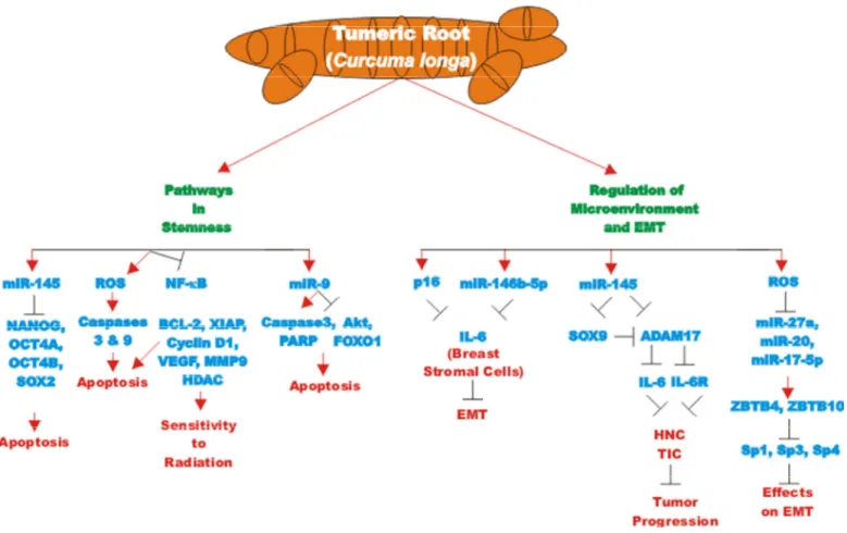
Figure 2. Pleiotropic effects of curcumin on signaling pathways involved in cell growth. Curcumin can induce many pathways which may result in suppression of cell growth, induction of apoptosis, autophagy, senescence or inhibition of cell cycle progression. These events are indicated by red arrows. In addition, CUR treatment can result in the suppression of many important proteins which result in decreased growth, EMT, metastasis or inflammation. These events are indicated by black closed arrows.
beta. These effects reduced the levels of alpha-smooth muscle actin (alpha-SMA) which was attributed to decreased migration and invasion of the cells. CUR suppressed Lamin B1 and induced DNA damage-independent senescence in proliferating but not quiescent breast stromal fibroblasts in a p16INK4A -dependent manner. The CUR-induced senescence was determined to occur in the absence of an inflammatory secretory phenotype, which was in some cases associated with procarcinogenic properties. Thus, CUR treatment can result in senescence in stromal fibroblasts [97]. An overview of the pleiotropic effects of CUR is presented in Figure 2.
Curcumin and memory loss diseases
CUR may be useful in the prevention of AD. AD results, in part, from the overproduction and accumulation in the brain of Abeta. CUR has recently been shown to suppress memory decline by suppressing beta-site amyloid precursor protein cleaving enzyme 1 (BACE1= Beta-secretase 1, an important gene in AD) expression which is implicated in beta-amyoid pathology in 5xFAD transgenic mice. CUR was determined to suppress synaptic degradation and improved spatial learning in the 5xFAD mice [98]. The ability of CUR to affect adiposity and obesity-associated cognitive impairment has been examined in mouse models. CUR was found to decrease adiposity and improve cognitive function in a similar fashion as CR in 15-month-old mice. The effects of CUR and CR were positively linked with anti-inflammatory or antioxidant actions [99].
The effects of CUR on the prevention of memory decline have been investigated in old mice. These studies have focused on the effects of CUR on the neuronal nitric oxide synthase (nNOS)/nitric oxide (NO) pathway. Treatment of mice with CUR aided the memory acquisition ability of old mice. CUR treatment increased nNOS expression, acidity and NO concentration [100].
CUR also has been shown to have positive effects on spatial memory by studies with the Morris water maze model with old female rats. In addition, CUR treatment had positive effects on the oxidative stress induced by aging by analysis of malondialdeyde (MDA), protein carbonyl and glutathione levels. CUR treatment resulted in decreased levels of MDA [101].
Curcumin and macular degeneration
The effects of CUR on age-related macular degenera-
tion (AMD) have been examined in an aging retinal pigment epithelial cell (RPE) model. The effects of CUR on H2O2-treated RPE cells were determined. CUR treatment improved cell viability and decrease both apoptosis and oxidative stress [102].
Damage induced by oxidative stress to RPE cells may be responsible for the aging of the retina and AMD which can lead to irreversible loss of vision in the elderly. Patient-derived RPEs were isolated by reprogramming T cells from patients with dry-type AMD. The cells were induced to become pluripotent stem cells (iPSCs) and then they were differentiated into RPE cells. Interestingly, the AMD-RPEs had decreased abilities to defend against oxidative stress which may have been responsible for their susceptibility to oxidative damage and AMD. These cells have been used in vitro drug screening assays. CUR treatment was determined to cause reduction of ROS in the AMD-RPEs and protected the cells from H2O2-induced cell death by reduction of ROS levels. CUR also altered the levels of: PDGF, VEGF, insulin like growth factor binding protein 2 (IGFBP-2), heme oxygenase 1 (HO1), superoxide dismutase 2 (SOD2), and glutathione peroxidase 1 (GPX1). Thus, CUR treatment might eventually be useful in the treatment of patients with or susceptible to the development of AMD [103].
Curcumin and miRs
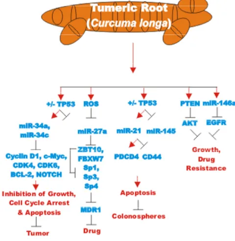
The effects of many drugs on the expression of various miRs is an important area of scientific investigation as miRs have been shown to play key roles in both positive and negative regulation of gene expression. A diagram of some of the effects of CUR on miRs important in TP53-mediated responses and apoptotic pathways is presented in Figure 3. Certain miRs can act as oncogenes and stimulate abnormal gene expression while other miRs can act as tumor suppressors and inhibit gene expression. Often the promoter regions of miR tumor suppressor genes becomes hypermethylated in cancer cells and the miR is turned off. Approaches to induce the expression of miRs with drugs that induce demethylation are an important area of scientific research.
CUR has been shown to regulate miR-21 in various types of cancer. miR-21 can have effects on PI3K/PTEN/AKT, NF-kappaB, programmed cell death protein 4 (PDCD4) and other signaling pathways. CUR decreases miR-21 levels by increasing miR-21 exosomes. CUR also decreases miR-21 expression by a transcriptional mechanism by binding the promoter region of the miR-21 gene [104].
Effects of curcumin on bladder cancer
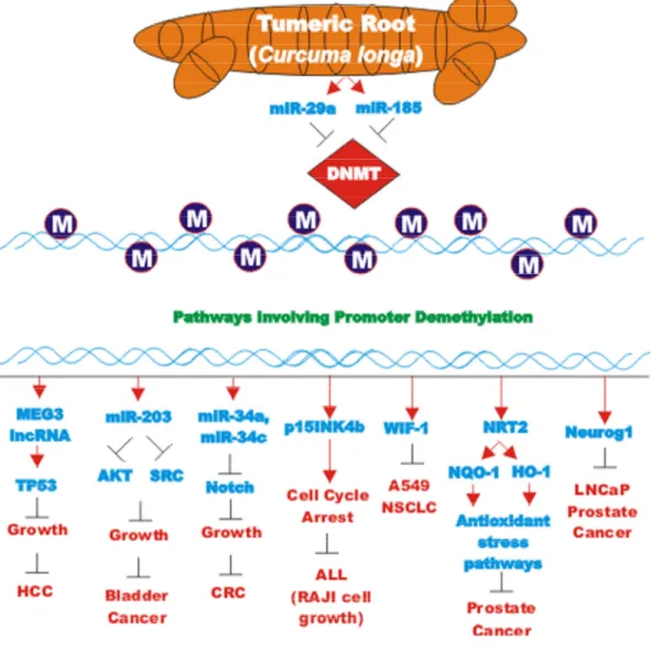
CUR also has been shown to have anti-cancer effects on bladder cancer. CUR has been shown to induce the expression of the tumor suppressive miR-203 in bladder cancer. Normally miR-203 is repressed in bladder cancer perhaps due to hypermethylation of its promoter region. CUR could induce the demethylation of the miR-203 promoter region in bladder cancer cell lines. AKT2 and SRC have been determined to be targets of miR-203 in bladder cancer and their expression was decreased after CUR treatment [105]. In some cancers, CUR has been shown to downregulate the expression of DNA methyl transferase I (DNMT1) and induce DNA hypomethylation [106]. A diagram illustrating the effects of CUR on chromatin structure is presented in Figure 4.
Effects of curcumin on brain cancers
Demethoxycurcumin (DMC) has been shown to have anti-tumor effects against glioblastoma (GMB). Temozolomide (TMZ) is used for treating GMB, however, TMZ does not prevent GMB recurrence which may be due to the presence of glioma stem cells (GSC). The effects of DMC and TMC on the induction of apoptosis and cell growth of GSC were examined. Combining the two drugs was determined to elicit more anti-GSC effects. Some of the effects were determined alterations in multiple signaling pathways including: JAK/STAT-3 signaling, induction of ROS and caspase-3-mediated apoptosis. Addition of DMC prior to TMZ treatment was determined to be more effective [107].
Figure 3. Effects of curcumin on TP53 and apoptosis pathways. An overview of the effects of CUR on TP53 and apoptotic pathways and the effects of miRs are indicated. miRs in magenta font indicate oncomirs. miRs in blue font are tumor suppressor miRs. Red arrows indicate induction of an event; black closed arrows indicate suppression of an event.
Interestingly, CUR was shown to both inhibit glioma cell proliferation and induce glioma cells to form spheres which were positive for CD133 and Nestin expression [108]. CUR suppressed G1 to S phase transition and enhanced the expression of OCT-4, SOX-2 and SOX-4. The expression of these genes was essential for retaining the stemness of the glioma-initiating cells [108].
Progranulin (PGRN) is a protein involved in regulation of cell growth. PGRN is overexpressed in GBM and has been shown to promote temozolomide-resistance by inducing DNA repair and stemness. PGRN upregulated the expression of many genes involved in DNA repair
including: Ataxia telangiectasia mutated (ATM, a S/T kinase involved in DNA repair response), breast cancer type 1 susceptibility protein (BRCA1, a protein involved in DNA repair), PARP, Rad51 (a protein involved in DNA repair), X-ray repair cross complementing 1 (XRCC1) and others. PGRN also upregulated the expression of many genes involved in stemness including: CD44 and CD133 and others involved in drug resistance such as ABCG2. The increased expression of these genes was due to their trans-criptional activation by c-FOS/JUNB present in the AP-1 transcription factor complex. CUR can down regulate AP-1 activity. CUR regulates both AP-1 and PGRN activity. Suppression of PGRN inhibited the stemness,
Figure 4. Effects of curcumin on chromatin structure. An overview of the effects of CUR on demethylation of genes and the effects on the various genes are indicated. Red arrows indicate induction of an event; black closed arrows indicate suppression of an event. M = methylation of a sequence.