doi:10.1152/japplphysiol.00126.2008 106:293-297, 2009. First published 8 May 2008;
J Appl Physiol
L'Abbate and Remo Bedini
Claudio Marabotti, Alessandro Scalzini, Danilo Cialoni, Mirko Passera, Antonio
breath-hold diving in humans
Cardiac changes induced by immersion and
You might find this additional info useful...
30 articles, 16 of which can be accessed free at:
This article cites
http://jap.physiology.org/content/106/1/293.full.html#ref-list-1 1 other HighWire hosted articles
This article has been cited by
[PDF] [Full Text] [Abstract] , November , 2009; 107 (5): 1526-1531. J Appl Physiol Bedini
Arne Sieber, Antonio L'Abbate, Mirko Passera, Erika Garbella, Antonio Benassi and Remo
Underwater study of arterial blood pressure in breath-hold divers
including high resolution figures, can be found at:
Updated information and services
http://jap.physiology.org/content/106/1/293.full.html
can be found at: Journal of Applied Physiology
about
Additional material and information
http://www.the-aps.org/publications/jappl
This information is current as of April 18, 2012.
ISSN: 0363-6143, ESSN: 1522-1563. Visit our website at http://www.the-aps.org/.
Physiological Society, 9650 Rockville Pike, Bethesda MD 20814-3991. Copyright © 2009 by the American Physiological Society. those papers emphasizing adaptive and integrative mechanisms. It is published 12 times a year (monthly) by the American
publishes original papers that deal with diverse areas of research in applied physiology, especially
Journal of Applied Physiology
on April 18, 2012
jap.physiology.org
HIGHLIGHTED TOPIC
The Physiology and Pathophysiology of the Hyperbaric and
Diving Environments
Cardiac changes induced by immersion and breath-hold diving in humans
Claudio Marabotti,1,2Alessandro Scalzini,1Danilo Cialoni,3Mirko Passera,2Antonio L’Abbate,2,4
and Remo Bedini2
1Unita` Operativa Cardiovascolare-Utic Ospedale di Cecina (Livorno), Padua;2Consiglio Nazionale delle Ricerca Institute
of Clinical Physiology, Pisa;3Apnea Academy Research, Padua; and4Scuola Superiore Sant’Anna, Pisa, Italy
Submitted 4 February 2008; accepted in final form 7 May 2008
Marabotti C, Scalzini A, Cialoni D, Passera M, L’Abbate A, Bedini R. Cardiac changes induced by immersion and breath-hold diving in humans. J Appl Physiol 106: 293–297, 2009. First published May 8, 2008; doi:10.1152/japplphysiol.00126.2008.—To evaluate the separate cardiovascular response to body immersion and increased environmental pressure during diving, 12 healthy male subjects (mean age 35.2⫾ 6.5 yr) underwent two-dimensional Doppler echocardiog-raphy in five different conditions: out of water (basal); head-out immersion while breathing (condition A); fully immersed at the surface while breathing (condition B) and breath holding (condition
C); and breath-hold diving at 5-m depth (condition D). Heart rate, left
ventricular volumes, stroke volume, and cardiac output were obtained by underwater echocardiography. Early (E) and late (A) transmitral flow velocities, their ratio (E/A), and deceleration time of E (DTE) were also obtained from pulsed-wave Doppler, as left ventricular diastolic function indexes. The experimental protocol induced signif-icant reductions in left ventricular volumes, left ventricular stroke volume (P⬍ 0.05), cardiac output (P ⬍ 0.001), and heart rate (P ⬍ 0.05). A significant increase in E peak (P⬍ 0.01) and E/A (P ⬍ 0.01) and a significant reduction of DTE (P ⬍ 0.01) were also observed. Changes occurring during diving (condition D) accounted for most of the changes observed in the experimental series. In particular, cardiac output at condition D was significantly lower compared with each of the other experimental conditions, E/A was significantly higher during
condition D than in conditions A and C. Finally, DTE was
signifi-cantly shorter at condition D than in basal and condition C. This study confirms a reduction of cardiac output in diving humans. Since most of the changes were observed during diving, the increased environ-mental pressure seems responsible for this hemodynamic rearrange-ment. Left ventricular diastolic function changes suggest a constric-tive effect on the heart, possibly accounting for cardiac output reduc-tion.
diving response; hemodynamics
PREVIOUS STUDIES ON NATURAL divers (mainly marine
mam-mals) showed that breath-hold diving is associated with energy-saving cardiovascular changes, i.e., a marked reduc-tion in cardiac output [due to the reducreduc-tion of both stroke volume and heart rate (HR)] and the redistribution of blood flow away from skin and myoglobin-rich muscles in favor of brain and heart (7, 28, 30). For years, however, technical difficulties have prevented a comprehensive assessment of cardiovascular changes during breath-hold diving in
hu-mans. Most of the knowledge on human diving physiology has been obtained from the study of head-out immersed subjects (9, 27), or extrapolated from the results obtained in breath-holding subjects, either with or without face immersion (2, 8, 10). The recent wide diffusion of recreational and competitive breath-hold diving (with a progressive increase of attained depths) highlighted the presence of serious diving-related pathologies, like syncope (ascent blackout), decompres-sion illness, hemoptysis, and pulmonary edema, whose patho-physiology is not yet completely understood (13, 31). A deeper knowledge of breath-hold diving physiology in humans seems thus needed.
Using a submersible echocardiographic machine, we re-cently showed, in humans, during short breath-hold dives up to 10-m depth, a hemodynamic pattern qualitatively similar to that described in marine mammals (22), with a significant decrease in HR, stroke volume, and, hence, cardiac output.
Breath-hold diving involves progressive decrease of O2and increase of CO2 blood content, changes in temperature and thermal conductivity, evocation of neural reflexes (induced by face immersion), change in environmental pressure (linearly increasing with depth), which, in turn, modulates the venous return to the heart (21). The relative role of these factors in determining the cardiovascular response to diving is still to be elucidated.
The aim of the present study was to separately evaluate, in humans, the cardiovascular response to body immersion, with or without face immersion, breath holding, and diving. MATERIALS AND METHODS
Subjects. A group of 12 healthy male subjects (age 35.2⫾ 6.5 yr;
range 24 –51 yr; height 180.2⫾ 6.8 cm; weight 77.4 ⫾ 10.2 kg; body mass index 23.8⫾ 2.3 kg/m2) was studied. The absence of female subjects was casual and not due to a selection criterion. All subjects were experienced, active breath-hold divers (practicing breath-hold diving from 5.8⫾ 3.5 yr), undergoing at least 2 h/wk of breath-hold diving training; no subject was engaged in regular physical activity besides underwater training. Each diver had the ability to reach a depth of at least 30 m under constant weight (i.e., with no ballast aid for descent); their maximum static breath-hold time at surface was 4.5⫾ 0.8 min (range 4–6 min). No subject had historical, clinical, or instrumental (resting ECG, Doppler echocardiography) evidence of
Address for reprint requests and other correspondence: C. Marabotti, UO Cardiovascolare-UTIC, Ospedale di Cecina, Via Montanara, 57023 Cecina (LI), Italy (e-mail: [email protected]).
The costs of publication of this article were defrayed in part by the payment of page charges. The article must therefore be hereby marked “advertisement” in accordance with 18 U.S.C. Section 1734 solely to indicate this fact.
First published May 8, 2008; doi:10.1152/japplphysiol.00126.2008.
on April 18, 2012
jap.physiology.org
arterial hypertension or cardiac or pulmonary diseases. All subjects were nonsmokers and had been fasting from at least 2 h before the study.
The study protocol was approved by the Scientific Committee of the Consiglio Nazionale delle Ricerca Institute of Clinical Physiology. All participants were informed about the aims and procedures of underwater ultrasound examination and gave their written consent.
Underwater echocardiographic equipment.
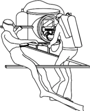
Doppler-echocardio-graphic examination was performed by a commercially available instrument (MyLab 30, Esaote SPA, Florence, Italy) as part of a submersible echograph previously described elsewhere (5). Briefly, a special, patented, water-tight container, made by two steel cylinders (60-cm diameter) intersecting each other (Fig. 1) contained the echo-cardiograph (6). A rubber glove sealed to the front Plexiglas panel allowed the user to access the instrument’s control keys. A pressure regulator connected to a standard 200-ATA compressed air cylinder (normally used for scuba diving) maintained the pressure inside the container of the echocardiographic instrument at the same level of external pressure, reducing the risk of water leakage and preventing rubber glove inflation. Ultrasound reflection in water was prevented by the rubber and silicon backing of the probe, ensuring the extinction of backward ultrasound radiation. The absence of ultrasound interfer-ences in free water was confirmed by tests performed by the manu-facturing company.
Experimental protocol. The study was performed, between 10 AM
and 2 PM, in a 10-m-deep pool (water temperature 29°C; air
temper-ature 27°C). Subjects were studied, by two-dimensional Doppler echocardiography, in five different conditions: out of water (basal), head-out immersion during normal breathing (condition A), fully immersed (head-in) at the surface while breathing by a snorkel (condition B), fully immersed (head-in) at the surface while breath holding (condition C), and, finally, breath-hold diving at 5-m depth (condition D). Subjects were simply wearing a bathing suit and, during full immersion (conditions B, C, and D), a diving mask. Echocardiogram was recorded, in each condition, with the subject lying on his left side during normal breathing (basal, conditions A and
B) or after a maximal inspiration (conditions C and D). Each dive was
preceded by 2– 4 min of surface floating preparation, during which subjects were breathing normally (no preliminary hyperventilation was done). Descents were done by using 10-kg ballast, thus reducing the cardiovascular effects of muscular work. Subjects equilibrated the pressure in the medium ear by Frenzel technique (in most cases) or by a mild Valsalva maneuver. As soon as the diver reached the echo station, he positioned himself on a metallic bracket, lying on his left side (Fig. 1). Cardiac imaging started during the first minute of apnea and lasted ⬍90 s in all subjects, so that echo-Doppler data were acquired in a cardiovascular steady state (not influenced by the possible, if any, effects of Valsalva maneuver, nor by those of hypoxia).
The time necessary for handling and positioning the underwater echocardiograph in the different positions prevented the possibility of performing, in each subject, the entire series of tests consecutively. The sequence of echocardiographic studies was the following: basal for all subjects; after the launch of the echocardiograph, the three studies at surface (conditions A, B, C) were performed consecutively in each subject. Finally, after echocardiograph was positioned at depth, ultrasonic study during diving (condition D) was made in all subjects. During the periods of instrumentation handling, subjects stayed out of the water, at rest.
Doppler-echocardiographic parameters. To minimize the duration
of the echocardiographic study during diving, we recorded only an apical four-chamber view loop (4-s duration) and a pulsed-wave Doppler tracing of transmitral blood flow. Analysis was made offline, according to the American Society of Echocardiography recommen-dations (19), by an expert in Doppler-echocardiography, unaware of the identity of the subjects and of the condition of recording. From the four-chamber view, the following parameters were obtained: systolic and diastolic left ventricular volumes (calculated by area-length method) (14, 19), and right ventricular internal dimension (maximal diastolic distance from right-side interventricular septum to right ventricular free wall). From the same view, maximal transversal (from interatrial septum to the opposite atrial wall) and supero-inferior (from the mitral valve plane to the opposite wall) dimensions were calcu-lated for left atrium during ventricular systole. Early (E) and late (A) peak transmitral diastolic flow velocities, as well as deceleration time of E velocity (DTE), were obtained from pulsed-wave Doppler trac-ings, by sampling blood velocities at the level of mitral valve tips; E-to-A ratio (E/A) was then calculated. Such indexes allowed the characterization of left ventricular diastolic function, as different filling patterns have been described in case of delayed ventricular Table 1. Doppler-echocardiographic data in basal (dry) and in the four different immersion conditions
Conditions LV EDV, ml LV ESV, ml LV SV, ml CO, l/min HR, beats/min RVD, mm E Peak, cm/s A Peak, cm/s E/A DTE, msec
Basal 169.9⫾27.6 75.8⫾17.3 94.1⫾19.5 6.3⫾1.5 66.7⫾8.5 38.4⫾7.1 69.8⫾12.8 51.8⫾11.0 1.39⫾0.3 214.2⫾46.5
A 186.8⫾29.9 86.9⫾22.8 99.9⫾21.6 6.8⫾2.0 68.3⫾14.0 36.1⫾3.1 75.9⫾16.6 53.2⫾11.9 1.39⫾0.3 193.8⫾33.4
B 173.2⫾27.1 83.2⫾12.2 90.0⫾23.2 6.1⫾1.9 67.7⫾11.0 42.8⫾1.8 76.8⫾13.6 48.9⫾8.7 1.6⫾0.29 183.5⫾38.9
C 152.7⫾21.2 60.6⫾11.5 93.1⫾18.3 5.2⫾1.5 57.3⫾16.0 40.0⫾2.1 77.7⫾26.9 45.6⫾15.7 1.89⫾1.06 191.7⫾34.5
D 138.4⫾27.0 67.2⫾15.2 71.1⫾16.2 3.8⫾1.2 55.6⫾22.1 39.5⫾2.7 99.2⫾25.9 46.4⫾19.8 2.49⫾1.29 143.7⫾19.3 Values are means⫾ SD. SeeMATERIALS AND METHODSfor definition of basal and conditions A, B, C, and D. LV, left ventricle; EDV, end-diastolic volume; ESV, end-systolic volume; SV, stroke volume; CO, cardiac output; HR, heart rate; RVD, right ventricular dimension; E peak, early transmitral flow velocity; A peak, late transmitral flow velocity; E/A, ratio of E to A; DTE, deceleration time of E peak.
Fig. 1. Schematic representation of the underwater echocardiographic equipment.
294 CARDIAC CHANGES DURING DIVING
J Appl Physiol•VOL 106 • JANUARY 2009 •www.jap.org
on April 18, 2012
jap.physiology.org
relaxation (as in early hypertensive heart disease or during aging), as well as in situations of increased wall stiffness (as in advanced hypertensive heart disease or in constrictive/restrictive heart diseases) (24). Duration of cardiac cycle (R-R interval) was measured as the time interval between two consecutive mitral A peaks; HR was then calculated (60/R-R interval expressed in seconds); the mean value of three consecutive cardiac cycles was considered. Left ventricular stroke volume was calculated as the difference between diastolic and systolic left ventricular volumes. Cardiac output was obtained as the product of stroke volume and HR.
Statistical analysis. Data are reported as means ⫾ SD. Normal
distribution of the parameters was evaluated preliminarily by the nonparametric Kolmogorov-Smirnov test. All parameters were nor-mally distributed. Analysis of variance for repeated measures was used to evaluate the global effect of the experimental protocol. A post hoc analysis, according to Bonferroni’s method, was then imple-mented, to evaluate the differences between each experimental con-dition. A probability⬍5% was assumed as threshold to reject the null hypothesis.
RESULTS
Mean values of Echo-Doppler cardiac parameters observed in the different experimental conditions are reported in Table 1. The analysis of variance showed, along the series of immer-sion and diving experiments, significant reductions in left
ventricular volumes, both diastolic and systolic (P⬍ 0.01 for both), left ventricular stroke volume (P⬍ 0.05), cardiac output (P⬍ 0.001), and HR (P ⬍ 0.05). As concerns Doppler indexes of left ventricular diastolic function, an increase in E peak (P⬍ 0.01) and E/A (P⬍ 0.01) and a reduction in DTE (P ⬍ 0.01) were observed.
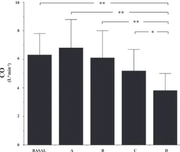
The results of post hoc analysis (Table 2) showed that changes occurring during 5-m breath-hold diving (condition D step) accounted for most of variance observed in the experi-mental series. In particular, cardiac output in condition D was lower than at any other experimental conditions (Fig. 2); left ventricular diastolic volume and stroke volume were lower in
condition D than in A (head-out immersion), whereas left
ventricular systolic volume results were lower in condition D than in B (surface submersion while breathing). As concerns diastolic function indexes, E/A was higher in condition D than in condition A (head-out immersion) and C (full body immer-sion at the surface while breath-holding). Finally, DTE was significantly shorter in condition D than in basal and in
con-dition C (Fig. 3).
No significant changes were observed in the dimensions of right ventricle at end diastole and of left atrium.
Fig. 2. Cardiac output (CO) in basal (dry) conditions and in the different immersion states. Values are means⫾ SD. SeeMATERIALS AND METHODSfor definition of conditions A, B, C, and D. *P⬍ 0.05; **P ⬍ 0.01.
Table 2. P values for comparisons between the different experimental conditions
LV EDV LV ESV LV SV CO HR RVD E Peak A Peak E/A DTE
Basal vs. A NS NS NS NS NS NS NS NS NS NS Basal vs. B NS NS NS NS NS NS NS NS NS NS Basal vs. C NS NS NS NS NS NS NS NS NS NS Basal vs. D NS NS NS 0.004 NS NS NS NS NS 0.009 A vs. B NS NS NS NS NS NS NS NS NS NS A vs. C NS NS NS NS NS NS NS NS NS NS A vs. D 0.012 NS 0,03 0.001 NS NS NS NS 0.001 NS B vs. C NS 0.028 NS NS NS NS NS NS NS NS B vs. D NS 0.003 NS 0.006 NS NS NS NS NS NS C vs. D NS NS NS 0.045 NS NS NS NS 0.033 0.037 NS, nonsignificant.
Fig. 3. Deceleration time of E peak (DTE) in basal (dry) conditions and in the different immersion states. Values are means⫾ SD. *P ⬍ 0.05; **P ⬍ 0.01.
on April 18, 2012
jap.physiology.org
DISCUSSION
The present study confirms and extends previous observa-tions of a clearly appreciable diving response that leads to a reduction in cardiac output during breath-hold diving in hu-mans (22). The sequence of immersions actually induced a significant reduction in cardiac output (due to a decrease in both HR and stroke volume) and in left ventricular diastolic and systolic volumes. Such a hemodynamic pattern is consis-tent with a preload reduction (since both increased afterload and/or reduced myocardial contractility would have implied increased left ventricular volumes).
The design of the study aimed to discriminate among the possible determinants of the diving cardiovascular response, as breath-hold diving exposes the organism to a series of stimuli (body immersion, breath holding, diving reflex elicitation, environmental pressure effects), overlapping each other. Most significant cardiac changes were observed during diving at depth, while surface immersion (irrespective of head in or out, breathing or breath holding) had, in our series, trivial effects on cardiac function. These data may be explained by several reasons. The progressive application of immersion stimuli (from head-out to diving at depth) may have attenuated car-diovascular changes at each step. Moreover, the reduced stim-ulation of facial receptor during submersion (subjects were wearing a diving mask), the small difference between air and water temperatures, and the relatively comfortable water tem-perature, not far from thermoneutrality, may also contribute to explaining this observation, since both diving-induced brady-cardia and peripheral vasoconstriction are marked in cold water (1, 23, 26). On the other hand, full body immersion in colder water can elicit sympathetic activation, potentially affecting cardiovascular response to immersion and diving (18). Thus body immersion, breath holding, and elicitation of diving reflex seem to have, per se, a relatively minor role in humans compared with the effect of diving at depth. Therefore, the increase in hydrostatic pressure seems to be essential in induc-ing cardiovascular changes durinduc-ing breath-hold divinduc-ing. A pre-vious study in humans, evaluating immersion, submersion, and simulated diving at depth in a pressure chamber (12), obtained different results, with cardiac output during diving significantly higher compared with both dry measurement at 1 ATA and surface breath holding. Methodological differences in cardiac output measurement and the possible influences of a sympa-thetic activation due to the unfamiliar experience represented by compression in a confined space might explain this discrep-ancy (25).
An increase in E/A with reduction of deceleration time of early filling peak was observed, during diving at depth, at Doppler evaluation of transmitral blood flow. This change, already observed in breath-hold diving athletes (22), is, in the clinical setting, typical of a restrictive/constrictive left ventric-ular diastolic dysfunction (24). It may be hypothesized that the reduction of chest volume (due to the increased environmental pressure), combined with an increase in intrathoracic blood content (3, 16), may exert a constraint on the heart, able to induce an impairment of left ventricular filling and, in turn, a relative reduction in preload (transmural filling pressure) and cardiac output (22). It may be speculated that these changes might contribute to the pathophysiology of diving-induced acute pulmonary edema. During deep and prolonged dives,
pulmonary vascular bed congestion (due to both an increased venous return and an impaired left ventricular filling), com-bined with an uneven hypoxic pulmonary vasoconstriction caused by hypoxia (4, 15), could lead to a pulmonary capillary stress failure (33) and pulmonary edema (34). The unfavorable consequences of a large intrathoracic blood redistribution, combined with thoracic squeeze during diving, are supported by two observations. On one hand, a recent report by Lindholm et al. (20) showed that breath-hold divers may have hemoptysis and instrumental signs of lower airway edema after shallow diving (6 m) performed at residual volume. On the other hand, animals highly adapted to diving (like pinnipeds) have special-ized anatomical structures devoted to reduce the intrathoracic venous return during immersion (caval sphincter, hepatic si-nus) (11, 29).
It is noteworthy to mention that changes in right ventricular diastolic dimension were not observed at any stage of the protocol, while intrathoracic blood displacement induced by immersion (3, 16) should, theoretically, be associated with right ventricular volume overload (17). Our negative finding might be explained by circulatory adjustments that rapidly occur in the period preceding cardiac imaging during the different experimental conditions. Alternatively, it might re-flect the intrinsic inefficiency of echocardiography in accu-rately detecting small changes in right ventricle dimensions, owing to its complex three-dimensional anatomy (32).
In conclusion, our study documents, in humans performing breath-hold diving at shallow depth (5 m), a cardiovascular response qualitatively similar to marine mammals. Body im-mersion at surface, diving reflex elicitation, and breath holding all seem to contribute only marginally to cardiac changes observed at depth, where the hydrostatic pressure on the chest becomes sufficiently high to constrict the heart, hampering its diastolic filling and reducing stroke work.
ACKNOWLEDGMENTS
The authors acknowledge Dr. Andrea Ripoli for invaluable assistance in statistical analysis, and the DiveSystem company (Follonica, Italy) for making available underwater equipment and swimming pool.
REFERENCES
1. Aellig WH. Clinical pharmacology, physiology and pathophysiology of superficial veins-1. Br J Clin Pharmacol 38: 181–186, 1994.
2. Andersson JPA, Line´r MH, Fredsted A, Schagatay EKA. Cardiovas-cular and respiratory responses to apneas with and without face immersion in exercising humans. J Appl Physiol 96: 1005–1010, 2004.
3. Arborelius M, Balldin UI, Lidja B, Lundgren CEG. Hemodynamic changes in man during immersion with the head above water. Aerospace
Med 43: 592–598, 1972.
4. Bartsch P, Gibbs SR. Effect of altitude on the heart and the lungs.
Circulation 116: 2191–2202, 2007.
5. Bedini R, Reale A, Belardinelli A, Passera M, Guerriero L, Navari A,
Dalle Luche A, Benassi A, Giuffre´ E, Cialoni D, Pingitore A, Ma-rabotti C, Data PG. Technologies for underwater biotelemetry during
diving. In: Blue 2005 Human Behaviour and Limits in Underwater
Environment. Special Conference on Breath-hold Diving, edited by Bedini
R, Belardinelli A, and Reale L. Pisa, Italy; STAR CNR Research Campus, 2005, p. 61.
6. Bedini R, Belardinelli A, Passera M, Reale L. Apparecchiatura per
ecografie subacquee. Italian Patent N. PI2005A000052, May 12, 2005;
PCT Application PCT/IB2006/001186, May 9, 2006.
7. Blix AS, Elsner R, Kjekshus JK. Cardiac output and its distribution through capillaries and A-V shunts in diving seals. Acta Physiol Scand 118: 109 –116, 1983.
8. Brick I. Circulatory responses to immersing the face in water. J Appl
Physiol 21: 33–36, 1966.
296 CARDIAC CHANGES DURING DIVING
J Appl Physiol•VOL 106 • JANUARY 2009 •www.jap.org
on April 18, 2012
jap.physiology.org
9. Dahlback GO, Jonsson E, Liner MH. Influence of hydrostatic compres-sion of the chest and intrathoracic blood pooling on static lung mechanics during head-out immersion. Undersea Biomed Res 5: 71– 85, 1978. 10. Duprez D, De Buyzere M, Trouerbach J, Ranschaert W, Clement DL.
Continuous monitoring of haemodynamic parameters in humans during the early phase of simulated diving with and without breatholding. Eur
J Appl Physiol 81: 411– 417, 2000.
11. Elsner R, Hanafee WN, Hammond DD. Angiography of the inferior vena cava of the harbor seal during simulated diving. Am J Physiol 220: 1155–1157, 1971.
12. Ferrigno M, Hickey DD, Liner MH, Lundgren CEG. Simulated breath-hold diving to 20 meters: cardiac performance in humans. J Appl Physiol 62: 2160 –2167, 1987.
13. Fitz-Clarke JR. Adverse events in competitive breath-hold diving.
Un-dersea Hyperb Med 33: 55– 62, 2006.
14. Folland ED, Parisi AF, Moynihan PF, Ray Jones D, Feldman CL, Tow
DE. Assessment of left ventricular ejection fraction and volumes by
real-time, two-dimensional echocardiography. A comparison of cinean-giographic and radionuclide techniques. Circulation 60: 760 –766, 1979. 15. Heistad DD, Abboud FM. Circulatory adjustments to hypoxia.
Circula-tion 61: 463– 470, 1980.
16. Hong SK, Cerretelli P, Cruz JC, Rahn H. Mechanics of respiration during submersion in water. J Appl Physiol 27: 537–538, 1969. 17. Hong Suk-Ki. Breath-hold diving. In: Bove and Davis’ Diving Medicine,
edited by Bove AA. Philadelphia, PA: Saunders, 1997 p. 65–74. 18. Jay O, Christensen JPH, White MD. Human face-only immersion in
cold water reduces maximal apnoeic times and stimulates ventilation. Exp
Physiol 92: 197–206, 2007.
19. Lang RM, Bierig M, Devereux RB, Flachskampf FA, Foster E,
Pellikka PA, Picard MH, Roman MJ, Seward J, Shanewise JS, Solomon SD, Spencer KT, St. John Sutton M, Stewart WJ.
Recom-mendations for chamber quantification: a report from the American Soci-ety of Echocardiography’s Guidelines and Standards Committee and the Chamber Quantification Writing Group, developed in conjunction with the European Association of Echocardiography, a branch of the European Society of Cardiology. J Am Soc Echocardiogr 18: 1440 –1463, 2005. 20. Lindholm P, Ekborn A, Oberg D, Gennser M. Pulmonary edema and
haemoptysis after breath-hold diving at residual volume. J Appl Physiol 104: 912–917, 2008.
21. Marabotti C, Pingitore A, L’Abbate A, Bedini R. Cardiovascular changes during diving. In: New Insight into Cardiovascular Apparatus
During Exercise. Physiological and Physio-pathological Aspects, edited
by Crisafulli A and Concu A. Kerala, India: Research Signpost, 2007 p. 205–221.
22. Marabotti C, Belardinelli A, L’Abbate A, Scalzini A, Chiesa F,
Cialoni D, Passera M, Bedini R. Cardiac function during breath-hold
diving in humans. An Echocardiographic study. Undersea Hyperb Med 35: 83–90, 2008.
23. Marsh N, Askew D, Beer K, Gerke M, Muller D, Reichman C. Relative contributions of voluntary apnoea, exposure to cold and face immersion in water to diving bradycardia in humans. Clin Exp Pharmacol Physiol 22: 886 – 887, 1995.
24. Nishimura RA, Tajik AJ. Evaluation of diastolic filling of left ventricle in health and disease: Doppler echocardiography is the clinician’s Rosetta stone. J Am Coll Cardiol 30: 8 –18, 1997.
25. Ross A, Steptoe A. Attenuation of the diving reflex in man by mental stimulation. J Physiol 302: 387–393, 1980.
26. Schagatay E, Holm B. Effects of water and ambient air temperatures on human diving bradycardia. Eur J Appl Physiol 73: 1– 6, 1996.
27. Shiraki K, Konda N, Sagawa S, Lin YC, Hong SK. Cardiac output by impedance cardiography during head-out water immersion. Undersea
Biomed Res 13: 247–256, 1986.
28. Thompson D, Fedak MA. Cardiac responses of grey seals during diving at sea. J Exp Biol 174: 139 –154, 1993.
29. Thornton SJ, Spielman DM, Pelc NJ, Block WF, Crocker DE, Costa
DP, LeBoeuf BJ, Hochachka PW. Effects of forced diving on the spleen
and hepatic sinus in northern elephant seal pups. Proc Natl Acad Sci USA 98: 9413–9418, 2001.
30. Thornton SJ, Hochachka PW, Crocker DE, Costa DP, LeBoeuf BJ,
Spielman DM, Pelc NJ. Stroke volume and cardiac output in juvenile
elephant seals during forced dives. J Exp Biol 208: 3637–3643, 2005. 31. Thorsen HC, Zubieta-Calleja G, Paulev PE. Decompression sickness
following seawater hunting using underwater scooters. Res Sports Med 15: 225–239, 2007.
32. Vieillard-Baron A, Prin S, Chergui K, Dubourg O, Jardin F. Echo-Doppler demonstration of acute cor pulmonale at the bedside in the medical intensive care unit. Am J Respir Crit Care Med 166: 1310 –1319, 2002.
33. West JB. Pulmonary capillary stress failure. J Appl Physiol 89: 2483– 2489, 2000.
34. Wilmshurst PT, Nuri M, Crowther A, Webb-Peploe MM. Cold-induced pulmonary oedema in scuba divers and swimmers and subsequent development of hypertension. Lancet 14: 62– 65, 1989.
on April 18, 2012
jap.physiology.org