Università degli Studi di Ferrara
DOTTORATO DI RICERCA IN
BIOCHIMICA, BIOLOGIA MOLECOLARE
E BIOTECNOLOGIE
CICLO XXII
COORDINATORE Prof. Francesco Bernardi
ABCA1 TRANSPORTER IN MONOGENIC
DISORDERS OF CELL CHOLESTEROL
METABOLISM: TANGIER DISEASE AND
FAMILIAL HYPERCHOLESTEROLEMIA
Settore Scientifico Disciplinare BIO/11
Dottorando Tutore
Dott. Bocchi Letizia Prof. Bernardi Francesco
Table of Contents
Abstract xi
1 INTRODUCTION 1
1.1 ABCA1 protein . . . 1
1.1.1 ABC proteins family . . . 1
1.1.2 ABCA1 protein structure . . . 2
1.1.3 Cell cholesterol efflux . . . 4
1.1.4 Reverse Cholesterol Transport . . . 5
1.1.5 Mechanism of action of ABCA1 protein . . . 7
1.1.6 ABCA1 intracellular traffic . . . 10
1.2 ABCA1 gene . . . 11
1.2.1 Gene expression regulation . . . 13
1.2.2 Post-transcriptional regulation . . . 17
1.2.3 Post-translational regulation . . . 17
1.3 Tangier Disease . . . 19
1.3.1 Historical Notes . . . 19
1.3.2 Case history of Tangier Disease . . . 20
1.3.3 Familial HDL Deficiency . . . 22
1.3.4 Cellular defects in Tangier Disease and Familial HDL deficiency . . . 22
1.3.5 Mutations on ABCA1 gene, Tangier Disease and Fa-milial HDL deficiency . . . 22
1.3.6 ABCA1, Ischemic Heart Disease and atherosclerosis . 23 1.4 ABCA1 gene screening . . . 25
1.4.1 Mutations identified up to now in ABCA1 gene . . . . 25 iii
1.4.2 Biological impact of ABCA1 mutations . . . 26
1.4.2.1 Polymorphisms and their biological value . . 27
1.4.2.2 Biological effect of mutations . . . 28
1.5 Cell cholesterol homeostasis . . . 30
1.5.1 De novo cholesterol synthesis . . . 30
1.5.2 Cholesterol uptake and endosomal cholesterol traffic . 31 1.5.2.1 LDLR structure . . . 32
1.5.2.2 The SREBP-SCAP system . . . 33
1.5.2.3 LDLR regulation . . . 35
1.5.3 Intracellular cholesterol transport . . . 37
1.5.4 Oxysterols . . . 38
1.5.5 Cholesterol efflux . . . 39
1.6 Monogenic disorders of cholesterol metab. . . 40
1.6.1 The NPC and CTX example . . . 40
1.6.2 Familial Hypercholesterolemia . . . 42
2 AIM OF THE STUDY 45 3 PATIENTS, MATERIALS AND METHODS 47 3.1 Subjects analyzed between 2005 and 2009 . . . 47
3.2 Materials . . . 48
3.2.1 Buffers and media . . . 48
3.2.2 Instruments and softwares . . . 51
3.3 Methods . . . 51
3.3.1 Isolation of DNA from peripheral blood . . . 51
3.3.2 Polymerase Chain Reaction (PCR) . . . 52
3.3.3 DHPLC . . . 53
3.3.3.1 Basic Principles . . . 54
3.3.4 Electrophoresis on Agarose gel . . . 55
3.3.5 DHPLC sample preparation . . . 55
3.3.6 Purification of PCR products . . . 55
3.3.7 Sequence reaction . . . 56
3.3.8 Purification of sequence reactions . . . 56
TABLE OF CONTENTS v
3.3.10 In silico prediction . . . 57
3.3.11 Minigene construction of ABCA1 Ex19-23 . . . 57
3.3.12 Creation of the pcDNA3.1-ABCA1-GFP vector . . . . 58
3.3.12.1 pcDNA3.1 and pEGFP-C3 . . . 58
3.3.12.2 pcDNA3.1-ABCA1-GFP . . . 58
3.3.12.3 pcDNA3.1-ABCA1-GFP ∆11-33 . . . 59
3.3.13 Transformation chemically competent E.coli . . . 60
3.3.14 Amplification of plasmidic cDNA in prokaryotic cells . 61 3.3.14.1 Small scale isolation of plasmid DNA . . . . 61
3.3.14.2 DNA selection via sequencing . . . 62
3.3.14.3 Large scale isolation of plasmid DNA . . . . 63
3.3.14.4 Control of plasmid DNA . . . 63
3.3.15 Cells . . . 64
3.3.15.1 HEK293 cell line . . . 64
3.3.15.2 COS-1 cell line . . . 64
3.3.15.3 Fibroblasts . . . 65
3.3.15.4 Monocyte isolation from whole blood . . . . 65
3.3.16 Cells treatment . . . 67
3.3.17 Transfection with LipofecTAMINE 2000 . . . 67
3.3.18 RNA extraction from cells . . . 68
3.3.19 ABCA1 cDNA analysis . . . 68
3.3.20 Cells Staining and Confocal Microscopy . . . 70
3.3.20.1 Fixation of HEK293 cells . . . 70
3.3.20.2 Labeling with anti-Calnexin antibody . . . . 70
3.3.21 Protein extraction from cells . . . 71
3.3.22 SDS-PAGE and Western Blot . . . 72
3.3.23 Efflux studies on transfected HEK293 cells . . . 74
3.3.24 Efflux studies on fibroblasts . . . 75
3.3.25 Oxidase treatment and cholestenone measurement . . 76
4 RESULTS 77 4.1 ABCA1 gene analysis . . . 77
4.1.1 List of the identified mutations . . . 78
4.3 Mutations affecting splice sites . . . 83
4.3.1 Ex-vivo investigation of the c.814-14 insA . . . 84
4.3.2 Ex-vivo investigation of the c.4773 + 1 G>A . . . 85
4.3.3 Analysis of the splice site mutation c.2961 –2 A>C . . 87
4.3.3.1 Ex vivo analysis of c.2961 -2A>C . . . 88
4.3.3.2 In vitro analysis of c.2961 –2 A>C . . . 89
4.4 In vitro functional characterization . . . 91
4.5 Ex vivo studies . . . 95
4.5.1 Protein extraction from fibroblasts and Western blot for ABCA1 protein . . . 95
4.5.2 Efflux studies . . . 97
4.5.3 Quantification of membrane cholestenone content . . . 99
4.6 Study of ABCA1 protein in FH fibroblasts . . . 100
4.6.1 Western blot for LDLR . . . 101
4.6.2 ABCA1 protein expression in FH cells . . . 102
4.6.3 Efflux studies . . . 105
5 DISCUSSION 109
6 REFERENCES 117
A Database of mutations 135 B Database of polymorphisms 141
List of Figures
1.1 ABCA1 protein structure . . . 3
1.2 Reverse Cholesterol Transport. . . 6
1.3 Mechanism of action of ABCA1. . . 9
1.4 ABCA1 Intracellular traffic. . . 12
1.5 Regulation of the ABCA1 gene promoter . . . 16
1.6 Tangier Island . . . 20
1.7 Cholesterol synthesis . . . 32
1.8 LDLR structure . . . 34
1.9 Two Actions of Insigs in Cholesterol Homeostasis . . . 35
1.10 SREBP regulation of cholesterol metabolism. . . 36
1.11 Expression of ABCA1 in human NPC1-deficient fibroblasts . 41 1.12 ABCA1 and SREBP-1c expression in CYP27 deficient fibrob-lasts . . . 42
1.13 ABCA1 expression and function in LDLR_/_ macrophages . 44 3.1 Overlapping PCR strategy . . . 60
4.1 PCR products for c.814 -14 ins A analysis. . . 84
4.2 Sequence of exon 8/exon 9 junction in ABCA1 cDNA. . . 86
4.3 Analysis of transcripts generated by c.4773+1G>A mutation 87 4.4 Analysis of transcripts found in monocytes of c.2961-2 A>C proband. . . 89
4.5 Analysis of transcripts generated by c.2961-2 A>C minigene. 91 4.6 Partial sequence of transcripts generated by c.2961-2 A>C minigene. . . 92
4.7 Scheme of abnormal mRNA splicing. . . 93 vii
4.8 Partial sequence of endogenous ABCA1 cDNA from COS-1cells. 94 4.9 Confocal microscopy image of transfected HEK293. . . 95 4.10 Cholesterol efflux from transfected HEK293. . . 95 4.11 Immunoblot of ABCA1 protein from cultured skin fibroblasts. 97 4.12 Cholesterol efflux to Apo A-I in skin fibroblasts. . . 98 4.13 Quantification of membrane cholestenone content . . . 100 4.14 Immunoblot of LDLR protein from FH fibroblasts. . . 103 4.15 Immunoblot of ABCA1 protein from FH fibroblasts. Basal
conditions. . . 104 4.16 Measure of intracellular cholesterol mass in control, FH and
Tangier fibroblasts. . . 104 4.17 Immunoblot of ABCA1 protein from FH fibroblasts.
Choles-terol loading. . . 105 4.18 Immunoblot of ABCA1 protein from FH fibroblasts. LXR
stimulation. . . 106 4.19 Immunoblot of ABCA1 protein from FH fibroblasts. LXR
stimulation with T0901317. . . 107 4.20 Cholesterol efflux from FH fibroblasts. . . 107 4.21 Linear correlation ABCA1 protein - cholesterol efflux. . . 108
List of Tables
3.1 Buffers and Media . . . 50
3.2 Instruments and softwares . . . 51
3.3 FH fibroblasts list. . . 66
4.1 List of mutations identified in TD/FHD patients. . . 79
4.2 Position and in silico prediction of the missense mutations. . . 81
4.3 FH fibroblasts cell lines. . . 101
Abstract
Background: ABCA1 is an ubiquitous plasma membrane transporter that promotes the efflux of cholesterol and phospholipids from the cell membrane to an extracellular acceptor, known as apolipoprotein A-I (apoAI), the main protein constituent of plasma high density lipoprotein (HDL). ABCA1 me-diated cholesterol/phospholipids efflux leads to the extracellular formation of HDL. Loss of function mutations of both ABCA1 alleles is the cause of Tangier Disease (TD), a recessive disorder characterized by extremely low levels of plasma HDL, increased risk of cardiovascular disease (CAD) and other clinical manifestations. Heterozygous mutations of ABCA1 gene cause Familial HDL Deficiency (FHD), characterized by half normal level of HDL-C and increased CAD risk. Familial Hypercholesterolemia (FH) is a monogenic disorder due to mutations of LDLR gene. In addition to the striking elevation of plasma LDL, FH patients (especially homozygotes) have greatly reduced plasma HDL levels, suggesting that they may have a defect in ABCA1-mediated cholesterol efflux pathway. Aim: Specific aims of this work were: i) the identification and functional characterization of several new ABCA1 mutations found in TD and FHD; ii) the study of ABCA1 regulation in fibroblasts derived from FH patients. Methods: ABCA1 gene was analyzed in 27 subjects with familial low HDL-C levels, by DHPLC and direct sequencing. Among the novel 22 mutations identified, 3 splice site mu-tations, 6 missense mutations and 1 frameshift mutation were investigated. The effect of splice site mutations was investigated ex-vivo, using patients’ fibroblasts or blood mononuclear cells, or in vitro by looking at transcripts generated by the expression of a mutant ABCA1 minigene in a heterologous cell system. ABCA1 protein expression was studied in eight FH derived
broblast cell lines under different conditions: basal culture conditions, stim-ulation with free cholesterol (FC), 22-hydroxycholesterol/9cis-Retinoic Acid (22OH/9cRA), or the LXR agonist T0901317. Cholesterol efflux to ApoA-I was also measured in these fibroblasts. Main results: 22 novel ABCA1 mu-tations were reported. The effect of 3 splice site mumu-tations (c.814-14InsA, c.2961-2A>C, c.4773+1G>A) was investigated with an ex-vivo or with a minigene approach. The c.814-14InsA resulted to be non-pathogenetic, the c.2961-2A>C resulted in the generation of three abnormally spliced mRNA species, the c.4773+1G>A caused the formation of an ABCA1 protein con-taining an in-frame deletion, which was poorly synthesized by cells. Five ad-ditional fibroblast cell lines derived from TD or FHD patients were studied in terms of ABCA1 protein expression and function. Fibroblasts carrier of R587W, A1046D, D1099Y, H1600R/M586Fs.629X, R130K/N1800H showed a null ABCA1-mediated cholesterol efflux. ABCA1 protein expression was studied in eight FH fibroblasts under different stimuli. Under basal culture conditions ABCA1 expression resulted lower in FH cells with respect to con-trol cells. This reduction was observed also after stimulation of cells with a load of FC or with 22OH/9cRA, known to stimulate ABCA1 expression via the transcription factor LXR. Treatment of cells with a non-steroidal LXR agonist (T0901317) was able to restore an ABCA1 expression similar to that of control fibroblasts. Also ABCA1-mediated cholesterol efflux in FH fibroblasts was lower than in control fibroblasts. Conclusions: Among 27 individuals with low HDL-C, 22 novel mutations in ABCA1 gene were identified. Three splice site, 6 missense and 1 frameshift mutation were functionally characterized in TD/FHD derived cells, allowing us to gain in-sight on genotype-phenotype correlations. Furthermore we demonstrated a defect in ABCA1 protein expression and function in FH, which may be partly responsible for the low plasma HDL-C levels exhibited by FH patients.
Chapter 1
INTRODUCTION
1.1
ABCA1 protein
1.1.1 ABC proteins family
ATP-binding cassette (ABC) transporters constitute one of the largest known protein superfamilies. These evolutionary highly conserved multispan trans-membrane molecules use the energy of ATP hydrolysis to translocate a broad spectrum of molecules across the cell membrane. Substrates that are trans-ported by ABC molecules include lipids, peptides, amino acids, carbohy-drates, vitamins, ions, glucuronide and glutathione conjugates and xenobi-otics. In eukaryotes, ABC transporters are located in the plasma membrane, in the membranes of intracellular compartments such as the Golgi, endo-somes, multivesicular bodies, endoplasmic reticulum, peroxisomes and in mitochondria. To date, 59 members of the human family of ABC trans-porters have been identified which, based on their structural relatedness, are subdivided into 7 families, designated ABC A–G [1,2].
Among these we cite:
• Subfamily ABCA, including 12 transporters, which is distinguished from others for being expressed only by multicellular organisms. In this subfamily, transmembrane domains and ATP binding domains consti-tute a single polypeptide.
• Subfamily ABCB, including transporters involved in multidrug resis-1
tance.
• Subfamily ABCC, including proteins which mediate ions transport, toxins elimination and signal transmission. The most famous is CFTR (Cystic Fibrosis Transport Regulator), which mediate Cl2 export: func-tional defects in this transporter are responsible of Cystic Fibrosis. Functional ABC transporters show a common global organization. So-called “full-size ABC transporters” are typically composed of two tandemly linked functional units while “half-size transporters” are usually composed by a sin-gle unit; each ABC unit (200-250 aa) contains 2 short, highly-conserved pep-tide motives directly involved in ATP binding (called Walker A and Walker B) and an “ABC signature” localized between them.
Being membrane transporters, ABC proteins contain one or more trans-membrane domains, tipically constituted by 6 helices, which can either be part of a single polypeptide (full-transporters) or be involved in a multi-protein complex bound to the membrane, so that they can form homo- or heterodimers, which consist of two half-size transporters that are encoded by distinct genes.
It is thus conceivable that full-size ABC transporters, as large compo-nents of membrane associated multiunit complexes, are not necessarily re-stricted to serve one defined function but are likely involved in a spectrum of biologic activities depending on the specific assembly of the functional complex and the biologic activities of their interaction partners [3].
1.1.2 ABCA1 protein structure
ABCA1 (formerly known as ABC1) is the prototypic member of ABCA sub-family and was initially identified by Luciani et al. [4] in mouse in 1994; 5 years later it was found to be responsible of two human genetic diseases [5], familial high-density lipoprotein (HDL)-deficiency (FHD) and Tangier dis-ease (TD), which identified ABCA1 as a major regulator of HDL metabolism [6-8].
ABCA1 is an integral membrane full-size transporter formed by two mir-ror halves each one containing 6 transmembrane domains, one large, highly-glycosylated, extracellular loop and one Nucleotide Binding Domain (NBD)
1.1. ABCA1 PROTEIN 3 composed by two conserved peptide motifs known as Walker A and B (thus it binds two ATP molecules). Both amino and carboxi-terminals are intracito-plasmatic [9]. The two large extracellular loops are linked by a disulfide bond.
It is a 2261-amino acid polypeptide with a molecular weight of 220 kDa [10,11].
Figure 1.1: ABCA1 protein structure.
ABCA1 is a ubiquitous protein and promotes the unidirectional active transport of two major components of the plasma membrane, cholesterol and phospholipids, to an extracellular acceptor represented by some apolipopro-teins (the main one is apolipoprotein A-I, apoA-I) [12,13]. This ABCA1-mediated efflux is one of the first steps involved in the Reverse Cholesterol Transport (RCT), a process by which cholesterol is eliminated from periph-eral cells and transferred to the liver via the plasma compartment.
A fraction of ABCA1 is located in internal endocytic compartments where it may facilitate cholesterol efflux from early/late endosomes and lysosome [14]; it is also likely that ABCA1 influences vesicles trafficking from Golgi apparatus, maybe through its phospholipid-transferase activity [15].
Recent studies, however, suggested that ABCA1 transports cholesterol to apolipoproteins exclusively at the cell surface [16-18] thus different mech-anisms of action are still proposed and have to be considered.
1.1.3 Cell cholesterol efflux
The intracellular cholesterol content is the result of three different metabolic processes: endogenous synthesis, LDL-mediated internalization and elimina-tion (see 1.5.2). The combinaelimina-tion of these mechanisms is highly dynamic to meet cellular requirements and it is important that the equilibrium is main-tained to avoid a damaging over-accumulation of free, unesterified cholesterol (FC). Even a too large accumulation of esterified cholesterol (EC) is respon-sible of cellular damage as it happens in macrophages. This is the reason why a redundancy and complementarity of mechanisms exist to manage cell cholesterol efflux.
Four are the main processes involved:
i) the first one is a bidirectional passive diffusion mechanism, where free-cholesterol molecules spontaneously dissociate from the plasma membrane, diffuse through the aqueous phase and are incorporated in high density lipoprotein (HDL) particles by collision [19].
ii) The second mechanism is a bidirectional transport mediated by scav-enger receptor class B type 1 (SR-B1): on one side SR-B1 is the receptor for HDL, mediating the transport of cholesterol and cholesteryl esters from HDL particles [20,21]; on the other side it favours the excretion of cholesterol via a facilitate passive transport. It is primarily expressed in liver and nonplacen-tal steroidogenic tissues where SR-B1 directly interacts with HDL particles, releasing cholesterol and phospholipids localized on the plasma membrane [22-24].
iii) The third mechanism is an ABCA1-mediated unidirectional trans-port: ABCA1 is involved in ATP-dependent cholesterol efflux and its main acceptor is lipid poor apoA-I, the main peptide component of HDL particles [25].
iv) A fourth, recently identified, mechanism proposes the involvement of the ABC transporter G1 (ABCG1), widely expressed in human tissues, in promoting the efflux of cholesterol to mature HDLs. This ABCG1-mediated efflux has not been completely characterized yet but it probably contributes and complements ABCA1 transport.
1.1. ABCA1 PROTEIN 5 due to the fact that they have different roles in different types of cells. The most efficient seems to be the ABCA1-mediated cholesterol efflux: phospho-lipids and unesterified cholesterol are transferred to a monomolecular, pre-β-migrating, lipid-poor or lipid-free form of apoA-I. This mononuclear form of apoA-I is quite distinct from the pre-β-migrating discoidal HDL which con-tain 2 or 3 molecules of apoA-I per particle and which are present as minor components of the HDL fraction in human plasma [26,27]. The cholesterol-phospholipid complex transferred to apoA-I forms or increases its lipid con-tent, creating nascent, pre-β-HDL which increase their lipid concon-tent, during their movement in lymphatic and blood circulation. Free cholesterol taken up by HDL is then esterified by lecithin:cholesterol acyltransferase (LCAT) and hydrophobic cholesteryl esters are retained into the core of HDL so that new cholesterol molecules can be translocated on the HDL surface. The en-richment of cholesterol and localization of cholesteryl esters in the core of the particles lead to transformation of discoidal pre-β-HDL into spherical mature HDL.
If ABCA1-mediated cholesterol and phospholipid transport doesn’t oc-cur, then apoA-I is not enriched in lipids and is rapidly catabolised by the kidney. The total amount of HDL in the plasma therefore shows a large decrease.
1.1.4 Reverse Cholesterol Transport
Reverse Cholesterol Transport is a process where exceeding cholesterol is removed from peripheral, extra-hepatic tissues and transported to the liver for reuse in enterohepatic cycle where it can be converted in biliary acids or eliminated via biliary excretion [28].
HDL and apoA-I ability to promote Reverse Cholesterol Transport is believed to be one of the main atheroprotective mechanisms.
Atherosclerosis is a progressive disease characterized by the accumulation of cholesterol in the arterial walls. Increased levels of low-density lipopro-tein cholesterol (LDL) are causally related to atherosclerosis, supported by substantial evidence from large-scale intervention trials, from animal studies, and from genetic studies of families and of the general population. LDL
par-Figure 1.2: Reverse Cholesterol Transport.
ticles in plasma enter the arterial wall where they are oxidized. In response to oxidized lipids, arterial-wall cells secrete proteins that attract monocytes into the intima and lead to their locally differentiation into cholesterol-engorged macrophages, the so-called foam cells which continuously accumulate lipids. These foam cells constitute the initial lesions that subsequently develop into more advanced atherosclerotic plaques, characterized by the accumulation of lipid-rich necrotic debris and smooth muscle cells. Plaque rupture and to-tal occlusion due to thrombus formation result in clinical ischemic vascular events.
In this process, the Reverse Cholesterol transport assures mobilization of excess cellular cholesterol from arterial-wall macrophages preventing or reducing foam cell formation [29].
1.1. ABCA1 PROTEIN 7
1.1.5 Mechanism of action of ABCA1 protein
Many different models have been proposed so far to explain ABCA1 mech-anism of action.
The three main processes are the following:
• In a first model a direct interaction with a small regulatory pool of apoA-I is required in order to transfer lipids outside the cell [30]; ABCA1 recognizes and interacts with a cluster of amphipathic helices in the apolipoprotein structure. This interaction likely involves the first and forth extracellular loop [31,32]. Formation of novel apolipoprotein binding structures protruding from the cell surface results as an inter-mediate step in the pathway by which apolipoproteins remove excess cholesterol from cells [33]. In this model ABCA1 moves cholesterol and phospholipids to the outer leaflet, but it isn’t known if this transfer is either simultaneous or phospholipids are transferred before cholesterol [34] or, in a third option, the translocation is simultaneous unless phos-pholipid and cholesterol-rich domains are too distant so that the two phases are dissociated.
• In the second model ABCA1 is able to reach and bind the plasma membrane without necessarily interact with apoA-I. The two symmet-rical ABCA1 transmembrane bundles come together to form a cham-ber that scans the inner leaflet of the membrane for substrates, in-corporates them into the chamber, and flips them to the outer leaflet for extrusion from the cell. This involves a series of conformational changes in the ABC protein that is driven by ATP hydrolysis in the NBD domains. ABCA1 amino acid motifs in the chamber are likely to bind phospholipids. Again, it is unknown whether inner leaflet cholesterol is co-transported with these phospholipids or phospholipids translocation alone increases the accessibility of cholesterol that flips to the outer leaflet by other processes. The observation that ABCA1 can affect the cholesterol content of lipid rafts, which probably relies on lateral diffusion of cholesterol along the membrane, supports the idea that ABCA1 also translocates cholesterol [35]. When induced
by cholesterol loading of cells, ABCA1 therefore constitutively gen-erates cholesterol and phospholipid-rich membrane domains, even in the absence of apolipoproteins [36] and these domains bend from the plasma membrane to relieve the strain of the densely packed phos-pholipids, generating curved and disordered lipid surfaces that favour apolipoprotein interactions. During the first several minutes of ex-posure to apolipoproteins, JAK2 is activated by autophosphorylation, which in turn increases binding of apolipoproteins to ABCA1. This binding facilitates the interaction of apoA-I with the protruding lipid domains, promoting their solubilization and release from the cells. • The third model involves retroendocytosis; the pioneering study of
Takahashi and Smith [37] showed that following internalization, apoA-I is recycled back to the cell surface to be re-secreted. Other stud-ies showed that apoA-I and ABCA1 are co-localized in endosomal compartments, and that ABCA1 rapidly shuttles between intracellu-lar compartments and the plasma membrane [38,39]. These results support the idea that ABCA1-bound apoA-I is delivered from the plasma membrane to early and late endosomal and/or lysosomal com-partments, where it forms nascent lipoprotein particles that are sub-sequently secreted from the cell. However, several groups examining internalization and recycling of apoA-I concluded that the retroendo-cytosis pathway does not contribute significantly to HDL formation and that the majority of internalized apoA-I is directly transported to late endosomes and lysosomes for degradation while only a small frac-tion is re-secreted from cells and the majority of re-secreted apoA-I seems to be degraded in the medium. These results suggest that the mass of retroendocytosed apoA-I is not sufficient to account for HDL produced by cholesterol efflux [17].
On the other side, it has been recently demonstrated that apoA-I exhibits saturable association with the plasma membrane and intracellular compart-ments (ICCs), that apoA-I induces ABCA1 endocytosis and co-localizes with cell-surface derived ABCA1 on endosomal compartments; moreover apoA-I dissociation from apoA-ICCs is 4-fold faster than from the plasma membrane
1.1. ABCA1 PROTEIN 9 and approximately 30% of endocytosed ABCA1 is recycled to the cell sur-face giving a critical contribution to HDL formation under conditions where cells have accumulated excess lipoprotein-derived cholesterol in endosomal compartments [39,40].
In conclusion the mechanism of active cellular lipids ABCA1-mediated efflux to apoA-I is not yet clear; it may involve 1) binding of apoA-I either directly to ABCA1 or indirectly to a lipid site created by ABCA1 activity, 2) either simultaneous or sequential release of membrane phospholipid and cholesterol to apoA-I, and 3) assembly of nascent HDL particles either at the cell surface or at intracellular sites during the retroendocytosis of ABCA1 [30].
The precise mechanism of action of ABCA1 in promoting delivery of cellular cholesterol and phospholipids to apoA-I remains to be determined.
Figure 1.3: Mechanism of action of ABCA1.The three mechanisms of action of lipid-efflux ABCA1-mediated. Image adapted from [14].
1.1.6 ABCA1 intracellular traffic
An important intracellular mechanism that seems to characterize ABCA1 pathway is the formation of multiple structures like dimers, tetramers and oligomers.
In 2004 Denis et al. [41] demonstrated that the majority of ABCA1 exists as a tetramer which binds apoA-I, that neither apoA-I nor lipid molecules af-fect ABCA1 oligomerization and that apoA-I is associated with both dimeric and tetrameric, but not monomeric forms of ABCA1. These findings sup-port the concept that the homotetrameric ABCA1 complex constitutes the minimum functional unit required for the biogenesis of HDL particles.
Trompier et al [42] further highlighted that ABCA1 is associated in dimeric structures that undergo transition into higher order structures, i.e. tetramers, during the ATP catalytic cycle. The assembly in homodimers takes place in the endoplasmic reticulum, which is a rather common rule in the assembly of multimolecular channel structures; the ABCA1 dimers are oriented so that the C-terminal extremities of ABCA1 are in close proxim-ity. Both a putative PDZ binding motif present at the C terminus and the VFVNFA sequence located at positions 2210–2215 have been identified as po-tential interaction sites; they may be required either to stabilize the dimeric interaction or to introduce additional molecular partners in the complex. These assemblies are transient in nature but can be trapped experimentally by interfering with the ATP catalytic cycles, namely by stabilizing the ATP-bound state; they are tetramers, rather than dimers, undergoing major con-formational changes upon ATP binding. Resetting of the transporter in the initial conformation takes place after effluxes of membrane phospholipids.
As explained in 1.1.3, current evidences suggest that ABCA1-mediated cholesterol efflux to apoA-I involves mobilization of cholesterol from plasma membrane, endoplasmic reticulum, trans-Golgi network, late endocytic and lysosomal compartments and cholesteryl ester droplets.
Neufeld et al [38] indeed demonstrated that ABCA1 resides on the surface of both small, mobile vesicles as well as on large, relatively immobile perin-uclear vesicles. The small, fast-moving ABCA1-containing vesicles are likely to represent early endosomes, whereas the ABCA1-containing large, static,
1.2. ABCA1 GENE 11 perinuclear vesicles are likely to represent late endocytic compartments (late endosomes/lysosomes). The ABCA1-containing early endosomes undergo fu-sion and fisfu-sion events: they transiently interact with one another, with the ABCA1-containing late endocytic vesicles, and with the cell surface. They shuttle and tubulate while moving vectorially, consistent with directed move-ment along cytoskeletal elemove-ments such as microtubules or actin filamove-ments. In addition to ABCA1, the membrane components that may be transferred from the ABCA1 early endosomes to other cellular compartments may in-clude substrate lipids for ABCA1 [43,44] and/or acceptor apolipoproteins [38-40]. Cholesteryl Esters (CE) in LDL are hydrolyzed into unesterified cholesterol in late endosomes and lysosomes and this cholesterol may serve as a direct substrate pool for ABCA1-mediated cholesterol efflux to apoA-I, suggesting that late endosome/lysosome cholesterol might represent a signif-icant substrate pool for ABCA1 and HDL formation and this provides the opportunity for the removal of excess endocytosed LDL-derived cholesterol from the cell, prior to its potential distribution to other sites by NPC1.
Moreover, ABCA1 mobilizes endogenously synthesized cholesterol in the Golgi apparatus to lipidate de-novo synthesized apoA-I [44]; it also depletes cholesterol released from cytoplasmic cholesteryl ester droplets but since there is no evidence of ABCA1 localization or association with cholesteryl ester droplets, it is more likely that the unesterified cholesterol released from cholesteryl ester droplets is transported rapidly to the plasma membrane, via the Golgi apparatus, to become part of the ABCA1 substrate pool [45].
1.2
ABCA1 gene
The gene encoding ABCA1 transporter is located on the long arm of chro-mosome 9 (9q31.1) [46], has a total length of 149 kb and includes 50 exons and 49 introns. Exons dimensions vary between 35 bp (Ex 33) and 244 bp (Ex 49). Introns dimensions are more variable, from a minimum length of 111 bp (IVS 38) to a maximum length of 24000 bp (IVS 1) [47].
Exon 1 encodes for part of the 5’ UnTraslated Region (5’UTR). Exon 2 encodes for the remaining part of 5’UTR, for the first methionine and the subsequent 21 aa of ABCA1 amino-terminal region. The putative
tran-Figure 1.4: ABCA1 intracellular traffic between early, late endosomes, lyso-somes and the plasma membrane.
scription starting site is localized 315 bases upstream of the ATG in exon 2. The open reading frame length is 6723 nucleotides. The nucleotide sequence around the initial ATG in exon 2 is conformed to the Kozak consensus se-quence (RNNATGG), with a purine in position -3 and a G in position +4 [47].
If translation starts at the ATG in exon 2, the protein obtained from the corresponding mRNA contains 2261 aa [6,7,10,48]. The first 45 aa have chemical and physical properties similar to those of the signal peptide of many secreted proteins.
Another possible translation starting point is located in exon 4, where a 2201aa-long protein could originate. This ATG, however, is positioned in a context not related to the Kozak consensus sequence; it is therefore unlikely that this starting point is active [47].
With regard to the end of translation, the first polyadenylation site is about 3000 bp downstream from the stop codon. Thus the 3’UTR of ABCA1
1.2. ABCA1 GENE 13 mRNA is quite long [11,46,47].
ABCA1 gene promoter contains many motifs with a relevant role in ex-pression regulation.
Regulation sequences are located for 221 bp upstream of exon 1, as com-mon as in many genes, and for 24 kb inside IVS1.
Between exon 1 and 2 there is an alternative exon, called exon 1a, 136 bp in length and localized 2210 bp upstream of exon 2 [49]. This alternatively spliced transcript encloses the complete coding sequence and is expressed in testis and liver but not in macrophages [50]. Upstream of exon 1a an alter-native promoter containing a TATA box and a CAAT box sites is present, together with other potential binding sites for nuclear receptors containing sterol regulatory elements.
1.2.1 Gene expression regulation
ABCA1 gene has ubiquitous expression and in basal conditions is mostly translated in tissues like placenta, lung, liver, kidney, intestine, adrenal gland and some areas of the nervous system. A high expression is present in the same tissues also during the fetal development [11].
Many different factors are responsible of ABCA1 gene expression or in-hibition and some of them are tissue-specific.
First of all, ABCA1 expression is markedly induced by overloading cells with cholesterol, especially in macrophages [51,52]. This induction occurs ex-clusively through activation of the nuclear receptors liver X receptor (LXRα and/or LXRβ) and retinoid X receptor (RXR). LXR and RXR form obli-gate heterodimers that preferentially bind to response elements within the ABCA1 gene promoter and the first intron. LXRs and RXRs bind to and are activated by oxysterols and retinoic acid, respectively. 9-cis retinoic acid (9-cisRA), either alone or in combination with oxysterols, is the major retinoic acid that upregulates ABCA1 mRNA levels. The LXRα gene pro-moter in human macrophages contains a LXR responsive element, indicating that LXRα can autoregulate its own expression; this would serve to amplify the effects of oxysterols on the ABCA1 lipid efflux pathway. Because uptake of non-oxidized cholesterol by cells increases ABCA1 expression, cholesterol
must be converted to oxysterols before inducing ABCA1. It is believed that 22-hydroxycholesterol, 24-hydroxycholesterol, and 24(s),25-epoxycholesterol are the major naturally occurring liver LXR ligands. Most oxysterols are generated by cytochrome P-450 enzymes that are particularly prevalent in the liver and play a role in bile acid metabolism. One of these enzymes, sterol 27-hydroxylase (Cyp27), is broadly distributed in various tissues and cell types, including macrophages, suggesting that 27-hydroxycholesterol is the major LXR ligand in macrophages and other peripheral cells.
A sterol-responsive element has been mapped to an imperfect Direct Repeat (AGGTCA) spaced by four nucleotides (DR-4) which was shown to bind LXR/RXR heterodimers; mutations of the DR4 element strongly reduced oxysterol-responsive ABCA1 gene activation [53,54].
The induction of ABCA1 gene transcription activity by 25-hydroxycholesterol, another important stimulator of ABCA1 expression, is critically dependent on Sp1 elements and an E-box motif present in the proximal promoter region of ABCA1, 147 bp upstream of the originally identified transcriptional start site.
The two ABCA1 promoters, upstream exon1 and inside intron 1 respec-tively, contain other transcription regulators like a TATA box located 24 bp upstream the transcriptional start site, essential for the promoter activity.
Lipid metabolites other than sterols can modulate ABCA1 expression by the LXR system [49]:
• Polyunsaturated fatty acids act as antagonists to oxysterol binding to response elements in the LXRα gene, potentially interfering with induction of ABCA1 by sterols.
• Geranylgeranyl pyrophosphate (GGPP), a product of the mevalonate pathway which isoprenylates proteins, was shown to suppress LXR-induced ABCA1 synthesis by two mechanisms: as an antagonist of the interaction of LXR with its nuclear co-activator SRC-1 and as an activator of Rho GTP-binding proteins. This second mechanism might alter sterol trafficking in cells, reducing their availability as ligands for LXR.
1.2. ABCA1 GENE 15 • Activators of Peroxisome Proliferator Activating nuclear Receptors (PPARs)
also enhance ABCA1 transcription in some cells and stimulate choles-terol efflux by activating transcription of the LXRα gene through a complex interaction between PPARα, PPARγ and LXRα, which in turn induces ABCA1 transcription [55,56].
• Thyroid hormone receptor was reported to suppress ABCA1 transcrip-tion by forming a complex with RXR that competes for LXR/RXR binding to its response elements.
• ABCA1 transcription is also regulated by factors independent of LXR/RXR: membrane-permeable analogs of cAMP and inhibitors of cAMP-specific phosphodiesterase 4 stimulate ABCA1 transcription in macrophages by unknown mechanisms; cAMP act in these cells by promoting the binding of Sp1 and E-box binding factors to the proximal promoter of ABCA1 thus stimulating ABCA1 gene transcription and surface expression in absence of cholesterol, whereas ABCA1 expression in fi-broblasts is critically dependent on cholesterol loading [57,58].
• The calcium channel blocker verapamil can also enhance ABCA1 tran-scription by an LXR-independent process [59].
The ABCA1 gene promoter contains other transcriptional response elements that are potential sites of regulation, some of which may influence constitu-tive and tissue-specific expression of ABCA1: cytokines such as interferon-γ (IFN-γ), along with a two-fold increase in the activity of acylcoenzyme A: cholesterol acyltransferase (ACAT) decreases ABCA1 mRNA in macrophages under basal conditions as well as after sterol-loading. This results in a two-fold decrease of cholesterol effux [60,61].
Transforming growth factor β1 (TGF-β) and oncostatin M have been re-ported to modulate ABCA1 transcription in cultured macrophages and hep-atoma cells. Sterol regulatory element binding protein 2 (SREBP2), a tran-scription factor that regulates sterol synthesis, appears to interact with the ABCA1 promoter in vascular endothelial cells and to suppress ABCA1 tran-scription, thus providing an additional mechanism for suppressing cholesterol efflux when cells are sterol depleted [13].
It has also been demonstrated a role of LDLR in regulation of ABCA1 expression as illustrated below (see 1.6.2).
Moreover negative control elements binding the transcriptional repres-sor zinc-finger protein 202 (ZNF202) are thought to counterbalance ABCA1 mRNA expression and may be influenced by the genetic background. ZNF202 requires the presence and of a SCAN domain that mediates selective hetero-and homodimerization hetero-and of a functional TATA box to bind to in order to repress ABCA1 expression.
[Warning: Image ignored]
Figure 1.5: Regulation of the ABCA1 gene promoter.
Catepsine D (CTSD), a lysosomal proteinase, is another regulator of ABCA1 expression: it increases both ABCA1 mRNA expression and cel-lular ABCA1 protein, while its inhibition results in ABCA1 retention in lysosomal compartments, reducing its traffic to the plasma membrane [62]. CTSD expression is regulated by NPC1 activity in hepatocytes: NPC1-null hepatocytes show increased CTSD expression, both transcriptionally and post-transcriptionally and this therefore has an effect on ABCA1 expression and protein production [63].
Recently, ORP8, a protein belonging to OSBP (oxysterol-binding pro-tein) and ORP (OSBP-related) gene family, was shown to negatively regulate ABCA1 transcription by a mechanism involving DR4 and E-box elements [64]. Since ORP8 is anchored to the ER by a C-terminal transmembrane
do-1.2. ABCA1 GENE 17 main, transcriptional inhibition is probably indirect and related to the sterol binding or metabolic activity of this ORP.
1.2.2 Post-transcriptional regulation
The first proposed mechanism of ABCA1 post-transcriptional regulation oc-curs by phosphorylation of residues in the regulatory domain by protein kinase A (PKA) following cAMP stimulation.
IFNγ appears to suppress ABCA1 expression by further destabilizing its mRNA: the kinetics of ABCA1 mRNA and protein decrease is consistent with the early IFN-γ-induced changes in Stat1 phosphorylation and nuclear translocation. Therefore, down-regulation of ABCA1 by IFN-γ is a post-transcriptional response that occurs early in the process of IFN-γ-induced macrophage activation [65].
1.2.3 Post-translational regulation
Several processes that modulate ABCA1 protein stability have been de-scribed and there is not always a strict correlation between ABCA1 mRNA and protein levels.
When ABCA1 inducers are removed in the absence of apolipoproteins, ABCA1 mRNA and protein are degraded at a fast rate (half-life of 1–2 h) [66,67].The rapid protein turnover is largely due to a sequence at amino acids 1283–1306 in the first intracellular loop that is enriched in proline-glutamate-serine-threonine (PEST motif). Phosphorylation of T1286 and T1305 in this motif promotes ABCA1 proteolysis by an unknown member of the calpain protease family. There are several metabolic factors that modulate the rate of ABCA1 protein degradation by either this calpain system or other processes. The interaction of apolipoproteins with ABCA1-expressing cells dramat-ically reduces the rate of ABCA1 protein degradation by inhibiting ABCA1 proteolysis by calpain [66] and activating other signaling events that sta-bilize the protein [68]. This regulation acts as a feedback mechanism to sustain ABCA1 levels when acceptors for cellular lipids are available. First, the cellular interactions of apolipoproteins interfere with the phosphoryla-tion of the PEST motif. Second, the removal of membrane sphingomyelin by
apolipoproteins activates phosphatidylcholine phospholipase C, which sig-nals phosphorylation of ABCA1 at a site that stabilizes the protein [69]. It is unknown if either or both of these mechanisms require apolipoprotein binding to ABCA1.
Unsaturated free fatty acids directly destabilize ABCA1 protein in cul-tured cells by a signaling pathway involving activation of phospholipase D2 and protein kinase Cδ and phosphorylation of ABCA1 serines [67,70-72] [71,74-76].
ABCA1 also interacts with β1- and α1-syntrophin, scaffolding proteins that regulate transport through linkage to the cytoskeleton, resulting in sta-bilization at intracellular sites and the plasma membrane [73,74].
Oxisterol binding protein (OSBP) is implicated in the integration of sterol sensing/transport with sphingomyelin synthesis and cell signaling. OSBP negatively regulates ABCA1 by decreasing its stability in the cytoplasm; this effect is not correlated with OSBP function in sphingomyelin synthesis or interaction of OSBP with the ER or Golgi apparatus but is dependent on OSBP sterol-binding activity [75].
The LXRβ/RXR complex also has a role in regulating ABCA1 activ-ity: this complex binds directly to ABCA1 on the plasma membrane of macrophages and modulates cholesterol secretion; when cholesterol does not accumulate, ABCA1-LXRβ/RXR localizes on the plasma membrane but is inert; when cholesterol accumulates, oxysterols bind to LXRβ and the LXRβ/RXR complex dissociates from ABCA1, restoring ABCA1 activity and allowing apoA-I dependent cholesterol-efflux. Upon binding oxysterols in the cytosol, LXRβ/RXR is translocated to the nucleus and activates the transcription of ABCA1 and other genes [76].
Moreover, recently Singaraja et al [77] highlighted that ABCA1 is ro-bustly palmitoylated by the palmitoyl transferase DHHC8 at cysteines 3, -23, -1110, and -1111 and that abrogation of palmitoylation of ABCA1 by mutation of the cysteines results in a reduction of ABCA1 localization at the plasma membranes and a reduction in the ability of ABCA1 to efflux lipids to apoA-I. Thus, palmitoylation regulates ABCA1 localization at the plasma membrane, and regulates its lipid efflux ability.
1.3. TANGIER DISEASE 19
1.3
Tangier Disease
1.3.1 Historical Notes
In the history of American literature, the name Tangier is usually associated with the eccentric writer William Borroughs, who conceived some of his novels in this Moroccan town on the Straight of Gibraltar.
At the beginning of 1960s, D. Fredrickson et al. [78] from the Na-tional Institute of Health (USA) described a particular syndrome in two sib-lings characterized by: hypertrophic yellow-grayish tonsils, limph adenopa-thy, hepathosplenomegaly and sensitive-motor like peripheral neuropaadenopa-thy, in association with almost complete absence of HDL-C in the plasma (Anal-phalipoproteinemia). These two siblings belonged to a family living in the Tangier Island, a small isle located in Chesapeake Bay, approximately 20 miles west of the eastern shore of Virginia (see Figure 6). The island was discovered by the English explorer Captain John Smith in 1608 and owes its name to the poetic imagination of the sailor, who was reminded by its wide, sandy shores of the white dunes of the port of Tangier in Morocco [79].
After its discovery, the island was without a single inhabitant for the next 78 years, until 1686 when John Crockett together with a few comrades founded a settlement there. Except for a calamitous outbreak of cholera in 1886, nothing particular happened for the next 300 years, and the inhabitants made a modest living from the fishery. Tangier Island was well insulated from the mainland both economically and socially, so when Dr Fredrickson traveled there in the early 1960s, he found only one road, one car and about 900 residents, many of them speaking a unique local dialect and bearing the surname Crockett [79].
Because other three couples of siblings were found affected by the same disease in Tangier Island, Fredrickson proposed the denomination “Tangier Disease” (TD). The presence of many individuals affected in one family with-out a vertical transmission of the phenotype, suggested a recessive transmis-sion of this syndrome, as explained in the big pedigree originally described by Fredrickson in 1964 [78].
The manifestation of this disease in the Tangier Island is likely the result of a geographic isolation and of endogamy that happened among the
in-habitants during 400 years, all descendents from the small group of English refugees.
After the first description of Tangier Disease in 1964, many others fol-lowed in different geographic regions (USA, UK, New Zeeland, Australia, Switzerland, Germany, Poland, Italy, Canada, France, Spain, The Nether-land, Pakistan, Japan and Egypt).
Figure 1.6: Tangier Island. North-East coast of United States and Chesapeake Bay with Tangier Island.
1.3.2 Case history of Tangier Disease
The phenotypic, pathognomonic case history that allowed the identification of the first cases of Tangier Disease was the association of almost total ab-sence of HDL-C in the plasma and preab-sence of yellow-grayish tonsils [46].
The clinical phenotype of Tangier Disease has autosomic recessive trans-mission. However the biochemical phenotype (very low HDL-C and apoA-I levels) has a co-dominant transmission [81], that obligate heterozygotes have HDL-C and apoA-I levels intermediate between normal values and those of Tangier patients, but do not present any clinical feature.
Tangier Disease is characterized by two different biochemical alterations: • Cholesteryl Esters (CE) accumulation in many cell types, especially in histiocytes and in the reticuloendothelial system (aka: mononuclear phagocyte system);
1.3. TANGIER DISEASE 21 • Almost total absence of HDL-C in plasma.
Cholesteryl Esters accumulation is particularly evident in tonsils, lymph nodes, bone marrow, liver, intestine and Schwann cells of the peripheral nervous system.
At a morphological level, histiocytic cells (e.g. macrophages) appear like foam cells with the cytoplasm full of lipid droplets, which can be highlighted with Sudan red/black, sometimes with crystal structures. This lipidic mate-rial is formed by esterified cholesterol accumulated outside lysosomes.
The serious HDL defect is considered as a predisposition factor for car-diovascular diseases (angina pectoris, myocardial infarction, stroke etc.) on atherosclerotic base [53].
The phenotypic manifestations differ among individuals but symptoms may be hypertrophic tonsils, hepatosplenomegaly, anemia, thrombocytope-nia, stomatocytes, peripheral neuropathy and increased incidence of cardio-vascular disease, whose clinical penetrance is however extremely variable [48]; the most represented feature is anyway tonsils hypertrophy, with typically yellow-grayish lobulated tonsils, and hepatosplenomegaly [84]. Obviously, tonsillectomy and therefore absence of tonsils can complicate the diagnosis. Two major reports that dealt with this issue, published prior to the time that the ABCA1 gene was identified, indicated that despite the near absence of HDL, CVD (cardiovascular disease) only represents a significant problem in middle age and elderly TD patients (over 35 years) [84,85]. Taken the epidemiological evidence that a drug-induced increase of 1% in HDL-C concentration may be associated with as much as 3% decrease in death or myocardial infarction incidence, it is nevertheless remarkable that TD patients with HDL-C below the 5th percentile (for age and sex) do not appear to develop CVD until the age of 35-40 and some of these patients live well into their seventies. One factor that has been brought forward to explain this is the very low level of LDL-C found in TD patients but this hypothesis has not yet been confirmed. Anyway the Italian patients with TD, diagnosed up to now by our group (≈ 30 patients), show a marked predisposition in developing premature atherosclerosis.
1.3.3 Familial HDL Deficiency
Familial HDL deficiency (FHD) is a more common disorder of lipid metabolism than Tangier disease, characterized by isolated moderate reductions of HDL-C in absence of the typical characteristics of clinically evident TD. Recently Frikke-Schmidt et al. revealed that i) approximately 10% of individuals with the lowest percentile of HDL cholesterol in the general population are het-erozygous for mutations in ABCA1 and ii) the frequency of FHD caused by ABCA1 mutations in the general population is about three in 1000 or considerably higher than previously assumed [87].
1.3.4 Cellular defects in Tangier Disease and Familial HDL deficiency
The Cholesteryl Esters accumulation in many different cell types of the retic-uloendothelial system suggested that in TD a defect of cholesterol intracellu-lar traffic is present. This hypothesis was sustained by a series of experimen-tal evidences obtained from dermal fibroblasts and monocyte-macrophages in TD patients. TD fibroblasts when overloaded with cholesterol (after in-cubation with acetylated LDL or free cholesterol) show a defect in phospho-lipid and cholesterol efflux to plasma apolipoprotein acceptors (e.g. apoA-I, apoA-II, apoC or apoE) [88-90].
1.3.5 Mutations on ABCA1 gene, Tangier Disease and Fa-milial HDL deficiency
Until genetic bases of TD and FHD were unknown, patients were diagnosed on the basis of their clinical phenotype. The discovery of the cellular defects characterizing TD and FHD patients initiated a virtual race to identify the molecular background responsible for the clinical and biochemical pheno-type, and in 1999 three groups independently reported that homozygosity and heterozygosity for mutations in ABCA1 caused Tangier disease and the milder form, Familial HDL deficiency (FHD), respectively [6-8].
Through the usage of genetic markers (wide genomic screening) in many Tangier families and the application of strict criteria for the biochemical
1.3. TANGIER DISEASE 23 phenotyping of heterozygotes, the “Tangier gene” was localized via linkage studies on the long arm of chromosome (9q31) [51].
The further refinement of linkage studies and the differential analysis of mRNA in wild type and TD macrophages when overloaded with cholesterol, led to the identification of ABCA1 as involved in TD [6-8].
Since this discovery, the diagnosis of TD and FHD has been obtained through analysis of genotype. Currently, the phenotypic variability of TD is evident, with some TD patients showing very low HDL levels (<1%) and others with HDL levels >10%.
Knowing the mutations characterizing affected patients, it is now possible to determine functional deficits for a certain phenotypic spectrum associated both with heterozygotes and homozygotes.
Nowadays ABCA1 genetic analysis is a key element in TD and FHD diag-nosis and can represent an important instrument for a wider comprehension of ABCA1 role in many pathways.
The investigation technique mainly used is the systematic sequencing of all exons: the spread of mutations identified along ABCA1 gene up to now forbids the restriction of the analysis area. The presence of 50 exons and 49 introns, for a total of 149 kb, transforms the complete sequencing in a long procedure that requires on average one month for the diagnosis of a single proband. For these reasons it is important to create a rapid, automated and standardized pre-screening system in order to reduce the sequencing phase to a low group of exons.
Some years ago we set up a technology that resembles these requirements through the usage of Denaturing High Performance Liquid Chromatography (DHPLC). This method allows the selection of those amplicons which show abnormal elution profiles (on average 10-15 amplicons in ABCA1 case) and thus the restriction of the following sequencing procedure [80].
1.3.6 ABCA1, Ischemic Heart Disease and atherosclerosis
Numerous population studies have shown an inverse relationship between plasma HDL levels and CVD risk, implying that HDL protects against atherosclerosis [81]. It is therefore clear that the relative activity of ABCA1
plays a role in this atheroprotective mechanism. The low plasma HDL levels identified in individuals with impaired ABCA1 pathway would also promote accumulation of cholesterol in tissue macrophages and perhaps increase local inflammation. It has been also demonstrated that ABCA1 over-expression protects against atherosclerosis in ApoE-/- mice, a strain that spontaneously develop atherosclerotic lesions [28].
Subjects with loss-of-function homozygous or compound heterozygous mutations in ABCA1 (Tangier disease) have a 4–6-fold higher than normal incidence of CVD, which applies equally to both men and women. This mod-erately high risk for atherosclerosis is not as dramatic as one would expect for individuals with a virtual absence of HDL. A thorough review of all known Tangier disease patients worldwide reported that only 20% of patients had atherosclerotic manifestations, thus emphasizing a huge discrepancy between the virtual absence of plasma HDL cholesterol in these homozygotes and the lack of the expected large increase in risk of cardiovascular disease, predicted from epidemiological studies [82].
Their low levels of LDL (40–60% normal) may partially protect these subjects from atherogenesis. Some subjects with common ABCA1 variants have been reported to have premature CVD. However, the number of iden-tified Tangier disease patients is very low and not all individuals with the same ABCA1 variant show signs of CVD. Therefore, it is difficult to draw firm conclusions about the cardio-protective role of ABCA1 so far.
Studies of ABCA1 heterozygotes, which tend to have more normal lev-els of LDL, have produced ambiguous results. Analyses of a small number of FHD patients showed significant increases in CVD and carotid artery intima media thickness when compared to control subjects and these in-creases were associated with impaired ABCA1-dependent cholesterol efflux from their cultured fibroblasts. These studies strongly support the idea that impaired ABCA1 increases atherosclerosis in humans, but not in all cases and by mechanisms that may not reflect reduced HDL levels. A huge study with total of 41,961 participants from the Danish population including 109 ABCA1 heterozygotes and 6666 IHD (Ischemic heart Disease) cases, were ascertained and it was showed that heterozygotes for four loss-of-function mutations had lower levels of HDL cholesterol and apoA-I than non-carriers
1.4. ABCA1 GENE SCREENING 25 but plasma triglycerides and remnant cholesterol did not differ; low plasma levels of HDL cholesterol due to heterozygosity for these four mutations were not associated with an increased risk of IHD [81].
There are several possible reasons for these variable responses to ABCA1 mutations. First, many ABCA1 heterozygotes have only modest reductions in plasma HDL levels and ABCA1 cholesterol export activities, thus the defects may not be significant enough to accelerate CVD in all individuals. Second, most of the HDL is produced by liver ABCA1, which may not reflect the activity of ABCA1 in arterial macrophages. Third, impairment of other ABCA1 functions, such as anti-inflammation, could have bigger impacts on atherogenesis than reduced cholesterol export, especially in individuals prone to enhanced inflammatory responses [34].
Recently Frikke-Schmidt highlighted that because data from observa-tional studies of the inverse relationship between low HDL-C levels and risk of IHD is confounded by high triglycerides, marking the presence of athero-genic remnant lipoproteins, and because genetically low HDL-C per se does not translate into the expected increase in risk, low levels of HDL cholesterol as a primary causal factor in the pathogenesis of IHD needs re-evaluation [82].
1.4
ABCA1 gene screening
1.4.1 Mutations identified up to the present in ABCA1 gene
Since 1999, when ABCA1 was identified as the main gene involved in TD (see 1.3), over than 112 different mutations have been described http:// www.uniprot.org/uniprot/O95477both in TD and FHD patients.
Among these variants, 76 are responsible of non conservative aminoacid substitutions (missense mutations), 10 introduce a stop codon (nonsense mu-tations), 20 are small insertions or deletions and 6 are splice-site variations. There is therefore a heterogeneity of mutant alleles that requires sequenc-ing of the entire ABCA1 gene when a new case of TD or FHD is found, in order to identify the causative mutation. All variants that cannot be referred as frequent polymorphisms (SNPs) usually lead to a reduction of lipid efflux
from peripheral cells.
Even if ABCA1 mutations are distributed along the entire gene, their localization is not random. Most of variants functionally studied so far are localized on the two large extracellular loops.
1.4.2 Biological impact of ABCA1 mutations
Due to the complexity of ABCA1 gene, the protein size and the high allelic heterogeneity observed, it is really hard to define the biological meaning of each mutation identified.
Despite these limitations, some hypothesis have been proposed to explain the functional defect depending on the position of a certain mutation.
Mutations in the extracellular loops: approximately half of missense mu-tations in ABCA1 gene associated with TD and FHD fall within the 2 extra-cellular loops. Mutations in the first and second extraextra-cellular loops might be expected to result in a lack of lipid efflux caused by dysfunctional interaction of ABCA1 with apoA-I; the lipid-poor pre-β-HDL particles would require ei-ther direct interaction or close proximity to ABCA1 for the lipid transfer to occur. It is also possible that mutations in the extracellular domains will result in a disruption of the tertiary structure of ABCA1, preventing its function at the plasma membrane where it is normally localized.
Mutations in the transmembrane domains: mutations in the transmem-brane domain region have been proven to disrupt the integration of ABCA1 into membranes and therefore prevent it from exiting the endoplasmic retic-ulum and the Golgi and its integration into the plasma membrane. This could result in the rapid turnover of the mutant ABCA1 protein.
Mutations in the Nuclear Binding Domains: it has been demonstrated that mutations at this level avoid ATP binding and hydrolysis and there-fore ABCA1 ability to mediate cholesterol efflux but this doesn’t mean that ABCA1 is unable to reach the cell surface. Moreover, mutations in the NBDs lead to an impairment of ABCA1 oligomerization as explained before [42].
Mutations at the C-terminus: ABCA1 C-terminus is critical because containing a PDZ binding domain [83,84] that, if mutated, leads to protein mislocalization. Both the PDZ binding motif and the VFVNFA sequence
1.4. ABCA1 GENE SCREENING 27 located at positions 2210–2215 have been identified as potential interac-tion sites for homodimers creainterac-tion, the predominant ABCA1 organizainterac-tion [73,84,85]. They may be required either to stabilize the dimeric interaction or to introduce additional molecular partners in the complex. It is therefore possible that an alteration of the C-terminus blocks ABCA1 oligomeriza-tion [42]. Only few mutaoligomeriza-tions in this domain have been identified and/or characterized so far.
1.4.2.1 Polymorphisms and their biological value
Due to its dimensions, it is not surprising that ABCA1 gene presents many polymorphisms, the most common shown in Appendix B. SNPs analyses have identified over 20 common polymorphisms (>1% allele frequency) in the coding, promoter and 5’UTR regions of ABCA1 and several of these are associated with either low of high HDL-C plasma levels [34]. It is likely that other polymorphisms are present in ABCA1 introns. The functional meaning of polymorphisms positioned in the promoter, especially in the proximity of the transcription start site, is unknown.
Many exonic polymorphisms lead to conservative aminoacid substitu-tions. The functional role of these common substitutions is not clear; some of them are related to changes in HDL plasma levels. For instance, ho-mozygotes for I883M variant have HDL plasma levels significantly increased compared to normal population [86,87]. Heterozygotes for R219K variant (46% of North-central European subjects) have lower triglyceride levels and higher HDL levels than controls. They also seem to show fewer vascular atherosclerotic alterations and a lower number of coronary events.
Polymorphisms make the genetic investigation for causative mutations more difficult. Moreover, intronic variants which meaning is unknown (pos-sible splice-site variations etc.) are quite common.
The study of extreme phenotypic groups from a general population sam-ple has increased the chance of identifying genetic variants with effects on a specific phenotype. Frikke-Schmidt et al. showed that common variants on ABCA1 gene may be differentially distributed in the extreme tails of HDL levels and that these SNPs are most likely to be associated with the level of
this specific trait in the general population. From this screening approach it was evident that common variants are present in both extreme groups but tend to differ in frequency if they affect the specific phenotype, whereas a rare variant with a strong phenotypic effect is more likely to be observed in only one extreme [88-91]
1.4.2.2 Biological effect of mutations: genotype to phenotype cor-relation
An investigation on the relationship between discrete mutation and pheno-type is instrumental in understanding what governs the variable penetrance of clinical signs in Tangier pedigrees.
As direct genotype/phenotype correlations are impossible due to the rar-ity of the disease, alternative approaches such as the generation of appro-priate animal models exclusively expressing informative ABCA1 mutants or ex vivo and in vitro studies have to be envisioned. Finally, as ABCA1 can be considered one of the most clearly identified therapeutic targets in the prevention of cardiovascular disease, several efforts have arisen in the aim of predicting how and whether genetic polymorphism may contribute to differ-ences in phenotypic traits.
Not always aminoacid variants on ABCA1 protein lead to a pathogenetic effect. In order to clarify the repercussion that a certain variant has on ABCA1 function, intracellular traffic and localization, it is necessary to func-tionally characterize it.
Knockout mice demonstrated that ABCA1 dysfunction is sufficient to generate hypolipidemic profiles [90,92-94]: they faithfully reproduce the hu-man syndrome; however, a few traits appear limited to the mouse models. First, the engulfment of cells dying by apoptosis is clearly impaired during embryonic development in the mice. This defect, which is devoid of major developmental consequences, has obviously not been investigated in humans. In addition, ABCA1−/− females are infertile due to impaired placental de-velopment, a phenotype related to the high expression level of ABCA1 in the pregnant uterus. A similar sign is lacking in human Tangier patients though an increased incidence of spontaneous abortions has been informally
1.4. ABCA1 GENE SCREENING 29 recorded. Although the alterations in metabolic profile and most phenotypes related to ABCA1 loss of function appear robust and background-insensitive, exquisite differences in the ABCA1 expression indeed exist between the var-ious mouse strains. The invalidation of ABCA1 gene in mice leads also to clinically relevant phenotypes unrelated to lipid metabolism; these span from aberrant responses to malaria infection to increased development of Alzheimer’s related degenerative lesions.
Another method involves patients’ derived cells, either fibroblasts or monocytes-macrophages or immortalized lymphocytes.
Immortalized lymphocytes show the advantage of a long-lasting mainte-nance but show a weak ABCA1 expression and are hard to immortalize and maintain in culture [95,96].
Fibroblasts are the most investigated patient-derived cells and show a good ABCA1 expression, but they are not always available, collection re-quires an invasive approach and their culture is complex [97-100]. The usage of dermal fibroblasts may produce some other limitations, especially if the proband is a simple heterozygote, thus carrying a wild type allele, or a com-pound heterozygote, thereby with two different mutant alleles.
Monocyte-macrophages instead are isolated from blood without invasive approaches and easily cultured [99,101,102].
Another way to investigate the effect of a novel ABCA1 variant on the protein function is that of using in vitro studies via transfection of heterol-ogous cells with mutant cDNA.
Transfection of mammal cells with mutant cDNA allows overcoming all the problems listed above, leading to the analysis of the only mutant desired. The main issue in choosing the transfection method is to obtain a specific mutant cDNA containing the only mutation of interest and able to create a mutant protein identifiable inside the cells. This problem arises in particular for ABCA1, due to the gene dimensions, a discouraging factor for the analysis of missense mutations. However the new mutagenesis technologies overcome this matter so that more and more researchers are applying in vitro studies for the functional analysis of novel ABCA1 missense mutations in order to clarify the biological role of specific ABCA1 protein domains (Walker domains, extracellular loops, apoA-I binding sites, carboxi-terminal region
etc.) [30,73,97,103-109].
It should be remarked anyway that transfection technologies induce an over-expression of ABCA1 gene in the host cells, which does not resemble the most physiological situation since it could generate some aspecific responses like ERAD (ER-Associated Degradation) and UPR (Unfolded Protein Re-sponse). Whenever possible, ex-vivo studies should be therefore preferred or at least performed in parallel with in vitro studies [110-113].
It is anyway difficult to generalize the relation between the position of a certain variant on ABCA1 protein domains and its biological effect on ABCA1 structure and function.
1.5
Cell cholesterol homeostasis
Mammalian cells require cholesterol for maintenance of membrane integrity and multiple cellular functions [114]. Cholesterol is vital to several cellular processes, including cell growth, gene regulation, and synthesis of lipopro-teins, steroid hormones, and bile acids [115]. However, an excess of intra-cellular free cholesterol could be toxic for the cell. Moreover, cholesterol is important in the pathogenesis of cardiac and brain vascular diseases, in de-mentias, diabetes and cancer as well as in several rare monogenic disorders. For these reasons the amount and distribution of intracellular and whole-body cholesterol are tightly controlled processes. Since lipids are not geneti-cally encoded, genome and transcriptome changes can only reflect cholesterol homeostasis indirectly; many of the proteins that preserve cellular cholesterol homeostasis exhibit considerable functional redundancy [116] thus leading to the conclusion that the organism needs a huge regulatory apparatus to finely tune cholesterol balance.
1.5.1 De novo cholesterol synthesis
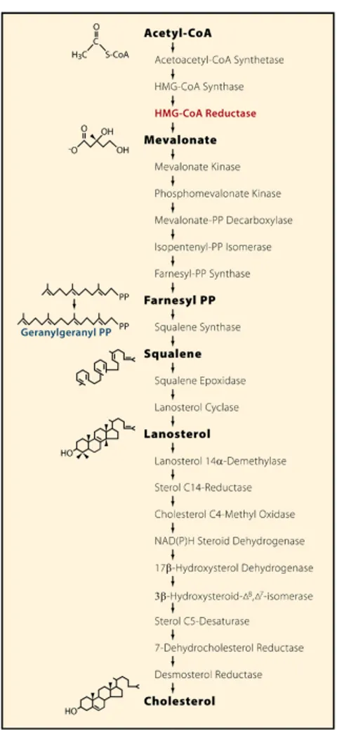
The major sources of cellular cholesterol are represented by: i) de novo synthesis and ii) uptake of cholesterol from circulating lipoproteins.
All cells of the body can synthesize cholesterol; synthesis occurs in the Endoplasmic Reticulum (ER) compartment where the synthesis apparatus
1.5. CELL CHOLESTEROL HOMEOSTASIS 31 is located. The rate-limiting step of the synthetic pathway is the conver-sion of HMG-CoA to mevalonate mediated by the HMG-CoA Reductase enzyme (Fig. 1.7). HMG-CoAR, similarly to other enzymes that function in the later steps of cholesterol synthesis, is an integral ER membrane protein [116]; it is composed of 8 TM domains and it contains (between TM and 6) a “Sterol Sensing Domain” (SSD), a conserved motif common also to sev-eral key proteins involved in cellular cholesterol metabolism regulation like SCAP, NPC1, SREBPs, Insigs. This domain allows to “sense” the cholesterol content of the ER membrane and to consequently regulate the synthetic rate of cholesterol.
HMG-CoAR is regulated, at the transcriptional level, by SREBPs (a group of proteins which mainly regulate cholesterol synthesis and uptake, as illustrated in 1.5.2.2); gene transcription is suppressed by intracellular cholesterol accumulation. At the post-transcriptional level the reductase is regulated by sterol-accelerated degradation: in particular in sterol-deprived cells HMG-CoAR is slowly degraded, while when sterols accumulate, the enzyme is rapidly ubiquitinated and degraded with an half-life of less than 1 hour. Both regulation processes are mediated by Insigs as described in section 1.5.2.2 [115].
1.5.2 Cholesterol uptake (influx) and endosomal cholesterol traffic
The alternative and preferred source for cellular cholesterol acquisition is the uptake of lipoproteins from the circulation through a receptor-mediated mechanism. The process is mediated by the cell surface LDL Receptor (LDLR) which binds plasma lipoproteins containing ApoB or ApoE (mainly LDL but also VLDL and chylomicron remnants). Lipoprotein particles, once bound to the receptor, are internalized via clathrin-coated pits; these vesicles then fuse with early endosomes where the lower pH allows the dissociation of LDL from LDLR. The LDLR is then localized to a recycling endocytic com-partment, which transfers back the receptor to the cell surface. Cholesteryl esters contained in the internalized LDLs are hydrolyzed in late endosomes and lysosomes by an acid activated enzyme, namely LAL (lysosomal acidic
Figure 1.7: Cholesterol synthesis.
lipase), which generates free cholesterol. 1.5.2.1 LDLR structure
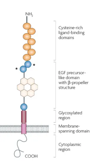
LDLR molecule is a glycoprotein of 839 aa, including 5 functional domains: i) a ligand binding domain at the NH2 side; ii) an Epidermal Growth Factor (EGF) homology region; iii) an “O-linked” oligosaccharide-rich region; iv) a TM domain; v) a cytoplasmatic tail at the COOH side (Fig. 1.8).
1. The ligand binding domain is organized in 7 repeated motifs, each one containing a cysteine-rich region important for the maintenance of the proper 3D folding. The conformation of the ligand binding domain ensures the exposure of negatively charged groups which allow


![Figure 1.11: Expression of ABCA1 in human NPC1-deficient fibroblasts. A, ABCA1 mRNA and protein levels; B, average ABCA1 protein levels in cells incubated in absence or presence of free cholesterol [126].](https://thumb-eu.123doks.com/thumbv2/123dokorg/4709154.45204/54.892.153.656.442.675/figure-expression-deficient-fibroblasts-average-incubated-presence-cholesterol.webp)
![Figure 1.12: ABCA1 and SREBP-1c expression in CYP27 deficient fibroblasts ABCA1 and SREBP-1c gene expression in normal and CYP27-deficient human fibroblasts: response to cholesterol loading [129].](https://thumb-eu.123doks.com/thumbv2/123dokorg/4709154.45204/55.892.226.732.475.822/expression-deficient-fibroblasts-expression-deficient-fibroblasts-response-cholesterol.webp)
