UNIVERSITÀ DEGLI STUDI DI SALERNO
Dipartimento di Farmacia
Dottorato di Ricerca
in
Scienze del Farmaco
Ciclo XXIX — Anno accademico 2016/2017
Tesi di Dottorato in
Design and synthesis of new integrase
inhibitors
Dottorando Tutore Dott. Antonia Spensiero Chiar.mo Prof. Pietro Campiglia
Index
Index
Chapter I: The Continuing Evolution of AIDS: HIV-1 integrase as a target for antiretroviral therapy
1. Acquired immune deficiency syndrome (AIDS) 1
1.1 Global distribution of HIV-1 infection 1
1.2 HIV genome 3
1.2.1 Replicative cycle of HIV-1 4
1.3 Antiretroviral Drugs 6
1.3.1 The Entry and Fusion Inhibitors 6
1.3.2 The Reverse Transcriptase (RT) Inhibitors 7
1.3.3. The Protease Inhibitors (PIs) 8
1.4 Highly Active Antiretroviral Therapy (HAART) and resistance effect to
drugs 9
1.5 References 12
Chapter II: The HIV-1 integrase as molecular target
2.1 Integrase and integration: biochemical activities of HIV-1 integrase 17
2.1.1 Integrase protein 17
2.1.2 The role of integrase in HIV-1 replication 19 2.1.3 HIV integrase structure and functional domains 22 2.1.4 IN interactions with host cell proteins 25
2.2 Integrase inhibitors 28
2.2.1 Integrase allosteric inhibitors 29
2.2.2 Integrase tetramerization inhibitors 30
2.2.3 Integrase inhibitors derived from N-terminal domain 32
2.3 References 33
Chapter III: Design, synthesis, biological evaluation and conformational studies of peptide derived from N-terminal domain HIV-1 integrase
3.1 Results 41
Index
3.1.2 Biochemical studies: HIV-1 IN catalytic activity inhibition (peptides 1-18)
43
3.1.3 Design of peptides 19-21 containing the Phe residue and Ala- scanning
approach (peptides 22-28) 44
3.1.4 Biochemical studies: HIV-1 IN catalytic activity inhibition (peptides
19-28) 45
3.1.5 Conformational Studies on peptides 22-28 46
3.1.6 IN-IN dimerization and IN multimerization studies on peptides 18-24-25 47 3.1.7 Cell-based HIV replication assay on the most active peptides 18,24 and
25 49
3.1.8 The structure-activity relationship studies on peptide 25 50 3.1.9 Biochemical studies: HIV-1 IN catalytic activity inhibition and IN-IN
dimerization and IN multimerization studies on peptides 29-39 52 3.1.10 NMR analysis on peptides 25 and 39 53 3.2 Improvement of pharmacokinetics properties on peptide 25 55 3.2.1 The head-to tail cyclization approach 55 3.2.2 Insertion of a lactam bridge on peptide 25 (peptides 45-46) 56
3.3 Chemistry 58
3.3.1 General procedure for synthesis 58
3.3.2 Synthesis of head-to-tail cyclic peptide 44: side reaction during the
sequence assembly 60
3.3.3 Synthesis of lactam analogues (peptides 54-55) 67
3.4 Discussion 69
3.5 Experimental section 73
3.5.1 Material and methods 73
3.5.1.1 Peptide synthesis 73
3.5.2 Synthesis peptide 44 75
3.5.3 Synthesis of lactam analogues (peptides 45-46) 76
3.5.4 Purification and characterization 76
3.5.5 IN Activity Inhibition assay 78
Index
3.5.7 HTRF LEDGF-dependent and -independent HIV-1 IN assay 79
3.5.8 Circular dichroism (CD) 79 3.5.9 Cell-based assays 80 3.5.10 NMR analysis 80 3.5.10.1 NMR assignment 81 3.5.10.2 Structure calculations 81 3.6 References 82
Chapter IV: Development and identification of a novel anti-HIV peptide: design, synthesis and biological evaluation
4.1 Cell penetrating peptides 89
4.1.1 Conjugation with fatty acids 91
4.2 Results 91
4.2.1 Design of peptides able to penetrate in cells 91
4.2.2 Cell-based HIV replication assay on peptides 5, TAT-5 and PA-5 92
4.3 Chemistry 94
4.3.1 General procedure for synthesis of PA-5 94
4.3.2 Synthesis of peptide TAT-5, TAT-47-55 96
4.4 Discussion 97
4.5 Experimental section 98
4.5.1 Material and methods 98
4.5.1.1 Peptide synthesis by microwaves 98
4.5.1.2 Synthetic Procedure: conjugation of the peptide Palmitic Acid 99
4.5.2 Purification and characterization 99
4.5.3 Cell-based assays 100
4.6 References 101
Chapter V: Summary
Figure captions
Figure captions
Chapter I:
Figure 1.1 Population of people living with HIV in 2015. 2
Figure 1.2 HIV virus structure. 3
Figure 1.3 Schematic overview of the HIV-1 replication cycle. 5
Chapter II:
Figure 2.1 Amino acid sequence alignment of retroviral integrase. 18
Figure 2.2 The role of integrase in the life cycle of HIV virus. 21
Figure 2.3 Functional domain of integrase. 22
Figure 2.4 The structures of the CCD in association with the CTD, and of the
NTD with the CCD. a and b | Side views showing the catalytic acidic triad in red (the canonical DDE motif consisting of residues D64, D116, E152; BOX 1, figure part a) in the catalytic core domain (CCD) of integrase. The two subunits of the dimer are shown in yellow and green. c and d | Front views of the same structures (after 90° anticlockwise rotation of panels a and b, respectively). a and c | Structure of the CCD–carboxy-terminal domain (CTD) dimer; (PDB codes: 1EXQ & 1EX4). b and d | Structure of the amino-terminal domain (NTD)–CCD (PDB code: 1K6Y). Combining the structures (a with b; c with d) indicates the positioning of each NTD into the cavity between the CCD and CTD in the full-size integrase dimer. The functional structure of integrase is probably tetrameric, and would therefore involve another dimer interface
Figure captions
Figure 2.5 Region of the CCD interaction with IBD. 27
Figure 2.6 Conventional drugs approved by the FDA for the treatment in
patients with HIV. a) Raltegravir; b) Elvitegravir; c) Tenofovir; d)
Emtricitabine; e) Cobicistat. 28
Figure 2.7 Structure of the catalytic domain dimerization of HIV-1 IN. The two monomers are shown in red and blue. The propellers a1 and α5, from which the inhibitory peptides INH1 and INH5 respectively, are indicated with the α3 helix. 30
Figure 2.8 IN secondary structure. The peptides derived from α1 (NL6) and α3
(NL9). 31
Chapter III:
Figure 3.1. Sequences of the designed peptides in this study. 42
Figure 3.2 CD Spectra of selected peptides in TFE/water 50% solution. Peptide
are represented in: 18 (solid blue line), 22 (solid red line), 23 (solid black line),
24 (solid purple line), 25 (dashed black line), 26 (solid orange line), 27 (dashed
blue line), 28 (dashed red line). 47
Figure 3.3 (1) NMR structures of peptides 25(A and B) and 39 (C and D) in 30
% TFE. (2) Overlay of the NMR structure of peptide 25 onto the NMR structure of peptide 39. (A) Backbone atoms for 25 are in blue and for 39 in black. (B) Backbone atoms for 25 are in cyan and side chains in blue, and for 39 backbone in grey and side chains in black. (C) Ribbon representation of the overlay of
representative conformers of 25 (in cyan) and 39 (in red). 54
Figure 3.4 The cyclic peptide 44 56
Figure 3.5 Cyclic peptide 46 with the lactam bridge. 57
Figure 3.6 Schematic representation of the peptide synthesis. 59
Figure 3.7 Schematic representation of diketopiperazine formation. 61
Figure 3.8 Schematic representation of the cyclic peptide synthesis. 62
Figure captions
Figure 3.10 Schematic representation of the cyclic peptide synthesis. 66
Figure 3.11 Schematic representation of the lactam cyclic peptide synthesis. 68
Figure 3.12 Analytical HPLC spectrum of peptide 25. 77
Chapter IV:
Figure 4.1. Activity in vitro assay of peptide 5. 89
Table captions
Table captions
Chapter III:
Table 3.1 Sequence, analytical data and effect of peptides 1-18 on HIV-1 IN
activities. 44
Table 3.2 Sequence, analytical data and effect of peptides 19-28 on HIV-1 IN
activities 46
Table 3.3 Effects of peptides 18, 24, and 25 on the HIV-1 IN dimerization and
multimerization process. 49
Table 3.4. Evaluation of the synthesized peptides against HIV-1 and HIV-2
replication in MT-4 cell cultures. 50
Table 3.5 Effects of peptides 1, 4, and 5 on the HIV-1 IN dimerization and
multimerization process, and effects on the HIV-1 IN LEDGF-dependent and –
independent catalytic activity. 53
Table 3.6 Cyclic peptides with lactam bridge 57
Chapter IV:
Table 4.1. Conjugated peptides. 92
Table 4.2. Evaluation of the synthesized peptides against HIV-1 and HIV-2
replication in MT-4 cell cultures. 92
Table 4.3. Evaluation of the synthesized peptides (47-55) against HIV-1 and
Abstract
The viral enzyme integrase (IN) is essential for the replication of human immunodeficiency virus type 1 (HIV-1) and represents an important target for the
development of new antiretroviral drugs. In this PhD project, we focused on the N-terminal domain of integrase (NTD) for the development and synthesis of a library
of overlapping peptide sequences, with specific length and specific offset covering the entire native protein sequence NTD IN 1-50. The most potent fragment,
VVAKEIVAH (peptide 18), inhibits the HIV-1 IN activity with an IC50 value of
4.5 µM. Amino acid substitution analysis on this peptide revealed essential residues for activity and allowed us to identify two nonapeptides (peptides 24 and 25), that show a potency of inhibition similar to peptide 18. Interestingly, peptide 18 does not interfere with the dynamic interplay between IN subunits, while peptides 24 and 25
modulate these interactions in different manners. In fact, peptide 24 inhibits the IN-IN dimerization, while peptide 25 promotes IN multimerization, with IC50 values
of 32 and 4.8 µM, respectively. In addition, peptide 25 has shown to have selective anti-infective cell activity for HIV-1. Moreover, the NMR analysis showed an alpha helix conformation of peptide 25, which could be essential for the interaction with IN. These results indicated peptide 25 as a hit for further development of new chemotherapeutic agents against HIV-1. In addition, we observed that the peptide 5, EKYHSNWRAM, conveniently conjugated with the cell-penetrating fragment TAT, inhibits replication of HIV-1 and HIV-2 in infected MT-4 cells.
Keywords: HIV-1, integrase, N-terminal domain, peptides, inhibitors.
Abbreviations
IN, integrase; HIV-1, human immunodeficiency virus type 1; PIC, preintegration complex; NTD, N-terminal domain; CCD, catalytic core domain; CTD, C-N-terminal domain; INSTIs, integrase strand transfer inhibitors (INSTIs); HOAt, 1-Hydroxy-7-azabenzotriazole; HOBt, hydroxybenzotriazole; HBTU, N,N,N′,N′-tetramethyl-O-(1H-benzotriazol-1-yl)uraniumhexafluorophosphate; DIEA N,N-diisopropylethylamine; DMF, dimethylformamide.
CHAPTER I:
The Continuing Evolution of AIDS: HIV-1 integrase as a target
for antiretroviral therapy
Chapter I: The Continuing Evolution of AIDS: HIV-1 integrase as a target for antiretroviral therapy
1
1. Acquired immune deficiency syndrome (AIDS)
1.1 Global distribution of HIV-1 infection
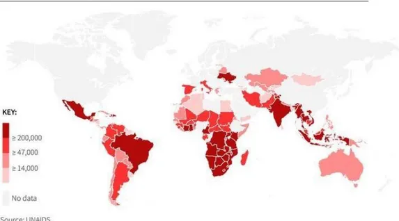
Acquired immune deficiency syndrome (AIDS), is induced by HIV infection and consists in a profoundly damage of immune system. Following the discovery of the HIV as the causative agent of AIDS, many advances were made in a short period of time, including the understanding of the modes of transmission; the sequencing of the HIV-1 genome;1 elucidation of the main cell targets, CD4 T cells and macrophages;2 the genomic heterogeneity of HIV with the innumerable micro variants within a single patient;3 identification of a SIV-monkey model to study and monitoring the epidemic events.4 Human immunodeficiency virus (HIV), is a retrovirus known for immune system degradation, transmitted by direct contact with infected blood, sexual contact or by direct transmission of mother to child. The HIV virus has remained a predominant health care concern for the past 30 years.5 Around 35 million people have died from AIDS-related illnesses since the first cases of HIV were reported, and, overall, 78 million have been infected with HIV, according to UNAIDS, the Joint United Nations Programme on HIV/AIDS. Thanks to a dramatic scale-up of antiretroviral treatment, AIDS-related deaths have fallen by 45% to1.1 million in 2015 from a peak of 2 million in 2005. Globally, 36.7 million people were living with HIV in 2015, the majority in poor and middle-income countries. As of June 2016, around 18.2 million were receiving antiretroviral therapy, up from 15.8 million at the same time last year, and 7.5 million in 2010 (figure 1.1).
Chapter I: The Continuing Evolution of AIDS: HIV-1 integrase as a target for antiretroviral therapy
2
Figure 1.1 Population of people living with HIV in 2015.
Adapted from: http://chartsbin.com/view/11852.
Last year, around 2.1 million people became newly infected with HIV. While the number of adults acquiring the virus is not falling, huge advances have been made in tackling mother to child transmissions. In the past five years, new infections among children halved 150.000 in 2015, down from 290.000 in 2010. UNAIDs warns that young women are particularly at risk of infection. In sub Saharan Africa, adolescent girls face a triple threat: a high risk of HIV infection, low rates of HIV testing and difficulty sticking with HIV treatment. The first approved therapy involved the use of multiple drugs (HAART, Highly Active Anti-Retroviral Therapy),6 which transform the HIV infection from lethal to chronic. Despite this, due to drug resistance that are highlighted below, the treatment regimen has not allowed the complete eradication of the disease itself. The treatment only reduced the number of new infections and subsequent deaths, but despite these promising advances, HIV infection remains a looming problem on global health and an important goal for scientific research.
Chapter I: The Continuing Evolution of AIDS: HIV-1 integrase as a target for antiretroviral therapy
3
1.2 HIV Genome
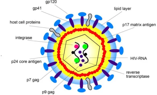
The HIV virus belongs to the retroviruses family and is a member of the genus of lentiviruses, which indicates a long incubation period. The essential genes for replication and then for virus activity are: gag, pol, and env. The gag gene encodes the nucleocapsid protein; the pol gene encodes the enzyme reverse transcriptase, protease and integrase; Finally, env encodes for the proteins of the pericapside.7,8 The HIV genome also contains, at the sides of the env gene, six more genes that code for regulatory proteins and accessory: tat (transactivation of Transcription), rev (Regulatory of Virus), vpu (Viral Protein U), nef (Negative Factor) , vif (viral infectivity factor) and vpr (Viral Protein R), involved in different steps in regulation and HIV pathogenesis. A conceptual representation of the virus architecture is represented in figure 1.2.
Figure 1.2 HIV virus structure.
Adapted from: http://healthfavo.com/hiv-virus-structure-anatomy-picture-reference.html.
Chapter I: The Continuing Evolution of AIDS: HIV-1 integrase as a target for antiretroviral therapy
4 1.2.1 Replicative cycle of HIV-1
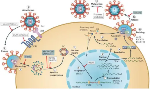
Replicative cycle of the virus can be divided into different stages: penetration of the virus within the host cell, reverse transcription of the viral genome and introduction into the host cell genome by the integrase enzyme, the production of second messengers and, finally, assembly of new infectious virions during the late phase. The infection begins when the envelope (Env) glycoprotein gp120 binds the receptor CD4 and the co-receptor CC-chemokine receptor 5 (CCR5) (step 1), leading to fusion of the viral and cellular membranes and entry of the viral particle into the cell (step 2). The role of the CD4 receptor in HIV cell entry was identified shortly after the isolation of HIV.9 The conformational changes in the gp120 protein leads to exposing an epitope which allows binding to a co-receptor, such as the chemokine receptors CCR5 or CXCR4, which are the most co-receptors used by HIV in vivo. The importance of this co-receptor binding was empathized by the compromised HIV-1 infectivity in the individuals harbouring mutant CCR5 proteins.10 Partial core shell uncoating (step 3) facilitates reverse transcription (step 4). This step is catalyzed by the viral enzyme reverse transcriptase (RT), which transcribes the viral single-stranded RNA genome which is converted into double-stranded DNA (dsDNA). Together with viral and host proteins the dsDNA forms the pre-integration complex (PIC), which is guided to the nuclear pore. This is an essential step in the HIV replication cycle of HIV because it prepares the viral genome for the subsequent integration into the host chromosome. Following of displacement into the cell nucleus (step 5), PIC-associated viral integrase forms the integrated provirus (step 6). Proviral transcription (step 7), mediated by host RNA polymerase II (RNA Pol II) and positive transcription elongation factor b (P-TEFb),
Chapter I: The Continuing Evolution of AIDS: HIV-1 integrase as a target for antiretroviral therapy
5
yields viral mRNAs of different sizes, the larger of which requires energy-dependent export to leave the nucleus via host protein CRM1 (step 8). mRNAs is used as templates for protein production (step 9) and genome-length RNA is incorporated into viral particles with protein components (step 10). Viral-particle budding (step 11) and release (step 12) from the cell is mediated by ESCRT (endosome sorting complex required for transport) complexes. Each step in the HIV-1 life cycle is a potential target for antiviral intervention;6 The sites of action of clinical inhibitors and cellular restriction factors are indicated in the figure 1.3.
Figure 1.3 Schematic overview of the HIV-1 replication cycle.
Adapted from: Engelman A and Cherepanov P. The structural biology of HIV-1: mechanistic and therapeutic insights. Nature Reviews Microbiology. 2012; 10, 279-290.
Chapter I: The Continuing Evolution of AIDS: HIV-1 integrase as a target for antiretroviral therapy
6
1.3 Antiretroviral Drugs
The significant advancement in the understanding of HIV replication and its pathogenesis has helped in the identification of various pharmacological targets. The first anti-HIV agent to be licensed for clinical use was Zidovudine (AZT), in 1987. Since then, several anti-HIV compounds have been approved for the treatment of AIDS by the FDA and EMEA (European Medicine Agency) for treating HIV infections.11 These compounds fall within different categories depending on the target within the HIV replicative cycle they interact with.
1.3.1 The Entry and Fusion Inhibitors
These inhibitors intercept the viral replication at the entry of the viral core into the cytosol of the host cell. The group of entry inhibitors can be subdivided into classes of agents that act at different stages of entry: attachment and CD4 binding, co-receptor binding, and fusion. In the recent years, important progress has been made in understanding the HIV-1 entry process in which the viral and cellular membranes are fused, resulting in the subsequent delivery of the viral genome into the host cell. These studies have led to the formulation of new approaches for therapeutic intervention. One of the first and clinically most advanced drug emerged from this effort is the fusion inhibitor T20. T20 acts by freezing a transient structural intermediate of the HIV-1 fusion process, thus blocking an essential step in viral entry. With phase III clinical trials already well underway, the success of T20 indicates that targeting the viral entry process will soon be an important component of antiretroviral therapy. Drug resistance mutations are usually located in
Chapter I: The Continuing Evolution of AIDS: HIV-1 integrase as a target for antiretroviral therapy
7
the ENF binding site on gp41 (direct resistance) or confer resistance indirectly via mutations in other regions of gp41 and even in gp120.12 Currently, only antagonists that block CCR5 binding (Maraviroc) and fusion (Enfuvirtide) have been approved by the FDA for treatment of HIV infected patients, although strategies to inhibit other aspects of HIV entry are under development.13 In addition to Maraviroc, other CCR5 and also CXCR4 inhibitors are being investigated.14
1.3.2 The Reverse Transcriptase (RT) Inhibitors
The Reverse Transcriptase (RT) inhibitors interfere with the generation of a DNA copy of the viral genome. RT functions as a heterodimer and catalyzes the conversion of the single-stranded genomic RNA into double-stranded DNA (with duplicated long terminal repeats), which is integrated into cellular DNA by the viral integrase.15 There are two classes of RT inhibitors, that exhibit different mechanism of action. First of them is the group of nucleoside/nucleotide reverse transcriptase inhibitors (NRTIs). These are nucleoside and nucleotide analogues that are incorporated by the viral RT into the newly synthesized DNA strand. They are inactive in their parent forms and require successive phosphorylation steps by host cell kinases and phosphotransferases to form deoxynucleoside triphosphate (dNTP) analogs responsible of viral inhibition. In their respective triphosphate (TP) forms, NRTIs compete with their corresponding endogenous dNTPs for incorporation by HIV RT. Once incorporated, they assist as terminators chain of viral RT, thus, acting early in the viral replication cycle by inhibiting a critical step of proviral DNA synthesis prior to integration into the host cell genome.16
Chapter I: The Continuing Evolution of AIDS: HIV-1 integrase as a target for antiretroviral therapy
8
The currently approved NRTIs are Zidovudine, Lamivudine, Didanosine, Zalcitabine, Stavudine, Abacavir, and Emtricitabine.
The second group of RT inhibitors is the Non-nucleoside reverse transcriptase inhibitors (NNRTIs). They are small molecules that carry out the inhibition of RT by binding to a hydrophobic pocket in the proximity of the active site of the enzyme. After the inhibitor is bound, it impairs the flexibility of the RT resulting in its inability to synthesize DNA. Mutations may induce resistance to NNRTIs resulting in reduced affinity of the inhibitor with the protein. Usually, a single mutation selected by one NNRTI is sufficient to confer complete resistance to all compounds of the drug class.17
1.3.3 The Protease Inhibitors (PIs)
Protease Inhibitors (PIs) interfere with the process of forming new infectious viral particles. The viral protease is involved in virion maturation. Protease targets the amino acid sequences in the gag and gag–pol polyproteins, which must be cleaved before nascent viral particles (virions) can mature. Cleavage of the gag polyprotein produces three large proteins (p24, p17, and p7) that contribute to the structure of the virion and to RNA packaging, and three small proteins (p6, p2, and p1) of uncertain function. PIs are small molecules that bind to the active site of the protease and compete with its natural substrates. PIs contain a synthetic analogue of the amino acid sequence of the gag–pol polyprotein that is cleaved by the protease. PIs prevent cleavage of gag and gag–pol protein precursors in acutely and chronically infected cells, arresting maturation and hence blocking the infectivity of nascent virions.18
Chapter I: The Continuing Evolution of AIDS: HIV-1 integrase as a target for antiretroviral therapy
9
The resistance of HIV against PIs can be achieved by two mechanisms. The first one involves the exchange of amino acids in the protease such that the affinity to the inhibitor is decreased while the natural substrates can be bound efficiently as opposed to the synthetic analogues.18 Modifications of the affinity to the natural substrate alter also the efficiency of the protease. Thus, the second mechanism introduces compensatory mutations aiming at re-establishing the efficiency of the enzyme while maintaining resistance against the inhibitor. These mutations can occur both in the protease or in its substrate, at cleavage sites.19 The four approved HIV-protease inhibitors are based on amino acid sequences: Indinavir, Nelfinavir, Ritonavir and Saquinavir.
1.4 Highly Active Antiretroviral Therapy (HAART) and resistance effect to drugs
The HIV virus develops resistance against individual drugs and inhibitors. This problem required a new pharmaceutical strategy. An approach of combination therapy, which involved combining several antiretroviral compounds, was developed. This approach benefited the most from the development of drugs in NNRTIs and PI classes. Combination therapy can block the resistance effect more effectively for two reasons; first, multiple mechanisms are required for resistance to occur to all drugs in the regimen and second; multiple drugs suppress viral replication more effectively than single agents. This marked the beginning of the era of highly active antiretroviral therapy (HAART) in 1995. HAART combines a minimum of three drugs from at least two different drug classes targeting distinct proteins.17 A typical HAART treatment combines two NRTIs plus either one PI or one NNRTI.20
Chapter I: The Continuing Evolution of AIDS: HIV-1 integrase as a target for antiretroviral therapy
10
Combinations of antiretrovirals create multiple barriers to the HIV replication process. This helps to keep the number of offspring low and reduce the possibility of a superior mutation.
In 2006, it was reported that the number of HIV related deaths declined as compared to the pre-HAART. Consequently, multidrug regimens are necessary for successful treatment. Since each HAART agent has its own unique adverse effect profile, selecting a regimen with a proper profile may be difficult. For example, certain PIs produce adverse metabolic effects that may increase the risk of developing cardiovascular disease. On the other hand, NNRTI-based therapies may result a different side effect profile. Once this therapy is initiated, it should never be stopped.
In 2006, FDA approved the combinations of antiretrovirals (for example, Atripla). These are multiple antiretroviral drugs combined into a single pill, helping to increase adherence and thus reducing potential development of viral resistance to the drugs. This may result in longer term effectiveness of the regimen. They may combine different classes of antiretrovirals or contain only a single class. Another milestone in HAART was the discovery that the protease inhibitor ritonavir interferes with the liver enzyme cytochrome P450.21
This enzyme is involved in the metabolic processing of most protease inhibitors. Thus, the use of a small dose of ritonavir inhibits the liver enzyme, and helps to maintain optimal levels of other protease inhibitors in the patient‟s circle for a longer period of time. The boosting of protease inhibitors with ritonavir is standard as of 2001 following the introduction of Kaletra (LPV+RTV) and is usually denoted by PI/r. Despite the increasing concerns regarding antiretroviral resistance, the death rate among HIV-infected people continued to decline.22
Chapter I: The Continuing Evolution of AIDS: HIV-1 integrase as a target for antiretroviral therapy
11
HAART suffers from certain limitations, despite of its success. HAART therapy is highly effective in delaying the onset of AIDS but its clinical utility is limited by viral resistance, non-adherence to therapy, and drug toxicity. Thus, the development of new inhibitors, targeting a distinct step in the retroviral life cycle, remains essential to reduce side effects and selection of drug-resistant viruses. In this context, integration of the proviral DNA into the host cell genome, a process carried out by a specific viral enzyme, integrase, has been recently recognised as site for therapeutic intervention. Development of new chemical entities able to interrupt this process, would allow to identify key biochemical sites of HIV-1 Integrase and increase the potency and selectivity of new Integrase inhibitors.
Chapter I: The Continuing Evolution of AIDS: HIV-1 integrase as a target for antiretroviral therapy
12
1.5 References
1. Sanchez-Pescador R, Power MD, Barr PJ, Steimer KS, Stempien MM,
Brown-Shimer SL, Gee WW, Renard A, Randolph A, Levy JA, et al. Nucleotide sequence and expression of an AIDS-associated retrovirus (ARV-2). Science. 1985; 227 (4686):484-92.
2.
Harper ME, Marselle LM, Gallo RC, Wong-Staal F. Detection of lymphocytes expressing human T-lymphotropic virus type III in lymph nodes and peripheral blood from infected individuals by in situ hybridization. Proc. Natl. Acad. Sci U S A. 1986; 83(3):772-6.
3. Starcich BR, Hahn BH, Shaw GM, McNeely PD, Modrow S, Wolf H,
Parks ES, Parks WP, Josephs SF, Gallo RC, et al. Identification and characterization of conserved and variable regions in the envelope gene of HTLV-III/LAV, the retrovirus of AIDS. Cell. 1986; 45 (5):637-48.
4.
Paul M, Sharp and Beatrice H. Origins of HIV and the AIDS Pandemic Hahn. Cold Spring Harb Perspect Med. 2011; 1(1): a006841.
5.
Metifiot M, Marchand C, Pommier Y. HIV integrase inhibitors: 20-year landmark and challenges. Adv Pharmacol. 2013; 67: 75-105
6.
Arts EJ and Hazuda DJ. HIV-1 Antiretroviral Drug Therapy. Cold Spring Harb Perspect Med. 2012; 2(4): a007161.
7. Frankel AD, Young JA. HIV-1: fifteen proteins and an RNA. Annu
Rev Biochem. 1998; 67:1–25.
8. Seelamgari A, Maddukuri A, Berro R, de la Fuente C, Kehn K, Deng
L, Dadgar S, Bottazzi ME, Ghedin E, Pumfery A, Kashanchi F. Role of viral regulatory and accessory proteins in HIV-1 replication. Front Biosci. 2004; 9:2388–2413.
9.
Dalgleish AG, Beverley P C L, Clapham PR, Crawford DH, Greaves MF and Weiss RA. The CD4 (T4) antigen is an essential component of the receptor for the AIDS retrovirus. Nature. 1984; 312, 763-767.
10.
Berger1 EA, Murphy PM and Farber JM. Chemokine receptors as hiv-1 coreceptors: roles in viral entry, tropism, and disease annual. Review of Immunology. 1999; 17: 657-700.
Chapter I: The Continuing Evolution of AIDS: HIV-1 integrase as a target for antiretroviral therapy
13
11. Broder SMD. The development of antiretroviral therapy and its impact
on the HIV-1/AIDS pandemic. Antiviral Res. 2010; 85(1): 1.
12. Miller MD, Hazuda DJ. HIV resistance to the fusion inhibitor
enfuvirtide: mechanisms and clinical implications. Drug Resist Updat. 2004; 7(2):89-95.
13. Tilton JC, Doms RW. Entry inhibitors in the treatment of HIV-1
infection. Antiviral Res. 2010; 85(1):91-100.
14. Esté JA, Telenti A. HIV entry inhibitors. Lancet. 2007;
370(9581):81-8.
15. Powers R, Clore GM, Stahl SJ, Wingfield PT, Gronenborn A. Analysis
of the backbone dynamics of the ribonuclease H domain of the human immunodeficiency virus reverse transcriptase using nitrogen-15 relaxation measurements. Biochemistry. 1992; 31 (38):9150–9157
16.
Cihlar T, Ray AS. Nucleoside and nucleotide HIV reverse transcriptase inhibitors: 25 years after zidovudine. Antiviral Res. 2010 J; 85(1):39-58.
17.
Clavel F, Hance AJ. HIV drug resistance. N Engl J Med. 2004; 350(10):1023-35.
18.
Flexner C. HIV-protease inhibitors. N Engl J Med. 1998; 338(18):1281-92.
19.
Nijhuis M1, van Maarseveen NM, Lastere S, Schipper P, Coakley E, Glass B, Rovenska M, de Jong D, Chappey C, Goedegebuure IW, Heilek-Snyder G, Dulude D, Cammack N, Brakier-Gingras L, Konvalinka J, Parkin N, Kräusslich HG, Brun-Vezinet F, Boucher CA. A novel substrate-based HIV-1 protease inhibitor drug resistance mechanism. PLoS Med. 2007; 4(1):e36.
20.
Dybul M, Bolan R, Condoluci D, Cox-Iyamu R, Redfield R, Hallahan CW, et al. Evaluation of initial CD4+T cell counts in individuals with newly diagnosed human immunodeficiency virus infection, by sex and race, in urban settings. Journal of Infectious Diseases. 2002; 185:1818–1821.
Chapter I: The Continuing Evolution of AIDS: HIV-1 integrase as a target for antiretroviral therapy
14
21. Kumar GN, Rodrigues AD, Buko AM and Denissen JF. Cytochrome
P450-mediated metabolism of the HIV-1 protease inhibitor ritonavir (ABT-538) in human liver microsomes. Journal of Pharmacology and Experimental Therapeutics. 1996; 277 (1) 423-431;
22.
Eras C, Riffenburgh NF, Robert H, Wegner S, Agan BK, Tasker SA, Spooner KM, Armstrong AW, Fraser S, Wallace MR. Epidemiology and Social Science Comparisons of Causes of Death and Mortality Rates Among HIV-Infected Persons: Analysis of the Pre-, Early, and Late HAART. JAIDS. 2006; 41(2):194-200.
CHAPTER II:
Chapter II: The HIV-1 integrase as molecular target
17
2.1 Integrase and integration: biochemical activities of HIV-1 integrase
2.1.1 Integrase protein
As mentioned above, the human immunodeficiency virus (HIV) that causes acquired immunodeficiency syndrome (AIDS), belongs to the family of Retroviridae, genus Lentivirinae. Lentiviruses represent a kind of slow viruses with a long incubation period (months, even years) and a propensity to induce a wide range of diseases in different animal species. According to the recent classification of the International Committee on Taxonomy of Viruses (ICTV), the genus Lentivirus consists of nine species, seven lentivirus animal and two human lentivirus. At the same time there exist different species of integrase. For example, PFV IN is significantly longer, comprises 392 residues and ASV IN is decoded into 323 amino acids and is only in post-translational phase modified polypeptide comprising 286 residues, which is the active form.1
Although the enzyme encoded by avian sarcoma virus (ASV) has been studied long before, it is clear that is more interesting to study HIV IN.2 The integrase of HIV-1 is structurally characterized by a single polypeptide chain consisting of 290 amino acid residues and consists of three clearly identifiable domains and inter-domain linker.3
Chapter II: The HIV-1 integrase as molecular target
18
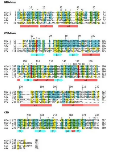
Figure 2.1 Amino acid sequence alignment of retroviral integrase.
Adapted from: Jaskolski M, Alexandratos JN, Bujacz G and Wlodawer A. Piecing together the structure of retroviral integrase, an important target in AIDS therapy. FEBS J. 2009; 276(11): 2926–2946.
As shown in the figure 2.1, the N-terminal domain (NTD) of HIV-1 IN contains the residues from 1 to 46, followed by a linker consisting of from 47-55 residues. The catalytic domain (CCD) contains 56-202 residues, and is followed by a linker sequence which comprises the amino acids from 203 to 219. Finally, the C-terminal domain (CTD) contains 220-288 residues. The number of domain residues of HIV-2 and SIV enzymes is approximately equal, which differs for ASV IN.
Chapter II: The HIV-1 integrase as molecular target
19
For the PFV IN, it is possible that there is an additional domain consists of approximately 50 residues preceding the NTD domain.
Analyzing for individual domains the ratio of the percentage identity/similarity are obtained the following data: for NTD 55/76% comparing HIV-1 IN to SIV, 26/46% comparing ASV IN; for the CCD are respectively 61/77% and 27/46% and for the CTD are 53/68% and 14/25% respectively. It is know that the lower sequence conservation is for the C-terminal domain.4
However, it should be emphasized, that the domains are characterized by a highly conserved sequence in all retroviral integrase:
• CCD has three highly conserved amino acid residues (DDE motif) • NTD sequence corresponds to HHCC residues.
2.1.2 The role of integrase in HIV-1 replication
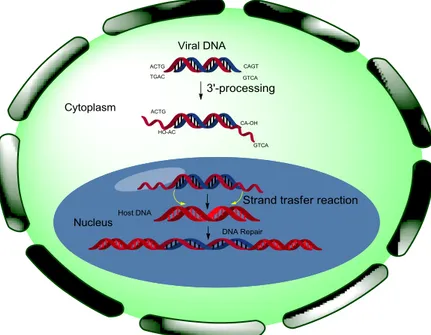
Integrase (IN) is a key enzyme for the integration of the HIV-1 genome into the host cell chromosome and, therefore, a very promising target for anti-AIDS drug design. IN performs two essential catalytic reactions.5 Following reverse transcription, the viral cDNA is primed for integration in the cytoplasm by integrase-mediated trimming of the 3′-ends of the viral DNA (figure 2.2). This step is referred to as 3′-Processing. It requires both fully functional integrase and the integrity of the last 10– 20 base pairs at both ends of the viral cDNA. 3′-processing consists of the endonucleolytic cleavage of the 3′-ends of the viral DNA.
This cleavage occurs immediately 3′ to a conserved CA dinucleotide motif. Alterations of this sequence prevent integrase from catalysing 3′-processing. This reaction generates CA-3′-hydroxyl DNA ends, which are the reactive intermediates required for Strand Transfer.
Chapter II: The HIV-1 integrase as molecular target
20
Following 3′-processing, integrase remains bound to the viral cDNA as a multimeric complex that bridges both ends of the viral DNA within the PIC.
The PIC contains both viral and cellular proteins in addition to the integrase–DNA complexes. The viral proteins reverse transcriptase (RT), matrix (Ma), nucleocapsid (Nc) and Vpr can contribute to the transport of PICs through the nuclear envelope. Some cellular proteins, packaged within PICs, can bind to integrase and stimulate the enzymatic activities of integrase. These proteins include interactor 1 (INI1)16 (the first integrase-binding protein discovered), lens epithelium derived growth factor (LEDGF, also known as p75);6 embryonic ectoderm-development protein 18 and heat shock protein 60 (HSP60).7
Promyelocytic leukaemia protein (PML) also co-localizes and co-migrates with PICs20. Two cellular proteins, high-mobility group
protein A1 (HMGA1, also known as HMG1(Y)) and barrier to auto-integration factor (BAF), regulate auto-integration by binding to DNA directly. HMGA1 stimulates integrase activity;8,9 BAF stimulates intermolecular integration and suppresses auto-integration. By contrast to other lentiviruses, such as the oncoretroviruses murine Moloney virus and Rous sarcoma virus, which require mitotic nuclear-envelope breakdown to access the chromosomes of infected cells, HIV-1 PICs are able to cross the nuclear envelope. The karyophilic property of the PICs enables HIV to replicate in not proliferative cells, such as macrophages.10 Once in the nucleus, integrase catalyzes the insertion of the viral cDNA ends into host chromosomes. This Strand Transfer reaction consists of the ligation of the viral 3′-OH DNA ends (generated by 3′-processing) to the 5′-DNA phosphate of a host chromosome.
Chapter II: The HIV-1 integrase as molecular target
21
Integrase can also catalyse the reverse reaction, referred to as Disintegration.11 Physiological integration requires the concerted joining of both ends of the viral cDNA on opposite DNA strands of the target (acceptor DNA) host chromosome with a canonical fivebase-pair stagger. The five-base stagger indicates that each viral cDNA end attacks the chromosomal DNA across its major groove. Completion of integration requires ligation of the 5′-end of the viral DNA. This last step of integration can only take place after trimming of the last two nucleotides at the proviral DNA 5′-ends and extension (gap filling) from the 3′-OH remodelling, and transcription complexes, such as those bound to integrase in the PICs (and described above), are implicated in the selection of the HIV integration sites within transcribing genes. The fully integrated viral genome is also referred to as the provirus.
Chapter II: The HIV-1 integrase as molecular target
22
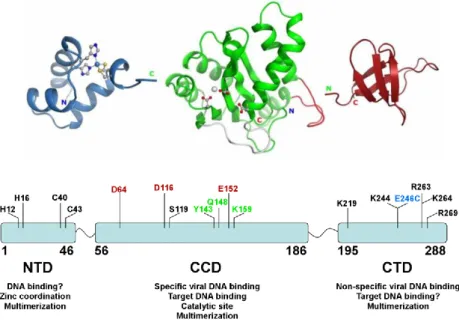
2.1.3 HIV integrase structure and functional domains
Integrase is generated during virus maturation by cleavage of the Pol polyprotein by HIV protease. HIV integrase is a 32-kDa protein comprising three structural domains: the N-terminal domain (NTD), the catalytic core domain (CCD) and the C-terminal domain (CTD) (figure 2.3). The atomic structure of each of these domains has been determined by X-ray diffraction or solution nuclear magnetic resonance (NMR).
Figure 2.3 Functional domain of integrase.
Adapted from: Kessl JJ, McKee CJ, Eidahl JO, Shkriabai N, Katz A and Kvaratskhelia M. HIV-1 Integrase-DNA Recognition Mechanisms. Viruses.
2009; 1(3), 713-736.
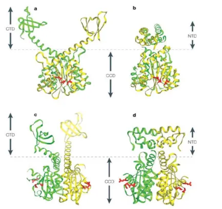
The figure 2.4 shows the structures of the CCD in association with the CTD,12 and of the NTD with the CCD.13 CCD, which encompasses residues 50–212, forms a dimer in all the structures examined. It is, structurally, remarkably similar to other retroviral integrases (MLV and avian sarcoma virus (ASV)), to the Tn5, to RNase H, to the Holiday junction recombinase RuvC35 and to the PIWI domain of Argonaute,14
Chapter II: The HIV-1 integrase as molecular target
23
the RNase associated with Dicer in RNA interference. This family of DNA processing enzymes (polynucleotide transferases) contains a canonical three amino acid, which formed in HIV-1 integrase the catalytic triad D64, D116 and E152. These residues are highly conserved in all integrases and retrotransposases (DDE motif).15 Mutation of any of these three acidic residues abolishes integrase‟s enzymatic activities and viral replication. The two D64 and D116 residues form a coordination complex (chemical bonds) with a divalent metal (Mg2+ or Mn2+). Because a second metal has been observed in an ASV integrase crystal structure,16 and because of the two-metal structure for polynucleotide transferases,17 it has been proposed that a second metal (Mg2+ or Mn2+) can be coordinated between D116 and E152 once HIV-1 integrase binds its DNA substrate(s).18 It is therefore likely that the metal(s) coordinate(s) integrase and the phosphodiester backbone of the DNA substrate(s) during the 3′-processing and strand-transfer steps. In most structures, the CCD contains a short disordered loop (encompassing residues 141–150), the structure of which can be stabilized by DNA. Although the CCD contains the enzyme catalytic site(s), in the absence of the NTD and CTD it can only catalyze the disintegration reaction the reverse of the strand-transfer reaction in vitro.19 Disintegration is the only reaction catalyzed by the isolated CCD. To catalyze 3′-processing and strand transfer, the CCD needs both the NTD and CTD in a dimeric complex.20 The NTD encompasses residues 1–50 and contains an HHCC motif that is common to all retroviral integrases. Binding of one Zn2+ atom to the HHCC motif stabilizes the folding of the NTD domain and is required for integrase activity. Single mutations of any of these four residues reduce integrase enzymatic activity.21 The NTD dimer interface is different in the crystal structures and the solution NMR structure (not
Chapter II: The HIV-1 integrase as molecular target
24
shown), which is indicative of multiple arrangements of the integrase multimers. The NTD is the preferential binding region for two cellular transcription factors in the PICs, INI1 and LEDGF/p75. The CTD, which encompasses residues 212–288, has an overall SH3 fold. It binds DNA nonspecifically and is required for integrase 3′-processing and strand-transfer activities. The CTD binds the cellular embryonic ectoderm development protein as well as RT, and this interaction seems to be required for reverse transcription. The figure 2.4 shows the structures of the CCD both with the NTD and the CTD. Together these two structures indicate the possibility that the NTD is positioned between the CCD and CTD, next to the extended α-helix joining the CCD and the CTD. The solution NMR dimer interfaces for the NTD and the CTD, which are different from those observed in the crystallographic structure might be used in higher-order complexes (tetramers and/or octamers), which have been proposed to correspond to the active enzyme.
Chapter II: The HIV-1 integrase as molecular target
25
Figure 2.4 The structures of the CCD in association with the CTD, and of the
NTD with the CCD. a and b | Side views showing the catalytic acidic triad in red (the canonical DDE motif consisting of residues D64, D116, E152; BOX 1, figure part a) in the catalytic core domain (CCD) of integrase. The two subunits of the dimer are shown in yellow and green. c and d | Front views of the same structures (after 90° anticlockwise rotation of panels a and b, respectively). a and c | Structure of the CCD–carboxy-terminal domain (CTD) dimer; (PDB codes: 1EXQ & 1EX4). b and d | Structure of the amino-terminal domain (NTD)–CCD (PDB code: 1K6Y). Combining the structures (a with b; c with d) indicates the positioning of each NTD into the cavity between the CCD and CTD in the full-size integrase dimer. The functional structure of integrase is probably tetrameric, and would therefore involve another dimer interface (unknown, and therefore not represented here).
Adapted from: Pommier Y, Johnson AA and Marchand C. Integrase inhibitors to treat HIV/Aids. Nature Reviews Drug Discovery. 2005; 4, 236-248.
2.1.4 IN interactions with host cell proteins
Purified IN proteins catalyzes 3′ processing and DNA strand transfer activities in vitro,22,23 indeed, the results obtained by numerous studies indicate that cell proteins play important roles, during virus infection. In particular, a crucial role for the IN-interacting protein lens epithelium-
Chapter II: The HIV-1 integrase as molecular target
26
derived growth factor (LEDGF)/p75 in HIV-1 replication and integration has highlighted.24 However, the function of LEDGF/p75 in viral replication is unknown.
The LEDGF/p75 belongs to the hepatoma-derived growth factor (HDGF) related protein (HRP) family, which is defined by the amino acid sequence conservation of an N-terminal Pro-Trp-Trp-Pro (PWWP) domain (figure 2.5).25 The binding occurs through a conserved IN-binding domain (IBD) found within the C-terminal portion of the larger p75 LEDGF splice variant. The IBD is essential for stimulation of IN activity in vitro and for LEDGF/p75 function during HIV-1 infection.26 LEDGF/p75 might therefore act as a critical costimulator of IN activity.27 Ectopically expressed HIV-1 IN is degraded by the proteasome in human cells, and LEDGF/p75 significantly increases its stability. The HIV-1 PIC can be degraded by the proteasome, so the IN-LEDGF/p75 interaction might help maintain PIC integrity during infection. The functional HIV-1 and feline immunodeficiency virus PICs were recovered from cytoplasmic extracts of infected cells using anti-LEDGF antibodies.28
Chapter II: The HIV-1 integrase as molecular target
27
Figure 2.5 Region of the CCD interaction with IBD.
Adapted from: Reddy KK, Singh P and Singh SK. Blocking the interaction between HIV-1 integrase and human LEDGF/p75: mutational studies, virtual screening and molecular dynamics simulations. Mol. BioSyst. 2014;10, 526-536.
Alternatively, LEDGF/p75 might function as an obligate chromatin acceptor for the PIC. This hypothesis indicates that LEDGF/p75 intimately associates with chromatin, and its N-terminal PWWP domain and AT-hook (ATh) DNA-binding motifs, which mediate chromatin binding, are required for HIV-1 infection.29
It was identified the NMR structure of the integrase-binding domain (IBD) in LEDGF and identified amino acid residues essential for the interaction. The IBD is a compact right-handed bundle composed of five alpha-helices. Based on folding topology, the IBD is structurally related to a diverse family of alpha-helical proteins that includes eukaryotic translation initiation factor eIF4G and karyopherin-beta. LEDGF residues essential for the interaction with IN were localized to interhelical loop regions of the bundle structure. Interaction-defective IN mutants were previously shown to cripple replication although they retained catalytic function. The initial structure determination of a host cell factor that tightly binds to a retroviral enzyme lays the groundwork
Chapter II: The HIV-1 integrase as molecular target
28
for understanding enzyme-host interactions important for viral replication.
2.2 Integrase inhibitors
Until now, only inhibitors targeting the catalytic site of IN with a specific effect on strand-transfer process (INSTIs) have been identified and developed.30 In 2007, the INSTI Raltegravir became the first IN inhibitor approved for use in the treatment of HIV infected patients.31 More recently, the FDA also approved Stribild, a single-tablet regimen HIV medication containing four drug combination of Elvitegravir, another HIV INSTI, Cobicistat, a CYP3A inhibitor, and Emtricitabine and Tenofovir DF, both HIV nucleoside analogue reverse transcriptase inhibitors (figure 2.6).32
Figure 2.6 Conventional drugs approved by the FDA for the treatment in
patients with HIV. a) Raltegravir; b) Elvitegravir; c) Tenofovir; d) Emtricitabine; e) Cobicistat.
However, the emergence of the resistance to these drugs gives raise to the pressing need of novel IN inhibitors.33 Absence of information on the structures of a full-sized enzyme, on its complex with DNA and also on
Chapter II: The HIV-1 integrase as molecular target
29
the PIC composition and operation complicates and slows the search for novel IN inhibitors.
To overcome these drawbacks, targeting allosteric sites of the protein including interaction site of IN with cellular co-factor essential for integration, for example the LEDGF/p75-IN interaction, or oligomerization/multi oligomerization sites might represent alternative approaches to IN inhibition.34
2.2.1 Integrase allosteric inhibitors
The design and development of compounds targeting integrase in a different way open a route to bypass the cross-resistance problematic of INSTIs.
Recently, a structure-based design approach resulted in the discovery of 2-(quinolin-3-yl)acetic acid derivatives.35 These first class of IN inhibitors are named “LEDGINs” since these compounds bind in the LEDGF/p75 binding pocket of IN and block the interaction of LEDGF/p75 with IN. LEDGINs likely also affect the catalytic activity of IN, since LEDGF/p75 binding allosterically modulates integrase activity. As a consequence, LEDGINs potently inhibit HIV replication in cell culture.36,37,38 Other studies have shown that Ledgin show an additive effect with INSTI. The binding site of the LEDGF/p75 is localized at the interface of the IN monomer-monomer, involving both of amino acid residues. So it was assumed its indirect role to promove oligomerization of IN.39,40
In fact, a study of LEDGIN-7, which mimics the amino acid sequence 365-368 of LEDGF/p75, showed effective inhibition of tetramerization of IN.
Chapter II: The HIV-1 integrase as molecular target
30
Other peptides derived from LEDGF/p75, which have amino acid sequence 353-378, 361-370, 402-411, hexhibited in vitro inhibitory activity for both binding IN-LEDGF/ p75 and the tetramerization of IN. In particular, this effect has been suggested since the peptides showed inhibition in the presence and in the absence of LEDGF/ p75.
Therefore, the formation of the tetramer, represent a very interesting target for the development of new allosteric inhibitors.
2.2.2 Integrase tetramerization inhibitors
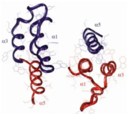
The first approach to the inhibition of integrase oligomerization process resulted the development of two peptides derived from the interface of the dimer IN CCD: INH1 (ATGQETAYFLLKLAGKA) and INH5 (DQAEHLKTAVQMAVFIHNYKA).
The first reproduces the amino acid sequence of the helix α1 (aa 93-107), the second reproduces the α5 helix and part of the loop which separates the α4 and α5 helices (aa167-187) (Figure 2.7). Experimental studies confirmed the dissociation of the tetramer by a INH1 and INH5, resulting in loss of enzymatic IN.41
Figure 2.7 Structure of the catalytic domain dimerization of HIV-1 IN. The two monomers are shown in red and blue. The propellers a1 and α5, from which the inhibitory peptides INH1 and INH5 respectively, are indicated with the α3 helix. Adapted from: Maroun RG, Gayet S, Benleulmi MS, Porumb H, Zargarian L, Merad H, Leh H, Mouscadet JF, Troalen F, Fermandjian S.
Chapter II: The HIV-1 integrase as molecular target
31
Peptide inhibitors of HIV-1 integrase dissociate the enzyme oligomers. Biochemistry. 2001; 40: 13840-13848.
Other studies, have led to the identification of two peptides, NL6 (TAYFLLKLAGRW) NL9 (ACWWAGIKQEF), with low values of IC50 for IN inhibition. These derived from the α1 helical domain (NL6)
and α3 helical domain respectively (NL9) (figure 2.8) .
Figure 2.8 IN secondary structure. The peptides derived from α1 (NL6) and α3 (NL9). Adapted from: Al-Mawsawi LQ, Neamati N. Allosteric inhibitor development targeting HIV-1 integrase. Chem. Med. Chem. 2011; 6: 228-41.
To determine the minimal active sequence, based on these sequences have been designed other peptides, only NL6-5 (YFLLKL), which contains the 6 central residues of NL6, has retained inhibitory activity. Furthermore they were synthesized analogues of NL6 and NL9 peptides with the amino acid sequence is inverted (RNL6, RNL9) and with the replacement of L-amino acids with D-amino acids (DNL6, RDNL6, DNL9, RDNL9). However, only analogues of NL6 have yet shown inhibitory activity.42
Chapter II: The HIV-1 integrase as molecular target
32
2.2.3 Integrase inhibitors derived from N-terminal domain
The N-terminal integrase plays an important role in the formation of tetramer integrase process. Therefore, this domain represents an important target for the design of drugs for inhibition of IN oligomerization process. In this context, compounds that target the IN N-terminal domain would be effective at disrupting IN function through a range of mechanisms, including allosteric or oligomerization inhibition. Recently, considering the amino acid sequence (1-50) of the N-terminal domain of integrase, the peptides NZ-1, NZ-2, N-ITFcc and NZ-4 were synthesized. Biological assays showed that peptide N-ITFcc, corresponding to the third α-helix (FNLPPVVAKEIVAS), inhibits viral replication almost 80%, indicating a possible use as an antiviral drug.43 Thus, we considered this domain an important starting point for the identification of peptide inhibitors in order to clarify some points on IN function in viral replication, such as the elucidation of HIV-1 IN polymerization state, or its potentiality in the structure-based design. In my PhD project, we present the design and synthesis of peptides targeting the integration events by directly inhibiting IN or its multimerization process. The synthesized peptides were assayed in vitro for their ability to inhibit IN strand transfer activity and for the capability to inhibit IN dimerization or to promote IN multimerization. Finally, the most potent compounds, conveniently conjugated with cell-penetrating fragment Tat, were assayed in MT-4 cells for determining anti-HIV infective activity.
Chapter II: The HIV-1 integrase as molecular target
33
2.3 References
1.
Katz RA, and Skalka AM. A C-terminal domain in the avian sarcoma-leukosis virus pol gene product is not essential for viral replication. J. Virol. 1988; 62 (2): 528-533.
2.
Grandgenett DP, Ajaykumar CV, Schiff RD. A 32,000-Dalton nucleic acid-binding protein from avian retravirus cores possesses DNA endonuclease activity. Virology. 1978; 89 (1): 119-132.
3. Bushman FD, and Wang B. Rous sarcoma virus integrase protein:
mapping functions for catalysis and substrate binding. J. Virol. April 1994; 68 (4): 2215-2223.
4.
Rhee SY, Liu TF, Kiuchi M, Zioni R, Gifford RJ, Holmes SP and Shafer RW. Natural variation of HIV-1 group M integrase: Implications for a new class of antiretroviral inhibitors. Retrovirology. 2008; 20085: 74.
5. Delelis O, Carayon K, Saïb A, Deprez E and Mouscadet JF. Integrase
and integration: biochemical activities of HIV-1 integrase. Retrovirology. 2008; 20085: 114.
6. Cherepanov P, Maertens G, Proost P, Devreese B, Van Beeumen J,
Engelborghs Y, De Clercq E, Debyser Z. HIV-1 integrase forms stable tetramers and associates with LEDGF/p75 protein in human cells. J.Biol. Chem. 2002; 278: 372–381.
7.
Parissi V, Calmels C, De Soultrait VR, Caumont A, Fournier M, Chaignepain S and Litvak S. Functional interactions of human immunodeficiency virus type 1 integrase with human and yeast HSP60. J. Virol. 2001; 75: 11344–11353.
8. Hindmarsh P, Ridky T, Reeves R, Andrake M, Skalka AM and Leis J.
HMG protein family members stimulate human immunodeficiency virus type 1 and avian sarcoma virus concerted DNA integration in vitro. J. Virol. 1999; 73:2994–3003.
9.
Farnet C and Bushman FD. HIV-1 cDNA integration: requirement of HMG I (Y) protein for function of preintegration complexes in vitro. Cell. 1997; 88: 483–492.
Chapter II: The HIV-1 integrase as molecular target
34
10. Bukrinsky MI, Sharova N, Dempsey MP, Stanwick TL, Bukrinskaya
AG, Haggerty S and Stevenson M. Active nuclear import of human immunodeficiency virus type 1 preintegration complexes. Proc. Natl. Acad. Sci. 1992; 89:6580–6584.
11.
Chow SA., Vincent, KA, Ellison V and Brown PO. Reversal of integration and DNA splicing mediated by integrase of human immunodeficiency virus. Science. 1992; 255: 723–726.
12. Chen JC, Krucinski J, Miercke LJ, Finer-Moore JS, Tang AH, Leavitt
AD, Stroud RM.. Crystal structure of the HIV-1 integrase catalytic core and C-terminal domains: a model for viral DNA binding. Proc. Natl Acad. Sci. 2000; 97:8233–8238.
13.
Wang JY, Ling H, Yang W and Craigie R. Structure of a two-domain fragment of HIV-1 integrase: implications for domain organization in the intact protein. EMBO J. 2001; 20:7333–7343.
14.
Yang W and Steitz TA. Recombining the structures of HIV integrase, RuvC and RNase H. Structure. 1995; 3:131–134.
15.
Song JJ, Smith SK, Hannon GJ and and Joshua-Tor L. Crystal structure of Argonaute and its implications for RISC slicer activity. Science. 2004; 305:1434–1437.
16.
Rice PA and Baker TA. Comparative architecture of transposase and integrase complexes. Nature Struct. Biol. 2001; 8:302–307.
17.
Bujacz G, Alexandratos J, Wlodawer A, Merkel G, Andrake M, Richard A, Skalka K and AM. Binding of different divalent cations to the active site of avian sarcoma virus integrase and their effects on enzymatic activity. J. Biol. Chem. 1997; 272: 18161–18168.
18. Beese LS and Steitz TA. Structural basis for the 3‟–5‟ exonuclease
activity of Escherichia coli DNA polymerase I: a two metal ion mechanism. EMBO J. 1991; 10: 25–33.
19.
Grobler JA, Stillmock K, Hu B, Witmer M, Felock P, Espeseth AS, Wolfe A, Egbertson Melissa, Bourgeois M, Melamed J, Wai JS, Young S, Vacca J and Hazuda DJ. Diketo acid inhibitor mechanism and HIV-1 integrase: implications for metal binding in the active site
Chapter II: The HIV-1 integrase as molecular target
35
of phosphotransferase enzymes. Proc. Natl Acad. Sci. 2002; 99: 6661– 6666.
20. Marchand C, Johnson AA, Karki RG, Pais GC, Zhang X, Cowansage
K, Patel TA, Nicklaus MC, Burke TR Jr, Pommier Y. Metal-dependent inhibition of HIV-1 integrase by β-diketo acids and resistance of the soluble double-mutant (F185K/C280S). Mol. Pharmacol. 2003; 64: 600–609.
21.
Chow SA and Brown PO. Juxtaposition of two viral DNA ends in a bimolecular disintegration reaction mediated by multimers of human immunodeficiency virus type 1 or murine leukemia virus integrase. J. Virol. 1994; 68: 7869–7878.
22. Engelman A, Bushman FD and Craigie R. Identification of discrete
functional domains of HIV-1 integrase and their organization within an active multimeric complex. EMBO J. 1993; 12: 3269–3275.
23.
Engelman A, and Craigie R. Identification of conserved amino acid residues critical for human immunodeficiency virus type 1 integrase function in vitro. J. Virol. 1992; 66, 6361–6369.
24.
Ming-Chieh S, Nidhanapati K, Raghavendra N, Vandegraaff J, Daigle SH, Kellam P, Cherepanov P, and Engelman A. LEDGF/p75 functions downstream from preintegration complex formation to effect gene-specific HIV-1 integration Genes Dev. 2007; 21(14): 1767–1778.
25.
Izumoto Y, Kuroda T, Harada H, Kishimoto T, Nakamura H. Hepatoma-derived growth factor belongs to a gene family in mice showing significant homology in the amino terminus. Biochem Biophys Res Commun 1997; 238(1):26-32.
26.
Cherepanov P, Devroe E, Silver PA, Engelman A. Identification of an evolutionarily conserved domain in human lens epithelium-derived growth factor/transcriptional co-activator p75 (LEDGF/p75) that binds HIV-1 integrase. J Biol Chem 2004; 279(47):48883-92.
27. Vandegraaff N and Engelman A. Molecular mechanisms of HIV
integration and therapeutic intervention. Expert Rev Mol Med 2007; 9(6):1-19.
Chapter II: The HIV-1 integrase as molecular target
36
28. Llano M, Vanegas M, Fregoso O, Saenz D, Chung S, Peretz M,
Poeschla EM. LEDGF/p75 determines cellular trafficking of diverse lentiviral but not murine oncoretroviral integrase proteins and is a component of functional lentiviral preintegration complexes. J Virol. 2004; 78(17):9524-37.
29. Llano M, Vanegas M, Hutchins N, Thompson D, Delgado S,
Poeschla EM. Identification and characterization of the chromatin-binding domains of the HIV-1 integrase interactor LEDGF/p75. J Mol Biol. 2006; 360(4):760-73
30.
Esposito F and Tramontano E. Past and Future. Current drugs targeting HIV-1 Integrase and Reverse Transcriptase-associated Ribonuclease H activity: single and dual active site inhibitors. Antiv. Chem Chemother. 2013; 23, 129-144.
31. Evering, TH and Markowitz M.. Raltegravir (MK-0518): an integrase
inhibitor for the treatment of HIV-1. Drugs Today. 2007; 43, 865-877.
32. Brinson, C and. Stribild. A Single Tablet Regimen for the Treatment
of HIV Disease. Comb. Prod. The. 2013; 3, 1-8.
33.
Mesplède T, Quashie PK, Wainberg MA. Resistance to HIV integrase inhibitors. Curr Opin HIV AIDS. 2012; 7, 401-408.
34.
Long YQ, Huang SX, Zawahir Z, Xu ZL, Li H, Sanchez TW, Zhi Y, De Houwer S, Christ F, Debyser Z, Neamati N. Design of Cell-Permeable Stapled Peptides as HIV-1 Integrase Inhibitors. J. Med. Chem. 2013; 56, 5601-5612.
35. Christ F, Voet A, Marchand A, Nicolet S, Desimmie BA, Marchand
D, Bardiot D, Van der Veken NJ.; Van Remoortel B, Strelkov SV, De Maeyer M, Chaltin P, Debyser Z. Rational design of small-molecule inhibitors of the LEDGF/p75-integrase interaction and HIV replication. Nat. Chem. Biol. 2010; 6: 442−448.
36. Christ F, Shaw S, Demeulemeester J, Desimmie BA, Marchand A,
Butler S, Smets W, Chaltin P, Westby M, Debyser Z, Pickford C. Small-molecule inhibitors of the LEDGF/p75 binding site of integrase block HIV replication and modulate integrase multimerization. Antimicrob. Agents Chemother. 2012; 56: 4365-4374.