A R T I C L E
O p e n A c c e s s
IFN
β enhances mesenchymal stromal
(Stem) cells immunomodulatory function
through STAT1-3 activation and
mTOR-associated promotion of glucose
metabolism
Tiziana Vigo
1, Claudia La Rocca
2, Deriggio Faicchia
2, Claudio Procaccini
2,3, Maddalena Ruggieri
4, Marco Salvetti
5,6,
Diego Centonze
6,7, Giuseppe Matarese
2,8and Antonio Uccelli
1,9, on behalf of the MSRUN Network
Abstract
Administration of mesenchymal stem cells (MSC) ameliorate experimental autoimmune encephalomyelitis (EAE), a mouse model of multiple sclerosis (MS), at both clinical and neuropathological levels. The therapeutic properties of MSC in EAE are mainly mediated by the modulation of pathogenic immune response, but other neurotropic effects, including decreased demyelination and axonal loss as well as promotion of tissue repair, play also a role. Properly controlled phase II clinical trials to explore the potential of MSC transplantation as a treatment for MS are underway. Interferon beta (IFNβ) is an approved treatment for relapsing-remitting and secondary progressive MS. Here, we explored the possibility that IFNβ might influence the therapeutic potential of MSC, in view of possible
synergistic effects as add-on therapy. IFNβ enhanced the immunomodulatory functions of MSC and induced the
expression of secretory leukocyte protease inhibitor (Slpi) and hepatocyte growth factor (Hgf), two soluble mediators involved in immune and regenerative functions of MSC. At molecular level, IFNβ induced a rapid and transient phosphorylation of STAT1 and STAT3, the transcription factors responsible for Slpi and Hgf induction. Concomitantly, IFNβ dynamically affected the activity of mTOR, a key checkpoint in the control of metabolic pathways. Indeed, the impairment of mTOR activity observed early upon exposure to IFNβ, was followed by a long-lasting induction of mTOR signaling, that was associated with an increased glycolytic capacity in MSC. When induced to switch their energetic metabolism towards glycolysis, MSC showed an improved ability to control T-cell proliferation. These results suggest that modifications of MSC energetic metabolism induced by IFNβ may contribute to promote MSC immunomodulatory function and support a role for metabolic pathways in the therapeutic function of MSC. Altogether, thesefindings support the idea of a combined treatment for MS, in which the immunomodulatory and possibly regenerative activity of MSC could be enhanced by the administration of IFNβ.
Introduction
Mesenchymal Stromal (Stem) Cells (MSC) are a het-erogeneous subset of stromal progenitors of mesodermal lineage that have been isolated from almost every tissue, mainly the bone marrow and adipose tissue. MSC are defined on the basis of their capability to grow as adherent
© The Author(s) 2019
Open Access This article is licensed under a Creative Commons Attribution 4.0 International License, which permits use, sharing, adaptation, distribution and reproduction in any medium or format, as long as you give appropriate credit to the original author(s) and the source, provide a link to the Creative Commons license, and indicate if changes were made. The images or other third party material in this article are included in the article’s Creative Commons license, unless indicated otherwise in a credit line to the material. If material is not included in the article’s Creative Commons license and your intended use is not permitted by statutory regulation or exceeds the permitted use, you will need to obtain permission directly from the copyright holder. To view a copy of this license, visithttp://creativecommons.org/licenses/by/4.0/.
Correspondence: Antonio Uccelli ([email protected]) 1
IRCCS Ospedale Policlinico San Martino, Genoa, Italy
2Laboratorio di Immunologia, Istituto di Endocrinologia e Oncologia Sperimentale, Consiglio Nazionale delle Ricerche (IEOS-CNR), Naples, Italy Full list of author information is available at the end of the article. Edited by G. Melino 1234567890() :,; 1234567890( ):,; 1234567890() :,; 1234567890( ):,;
cells on plastic, to display afibroblast-like morphology, to form colonies in vitro supporting hematopoiesis, to dif-ferentiate into cells of the mesodermal lineage and express stromal while lacking hematopoietic markers1. Several studies showed that MSC possess immunomodulatory properties exerted on cells populations of both adaptive and innate immunity2 and these features, together with their reported ability to protect neural cells from death and foster neural repair, account for their proposed therapeutic effect on multiple sclerosis (MS) and other
neurological diseases3. Intravenous infusion of MSC
improved the clinical course of EAE, inducing immune tolerance, reducing inflammation decreasing demyelina-tion and promoting tissue repair4–9. The mechanisms through which MSC exert their therapeutic function are heterogenous and probably pleiotropic. It is generally accepted that MSC immunomodulation is strongly influenced by cytokines in the inflammatory environment,
particularly by IFN gamma (IFNγ)10,11
and that their therapeutic effect is mediated by paracrine mechanisms through the release of soluble factors2. Particularly, intravenous injection of conditioned medium containing MSC-derived hepatocyte growth factor (HGF) promoted remyelination in vitro and tissue repair in vivo12. Auto-logous MSC have been safely administrated in a limited number of patients with MS13, and ongoing controlled clinical studies to explore the potential of MSC trans-plantation as a treatment for MS have been launched (Clinical Trial NCT01854957).
Interferon beta (IFNβ) is an approved treatment
for relapsing-remitting14 and secondary progressive
multiple sclerosis (MS). Interferons are a family of cytokines secreted by various cell types of the innate and adaptive immune systems as well as by other tissues. One major pathway in IFNβ signaling involves activa-tion of Signal Transducer and Activator of Transcrip-tion (STAT) proteins and formaTranscrip-tion of complexes that translocate to the nucleus and bind to specific elements to regulate gene transcription15. Efficacy of IFNβ for the treatment of MS is believed to be due to modulation
of immune responses16. Among its functions, in MS
subjects IFNβ modulates dendritic cells17, T and B
lymphocytes18, as well as regulatory NK cells and T
regulatory cells19.
While the potent immunomodulatory effect of IFNβ on cells of the immune system has been extensively studied, little is known on the interaction between IFNβ and MSC. We recently demonstrated that the pro-immunomodulatory effect of IFNγ on MSC is mediated by the phosphorylation of STAT1 and STAT3 and by the inhibition of mammalian target of rapamycin (mTOR) activity11. Thus, we sought to
address the effect of IFNβ on MSC immunomodulatory
functions keeping in mind that, based on the possibility of a
synergic effect, these two treatments could be effectively associated to treat MS.
Our data showed that IFNβ promoted the ability of MSC to control T-cell proliferation and enhanced the gene expression of Hgf and of secretory leukocyte protease inhibitor (Slpi), an important regulator of innate and adaptive immunity and a component of tissue regenerative programs20. At molecular level IFNβ induced a rapid activation of STAT1 and STAT3 in MSC, concomitant with an early inhibition followed, at 24 h, by a strong re-activation of mTOR pathway. The oscillatory switch of mTOR signaling induced by IFNβ was associated with an increased glycolytic capacity of MSC, that improved their ability to control T-cell proliferation.
Materials and methods
MSC culture and treatments
MSC isolated from mouse bone marrow were cultured in Mesencult medium as described [8]. MSC were treated with 1500 UI/ml of IFNβ (Rebif ® 44, Merk Serono) for 1 h in serum-free RPMI (Gibco).
Gene silencing
siRNA transfection was carried out using Lipofectamine 2000 in serum free RPMI for 24 h in the presence of specific siRNA (20 pmol). STAT1:(gauugaccuggagaccaccucucuu/ aagagagguggucuccaggucaauc)
SLPI: (caagugcugugaggguaua(dt)(dt)/uauacccucacagca-cuug(dt)(dt)
As negative control, MSC were transfected with AllStar siRNA (Qiagen) (CTRL-KD) according to manufacturer’s instructions. The efficacy of siRNA knock-down was tested by real time PCR.
Collection of MSC-conditioned medium
Confluent MSC were primed with IFNβ for 1 h in
serum-free RPMI. MSC were washed and cultured for an additional 24 h in serum-free RPMI with glucose 2 or 4.5 g/L, and the medium was collected (conditioned medium, CM).
In vitro T-cell proliferation assay
MSC CM equivalent to that of 2 × 10⁴MSC was assessed for its effect on proliferation of 2 × 10⁵ anti-CD3/CD28-activated spleen cells (1:10 ratio) labeled with CFSE (1 µM; Molecular Probes). Equivalent amount of CM from 3T3 fibroblast culture was used as control. Where the CM of MSC was collected in RPMI with different concentrations of glucose (2 or 4.5 g/L), thefinal glucose concentration in the T-cell activation medium was 4.5 g/L. Alternatively, MSC CTRL-KD and SLPI-KD were irradiated (5000 rad) and added to 2 × 10^5 anti-CD3/CD28-activated spleen cells (1:10 ratio) labeled with CFSE (1 µM) at the ratio of
1:40. After 72 h, splenocytes were collected and the mean fluorescence intensity of CD3-positive T cells was evaluated by flow cytometry. Statistical differences were evaluated using Student’s T-test.
Real-time PCR
Total RNA was extracted with Trizol (Invitrogen) from MSC, MSC RAPA, MSC CUC, MSC CTRL-KD and
STAT1-KD 24 h after treatment with IFNβ. Real-time
PCR experiments were conducted as per the
manu-facturer’s instructions (SYBR Green I Master, Roche)
using the following primers:
Cd274(aaatcgtggtccccaagc/tcctcatgttttgggaactatct) Ccl2(catccacgtgttggctca/gatcatcttgctggtgaatgagt) Hgf(caccccttgggagtattgtg/gggacatcagtctcattcacag) Il18bp(agctattcggggcttaggag/tgcaagcaagtctggtgtct) Slpi(cttgctctggggatcctg/ggctccgattttgatagcat)
Gene expression values were calculated as 2(−ΔCT)with hypoxanthine guanine phosphoribosyl transferase (Hprt) (tcctcctcagaccgctttt/cctggttcatcatcgctaatc) as reference gene. Statistical differences were evaluated using Student’s T-test.
Western blot
Total cell lysates and western blot analysis were per-formed as previously described21. The blots were probed
with antibodies specific for pSTAT1 (Tyr701), pSTAT3
(Tyr705), pS6 (all 1:1000 dilution and from Cell Signaling Technology, Beverly, MA) and with an actin antibody (1:1000 from Santa Cruz) to normalize for the amount of loaded protein.
Pathway inhibition by pharmacological agents
STAT3 inhibition was obtained by exposure of MSC to cucurbitacin I (CUC) (500 nM; Sigma Aldrich) for 3 h in serum-free RPMI; mTOR inhibition was obtained through exposure of MSC to rapamycin (100 nM; Sigma Aldrich) in serum-free RPMI for 1 h (RAPA). The inhi-bition of STAT3 and S6 phosphorylation following treatments was evaluated by Western blot analysis using phospho-specific antibodies (data not shown).
Immunofluorescence
MSC werefixed in 4% paraformaldehyde, permeabilized
with 0.5% Triton and labeled with antibodies specific for mouse SLPI (1:100 dilution from Cell Signaling) followed by a secondary antibody conjugated with AlexaFluor-594. Nuclei were stained with DAPI. The slides were analyzed with a Olympus microscopy equipped with 405 and 561 nmfilters.
Analysis of MSC metabolism
Real-time measurements of extracellular acidification rate (ECAR) and oxygen consumption rate (OCR) were
measured using an XFe-96 Extracellular Flux Analyzer (Seahorse Bioscience). Cells were counted using an automated Cell counter (Countess from Life Technolo-gies), seeded in XFe-96 plates (Seahorse Bioscience) at the density of 2 × 10⁴ cells/well and incubated overnight at 37 °C in 5% CO2atmosphere in the presence or absence
of IFNβ (1500UI/ml). ECAR was measured in XF media in basal condition and in response to 10 mM glucose, 4μM
oligomycin and 100 mM of 2-Deoxy-D-glucose (2-DG).
Basal glycolysis was calculated after glucose injection (subtracting the ECAR rate inhibited by 2DG). Maximal glycolysis was measured after oligomycin injection and glycolytic capacity as the difference of oligomycin-induced ECAR and 2-DG-oligomycin-induced ECAR. OCR was measured in XF media (non-buffered DMEM medium, containing 10 mM glucose, 2 mM L-glutamine, and 1 mM sodium pyruvate), under basal conditions and in response
to 5μM oligomycin, 1.5 μM of
carbonylcyanide-4-(tri-fluoromethoxy)-phenylhydrazone (FCCP) and 1 μM of antimycin A and rotenone (all chemicals from Sigma Aldrich). Basal OCR was calculated as the difference between baseline measurements and antimycin A/rote-none-induced OCR; maximal OCR was measured as the difference between FCCP-induced OCR and antimycin A/rotenone-induced OCR. Finally, the ATP-linked para-meter was calculated as the difference between baseline
measurements, without injection, and
oligomycin-induced OCR. Experiments with the Seahorse system were done with the following assay conditions: 3 min mixture; 3 min wait; and 3 min measurement. Then, metabolic parameters were calculated. Data are expressed as mean and S.E.M. from four independent experiments. Statistical differences were evaluated using the Wilcoxon matched-pairs test.
Results
IFNβ promoted MSC immunomodulation inducing the expression of secreted mediators
To verify the possibility that IFNβ may affect the
expression of immunomodulatory factors secreted by
MSC we analyzed, 24 h upon priming with IFNβ, the
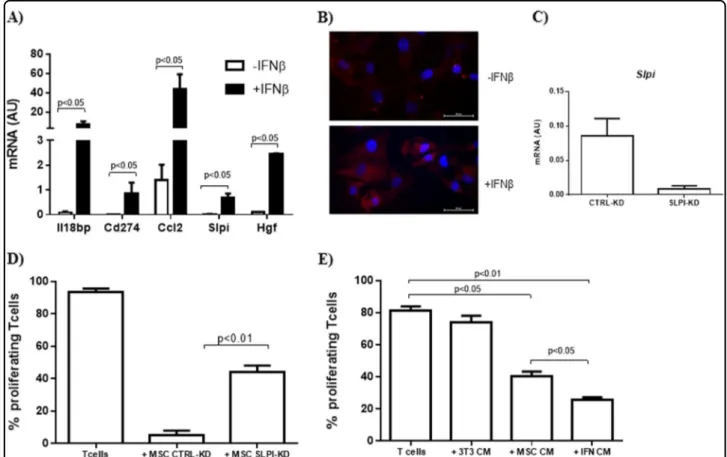
mRNA levels of Cd274, Ccl2, and Hgf, which are known to act in a paracrine way on immune cells. We also assessed the gene expression of Il18bp and Slpi, that we recently identified as induced in MSC upon interaction with activated T cells11. As shown Fig. 1a, IFNβ induced a
significant increase of mRNA level of all the genes ana-lyzed. We then focused on Slpi, whose role in MSC
immunomodulation has never been reported. We first
confirmed that MSC exposed to IFNβ increased Slpi expression at protein level (Fig.1b). To get some insight into the role of Slpi on MSC immunoregulatory function, we reduced its expression in MSC through gene silencing (SLPI-KD) and used these Slpi-silenced cells to inhibit
T-cell proliferation (Fig.1c). As shown in Fig. 1d, MSC SLPI-KD showed a reduced ability to inhibit T-cell pro-liferation in vitro as compared to MSC transfected with control siRNA (CTRL-KD).
Then we sought to verify if IFNβ was able to affect the ability of MSC to control T-cell proliferation priming
MSC with IFNβ and testing the effect of the derived
conditioned medium (CM) on activated T cells. As shown in Fig. 1e, IFNβ significantly enhanced the constitutive immunomodulatory effect of MSC CM.
These results demonstrated that IFNβ induced in MSC the expression of soluble immunomodulatory factors and concomitantly improved their ability to inhibit T-cell proliferation in vitro.
IFNβ induced dynamic changes in STAT1, STAT3, and S6 phosphorylation
As activation of STAT1 and STAT3, and inhibition of mTOR pathway positively affects MSC immunomodula-tory function upon administration of interferon gamma
(IFNγ)11
, we analyzed the activation state of these pathways following exposure to IFNβ. Analysis of phos-phorylated pathway components revealed that exposure
to IFNβ for 1 h dramatically increased the levels of
phosphorylated STAT1 (pSTAT1) and STAT3 (pSTAT3) and concomitantly reduced the amount of phosphorylated S6 (pS6), used as read-out of mTOR activity (Fig. 2b). Upon a prolonged exposure (24 h), pSTAT1 and pSTAT3
decreased as compared to 1 h, indicating that IFNβ
induced a a transient phosphorylation of STAT1 and STAT3. Concomitantly, the levels of pS6 strongly increased as compared to that observed upon exposure to IFNβ for 1 h, revealing that IFNβ induced an oscillatory modification of mTOR pathway activity (Fig.2a, b). IFNβ-dependent expression of Slpi and Hgf was mediated by STAT1 and STAT3
Engagment of STAT1 and STAT3 induces the expres-sion of immunomodulatory genes in MSC, that is further enhanced by inhibition of mTOR signaling11. To assess
Fig. 1 IFNβ induced the expression of soluble mediators and promoted MSC immunomodulatory function. a Real-time PCR analysis of immunoregulatory genes in MSC exposed or not to IFNβ. Data are mean ± SD (n = 3); b Immunofluorescence analysis of Slpi expression in MSC. Red, SLPI; blue, DAPI; c Real-time PCR analysis of Slpi expression was evaluated in MSC following transfection with specific siRNA (MSC SLPI-KD) and compared to that of control MSC (MSC CTRL-KD). Data are mean ± SD (n= 3); d In vitro T-cell proliferation assay in the presence of MSC CTRL-KD and MSC SLPI-KD. MSC were added at the ratio 1:40 with activated splenocytes. Data are presented as mean ± SD (n= 3); e In vitro T-cell proliferation in the presence of MSC conditioned medium (MSC CM) collected after priming with IFNβ (IFN CM). The CM of 3T3 fibroblast culture was used as control. Data are mean ± SD (n= 3)
the role of these pathways in the induction of Slpi and Hgf by IFNβ, we inhibited STAT1 through gene silencing, STAT3 using the chemical inhibitor Cucurbitacin I (CUC) and mTOR with its inhibitor rapamycin (RAPA) before the exposure to IFNβ and we analyzed Slpi and Hgf expression by real-time PCR. We observed that the induction of Slpi expression by IFNβ was significantly impaired by STAT1 and STAT3 inhibition, while the blockade of mTOR pathway did not exert any affect on their expression (Fig. 2c). Similarly, we observed that impairment of STAT1 and STAT3 signaling, but not that of mTOR, reduced the induction of Hgf upon exposure to IFNβ (Fig.2d).
Exposure to IFNβ modified MSC energetic metabolism As IFNβ affected the activity of mTOR pathway, a master regulator of intracellular metabolism, we assessed whether exposure to IFNβ could impact on the bioener-getic profile of MSC, by measuring extracellular acid-ification rate (ECAR) as an indicator of glycolysis, and oxygen consumption rate (OCR) indicator of mitochon-drial respiration. We observed that IFNβ did not modify basal glycolysis in MSC (Fig. 3a, b). However, maximal glycolysis (induced by oligomycin injection) and glycolytic capacity were significantly increased by IFNβ treatment,
as compared to untreated MSC (Fig. 3a, b). Then we
investigated mitochondrial respiration in terms of OCR. IFNβ did not affect maximal OCR and OCR coupled to ATP generation, but slightly enhanced the respiratory capacity of MSC in basal conditions (Fig.3c, d), indicating that IFNβ preferentially increased the glycolytic capacity of MSC.
Glycolisis engagement improved MSC immunomodulatory function
To link MSC glucose metabolism with their immuno-modulatory function, we assessed whether or not improvement of MSC glycolitic rate may impact on their capacity to inhibit T-cell proliferation. We boosted
gly-colitic flux by growing MSC in high glucose containing
medium (4.5 g/L), and we assessed the immunomodula-tory properties of the resulting CM in T-cell proliferation assay. We obtained a significant improvement in the constitutive ability of MSC CM to inhibit T-cell pro-liferation by collecting MSC CM in high glucose-containing medium as compared to MSC CM prepared in normal medium (2 g/L) (Fig.4).
These results clearly demonstrated that promoting glucose metabolism improved the immunomodulatory function of MSC, thus suggesting a new possible
mechanism through which IFNβ enhances MSC
immunomodulation.
Fig. 2 IFNβ induced dynamic changes in STAT1, STAT3, and S6 phosphorylation. a Western blot analysis conducted after 1 or 24 h of incubation with IFNβ. Actin served as loading control; b relative densitometric quantitation of the shown gel, after normalization on actin; c, d Real-time PCR analysis of Slpi and Hgf expression in MSC exposed to IFNβ upon pathway inhibition. Data are presented as mean ± SD (n = 3)
Discussion
While the potent immunomodulatory effect of IFNβ on cell types of the immune system has been extensively studied22, little is known on the interaction between IFNβ
and MSC. IFNβ was shown to influence the expression of chemokines and their receptors in human MSC23, but the effects of IFNβ on MSC immunomodulatory function are largely unknown. Our data demonstrated that IFNβ sig-nificantly increased the expression of molecules, secreted by MSC, with reported immunomodulatory activities. Among those, we observed that Slpi, a molecule never related to MSC biology, potentiated the ability of MSC to control T-cell proliferation in vitro.
Slpi is a polypeptide secreted by epithelial cells to pro-tect tissue from damage by leucocyte proteolytic enzymes. Nevertheless, the regulatory function of Slpi on adaptive immunity is independent to its anti-protease activity. Slpi has been demonstrated to affect the monocyte pattern secretion by preventing the interaction of p65 with the NF-kB binding sites24, to inhibit effector T-cell pro-liferation25 and to promote the generation of regulatory T cells26. Slpi is upregulated in macrophages, activated microglia, neuronal cells and astrocytes during EAE attack27. Interestingly, Slpi was shown to increase pro-liferation of adult neural stem cells promoting oligoden-droglial differentiation, as incubation of adult neural stem cells with recombinant Slpi resulted in an increase of cell
Fig. 3 Exposure to IFNβ modified MSC energetic metabolism. a Kinetic profile of ECAR in MSC treated or not with IFNβ for 12 h. The data are shown as mean ± S.E.M. of four independent experiments. ECAR was measured in real time, under basal conditions and in response to glucose, oligomycin and 2-DG; b parameters of glycolysis in MSC were calculated as detailed in Materials and methods. Data are expressed as mean ± S.E.M. of three measurements, from four independent experiments; c kinetic profile of OCR in MSC treated or not with IFNβ for 12 h. The data are shown as mean ± S.E.M. of four independent experiments. OCR was measured in real time, under basal conditions and in response to oligomycin, FCCP and Antimycin A+ Rotenone. d Parameters of mitochondrial respiration in MSC were calculated as detailed in material and methods. Data are expressed as mean ± S.E.M. of three measurements, from four independent experiments
Fig. 4 Promotion of MSC glycolisis improved their ability to inhibit T-cell proliferation in vitro. In vitro T-cell proliferation assay in the presence of conditioned medium of MSC (MSC CM) collected in serum-free RPMI at different concentration of glucose (2 g/L or 4, 5 g/L). Thefinal glucose concentration in the T-cell activation medium was 4.5 g/L. Data are mean ± SD (n= 3)
proliferation and of differentiation towards oligoden-drocytes27. Whether Slpi secreted by MSC may play a role also in tissue repair promoted by MSC requires further investigation. Interestingly IFNβ significantly enhanced also the expression of Hgf a pleiotropic cytokine with neuroprotective which has been demonstrated to enhance axonal outgrowth and oligodendroglial maturation12.
At molecular level, IFNβ induced a transient
phos-phorylation of STAT1 and STAT3, determining the induction of Slpi and Hgf expression, that was indeed impaired by both STAT1 and STAT3 inhibition.
Con-comitantly, IFNβ induced a dynamic modification of
mTOR activity, that was early reduced and then increased. In line with the role of mTOR in the control of glucose metabolism, a prolonged exposure to IFNβ increased MSC glycolytic capacity, thus improving their ability to engage glycolysis to convert glucose to pyruvate or lactate. Energy metabolism and functional activation are fully integrated in immune cells to determine their ability to divide, differentiate, and carry out effector functions28.
Here we found that inhibition of T-cell proliferation by MSC was strongly enhanced by boosting MSC glycolisis. As ECAR, monitored as readout of MSC glycolitic activity, was mainly due to production of lactate, our data suggest the possible involvement of lactic acid in MSC immuno-modulatory function. Lactic acid had been shown to suppress the proliferation and cytokine production of
human cytotoxic T lymphocytes29 and to inhibit TNF
secretion and glycolysis of human monocytes30.
Our data support that IFNβ can enhance MSC
ther-apeutic function through two different molecular
mechanisms: the early induction of STAT1-3-dependent immunomodulatory genes, and the long-lasting mTOR-associated promotion of glucose metabolism. Particularly, based on thefinding that IFNβ induced the expression of cytokines with both immunomodulatory and neuropro-tective features such as Hgf and Slpi, we speculate that MSC as add-on therapy to IFNβ treatment could promote
tissue repair beyond modulation of inflammatory
reac-tions. In line with these results, administration of IFN β-secreting engineered MSC has been demonstrated to be more effective than wild-type MSC in the attenuation of EAE31,32, further supporting the rationale for combined
protocols to treat MS. Moreover, the finding that MSC
immunomodulatory function is improved by glucose metabolism revealed a new possible mechanism under-lying MSC therapeutic effect.
Acknowledgements
This work was supported by grants from Fondazione Italiana Sclerosi Multipla (FISM) (grant MESEMS-FISM 2012/S/4 to A.U.), (grant FISM no. 2016/R/18, no. 2014/R/21 to G.M.), from the Compagnia di San Paolo (grant 2015-0646 to A.U.) and by a grant from Merck Serono SpA (Multiple Sclerosis Research University Network, MS-RUN project to A.U., M.R., M.S., D.C., and G.M.). T.V. was supported by Fondazione Italiana Sclerosi Multipla (FISM) (grant 2017/R/28). G.M. was supported by grants from European Research Council Grant
“menTORingTregs” no. 310496, from Fondazione Italiana Sclerosi Multipla (FISM) no. 2016/R/18 and from Telethon no. GGP17086. C.P. was supported by the Italian Ministry of Health, GR-2016-02363749. The sponsors had no role in study design, data collection and analysis, decision to publish or preparation of the paper.
Author details 1
IRCCS Ospedale Policlinico San Martino, Genoa, Italy.2Laboratorio di Immunologia, Istituto di Endocrinologia e Oncologia Sperimentale, Consiglio Nazionale delle Ricerche (IEOS-CNR), Naples, Italy.3IRCSS Fondazione Santa Lucia, Rome, Italy.4Department of Basic Medical Science, Neuroscience and Sense Organs, University of Bari, Bari, Italy.5Centre for Experimental Neurological Therapies (CENTERS), Sapienza University, Rome, Italy.6IRCCS Istituto Neurologico Mediterraneo (INM) Neuromed, Pozzilli, Italy.7Laboratory of Synaptic Immunopathology, Tor Vergata University, Rome, Italy. 8Dipartimento di Medicina Molecolare e Biotecnologie Mediche, Università degli Studi di Napoli“Federico II”, Naples, Italy.9Department of Neurosciences, Rehabilitation, Ophthalmology, Genetics, Maternal and Child Health Unit and Center of Excellence for Biomedical Research, University of Genoa, Genoa, Italy
Author contributions
Tiziana Vigo: conception and design, collection and/or assembly of data, data analysis and interpretation, manuscript writing,final approval of manuscript. Claudia La Rocca: collection and/or assembly of data, data analysis and interpretation,final approval of manuscript. Deriggio Faicchia: collection and/ or assembly of data, data analysis and interpretation,final approval of manuscript. Claudio Procaccini: collection and/or assembly of data, data analysis and interpretation,final approval of manuscript. Maddalena Ruggieri: conception and design,final approval of manuscript. Marco Salvetti: conception and design,final approval of manuscript. Diego Centonze: conception and design,final approval of manuscript. Giuseppe Matarese: conception and design, data analysis and interpretation, manuscript writing, final approval of manuscript. Antonio Uccelli: conception and design, financial support, manuscript writing,final approval of manuscript.
Conflict of interest
The authors declare that they have no conflict of interest. Publisher’s note
Springer Nature remains neutral with regard to jurisdictional claims in published maps and institutional affiliations.
Received: 22 October 2018 Revised: 31 December 2018 Accepted: 7 January 2019
References
1. Horwitz, E. M. et al. Clarification of the nomenclature for MSC: The Interna-tional Society for Cellular Therapy position statement. Cytotherapy 7, 393–395 (2005).
2. Uccelli, A., Moretta, L. & Pistoia, V. Mesenchymal stem cells in health and disease. Nat. Rev. Immunol. 8, 726–736 (2008).
3. Uccelli, A., Laroni, A. & Freedman, M. S. Mesenchymal stem cells for the treatment of multiple sclerosis and other neurological diseases. Lancet Neurol. 10, 649–656 (2011).
4. Bai, L. et al. Human bone marrow-derived mesenchymal stem cells induce Th2-polarized immune response and promote endogenous repair in animal models of multiple sclerosis. Glia 57, 1192–1203 (2009).
5. Gerdoni, E. et al. Mesenchymal stem cells effectively modulate pathogenic immune response in experimental autoimmune encephalomyelitis. Ann. Neurol. 61, 219–227 (2007).
6. Gordon, D. et al. Human mesenchymal stem cells abrogate experimental allergic encephalomyelitis after intraperitoneal injection, and with sparse CNS infiltration. Neurosci. Lett. 448, 71–73 (2008).
7. Rafei, M. et al. Mesenchymal stromal cells ameliorate experimental auto-immune encephalomyelitis by inhibiting CD4 Th17 T cells in a CC chemokine ligand 2-dependent manner. J. Immunol. 182, 5994–6002 (2009).
8. Zappia, E. et al. Mesenchymal stem cells ameliorate experimental auto-immune encephalomyelitis inducing T-cell anergy. Blood 106, 1755–1761 (2005).
9. Zhang, J. et al. Human bone marrow stromal cell treatment improves neu-rological functional recovery in EAE mice. Exp. Neurol. 195, 16–26 (2005). 10. Ren, G. et al. Mesenchymal stem cell-mediated immunosuppression occurs via
concerted action of chemokines and nitric oxide. Cell Stem Cell 2, 141–150 (2008).
11. Vigo, T. et al. IFN-gamma orchestrates mesenchymal stem cell plasticity through the signal transducer and activator of transcription 1 and 3 and mammalian target of rapamycin pathways. J. Allergy Clin. Immunol. 139, 1667–1676 (2017).
12. Bai, L. et al. Hepatocyte growth factor mediates mesenchymal stem cell-induced recovery in multiple sclerosis models. Nat. Neurosci. 15, 862–870 (2012).
13. Connick, P. et al. Autologous mesenchymal stem cells for the treatment of secondary progressive multiple sclerosis: an open-label phase 2a proof-of-concept study. Lancet Neurol. 11, 150–156 (2012).
14. Randomised double-blind placebo-controlled study of interferon beta-1a in relapsing/remitting multiple sclerosis. PRISMS (Prevention of Relapses and Disability by Interferon beta-1a Subcutaneously in Multiple Sclerosis) Study Group. Lancet 352, 1498–1504 (1998)..
15. Platanias, L. C. & Fish, E. N. Signaling pathways activated by interferons. Exp. Hematol. 27, 1583–1592 (1999).
16. Severa, M., Rizzo, F., Giacomini, E., Salvetti, M. & Coccia, E. M. IFN-beta and multiple sclerosis: cross-talking of immune cells and integration of immu-noregulatory networks. Cytokine Growth Factor Rev. 26, 229–239 (2015). 17. Severa, M. et al. IFN-beta therapy regulates TLR7-mediated response in
plas-macytoid dendritic cells of multiple sclerosis patients influencing an anti-inflammatory status. J. Interferon Cytokine Res. 35, 668–681 (2015). 18. Alenda, R. et al. Blood lymphocyte subsets identify optimal responders to
IFN-beta in. MS 265, 24–31 (2018).
19. Vandenbark, A. A. et al. Interferon-beta-1a treatment increases CD56 bright natural killer cells and CD4+CD25+Foxp3 expression in subjects with multiple sclerosis. J. Neuroimmunol. 215, 125–128 (2009).
20. Majchrzak-Gorecka, M., Majewski, P., Grygier, B., Murzyn, K. & Cichy, J. Secretory leukocyte protease inhibitor (SLPI), a multifunctional protein in the host defense response. Cytokine Growth Factor Rev. https://doi.org/10.1016/j.
cytogfr.2015.12.001(2015).
21. De Rosa, V. et al. A key role of leptin in the control of regulatory T cell proliferation. Immunity 26, 241–255 (2007).
22. Kasper, L. H. & Reder, A. T. Immunomodulatory activity of interferon-beta. Ann. Clin. Transl. Neurol. 1, 622–631 (2014).
23. Croitoru-Lamoury, J., Lamoury, F. M., Zaunders, J. J., Veas, L. A. & Brew, B. J. Human mesenchymal stem cells constitutively express chemokines and chemokine receptors that can be upregulated by cytokines, IFN-beta, and Copaxone. J. Interferon Cytokine Res. 27, 53–64 (2007).
24. Taggart, C. C. et al. Secretory leucoprotease inhibitor binds to NF-kappaB binding sites in monocytes and inhibits p65 binding. J. Exp. Med. 202, 1659–1668 (2005).
25. Guerrieri, D. et al. Serine leucocyte proteinase inhibitor-treated monocyte inhibits human CD4(+) lymphocyte proliferation. Immunology 133, 434–441 (2011).
26. Tateosian, N. L. et al. Neutrophil elastase treated dendritic cells promote the generation of CD4(+)FOXP3(+) regulatory T cells in vitro. Cell Immunol. 269, 128–134 (2011).
27. Mueller, A. M. et al. Novel role for SLPI in MOG-induced EAE revealed by spinal cord expression analysis. J. Neuroinflamm. 5, 20 (2008).
28. Norata, G. D. et al. The cellular and molecular basis of translational immuno-metabolism. Immunity 43, 421–434 (2015).
29. Fischer, K. et al. Inhibitory effect of tumor cell-derived lactic acid on human T cells. Blood 109, 3812–3819 (2007).
30. Dietl, K. et al. Lactic acid and acidification inhibit TNF secretion and glycolysis of human monocytes. J. Immunol. 184, 1200–1209 (2010).
31. Marin-Banasco, C. et al. Gene therapy with mesenchymal stem cells expressing IFN-ss ameliorates neuroinflammation in experimental models of multiple sclerosis. Br. J. Pharmacol. 174, 238–253 (2017).
32. Kim, M. J. et al. Effective combination of methylprednisolone and interferon beta-secreting mesenchymal stem cells in a model of multiple sclerosis. J. Neuroimmunol. 314, 81–88 (2018).