R E S E A R C H A R T I C L E
Open Access
Meripilus giganteus ethanolic extract
exhibits pro-apoptotic and anti-proliferative
effects in leukemic cell lines
Monia Lenzi
1†, Veronica Cocchi
1†, Aleksandra Novakovi
ć
2, Maja Karaman
3, Marijana Saka
č
2, Anamarija Mandi
ć
2,
Milica Poji
ć
2, Maria Cristina Barbalace
4, Cristina Angeloni
5, Patrizia Hrelia
1, Marco Malaguti
4*†and Silvana Hrelia
4†Abstract
Background: The interest towards botanicals and plant extracts has strongly risen due to their numerous biological effects and ability to counteract chronic diseases development. Among these effects, chemoprevention which represents the possibility to counteract the cancerogenetic process is one of the most studied. The extracts of mushroom Meripilus giganteus (MG) (Phylum of Basidiomycota) showed to exert antimicrobic, antioxidant and antiproliferative effects. Therefore, since its effect in leukemic cell lines has not been previously evaluated, we studied its potential chemopreventive effect in Jurkat and HL-60 cell lines.
Methods: MG ethanolic extract was characterized for its antioxidant activity and scavenging effect against different radical species. Moreover, its phenolic profile was evaluated by HPLC-MS-MS analyses. Flow cytometry (FCM) analyses of Jurkat and HL-60 cells treated with MG extract (0–750 μg/mL) for 24–72 h- allowed to evaluate its cytotoxicity, pro-apoptotic and anti-proliferative effect. To better characterize MG pro-pro-apoptotic mechanism ROS intracellular level and the gene expression level of FAS, BAX and BCL2 were also evaluated. Moreover, to assess MG extract selectivity towards cancer cells, its cytotoxicity was also evaluated in human peripheral blood lymphocytes (PBL).
Results: MG extract induced apoptosis in Jurkat and HL-60 cells in a dose- and time- dependent manner by increasing BAX/BCL2 ratio, reducing ROS intracellular level and inducing FAS gene expression level. In fact, reduced ROS level is known to be related to the activation of apoptosis in leukemic cells by the involvement of death receptors. MG extract also induced cell-cycle arrest in HL-60 cells. Moreover, IC50at 24 h treatment resulted
2 times higher in PBL than in leukemic cell lines.
Conclusions: Our data suggest that MG extract might be considered a promising and partially selective chemopreventive agent since it is able to modulate different mechanisms in transformed cells at concentrations lower than in non-transformed ones.
Keywords: Meripilus giganteus, Cytotoxicity, Apoptosis, Chemoprevention, Flow-cytometry, Leukemic cell lines Background
Mushrooms have been used for centuries as food all over the world, due to their unique taste and flavour [1]. Archaeological knowledge shows that humans have been
using mushrooms since the Palaeolithic [2] and
trad-itional uses in the treatment of infectious diseases have
been previously described especially in Asian countries [3]. Over the last few years the interest in therapeutic potential of different species of lignicolous mushrooms has increased, justified by the traditional use of these or-ganisms in the folk medicine of many countries [4,5].
Meripilus giganteus (MG) is a ligniculous saprobiontic or parasite mushroom, which fructifies from summer to autumn at the base of broad-leaved trees, on stumps and roots, especially on beech wood. It derives its name
* Correspondence:[email protected]
†Monia Lenzi, Veronica Cocchi, Marco Malaguti and Silvana Hrelia
contributed equally to this work.
4Department for Life Quality Studies, University of Bologna, Corso d’Augusto
237, 47921 Rimini, Italy
Full list of author information is available at the end of the article
© The Author(s). 2018 Open Access This article is distributed under the terms of the Creative Commons Attribution 4.0 International License (http://creativecommons.org/licenses/by/4.0/), which permits unrestricted use, distribution, and reproduction in any medium, provided you give appropriate credit to the original author(s) and the source, provide a link to the Creative Commons license, and indicate if changes were made. The Creative Commons Public Domain Dedication waiver (http://creativecommons.org/publicdomain/zero/1.0/) applies to the data made available in this article, unless otherwise stated.
from the remarkable dimensions that it is able to reach: up to a meter in diameter, protruding from the guest trunk for more than 30 cm, with a weight up to 10 kg. The upper portion is zoned, furrowed radially and con-centrically by streaks of light brown to dark colour, wrin-kled and covered with numerous scales. The tissue is initially soft and tenacious, and then becomes fibrous, leathery and whitish, blackening on contact or rubbing.
Although the young tops are edible after cooking, the completely grown mushroom is considered not edible due to its hard and tough consistency. For these reasons it is considered a species of little value in the culinary field.
Recently MG has drawn the attention of several scien-tists on its pharmacological properties such as antioxi-dant, antimicrobial, and anti-proliferative activities.
Karaman et al. [5, 6] investigated the antioxidant and antimicrobial activity of numerous lignicolous mush-room extracts. They demonstrated that MG extract
ex-erts both DPPH radical (DPPH·) and hydroxyl radical
(OH·) scavenging activity. Moreover, they demonstrated that the antioxidant activity of lignicolous mushroom ex-tracts directly correlate with their phenolic content, that in MG are mainly represented by gallic and protocate-chuic acids.
More recently, Maity et al. [7] isolated from the fruiting body of MG a polysaccharide (MGPS), which seems to possess an antioxidant capacity. In detail, it has been shown that increasing concentrations of MGPS are well correlated with the ability to scavenge OH·and superoxide anion radical (O2·-). In order to have a more complete un-derstanding of MGPS antioxidant mechanisms, the re-searchers also investigated its potential as a chelating agent of ferrous ions (Fe2+). Also in this case the ability of MGPS to chelate Fe2+ions was demonstrated [7].
The results obtained from this study seem to con-firm what was previously demonstrated by Rai et al.
[8], who investigated the antioxidant properties of
different MG extracts, finding a similar antiradical ac-tion against OH· and O2·-.
Researchers investigated the antimicrobial potential of several fungal species, including MG, against five species of gram-positive bacteria, and four of gram-negative bac-teria. The methanolic extracts of MG were shown to have a narrow spectrum of action against gram-negative bac-teria, while strongly inhibit the growth of gram-positive species [6]. These data implement results previously ob-tained by Rai and co-workers [9], who described a moder-ate antibacterial action of MG against E. coli and P. aeruginosa.
Many substances with anti-proliferative effect have been isolated from fungi. Tomasi. et al. [10] analysed the effects of numerous methanolic mushroom extracts and demonstrated that MG exerts antiproliferative activity
on 3LL murine lung cancer cell line. Previously Narbe et al. [11] isolated ergosterol peroxide from MG extract, which is known for its antiproliferative properties on both solid and liquid cancer models [12,13].
Aim of the study
Considering the numerous biological and pharmaco-logical properties of MG extracts, it is possible to hypothesize its application as a chemopreventive agent in leukaemia. Therefore, the aim of this study was to evaluate the antitumor potential of MG ethanolic ex-tract, fully characterized in its phenolic profile and anti-oxidant properties, in two different leukaemic cell lines. More specifically, its pro-apoptotic and anti-proliferative effects were analysed in human lymphoblastic leukaemia cells (Jurkat cells) and in human promyelocytic leukae-mia cells (HL-60 cells). Moreover, expression level of genes involved in apoptotic pathways was evaluated. In addition to evaluating its selectivity towards cancer cells, its cytotoxic effect on non-transformed human periph-eral blood lymphocytes (PBL) was also tested.
Methods
Materials
Sodium carbonate, Aluminium trichloride, Sodium acet-ate, Quercetin, Formic acid, FRAP reagent, Sodium nitro-prusside (SNP), Griess reagent, TBA-reagent, Nitro Blue
Tetrazolium (NBT), Ethylenediaminetetraacetic acid
(EDTA), Ascorbic acid (AA), Gallic acid (GA), DPPH so-lution, Thiobarbituric acid (TBA), Trichloroacetic acid (TCA), Dimethyl sulfoxide (DMSO), Fetal Bovine Serum (FBS), Formaldehyde, Hanks’ balanced salt solution (HBSS), Histopaque-1077, Hoechst 33342, L-Glutamine (L-GLU), Penicillin-Streptomicin (PS), Phitohemagglutinin (PHA), Phosphate buffered saline (PBS), Primers (BAX, BCL2, FAS, GADPH, 18S rRNA), Roswell Park Memorial
Institute (RPMI)1640 medium, Triton X-100,
2′-7′-dichlorodihydrofluorescin diacetate (DCFH-DA) were obtained from Sigma Co. (St. Louis, MO). Folin-Ciocalteu reagent (FC) was obtained from Merck
(Darmstadt, Germany). Methanol (MeOH), Ethanol
(EtOH) were purchased from Zorka (Šabac, Serbia). Guava Cell Cycle Reagent, Guava Nexin Reagent, Guava ViaCount Reagent (all from Merck, Darmstadt, Germany). RNeasy Mini Kit (from QIAGEN GmbH, Hilden, Germany), SsoAdvanced Universal SYBR Green Super-mix, iScript cDNA Synthesis Kit (both from BIO-RAD, Hercules, CA, USA).
Meripilus giganteus extract preparation
The extract was provided by the Institute of Food Tech-nology (FINS) (Novi Sad, Republic of Serbia) as a part of the collaborative activities included in the Horizon 2020 project, FOODSTARS.
Mushrooms were collected in 2012 in the Sikole area (Serbia), fungal material was identified by Professor Maja Karaman (University of Novi Sad), expert in mycology. A voucher specimen of the fungal material has been
de-posited at“Buns herbarium” (Department of Biology and
Ecology, University of Novi Sad, Serbia) with voucher number: 12–00697. After the exact determination of
specie, mushrooms were stored at − 20 °C, freeze dried
(Martin Christ GmbH, Germany) and ground to a fine powder. The extraction was obtained by macerating the powder (1 g) with 10 mL of 80% ethanol (EtOH) for 24 h in a shaker at room temperature (25 °C). The ex-tract was filtered through Whatman No. 4 filter paper and, subsequently, the solvent was evaporated to dryness in a Rotavapor at 40 °C (Büchi, Switzerland) and stored. For further analysis the dried extract was dissolved in ethanol to obtain 5% (w/w) solution.
Total phenolic content
Total phenolic content (TP) was determined in the etha-nolic extract according to the method by Singleton et al. [14] and modified by Novaković et al. [15]. Briefly,
125 μL of Folin–Ciocalteu (0.1 M) reagent were added
to 25μL of the extract. After 10 min incubation, 100 μL of 7.5% sodium carbonate was added and the reaction mixture was incubated for 2 h. Absorbance was read at 690 nm in a plate reader (Multiskan Ascent, Thermo Electron Corporation). A standard curve was con-structed using gallic acid in the range of 0 to
1000 μmol/L. Total phenolic content was expressed as
mg gallic acid equivalents (GAE)/g of extract on dry weight basis.
Total flavonoid content
The total flavonoid (TF) content of the ethanolic extract
was determined by Chang et al. [16] modified for the
measurements in a 96-well plate reader [15]. Briefly,
90 μL of methanol, 6 μL of aluminium trichloride
(0.75 M), 6 μL of sodium acetate (1 M) and 170 μL of
distilled water were added to 30 μL of the extract. After 30 min incubation absorbance was measured at 414 nm. A standard curve was constructed using quercetin in the range of 1.25–100 μg/mL. Results were expressed as mg quercetin equivalents (QE)/g of extract dry weight.
HPLC–MS/MS determination of the phenolic compounds
Phenolic compounds were determined in the ethanolic
extract according to the method of Orčić et al. [17],
using an Agilent 1200 series liquid chromatograph equipped with a Zorbax Eclipse XDB-C18 RR 4.6 mm × 50 mm × 1.8 mm column (Agilent Technologies) at 40 ° C. The separated compounds were detected by an Agi-lent series 6410A triple-quadrupole mass spectrometer with electrospray ionization (ESI). MassHunter ver.
B.03.01. Software (Agilent Technologies) was used for instrument control and data analysis. The mobile phase consisted of 0.05% formic acid (A) and methanol (B) with a flow rate of 1 mL/min. The following gradient elution was used: at 0 min, 30% B, at 6.00 min reaching 70% B, then at 9.00 min 100% B, holding until 12.00 min, followed by equilibration time of 3 min to the starting mixture of 30% B. Samples were injected automatically, the injection volume for all samples was
5 μL. ESI parameters were: drying gas (N2) at 350 °C,
flow of 9 L/min, nebulizer gas pressure of 40 psi, capil-lary voltage of 4 kV with negative polarity. All com-pounds were quantified in dynamic MRM mode (multiple reaction monitoring mode). The stock solution was prepared by mixing the solutions of 44 individual phenolic acids and flavonoids at concentration of
100 μg/mL each. Working standard solutions were
pre-pared by dilution of stock solution in methanol–water (1:1, v/v) to obtain final concentrations in the range of
0.0015 to 25.0 μg/mL. Concentrations of compounds in
the extract were determined from the peak areas using the equation for linear regression obtained from the cali-bration curves (r2> 0.995) and expressed as μg/g dry weight.
DPPH radical (DPPH·) scavenging activity
Free radical scavenging activity based on the monitoring of DPPH· radical transformation in the presence of the ethanolic extract was determined as previously described by Espin et al. [18]. Briefly, the reaction mixture in the
wells consisted of the extract (10 μL), DPPH solution
(60 μL) and methanol (180 μL). The reaction mixture
was incubated in the dark for 60 min at 25 °C. The ab-sorbance was measured at 540 nm using a plate reader (Multiskan Ascent, Thermo Electron Corporation). Each sample was tested at five different concentrations in the
range of 7.5–200 μg/mL to obtain IC50. The IC50 value
(μg/mL) was defined as the concentration of an antioxi-dant extract which was required to quench 50% of the initial amount of DPPH· under the experimental condi-tions given.
Ferric reducing antioxidant power (FRAP)
FRAP assay was performed on the ethanolic extract ac-cording to modified procedure of Benzie et al. [19]. The FRAP reagent is a mixture of 300 mM acetate buffer (pH 3.6): 10 mM 2,4,6-tris(2-pyridyl)-s-triazine (TPTZ)
in 40 mM HCl: 20 mM FeCl3 (10:1:1, v/v/v). The
reac-tion mixture in the wells consisted of the extract
(10 μL), FRAP reagent (225 μL) and distilled water
(22.5 μL). Absorbance was measured at 620 nm, after
6 min of incubation. Ascorbic acid was used to construct the standard curve and results were expressed as mg
ascorbic acid equivalents (AAE)/g of extract on dry weight basis.
Nitric oxide radical (NO·) scavenging capacity
NO scavenging capacity was determined according to the procedure of Green et al. [20]. Briefly, the reaction mixture in the test tubes consisted of the extract
(30 μL), sodium nitroprusside (SNP) (500 μL) and
0.067 mol/L phosphate buffer at pH 7.4 (500 μL). Test
tubes were incubated under light exposure at 25 °C for 90 min. After incubation, Griess reagent consisting of 0.2% solution of N-(1-naphthyl) ethylenediamine dihy-drochloride (NEDA): 2% solution of sulphanilamide in 4% of phosphoric acid (1:1, v/v) was added (1 mL).
Ali-quots of the reaction mixture (250 μL) were transferred
to the plate, and their absorbances were measured using a plate reader at 540 nm (Multiskan Ascent, Thermo Electron Corporation). Samples were tested at five differ-ent concdiffer-entrations in the range of 74–591 μg/mL to ob-tain IC25, defined as the concentration of the extract which scavenges 25% of the initial amounts of NO..
Hydroxyl radical (OH·) scavenging capacity
Determination of scavenging activity on OH·,
gener-ated in Fenton reaction, was performed from the re-action of 2-deoxyribose degradation [21]. The reaction
mixture contained 100 μL of 2-deoxyribose, 100 μL of
FeSO4 (127 mg FeSO4·7 H2O in 50 mL of phosphate
buffer, pH 7.4) and 10 μL of the tested extract.
Phos-phate buffer was added to the mixture to the final volume of 3 mL and incubated at 37 °C for 1 h. Two millilitres of TBA-reagent (10.4 mL of 10% HClO4, 3 g of thiobarbituric acid (TBA) and 120 g of 20% trichloroacetic acid (TCA) dissolved in 800 mL of dis-tilled H2O) and 0.2 mL of 0.1 M EDTA were added to terminate the reaction. The absorbance was mea-sured at 532 nm using a UV-Vis spectrophotometer (Agilent Technologies, Santa Clara, CA). To obtain
IC50 value, the range of concentrations of 0.7–4.7 μg/
mL MG extract was tested.
Superoxide anion radical (O2
-.
) scavenging activity
Superoxide anion radical scavenging activity of the etha-nolic extract was determined by measuring its ability to neutralize superoxide anion radicals generated during aerobic reduction of nitro blue tetrazolium (NBT) by NADH, mediated by 5-methylphenazin-5-ium methyl sulphate (PMS) [22]. The reaction mixture in a test tube
was composed by 677μM NADH (100 μL), 60 μM PMS
(100μL), 144 μM NBT (200 μL), 0.017 mol/L phosphate
buffer at pH 8.3 (1,1 mL) and the extract (10 μL). After 5 min of incubation, aliquots (250 μL) were transferred to the plate wells (Multiskan Ascent, Thermo Electron Corporation), and their absorbances were measured at
540 nm. Five different concentrations of each sample in the range of 2.4–33.1 μg/mL were tested to obtain IC50, defined as the concentration of the extract able to scav-enge 50% of O2·-.
Jurkat and HL-60 cell culture and treatments
Jurkat cells (acute T-cell lymphoblastic leukaemia) and HL-60 cells (acute promyelocytic leukaemia) were pur-chased at the“Istituto Zooprofilattico” of Lombardia and Emilia-Romagna (Brescia, Italy). Both cell lines were
cul-tured at 37 °C and 5% CO2 in Roswell Park Memorial
Institute (RPMI)1640 medium supplemented with 1% Penicillin-Streptomicin (PS), 1% L-Glutamine (L-GLU) and 10% of Fetal Bovine Serum (FBS) for Jurkat cells and 20% of FBS for HL-60 cells (all from Sigma Aldrich, Saint Luis, MO, USA).
The MG extract was dissolved in RPMI at 20% of DMSO (v/v), in order to obtain a Working Solution 50 mg/mL. The solution thus prepared has been stored
for a maximum of 72 h at − 20 °C and protected from
light. The concentrations of the different extracts tested
ranged from 0 to 750 μg/mL and the concentration of
DMSO was always within the 0.05–1% range in all ex-perimental conditions.
In particular, 3.75 × 10⁵ of Jurkat cells were treated with increasing concentrations of extract from 0 to
500 μg/mL and incubated for 24, 48 and 72 h. The cell
density never exceeded the critical value of 3.00 × 10⁶ cells/mL of medium. One hundred twenty-five thousand of HL-60 cells were treated with increasing
concentra-tions of extracts from 0 to 750μg/mL and incubated for
24, 48 and 72 h. The cell density never exceeded the critical value of 1.00 × 10⁶cells/mL of medium.
PBL culture and treatments
Authorization to the use of human blood samples (Buffy coat), for research purposes, was obtained from AUSL of Bologna, Italy, S. Orsola-Malpighi Hospital -PROT GEN No. 0051937, and written informed consent was ob-tained by AUSL of Bologna, Italy, S. Orsola-Malpighi Hospital from donors for the use of their blood for sci-entific research purposes. PBL were isolated from the whole peripheral blood of 5 AVIS healthy donors (Asso-ciation of Italian Blood Volunteers), by density gradient centrifugation with Histopaque-1077 (Sigma Aldrich, Saint Luis, MO, USA) [23].
PBL were cultured at 37 °C and 5% CO2 in
RPMI-1640 supplemented with 1% PS, 1% L-GLU, 15% FBS and in the presence of 0.5% Phitohemagglutinin (PHA) (Sigma Aldrich, Saint Louis, MO, USA) for 48 h to stimulate cell proliferation. Two hundred thousand PBL were than treated with increasing concentrations of
Flow cytometry (FCM)
All FCM analyzes reported below were performed using a Guava easyCyte 5HT flow cytometer equipped with a class IIIb laser operating at 488 nm (Merck, Darmstadt, Germany).
Cytotoxicity analysis by FCM
The cytotoxicity induced by MG was evaluated by the Guava ViaCount Assay protocol. In particular, the per-centage of viable cells was assessed by FMC using the Guava ViaCount Reagent (Merck, Darmstadt, Germany) that contains the dye Propidium Iodide (PI) and ana-lyzed by Guava ViaCount software [24].
The results obtained in the samples treated with differ-ent concdiffer-entrations of extracts were normalized on those obtained in control cultures and used to calculate the
IC50 by interpolation from the dose-response curve. In
the subsequent experiments concentrations ≤ IC50 were
used.
Analysis of apoptosis by FCM and optical microscopy
The discrimination of the death mechanism was assessed by the Guava Nexin Assay Protocol. In par-ticular, the percentage of live, apoptotic and necrotic cells was assessed by FCM using the Guava Nexin Reagent (Merck, Darmstadt, Germany) that containing 7-aminoactinomycin D (7-AAD) and Annexin-V-PE and analyzed by Guava Nexin software as previously reported [24, 25].
Moreover, the nuclear condensation and fragmenta-tion associated with the apoptotic process was
evalu-ated by fluorescence microscopy with 100X
magnification. After treatment, 1.00 × 10⁶ cells were
loaded into cytospin chamber and centrifuged ad 450 rpm for 10 min. Cells were than fixed in formal-dehyde 3.7%, washed in PBS pH 7.2, permeabilized in 0.15% triton X-100 (all Sigma-Aldrich, Saint Louis, MO, USA) and nuclei were stained using Hoechst 33342 500 nM as previously reported [26].
Cell-cycle analysis by FCM
The effect of MG on the cancer cells replication was evaluated by GUAVA Cell-Cycle Assay protocol. In par-ticular, the percentage of cells in the different phases of the cell-cycle (G0/G1, S, G2/M) was assessed by FCM
using the Cell-Cycle Reagent (Merck, Darmstadt,
Germany) that containing PI and analysed by Guava Cell-Cycle software [24].
RNA extraction
After 16 h treatments, total RNA was extracted from Jurkat and HL-60 cells by using RNeasy Mini Kit (QIA-GEN GmbH, Hilden, Germany), following the manufac-turer’s protocol. The yield and purity of the RNA were
measured using NanoVue Spectrophotometer (GE
Healthcare, Milan, Italy).
Analysis of FAS, BAX and BCL2 mRNA expression levels by reverse transcriptase polymerase chain reaction
cDNA was obtained by reverse transcribing mRNA
starting from 1 μg of total RNA using iScript cDNA
Synthesis Kit (BIO-RAD, Hercules, CA, USA), following the manufacturer’s protocol. The subsequent polymerase chain reaction (PCR) was performed in a total volume of
10μl containing 2.5 μl (12.5 ng) of cDNA, 5 μl
SsoAd-vanced Universal SYBR Green Supermix (BIO-RAD,
Hercules, CA, USA) and 0.5μl (500 nM) of each primer.
The primers used are reported in Table118S rRNA and
GAPDH were used as reference genes.
Intracellular ROS level
After 24 h treatment Jurkat and HL-60 cells (1.00 × 10⁶ cells/mL), were washed twice in HBSS, and incubated
with 5 μM 2′-7′-dichlorodihydrofluorescin diacetate
(DCFH-DA) (Sigma-Aldrich, Saint Louis, MO, USA), for 20 min at 37 °C. When inside the cell, DCFH-DA is dea-cetylated and can be oxidized by ROS to the highly fluorescent 2′,7′-dichlorofluorescein (DCF). DCF fluor-escence was measured using a multi-well plate reader (Wallac Victor2, PerkinElmer) at excitation and emission wavelengths of 485 and 535 nm, respectively [27]. Fluor-escence values were reported as percentage of intracellu-lar ROS with respect to control.
Statistical analysis
Results on TP and TF content, on DPPH·, OH·, O2·-,
NO· scavenging activity and FRAP are expressed as
mean ± standard deviation (SD). All results on cell viability, analysis of apoptosis, cell-cycle, gene expres-sion and intracellular ROS are expressed as mean ± standard error mean (SEM) of at least five independ-ent experimindepend-ents. For the statistical analysis of apop-tosis, cell-cycle and gene expression level we used the
Analysis of Variance for paired data (repeated
ANOVA), followed by Bonferroni as the post-test. For statistical analyses of ROS intracellular level we used the t-test for paired data. All the statistical analyses
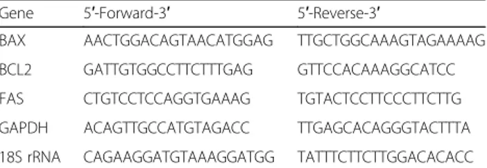
Table 1 Both forward and reverse primer sequences for BAX, BCL2, FAS, GAPDH and 18S rRNA genes are reported
Gene 5′-Forward-3′ 5′-Reverse-3′
BAX AACTGGACAGTAACATGGAG TTGCTGGCAAAGTAGAAAAG BCL2 GATTGTGGCCTTCTTTGAG GTTCCACAAAGGCATCC FAS CTGTCCTCCAGGTGAAAG TGTACTCCTTCCCTTCTTG GAPDH ACAGTTGCCATGTAGACC TTGAGCACAGGGTACTTTA 18S rRNA CAGAAGGATGTAAAGGATGG TATTTCTTCTTGGACACACC
were performed using GraphPad Software Prism 6 (GraphPad Software LLC, La Jolla, CA, USA).
Results
MG phenolic composition
TP content of MG ethanolic extract resulted in 106.33 ± 11.27 mg GAE/g extract, while TF content resulted in 13.84 ± 1.11 mg QE/g extract. HPLC-MS/MS analyses revealed the presence of p-OH-benzoic acid, protocate-chuic acid, p-coumaric acid and caffeic acid. Quantita-tive determination is reported in Table2.
Antioxidant activity of MG extract
Since different mechanisms are involved in the
neutralization of different radical species Table3reports the antioxidant activity of MG extract measured accord-ing to five methods, DPPH., NO., OH· and O2·- scaven-ging capacity and FRAP.
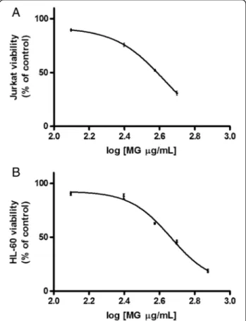
Cytotoxicity analysis on Jurkat and HL-60 cells
After 24 h treatment the IC50for MG was found to be
385 μg/mL in Jurkat cells and equal to 461 μg/mL in
HL-60 (Fig.1).
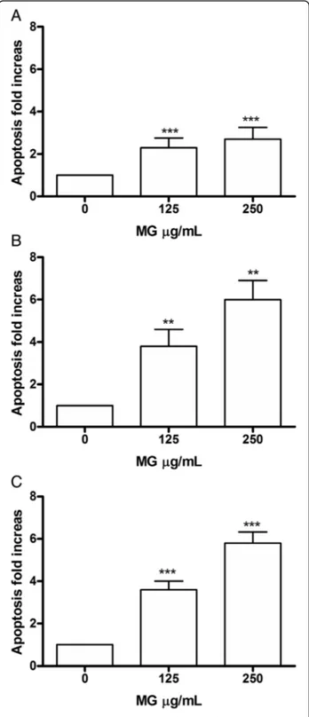
Apoptosis analysis on Jurkat and HL-60 cells
In order to evaluate the involvement of a specific cell death mechanism in the demonstrated cytotoxic action, the analysis of apoptosis possibly induced at different concentrations on Jurkat and HL-60 cells was per-formed, after 24, 48 and 72 h, in order to determine if the event was dose and/or time-dependent.
Specifically, the cells were treated with concentrations
≤ IC50 and the double staining Annexin V-PE / 7-AAD
allowed to measure the percentage of live, apoptotic and necrotic cells.
In Jurkat cells, MG showed a dose- and time-dependent induction of apoptosis. In fact after 24 h there was a sta-tistically significant increase of apoptotic cells
per-centage at the concentration 125 μg/mL equal to 2
times compared to the control (7.3% vs 3.1%) and an
increase equal to 3 times at 250 μg/mL (8.4% vs
3.1%). After 48 h, on the other hand, a doubling of the fraction of apoptotic cells compared to 24 h was
observed at both concentrations tested. Specifically
there was a 4 times increase at 125 μg/mL (13.3% vs
3.5%) and a 6 times increase at 250 μg/mL (21.2% vs
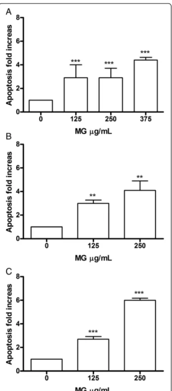
3.5%). At 72 h the trend is comparable (Fig. 2a, b, c). In HL-60 (Fig.3) after 24 h there was evidence at the concentration of 125μg/mL, a statistically significant in-crease in the fraction of apoptotic cells, equal to 3 times compared to control (11.3% vs. 3.8%), parallel to an
Table 2 Content of phenolic compounds in MG extract
Phenolic compounds μg/g dry weight
p-OH-benzoic acid 23.9 ± 3.66
Protocatechuic acid 1.26 ± 0.13
p-coumaric acid 0.2 ± 0.05
Caffeic acid 1.3 ± 0.11
Values are expressed as means ± SD of triplicate measurements. Phenolic acid contents were determined by HPLC-MS/MS as described in the
Methods section
Table 3 Antioxidant activity of MG extract
DPPH.(IC50) a μg/mL 57.77 ± 2.48 NO.(IC25)bμg/mL 148.04 ± 4.98 OH·(IC50)aμg/mL 1.74 ± 0.2 O2−(IC50)aμg/mL 24.28 ± 2.06 FRAPcmg (AAE)/g 22.54 ± 3.51
Values are expressed as means ± SD of triplicate measurements. Antioxidant activity was determined as described in the Methods section
a
IC50(μg/mL), concentration of extract that neutralized 50% of DPPH·, OH·
and O2 .-b
IC25(μg/mL), concentration of extracts that neutralized 25% of NO c
Ferric reducing antioxidant power (FRAP) is expressed as mg ascorbic acid equivalents/g extract dry weight (mg AAE/g d.w)
Fig. 1 Effect of MG on viability of Jurkat and HL-60 cells. Cell viability was determined as described in Methods section. IC50obtained by
curve fitting of viable cells after 24 h treatment with MG for Jurkat cells (a) and HL-60 cells (b). Data are presented as means ± SEM of five independent experiments
increase of 3 and 4 times, for 250 μg/mL and 375 μg/ mL, respectively (11.1% vs. 3.8, 16.2% vs 3.8%). After 48 h, the trend remained comparable to 24 h for the
125 μg/mL concentration (11.6% vs 3.7%), while for
250μg/mL a further increase in the number of apoptotic cells was observed, equal to at 4 times the control (15.2% vs. 3.7%); the same concentration, at the longest treatment time (72 h), was instead associated with an in-crease equal to 6 times (8.6% vs 1.4%). Therefore, these data demonstrate a dose- and time-correlated trend at all times analysed, particularly evident for the
concentra-tion of 250μg/mL.
Considering the demonstrated pro-apoptotic effect, we wanted to confirm the results obtained by FCM, also by fluorescence microscopy analysis, visualizing the mor-phological changes characteristic of apoptosis, such as nuclear condensation and fragmentation (Fig.4).
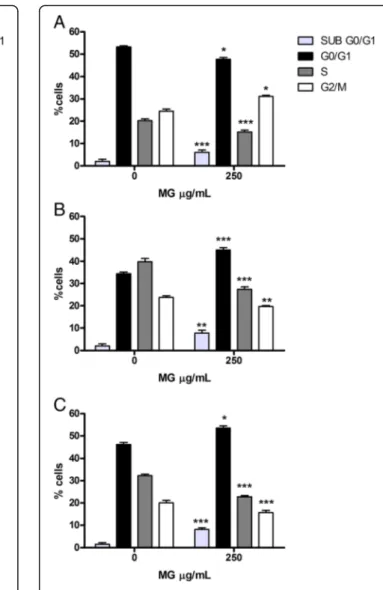
Cell-cycle analysis on Jurkat and HL-60 cells
In order to evaluate whether the induction of apop-tosis caused by MG was an independent event or subsequent a slowing/blocking of the cell-cycle, the Jurkat and HL-60 cells were treated with the concen-tration selected on the basis of the results obtained from the apoptosis analysis and incubated at the same treatment times. The PI staining allowed to highlight that MG does not show any activity on the cell-cycle
of Jurkat cells, at the tested concentration (250 μg/
mL), at any treatment time (Fig. 5a, b, c).
Conversely, on HL-60 cells, after 24 h a slowing of the cell-cycle is observed in the G2/M phase, underlined by an increase in the cell fraction equal to a 33.1% vs 25.2% in the control cultures. At 48 h instead, there was an
in-crease in the number of cells in the G0/G1phase (47.7%
vs 35.1%), with a consequent reduction in S phases (30.0% vs 40.4%) and G2/M (21.8% vs 24.5%); the same behaviour is observed at 72 h (Fig.6a, b, c).
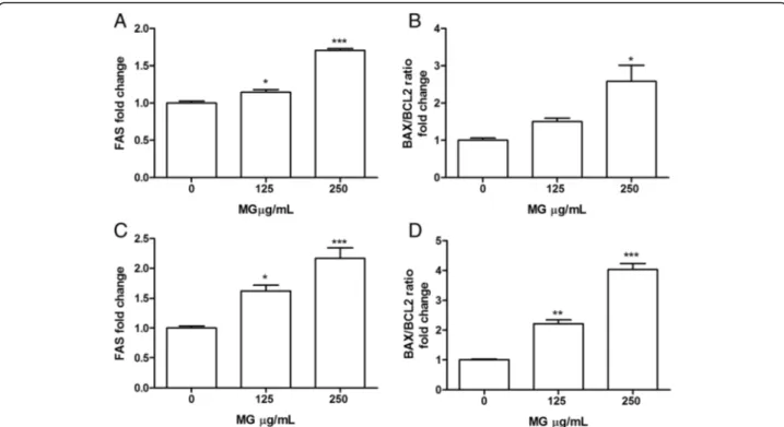
FAS, BAX and BCL2 expression analysis on Jurkat and HL-60 cells
mRNA expression analysis of three genes involved in apoptotic pathways regulation such as FAS, BAX, BCL2 were performed.
Figure 7 shows that MG extract after 16 h
treat-ment significantly induced, in both Jurkat and HL-60,
FAS mRNA expression level (Fig. 7a, c) and increases
the ratio between BAX and BCL2 mRNA expression (Fig. 7b, d).
Intracellular ROS level
To characterize the mechanism behind MG capability to induce apoptosis in leukaemia cells its potential modula-tory effect on intracellular ROS level was investigated in both Jurkat and HL-60 cells by the DCFH-DA assay. As
Fig. 2 Increase in apoptotic Jurkat cell fraction after MG treatment. Apoptosis was evaluated at 24 h (a), 48 h (b) and 72 h (c), as reported in Methods section. Each bar represents means ± SEM of five independent experiments. Data were analysed using repeated ANOVA followed by Bonferroni post-test.**p < 0.01 vs control;***p <
shown in Fig.8, ROS intracellular level was significantly
decreased respect to control by MG 250 μg/mL after
24 h treatment in both cell-lines.
Cytotoxicity analysis on PBL
The analysis of the viability of PBL following MG treat-ment for 24 h allowed to obtain an IC50value of 761μg/ mL, which demonstrated a cytotoxicity lower than that manifested on the two leukaemia cell lines (Fig.9).
Discussion
Great attention has been dedicated to the potential of many extracts and naturally occurring compounds in the prevention/counteraction of chronic diseases [28–30]. Numerous studies have demonstrated that molecules that exhibit antioxidant properties such as polyphenols, isothiocyanates and other compounds counteract cardio-vascular, neurodegenerative diseases and the carcino-genic process at various levels by acting through
multiple mechanisms [31–34]. Extracts from plant
origin may contain various compounds with useful biological properties, thus configuring themselves as complex mixtures with multi-target activities, capable of inhibiting or modulating simultaneously numerous critical targets [25, 35, 36].
Edible mushrooms, including MG, possess antioxidant, antimicrobial and anti-inflammatory properties [7, 37– 39], suggesting their potential application as chemopre-ventive agents. The purpose of this work was, therefore,
Fig. 3 Increase in apoptotic Jurkat cell fraction after MG treatment. Apoptosis was evaluated at 24 h (a), 48 h (b) and 72 h (c), as reported in Methods section. Each bar represents the mean ± SEM of five independent experiments. Data were analysed using repeated ANOVA followed by Bonferroni post-test.**p < 0.01 vs control;***p
< 0.001 vs control
Fig. 4 Fluorescence microscopy analysis of Jurkat and HL-60 after MG treatment. Nuclear condensation and fragmentation associated to apoptotic process on Jurkat and HL-60 cells was evaluated by fluorescence microscopy at 100x magnification after 24 h of 250μg/ mL MG treatment (b, d) respect to control culture (a, c). White arrows indicate condensed and/or fragmented nuclei as marker of apoptosis
to evaluate whether MG extract is able, thanks to its anti-oxidant activity to exhibit chemopreventive activities. The research focused on the evaluation of numerous in vitro end-points in two leukemic cell lines (Jurkat and HL-60 cells) and subsequently in healthy lymphocytes (PBL).
Among scientific literature, strong discrepancies can be observed when comparing TP and TF content of MG extracts., These differences are probably due to extraction methods, mushroom provenance and the fact that TP and TF might be expressed in different ways. Papers reporting the TP of wild edible mush-rooms other than MG differ in a very wide range in the order of mg/mL [6, 40–42].
HPLC-MS/MS analysis of MG ethanolic extract re-sulted in quantification of four phenolic acids, giving the
total sum of 26.66 μg/g. Dominant phenolic acid was
p-hydroxybenzoic acid, in accordance with previously published data on MG [6]. All the differences that can be observed when comparing the previously published data for MG and other wild mushrooms should be at-tributed to the changes that occur during the mush-rooms harvest and postharvest period of time, due to the enzymatic and oxidative processes [43,44].
It has been shown that the strong antiradical activity of mushroom extracts on different radical species is due to the presence of different phenolic compounds and flavonoids [45,46].
Fig. 5 Cell-cycle analysis of Jurkat cells treated with MG extract. Fraction of Jurkat cells in the different phases of the cell cycle after MG treatment for 24 h (a) 48 h (b) and 72 h (c) was evaluated as reported in Methods section. Each bar represents the mean ± SEM of five independent experiments. Data were analysed using repeated ANOVA followed by Bonferroni post-test and revealed no statistically significant differences
Fig. 6 Cell-cycle analysis of HL-60 cells treated with MG extract. Fraction of HL-60 cells in the different phases of the cell-cycle after MG treatment for 24 h (a) 48 h (b) and 72 h (c) was evaluated as reported in Materials and Methods section. Each bar represents the mean ± SEM of five independent experiments. Data were analysed using repeated ANOVA followed by Bonferroni post-test.*p < 0.05 vs control;**p < 0.01 vs control;***p < 0.001 vs control
Determination of DPPH·scavenging capacity is a com-monly employed assay in antioxidant studies, Ferreira et al. [47] analysed DPPH·scavenging capacity of different mushroom methanolic extracts finding that DPPH EC50 was in the range of 8–50 mg/mL. Similarly, Puttaraju et al. [48] analysed methanolic and water extracts of 23 dif-ferent edible mushrooms and found that DPPH EC50 values were in the same order of magnitude.
Only few studies have previously evaluated MG ex-tracts antioxidant activity. Our data show MG ethanolic extract ability to scavenge different radicals such as
DPPH·, O2·-, OH·, NO and to exert antioxidant activity by reducing Fe3+ions (FRAP). These data are in agree-ment with those previously published [5, 6, 8] which found that MG water, ethanolic, methanolic and chloro-formic extracts exert DPPH·and OH· radical scavenging
activity with EC50 values in the order of μg/mL.
Simi-larly, FRAP result is in agreement with previous report [6]. All together, these data suggest that MG extract is characterized by a strong radical scavenging activity
Fig. 7 Effect of MG extract treatment on FAS, BAX and BCL2 expression level in Jurkat (a, b) and HL-60 (c, d) cells. Total RNA was isolated, and the mRNA level of target genes was quantified using RT-PCR normalized to 18S rRNA and GAPDH as reference genes. Triplicate reactions were performed for each experiment. Each bar represents the mean ± SEM of three independent experiments. Data were analysed by one-way ANOVA followed by Bonferroni’s test.*p < 0.05 vs control;**p < 0.01 vs control;***p < 0.001 vs control
Fig. 8 Intracellular ROS level in Jurkat and HL-60 cells treated with MG at 250μg/mL for 24 h. Each bar represents the mean ± SEM of three independent experiments. Data were analysed by t-test,
**p < 0.01 vs control
Fig. 9 Effect of MG on viability of PBL. IC50was obtained by curve
fitting of viable cells after 24 h treatment. Data are presented as mean ± SEM of five independent experiments
mainly related to its phenols content as previously re-ported by Karaman et al. [6].
Due to the strong antioxidant activity, it was pos-sible to hypothesize a chemopreventive role for MG ethanolic extract.
A chemopreventive agent can act in different ways: by modulating the biotransformation enzymes in-volved in the activation/detoxification processes of carcinogens or by stimulating apoptosis and inhibiting the proliferation of transformed cells [49].
Even though some studies reporting pro-apoptotic and anti-proliferative effects of mushroom extract
have already been published [50, 51], to our
know-ledge no data on MG effects in leukemic cell lines are available. Therefore, specific mechanisms of cell death, apoptosis and/or necrosis and MG ability to modulate cell-cycle were investigated.
Results show that MG is able to significantly induce apoptosis in a dose- and time- related manner in both leukemic cell lines. Moreover, it does not modulate, in any way, Jurkat cell-cycle while it inhibits HL-60
prolif-eration by causing a slowdown in G2 /M phase after
24 h treatment, which results in a real block after 48 h
in G0/G1 phase with a corresponding decrease in the
percentage of cells in phase S and G2/M. This effect is confirmed after 72 h of treatment. These data are in agreement with previous reports showing that different flavonoids and polyphenols exert cell-cycle arrest in HL-60 [52]. A possible hypothesis to explain the differ-ences between MG treatment effect on Jurkat and HL-60 cell cycle resides in the substantial differences that exist between these two cell lines. Jurkat cells are a lymphocyte cell line in an advanced state of matur-ation and differentimatur-ation, while HL-60 are a highly undifferentiated immature promyelocytic cell line. Fu-ture studies are needed to clarify whether these differ-ences are responsible for the different effect of MG on the cell cycle [53].
In order to elucidate which mechanism is responsible for the pro-apoptotic effect we evaluated the expression level of genes such as FAS, BAX and BCL2.
FAS receptor belongs the family of death receptors, it is located on the cell membrane and the binding to its ligand leads to apoptosis through caspase-8 activation which dir-ectly activates caspase 3 and simultaneously promote Bid cleavage leading to mitochondrial membrane potential loss [54]. Moreover, it has been demonstrated that FAS over-expression induces apoptosis in Jurkat cell and other malignant T-cell lines [55]. In our study, 16 h treatment with MG induced FAS mRNA expression in both Jurkat and HL-60 cells. Bax and Bcl-2 are mitochondrial pro-teins, while Bax exhibits pro-apoptotic activity, Bcl-2 is considered an anti-apoptotic and is often overexpressed in different cancers [56,57]. In order to induce apoptosis in
cancer cells, most therapies are based on stimulating the expression of Bax and/or suppressing Bcl-2 protein. In this study we contemporaneously observed, in Jurkat cells, an increase of BAX and a decrease of BCL2 gene
expres-sion level after 250 μg/mL MG treatment. Otherwise, in
HL-60 cells a reduction of BCL2 level was found, while BAX was not affected. Our data demonstrate that BAX/ BCL2 mRNA expression ratio significantly increased in both cell lines suggesting the involvement of these two proteins in the progression of the apoptotic cascade in-duced by MG.
It is known, that high ROS intracellular level is a com-mon characteristic of leukaemic and other cancer cells
[58]. This feature has been observed in numerous
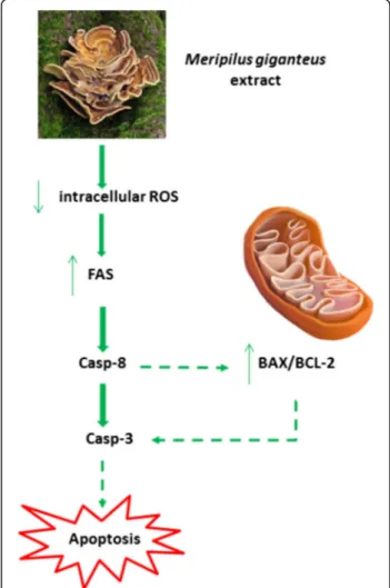
Fig. 10 Hypothesised pro-apoptotic mechanism of MG extract in leukemic cell lines. Due to its phenolic content and antioxidant activity, MG ethanolic extract induces a decrease of ROS intracellular level. In leukemic cell, ROS decrease has been related to FAS recruitment leading to the activation of the extrinsic apoptotic pathway. A possible explanation of the observed increase of BAX/ BCL-2 ratio consists in the fact that FAS ligands lead to apoptosis through caspase-8 activation which on one hand promote the extrinsic apoptotic pathway, while on the other promote mitochondrial membrane potential loss through BAX/BCL2 ratio increase
leukaemic cell lines and also in cells from patients with different types of leukaemia [59]. Therefore, it is gener-ally accepted that increased ROS production is import-ant for the proliferation of hematological malignancies [60,61]. Due to ROS importance in sustaining leukemic cell proliferation and survival, the reduction of their levels could represent an effective strategy to reduce leukemic cells proliferation [59]. Moreover, Aronis et al.
[62] demonstrated that a reduced ROS level could
in-duce apoptosis by the involvement of death receptor in Jurkat cells.
In this context, our data show MG ability to exert scavenging and antioxidant activity leading to a signifi-cant reduction of ROS intracellular level which contrib-utes to explain the pro-apoptotic effect induced by the activation of the extrinsic apoptotic pathway as indicated by FAS increased expression level.
The hypothesised pro-apoptotic mechanism of MG ex-tract is summarized in the scheme shown in Fig.10.
A fundamental feature of a good chemopreventive agent is the low toxicity towards healthy cells and the relative selectivity of action against tumor cells [63]. Therefore, the study was completed by evaluating MG cytotoxicity on PBL after 24 h of treatment.
MG demonstrated good selectivity as shown by the IC50, calculated for PBL by interpolation of the dose re-sponse curve, that resulted 2 and 1.7 times higher than those obtained on Jurkat and HL-60 cells respectively.
Conclusions
MG demonstrated good pro-apoptotic capacity in both Jurkat and HL-60 cell lines, with a predominant antipro-liferative effect in HL-60 and to be partially selective to-wards leukemic cells. These findings allow to propose MG extracts as possible candidate as chemopreventive agents.
Abbreviations
7-AAD:7-aminoactinomycin D; AAE/g: mg ascorbic acid equivalents; DCF: 2′,7′-dichlorofluorescein; DCFH-DA: 2′-7′-dichlorodihydrofluorescin diacetate; DMSO: Dimethyl sulfoxide; DPPH·: DPPH radical; EtOH: Ethanol;
FBS: Fetal bovine serum; FCM: Flow cytometry; Fe2+: Ferrous ions;
FRAP: Ferric reducing antioxidant power; GAE/g: mg gallic acid equivalents; HL-60 cells: Acute promyelocytic leukemia; Jurkat cells: Acute T-cell lympho-blastic leukemia; L-GLU: L-Glutamine; MG: Meripilus giganteus; MGPS: MG a polysaccharide; MRM mode: Multiple reaction monitoring mode; NBT: Nitro blue tetrazolium; NEDA: N-(1-naphthyl) ethylenediamine dihydrochloride; NO·: Nitric oxide; O
2·-: Superoxide anion radical; OH·: Hydroxyl radical;
PBL: Human peripheral blood lymphocytes; PHA: Phitohemagglutinin; PI: Propidium iodide; PMS: 5-methylphenazin-5-ium methyl sulfate; PS: Penicillin-strptomicin; QE/g: mg quercetin equivalents; RPMI 1640 medium: roswell park memorial institute; SNP: Sodium nitroprusside; TBA: Tiobarbituric acid; TCA: Trichloroacetic acid; TF: Total flavonoid; TP: Total phenolic content; TPTZ: 2,4,6-tris(2-pyridyl)-s-triazine
Acknowledgements Not applicable.
Funding
The work on this paper is supported by project that has received funding from the European Union’s Horizon 2020 Spreading Excellence and Widening Participation programme under grant agreement No 692276, and by the Ministry of Education, Science and Technological Development of the Republic of Serbia (Project No III 46001).
Availability of data and materials
Data presented in the manuscript are available upon motivated request.
Authors’ contributions
ML, MM, AM, SH designed the study. ML, VC, AN, MK, MS, AM, MCB, performed the experiments. ML, MM, VC, AN, AM, MK, MS, MP, AC, PH, SH analyzed and discussed the data. ML, VC, MM, AN, MS, AM, SH wrote the manuscript. All authors read and approved the final version of the manuscript.
Ethics approval and consent to participate
Authorization to the use of human blood samples (Buffy coat), for research purposes, has been obtained from AUSL Bologna IT, S. Orsola-Malpighi Hospital - PROT GEN n° 0051937, and written informed consent was obtained by AUSL Bologna IT, S. Orsola-Malpighi Hospital from donors for the use of their blood for scientific research purposes.
Consent for publication Not applicable.
Competing interests
The authors declare that they have no competing interests.
Publisher’s Note
Springer Nature remains neutral with regard to jurisdictional claims in published maps and institutional affiliations.
Author details
1Department of Pharmacy and Biotechnology, University of Bologna, Via San
Donato 15, 40127 Bologna, Italy.2Institute of Food Technology, University of Novi Sad, Bul. Cara Lazara 1, Novi Sad 21000, Serbia.3Faculty of Sciences,
Department of Biology and Ecology, University of Novi Sad, Trg Dositeja Obradovića 2, Novi Sad 21000, Serbia.4Department for Life Quality Studies,
University of Bologna, Corso d’Augusto 237, 47921 Rimini, Italy.5School of Pharmacy, University of Camerino, Via Madonna delle Carceri, 9 - 62032 Camerino, MC, Italy.
Received: 26 July 2018 Accepted: 30 October 2018
References
1. Tsai SY, Tsai HL, Mau JL. Antioxidant properties of Agaricus blazei, Agrocybe cylindracea, and Boletus edulis. LWT Food Sci Technol. 2007;40:1392–402. 2. Stamets P. Growing gourmet and medicinal mushrooms. New York: Ten
Speed Press; 2000.
3. Kalyoncu F, Oskay M, Saglam H, Erdogan TF, Tamer AU. Antimicrobial and antioxidant activities of mycelia of 10 wild mushroom species. J Med Food. 2010;13(2):415–9.
4. Lindequist U, Niedermeyer TH, Julich WD. The pharmacological potential of mushrooms. Evid Based Complement Alternat Med. 2005;2(3):285–99. 5. Karaman M, Jovin E, Malbasa R, Matavuly M, Popovic M. Medicinal and
edible lignicolous fungi as natural sources of antioxidative and antibacterial agents. Phytother Res. 2010;24(10):1473–81.
6. Karaman M, Stahl M, Vulic J, Vesic M, Canadanovic-Brunet J. Wild-growing lignicolous mushroom species as sources of novel agents with antioxidative and antibacterial potentials. Int J Food Sci Nutr. 2014;65(3):311–9. 7. Maity P, Nandi AK, Manna DK, Pattanayak M, Sen IK, Bhanja SK, Samanta S,
Panda BC, Paloi S, Acharya K, et al. Structural characterization and antioxidant activity of a glucan from Meripilus giganteus. Carbohydr Polym. 2017;157:1237–45.
8. Acharya K, Rai M. Proximate composition, free radical scavenging and NOS activation properties of a wild edible mushroom. Int J Pharm Pharm Sci. 2013;5(1):67–72.
9. Rai M, Sen S, Acharya K. Antimicrobial activity of four wild edible mushrooms from Darjeeling hills, West Bengal, India. Int J PharmTech Res. 2013;5(3):949–56.
10. Tomasi S, Lohezic-Le Devehat F, Sauleau P, Bezivin C, Boustie J. Cytotoxic activity of methanol extracts from Basidiomycete mushrooms on murine cancer cell lines. Pharmazie. 2004;59(4):290–3.
11. Narbe G, Lindequist U, Teuscher E, Franke P, Vainiotalo P, Basner R. The chemistry of immunosuppressive acting fractions of Meripilus giganteus (PERS ex. FR.) KARST., the giant spore. Pharmazie. 1991;46(10):738–40. 12. Nam KS, Jo YS, Kim YH, Hyun JW, Kim HW. Cytotoxic activities of
acetoxyscirpenediol and ergosterol peroxide from Paecilomyces tenuipes. Life Sci. 2001;69(2):229–37.
13. Kahlos K, Kangas L, Hiltunen R. Ergosterol peroxide, an active compound from Inonotus radiatus. Planta Med. 1989;55(4):389–90.
14. Singleton VL, Orthofer R, Lamuela-Raventós RM. Analysis of total phenols and other oxidation substrates and antioxidants by means of folin-ciocalteu reagent. Methods Enzymol. 1999;299:152–78.
15. Novaković AR, Karaman MA, Milovanović IL, Belović MM, Rašeta MJ, Radusin TI, Ilić NM. Edible mycorrhizal species Lactarius controversus Pers. 1800 as a source of antioxidant and cytotoxic agents. Hemijska Industrija. 2016;70(2): 113–22.
16. Chang CC, Yang MH, Wen HM, Chern JC. Estimation of total flavonoid content in propolis by two complementary colorimetric methods. J Food Drug Anal. 2002;10(3):178–82.
17. Orcic D, Franciskovic M, Bekvalac K, Svircev E, Beara I, Lesjak M, Mimica-Dukic N. Quantitative determination of plant phenolics in Urtica dioica extracts by high-performance liquid chromatography coupled with tandem mass spectrometric detection. Food Chem. 2014;143:48–53. 18. Espin JC, Soler-Rivas C, Wichers HJ. Characterization of the total free
radical scavenger capacity of vegetable oils and oil fractions using 2,2-diphenyl-1-picrylhydrazyl radical. J Agric Food Chem. 2000;48(3):648–56. 19. Benzie IF, Strain JJ. Ferric reducing/antioxidant power assay: direct
measure of total antioxidant activity of biological fluids and modified version for simultaneous measurement of total antioxidant power and ascorbic acid concentration. Methods Enzymol. 1999;299:15–27. 20. Green LC, Wagner DA, Glogowski J, Skipper PL, Wishnok JS, Tannenbaum
SR. Analysis of nitrate, nitrite, and [15N]nitrate in biological fluids. Anal Biochem. 1982;126(1):131–8.
21. Cheeseman KH, Beavis A, Esterbauer H. Hydroxyl-radical-induced iron-catalysed degradation of 2-deoxyribose. Quantitative determination of malondialdehyde. Biochem J. 1988;252(3):649–53.
22. Nishikimi M, Appaji N, Yagi K. The occurrence of superoxide anion in the reaction of reduced phenazine methosulfate and molecular oxygen. Biochem Biophys Res Commun. 1972;46(2):849–54.
23. Fimognari C, Berti F, Cantelli-Forti G, Hrelia P. Effect of sulforaphane on micronucleus induction in cultured human lymphocytes by four different mutagens. Environ Mol Mutagen. 2005;46(4):260–7.
24. Lenzi M, Cocchi V, Malaguti M, Barbalace MC, Marchionni S, Hrelia S, Hrelia P. 6-(Methylsulfonyl) hexyl isothiocyanate as potential chemopreventive agent: molecular and cellular profile in leukaemia cell lines. Oncotarget. 2017;8(67):111697–714.
25. Lenzi M, Malaguti M, Cocchi V, Hrelia S, Hrelia P. Castanea sativa Mill. bark extract exhibits chemopreventive properties triggering extrinsic apoptotic pathway in Jurkat cells. BMC Complement Altern Med. 2017;17(1):251. 26. Henry CM, Hollville E, Martin SJ. Measuring apoptosis by microscopy and
flow cytometry. Methods. 2013;61(2):90–7.
27. Angeloni C, Malaguti M, Rizzo B, Barbalace MC, Fabbri D, Hrelia S. Neuroprotective effect of sulforaphane against methylglyoxal cytotoxicity. Chem Res Toxicol. 2015;28(6):1234–45.
28. Angeloni C, Hrelia S, Malaguti M. Neuroprotective effects of Glucosinolates. In: Mérillon JM, Ramawat KG, editors. Glucosinolates, reference series in phytochemistry. Switzerland: Springer International Publishing; 2016. p. 1–25.
29. Jurikova T, Mlcek J, Skrovankova S, Sumczynski D, Sochor J, Hlavacova I, Snopek L, Orsavova J. Fruits of black chokeberry Aronia melanocarpa in the prevention of chronic diseases. Molecules. 2017;22(6):944.
30. Fimognari C, Ferruzzi L, Turrini E, Carulli G, Lenzi M, Hrelia P, Cantelli-Forti G. Metabolic and toxicological considerations of botanicals in anticancer therapy. Expert Opin Drug Metab Toxicol. 2012;8(7):819–32.
31. Zhang H, Tsao R. Dietary polyphenols, oxidative stress and antioxidant and anti-inflammatory effects. Curr Opin Food Sci. 2016;8:33–42.
32. Zhang C, Shu L, Kim H, Khor TO, Wu R, Li W, Kong AN. Phenethyl isothiocyanate (PEITC) suppresses prostate cancer cell invasion
epigenetically through regulating microRNA-194. Mol Nutr Food Res. 2016; 60(6):1427–36.
33. Angeloni C, Malaguti M, Barbalace MC, Hrelia S. Bioactivity of olive oil phenols in neuroprotection. Int J Mol Sci. 2017;18(11):E2230. 34. Malaguti M, Angeloni C, Hrelia S. Nutraceutical bioactive compounds
promote healthspan counteracting cardiovascular diseases. J Am Coll Nutr. 2015;34(Suppl 1):22–7.
35. Chiarini A, Micucci M, Malaguti M, Budriesi R, Ioan P, Lenzi M, Fimognari C, Gallina Toschi T, Comandini P, Hrelia S. Sweet chestnut (Castanea sativa Mill.) bark extract: cardiovascular activity and myocyte protection against oxidative damage. Oxidative Med Cell Longev. 2013; 2013:471790.
36. Micucci M, Malaguti M, Toschi TG, Di Lecce G, Aldini R, Angeletti A, Chiarini A, Budriesi R, Hrelia S. Cardiac and vascular synergic protective effect of Olea europea L. Leaves and Hibiscus sabdariffa L. Flower Extracts. Oxidative Med Cell Longev. 2015;2015:318125.
37. Klaus A, Kozarski M, Niksic M, Jakovljevic D, Todorovic N, Stefanoska I, Van Griensven LJ. The edible mushroom Laetiporus sulphureus as potential source of natural antioxidants. Int J Food Sci Nutr. 2013;64(5):599–610. 38. Zhang S, Liu X, Yan L, Zhang Q, Zhu J, Huang N, Wang Z. Chemical
compositions and antioxidant activities of polysaccharides from the sporophores and cultured products of Armillaria mellea. Molecules. 2015; 20(4):5680–97.
39. Trumbeckaite S, Benetis R, Bumblauskiene L, Burdulis D, Janulis V, T A, Viskelis P, Jakstas V. Achillea millefolium L.s.l. herb extract: antioxidant activity and effect on the rat heart mitochondrial functions. Food Chem. 2011;127:1540–8. 40. Keles A, Koca I, Gençcelep H. Antioxidant properties of wild edible
mushrooms. J Food Process Technol. 2011;2(6):130.
41. Yildiz O, Can Z, Laghari AQ, Ahin SH, Malkoç M. Wild edible mushrooms as a natural source of Phenolics and antioxidants. J Food Biochem. 2015;39: 148–54.
42. Alvarez-Parrilla E, de la Rosa LA, Martínez NR, Aguilar-González GA. Total phenols and antioxidant activity of commercial and wild mushrooms from Chihuahua, Mexico. CYTA J Food. 2007;5(5):329–34.
43. Ribeiro B, Valentao P, Baptista P, Seabra RM, Andrade PB. Phenolic compounds, organic acids profiles and antioxidative properties of beefsteak fungus (Fistulina hepatica). Food Chem Toxicol. 2007;45(10):1805–13. 44. Vaz JA, Barros L, Martins A, Morais JS, Vasconcelos MH, Ferreira IC. Phenolic
profile of seventeen Portuguese wild mushrooms. LWT Food Sci Technol. 2011;44(1):343–6.
45. Barros L, Cruz T, Baptista P, Estevinho LM, Ferreira IC. Wild and commercial mushrooms as source of nutrients and nutraceuticals. Food Chem Toxicol. 2008;46(8):2742–7.
46. Heleno SA, Martins A, Queiroz MJ, Ferreira IC. Bioactivity of phenolic acids: metabolites versus parent compounds: a review. Food Chem. 2015;173:501–13.
47. Ferreira ICFR, Baptista P, Vilas-Boas M, Barros L. Free-radical scavenging capacity and reducing power of wild edible mushrooms from Northeast Portugal: individual cap and stipe activity. Food Chem. 2007;100(4):1511–6. 48. Puttaraju NG, Venkateshaiah SU, Dharmesh SM, Urs SM, Somasundaram R. Antioxidant activity of indigenous edible mushrooms. J Agric Food Chem. 2006;54(26):9764–72.
49. Lenzi M, Fimognari C, Hrelia P. Sulforaphane as a promising molecule for fighting cancer. Cancer Treat Res. 2014;159:207–23.
50. Arora S, Tandon S. Mushroom extracts induce human colon cancer cell (COLO-205) death by triggering the mitochondrial apoptosis pathway and go/G1-phase cell cycle arrest. Arch Iran Med. 2015;18(5):284–95. 51. Leon F, Quintana J, Rivera A, Estevez F, Bermejo J. Lanostanoid triterpenes
from Laetiporus sulphureus and apoptosis induction on HL-60 human myeloid leukemia cells. J Nat Prod. 2004;67(12):2008–11.
52. Abubakar MB, Abdullah WZ, Sulaiman SA, Suen AB. A review of molecular mechanisms of the anti-leukemic effects of phenolic compounds in honey. Int J Mol Sci. 2012;13(11):15054–73.
53. Fimognari C, Lenzi M, Cantelli-Forti G, Hrelia P. Induction of differentiation in human promyelocytic cells by the isothiocyanate sulforaphane. In Vivo. 2008;22(3):317–20.
54. Harrington HA, Ho KL, Ghosh S, Tung KC. Construction and analysis of a modular model of caspase activation in apoptosis. Theor Biol Med Model. 2008;5:26.
55. Li L, Zhang R, Chen Z, Xue S, Wang X, Ruan C. Over-expressed Fas improves the apoptosis of malignant T-cells in vitro and vivo. Mol Biol Rep. 2011;38(8):5371–7.
56. Autret A, Martin SJ. Emerging role for members of the Bcl-2 family in mitochondrial morphogenesis. Mol Cell. 2009;36(3):355–63.
57. Wang Z, Tang X, Zhang Y, Qi R, Li Z, Zhang K, Liu Z, Yang X. Lobaplatin induces apoptosis and arrests cell cycle progression in human
cholangiocarcinoma cell line RBE. Biomed Pharmacother. 2012;66(3):161–6. 58. Szatrowski TP, Nathan CF. Production of large amounts of hydrogen
peroxide by human tumor cells. Cancer Res. 1991;51(3):794–8. 59. Prieto-Bermejo R, Romo-Gonzalez M, Perez-Fernandez A, Ijurko C,
Hernandez-Hernandez A. Reactive oxygen species in haematopoiesis: leukaemic cells take a walk on the wild side. J Exp Clin Cancer Res. 2018; 37(1):125.
60. Sallmyr A, Fan J, Rassool FV. Genomic instability in myeloid malignancies: increased reactive oxygen species (ROS), DNA double strand breaks (DSBs) and error-prone repair. Cancer Lett. 2008;270(1):1–9.
61. Hole PS, Darley RL, Tonks A. Do reactive oxygen species play a role in myeloid leukemias? Blood. 2011;117(22):5816–26.
62. Aronis A, Melendez JA, Golan O, Shilo S, Dicter N, Tirosh O. Potentiation of Fas-mediated apoptosis by attenuated production of mitochondria-derived reactive oxygen species. Cell Death Differ. 2003;10(3):335–44.
63. Fimognari C, Lenzi M, Hrelia P. Apoptosis induction by sulfur-containing compounds in malignant and nonmalignant human cells. Environ Mol Mutagen. 2009;50(3):171–89.