To my scientific lighthouses
Anna Maria Musti
and
Marcello Maggiolini
“When the going gets tough
The tough get going…..”
INDEX
LIST OF ABBREVIATIONS
SOMMARIO
1
SUMMARY
3
INTRODUCTION
7
RESULTS
1-
G Protein–Coupled Receptor 30 (GPR30) Mediates Gene ExpressionChanges and Growth Response to 17Beta-Estradiol and Selective GPR30
Ligand G-1 in Ovarian Cancer Cells
26
2-
G Protein-Coupled Receptor 30 and Estrogen Receptor α are involved inthe proliferative effects induced by Atrazine in ovarian cancer cells
.
32
3-
Epidermal Growth Factor Induces G Protein-Coupled Receptor 30Expression in Estrogen Receptor-Negative Breast Cancer cells.
37
MATERIAL
and
METHODS
42
DISCUSSION and CONCLUSIONS
54
REFERENCES
63
List of Abbreviations
17β-Estradiol (E2) 2-chloro-4-(ethylamine)-6-(isopropylamine)-s-triazine (Atrazine) (1-[4-(6-bromobenzo[1,3]diodo-5yl)-3a,4,5,9b-tetrahydro-3H-cyclopenta[c]quinolin -8-yl]-ethanone) (G-1) 5α-Dihydrotestosterone (DHT) 4-Hydroxytamoxifen (OHT) Activating Protein-1 (AP-1) Activation Function (AF2) AG1478 (AG)Dexamethasone (DEX)
DNA-Binding Domain (DBD); Dominant Negative (DN) Epidermal Growth Factor (EGF)
Epidermal Growth Factor Receptor (EGFR) Estrogen Receptor (ER)
Estrogen Response Element (ERE)
Extracellular Signal-Regulated protein Kinase (ERK) G protein coupled receptor (GPCR)
Genistein (G) GF109203X (GFX)
Heat shock protein (hsp)
Hormone Response Elements (HRE) ICI 182,780 (ICI)
Insulin-Like Growth Factor-1 (IGF-1) Ligand-Binding Domain (LBD) LY 294,002 (LY)
Mitogen Activated Protein Kinase (MAPK) PD98059 (PD)
Progesterone Receptor (PR) Progesterone (PRG)
Pertussis Toxin (PT)
Serum Response Factor (SRF) Tamoxifen (TAM)
Ternary Complex Factor (TCF) Transactivation Domain (AF-1) Wortmannin (WM)
SOMMARIO
Gli estrogeni (E2) regolano la crescita e la differenziazione di molti tessuti. Agendo da mitogeni sono inoltre in grado di promuovere lo sviluppo di tumori estrogeno-sensibili come il tumore mammario ed ovarico. Gli effetti biologici degli estrogeni sono mediati dal Recettore Estrogenico (ER) α e β, che agendo da fattori di trascrizione, legano le Sequenze Responsive agli Estrogeni (ERE) presenti nelle regioni promoter di geni target. Diversi studi hanno dimostrato che segnali estrogenici possono alterare l’espressione genica attraverso meccanismi indipendenti dai classici ERs. In particolare, è stato visto che gli estrogeni possono indurre importanti effetti biologici attraverso il recettore di membrana GPR30 che è in grado di attivare la via trasduzionale del Recettore dell’ Epidermal Growth Factor (EGFR). Se gli estrogeni agiscono sul pathway trasduzionale EGFR/ERK solo mediante il legame con GPR30 o attivando anche ERα è poco chiaro, poichè gli estrogeni sono in grado di legare entrambi i recettori. La recente identificazione di un ligando selettivo per GPR30, G-1, ha fornito nuove possibilità per differenziare le funzioni di ERs e GPR30. Per valutare i meccanismi molecolari coinvolti nell’azione proliferativa degli estrogeni attraverso GPR30, abbiamo analizzato la capacità di G-1 di indurre effetti proliferativi in cellule tumorali ovariche estrogeno-sensibili, esprimenti ERα e in cellule tumorali mammarie estrogeno-sensibili ma non esprimenti ERα. Abbiamo inoltre valutato gli effetti di G-1 sull’attivazione delle ERK e sull’induzione di c-fos e altri geni coinvolti nella progressione del ciclo cellulare. Abbiamo dimostrato che G-1 induce la proliferazione di entrambe le linee tumorali, indipendentemente dalla presenza di ERα. Considerato che in cellule tumorali ovariche l’espressione di ERα è richiesta sia per la proliferazione cellulare che per l’induzione di c-fos e l’attivazione delle ERK, la capacità di GPR30 di mediare segnali estrogenici indipendentemente da ERα potrebbe costituire una condizione di adattamento funzionale in cellule tumorali ERα negative.
In un ulteriore studio abbiamo valutato la regolazione dell’espressione di GPR30 in cellule tumorali mammarie ER-negative SKBR3. Trasfezioni transienti effettuate con un plasmide codificante per la regione promotore di GPR30 hanno mostrato che un sito AP-1 situato nella regione è necessario per l’attivazione del promoter di GPR30 in risposta al trattamento con EGF. EGF è inoltre in grado di up-regolare i livelli proteici di GPR30 che si accumulano prevalentemente nel compartimento intracellulare. Questo effetto stimolatorio indotto dall’EGF sull’espressione di GPR30 è innescato attraverso la rapida fosforilazione delle ERK e l’induzione di c-fos. Inoltre sia l’abrogazione dell’espressione di GPR30 che un dominante negativo di c-fos hanno ridotto la proliferazione cellulare indotta dall’E2 nelle cellule SKBR3 e BT20.
Abbiamo infine dimostrato che GPR30 è coinvolto nell’induzione degli effetti stimolatori esercitati dall’atrazina in cellule tumorali ovariche. L’atrazina è il più comune contaminante di falde acquifere e di acque di superficie. Fra gli effetti sul sistema endocrino, è stato descritto come l’atrazina interferisca nei processi mediati da androgeni ed estrogeni. Studi di binding hanno dimostrato che tali fenomeni sono indotti dall’atrazina in assenza di un diretto agonismo o antagonismo sui recettori steroidei. Utilizzando come sistema modello la linea di tumore ovarico BG-1 abbiamo dimostrato che l’atrazina non è in grado di indurre alcun effetto diretto attraverso ERα. ma stimola la fosforilazione delle ERK e l’espressione di c-fos, fenomeni aboliti dall’antagonista di ERs ICI 182,780, dall’inibitore delle MAPK, PD98059 e dall’inibitore del recettore dell’EGF AG1478. Silenziando l’espressione di ERα e di GPR30 la fosforilazione delle ERK e l’induzione di c-fos venivano notevolmente ridotte così come la proliferazione cellulare indotta dall’atrazina.
I nostri studi su GPR30 hanno consentito nuove conoscenze sui meccanismi molecolari coinvolti dagli estrogeni nella progressione di tumori ormono-sensibili.
SUMMARY
Estrogens are pleiotropic hormones that regulate the growth and differentiation of many tissues. By acting as mitogens they also promote the development of breast and ovarian tumors. The biological effects of estrogens are classically mediated by the estrogen receptor (ER)s α and β which function as hormone-inducible transcription factors binding to the estrogen-responsive element (ERE) located within the promoter region of target genes. Several studies have demonstrated that membrane-associated estrogen signals may alter gene expression through non-genomic mechanisms that are independent of nuclear ERs. In particular, it has been shown that estrogens can signal through the membrane G-protein coupled receptor 30 (GPR30). GPR30 mediates non genomic signaling of E2 in a variety of estrogen-sensitive cancer cells through activation of the Epidermal Growth Factor Receptor (EGFR) pathway.
Whether E2 acts on the EGFR/ERK transduction pathway only through GPR30 binding or also through ERα binding is less clear, since E2 binds to both receptors although with different affinity. G-1 is the first well-known GPR30-selective ligand and its recent identification has provided new opportunities to differentiate between ERs and GPR30 function. To better understand the molecular mechanisms involved in the proliferative action of E2-GPR30 signaling, we evaluated the ability of G-1 to induce cell growth of E2-responsive ovarian cancer cells expressing ERα as well as of E2-responsive breast cancer cells not expressing ERα. We have also investigated the effect of G-1 on ERK activation and on induction of c-fos and other genes involved in the progression of the cell cycle. We found that G-1 induces the proliferation of both positive and negative ERα cancer cells. However, in ovarian tumor cells, ERα expression was required for cell proliferation as well as for c-fos stimulation and ERK activation, suggesting that the capacity of GPR30 to signal independently of ERα is a specific feature of ERα negative tumors. Next, we investigated the molecular mechanism involved in GPR30 expression. To this end we assessed GPR30 expression and promoter activity in SkBr3 and BT20 breast cancer cells
(lacking the classical ERs), by either E2, or G-1, or Insulin like Growth Factor-I (IGF-I), or Epidermal Growth Factor (EGF). Transient transfections with an expression plasmid encoding a short 5'-flanking sequence of the GPR30 gene revealed that an AP-1 site located within this region is required for the GPR30 promoter activity in response to EGF. Accordingly, EGF up-regulated GPR30 protein levels, which accumulated predominantly in the intracellular compartment. The stimulatory role elicited by EGF on GPR30 expression was triggered through rapid ERK phosphorylation and c-fos induction which was strongly recruited to the AP-1 site found in the short 5'-flanking sequence of the GPR30 gene. Furthermore, either the abrogation of GPR30 expression or the expression of Dominant Negative DN/c-fos reduced E2-dependent proliferation of SkBr3 and BT20 cancer cells.
After having investigated the molecular mechanisms linking E2/GPR30 signaling to cancer cell proliferation, we examined whether such a pathway is also involved in the cancerogenic effect of the synthetic compound atrazine. Atrazine is the most common pesticide contaminant of groundwater and surface water. Among the endocrine-disrupting effects, atrazine interferes with androgen- and estrogen-mediated processes. Based on binding affinity studies, this occurs without direct agonism or antagonism of the cognate receptors for these steroids. Epidemiologic studies have associated long-term exposure to triazine herbicides with an increased risk of ovarian cancer in female farm workers in Italy.
We used BG-1 ovarian cancer cells as a model system and found that ERα was modulated at both mRNA and protein levels by E2, whereas atrazine did not produce any effect and did not show any binding affinity for ERα. Furthermore, atrazine did not stimulate aromatase activity in BG-1 cells, but was able to induce ERK phosphorylation (which was abolished by the ER antagonist ICI182,780, PD98059 and AG1478) and c-fos expression. Both ERK and c-fos stimulation induced by atrazine were abolished knocking-down ERα and GPR30 in BG-1 cancer cells. Furthermore, we found that atrazine induced ovarian cancer cell proliferation, which was inhibited by silencing the
expression of either GPR30 or ERα.
Our results have contributed to provide new insights into the molecular mechanisms implicated in tumor progression.
Estrogens (E2) are important hormones in mammalian physiology, regulating the development and homeostasis of many organs. Estrogen is the best characterized member of the family of steroid hormones that includes progesterone, testosterone, glucocorticoids, and mineralocorticoids. The highly hydrophobic nature of steroid ligands allows them to pass through cellular membranes by passive diffusion. Estrogen action is required for normal development and growth of female reproductive tissue (Couse and Korach 1999), but also to regulate bone integrity (Termine and Wong 1998), cardiovascular function (Guzzo 2000), the central nervous system (Hurn and Macrae 2000) and the immune system (Kovacs et al., 2002).
The major estrogen-producing organ is the ovary but recent studies have revealed the synthesis of estrogen at multiple discrete sites where it may have highly localized effects (Baquedano et al,. 2007). Plasma concentrations of estrogen in women are commonly in the 1 nM range, although the normal concentration in breast tissue of postmenopausal women, has been reported to be 10-20-fold higher than serum concentration, suggesting local production or concentration of the hormone (Geisler 2003). The biological effects of estrogens are mediated by a specific nuclear receptor (ER) that recognizes and binds the hormone, transmitting this information to downstream effectors. The first described ER, ERα, was characterized in 1973 on the basis of specific binding activity in rat uterus/vagina extracts (Jensen and Desombre 1973). Its DNA sequence was determined in 1986 (Greene et al., 1986) and the first crystal structure of an ER ligand-binding domain was described in 1997 (Brzozowski et
al., 1997). A second related ER, ΕRβ was identified in 1996 (Kuiper et al. 1997). The ERs are coded from two separate genes: ERα is located at chromosomal locus 6q25.1 (Menasce et al., 1993), and encodes a 66kDa protein of 595 amino acids, whereas ERβ is found at position 14q22-24 (Enmark et al., 1997) encoding a 54kDa protein of 485 aminoacids.
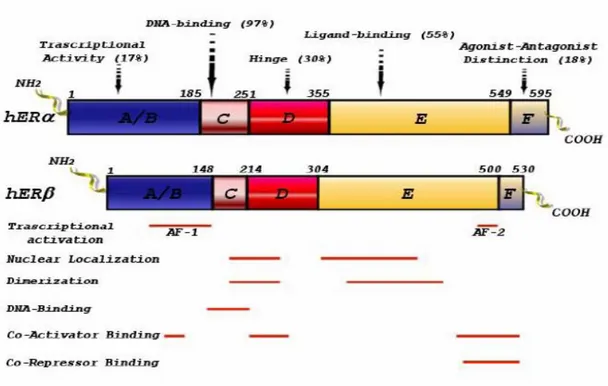
As for the other members of the steroid/thyroid hormone superfamily of nuclear receptors, ERα and ERβ are composed of three independent but interacting functional domains: the NH2-terminal or A/B domain, the C or DNA-binding domain, and the D/E/F or ligand-binding domain (Nilsson et al, 2001). Binding of a ligand to ER triggers conformational changes in the receptor and this leads to changes in the rate of transcription of estrogen-regulated genes. These events include receptor dimerization, receptor-DNA interaction, recruitment of and interaction with co-activators and other transcription factors, and formation of a pre-initiation complex (Nilsson, 2001). The N-terminal domain of nuclear receptors encodes a ligand-independent activation function (AF1) involved in protein-protein interactions, and transcriptional activation of target-gene expression. Comparison of the AF1 domains of the two ERs has revealed that, in ERα, this domain is very active in stimulation of reporter-gene expression from a variety of estrogen response element (ERE)-reporter constructs, in different cell lines (Cowley and Parker, 1999). Differences in the NH2-terminal regions of ERα and ERβ may explain the differences between the two receptors in their response to various ligands. In ERα, two distinct parts of AF1 are required for agonism of E2 and the partial agonism of tamoxifen,
respectively (McDonnel et al, 1995) whereas in ERβ, this dual function of AF1 is missing (McInerney et al, 1998). The DBD contains a two zinc finger structure, important in receptor dimerization and in binding of receptors to specific DNA sequences (Nilsson et al, 2001). The DBDs of ERα and ERβ are highly homologous (Nilsson et al, 2001). In particular, the P box sequence, critical for target-DNA recognition and specificity, is identical in the two receptors. Thus ERα and ERβ can be expected to bind to various EREs with similar specificity and affinity.
The COOH-terminal, E/F-, or LBD mediates ligand binding, receptor dimerization, nuclear translocation, and transactivation of target gene expression (Nilsson et al, 2001).
The ligand binding domains (LBD) of ERα and ERβ share a high degree of homology in their primary amino acid sequence and are also very similar in their tertiary architecture. Many compounds tested so far bind to ERα and ERβ with similar affinities and similar potencies in activation of ERE-mediated reporter gene expression (Kuiper et al, 1998). ERβ shares considerable homology in the DNA binding region (97%) with ERα, while this homology is markedly lower (55%) in the LBD, but the trans-activation mode of action of both ERs, is similar (Petterson et al., 1997). In the absence of its cognate ligand, ERs are recovered in the cytosolic fraction of target cell homogenates in inactive untransformed hetero-oligomeric complexes which contain one steroid-binding subunit and a non steroid, non-DNA-binding component, identified as a heat shock protein (hsp90). An important physiologic role for hsp90 is that of
maintaining the receptor in a non functional state: interaction of hsp90 and LBD of the receptor, would interfere with several LBD and DNA binding domain (DBD) functions, resulting in the repression of the transcriptional activity of ER (Picard 1990 and 2002; Pratt 2003). Another essential characteristic of hsp90 is to mediate receptor trafficking from the cytoplasmatic fraction to the nucleus, through a microtubule dependent mechanism (Pratt and Toft 1990).
Fig. 1: ERα and ERβ functional domains.
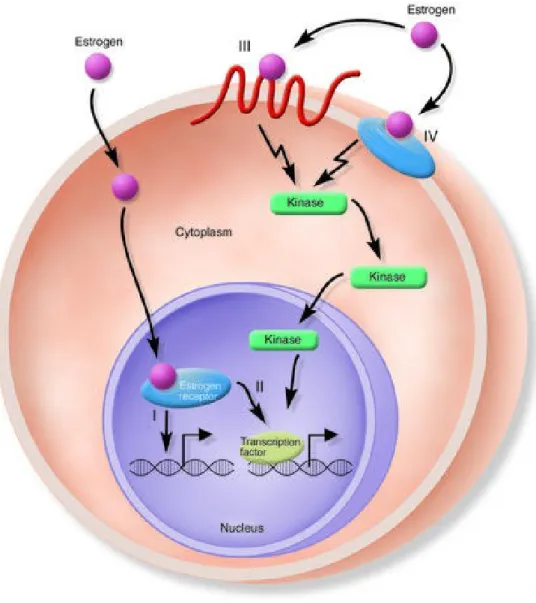
Both ERs are widely distributed throughout the body, displaying distinct but overlapping expression patterns in a variety of tissues (Petterson and Gustafsson 2001). ERα is expressed primarily in the uterus, liver, kidney and heart, whereas ERβ is expressed principally in the ovary, prostate, lung, gastrointestinal tract, bladder and hematopoietic and central nervous systems. ERs are, however, co-expressed in a number of tissues including the mammary gland, epididymis, thyroid, adrenal, bone and certain regions of the brain (Matthews and Gustafsson 2003). Cellular responses to estrogens are often divided into two broad categories: Genomic and Non-Genomic Responses. Genomic responses are characterized by gene transcription changes and occur in the time frame of hours to days, while non-genomic responses are generally rapid signaling events. Classical ERs, mediate their primary effects at the genomic level, but in recent years, it has become clear that not all effects of estrogens and compounds with estrogen-like activity can be explained by the classic genomic mechanism. In addition, the growth of estrogen-dependent tumors may also have an important non-genomic component (Singleton et al. 2003). It has been shown that estrogens act rapidly by activating membrane receptors coupled to G proteins (GPCRs) (Kelly et al., 2001; Acconcia et al. 2004; Li et al. 2003; Razandi et al. 2004). These receptors are able to mediate estrogen function including transcriptional signaling as well as non-genomic or rapid signaling (Govind and Thampan 2003). Some reports described estrogen binding sites on intracellular membrane (Evans and Muldoon 1991), other reports suggest that palmitoylation (Acconcia et al. 2004; Li et al.2003) or
phosphorylation (Balasenthil et al., 2004) may transfer ERs to the cytoplasmic face of the plasma membrane. Also adaptor proteins, such as Shc (Evinger and Levin 2005) and NMAR, (Boonyaratanakornkit and Edwards 2004) can recruit ERα to the plasma membrane. Classical steroids receptors, bind DNA after ligand stimulation, but they can also act in the presence or absence of ligand (Lu et al., 2006), independently of direct DNA binding to scaffold transcription factors, like AP1 (Barkhem et al., 2004; Kushner et al., 2000), or induce the activation of kinases, like MAPKs, phosphatidylinositol 3-kinase (PI3K), Src or lead to phosphorylation and transcriptional events through transcription factors like Elk-1 (Duan et al., 2001) and serum response factor (SRF) (Duan et al., 2002). Therefore, in addition to transcriptional regulation estrogens can also mediate cellular effects including the generation of the second messengers like Ca2+, cAMP and NO, as well as activation of receptor tyrosine kinases, EGFR
and IGF-1R and protein/lipid kinases (Hall et al., 2001; Ho and Liao 2002; Kelly and Levin 2001; Levin 2001-2002; Razandi et al., 2003). The AP-1 transcription factor participates in the control of cellular responses to stimuli that regulate proliferation, differentiation, immune responses, cell death and the response to genotoxic agents or stress (Angel 1991). AP-1 is composed of Jun family members (c-Jun, JunB and JunD) that can form either homo- or hetero-dimers among themselves. Jun proteins also dimerize with fos family members (c-fos, fosB, Fra1 and Fra2) (Curran and Franza 1988) and with members of the Activating Transcription Factor (ATF) family of proteins (Karin 1994). These proteins are characterized by a highly charged, basic DBD, immediately
adjacent to an amphipathic dimerization domain, referred to as the “leucine zipper”. The composition of the subunit is determined by the nature of the extracellular stimulus and the MAPK signaling pathway that is activated: the expression and activity of c-Jun and c-fos are tightly regulated by members of the mitogen-activated protein kinase (MAPK) family, including c-Jun N-terminal kinases (JNKs), extracellular signal-regulated protein kinase 5 (ERK5), and p38MAPK kinases and by acting on transcription factors of the TCF family such as Elk-1, can cause induction of the c-fos gene. Upon stimulation, the regulation of AP-1 activity occurs by activating the transcription of these genes as well as by phosphorylation of existing Jun and Fos proteins at specific serine and threonine sites (Vinciguerra et al., 2008; Shaulian and Karin 2001). AP-1 activity is regulated by a broad range of physiological and pathological stimuli, including cytokines, growth factors, stress signals and infections, as well as by oncogenic stimuli (Karin and Shaulian 2001; Shaulian and Karin 2001). The proto-oncogene c-fos plays a relevant role in the regulation of normal cell growth, differentiation, and cellular transforming processes (Curran 1988). In particular, c-fos is classified as a prototypical “immediate early gene” since its expression is rapidly induced by numerous extracellular stimuli, including hormones and mitogens (Weisz and Bresciani 1993; Ginty et al, 1994; Hill and Treisman 1995; Bonapace et al, 1996).
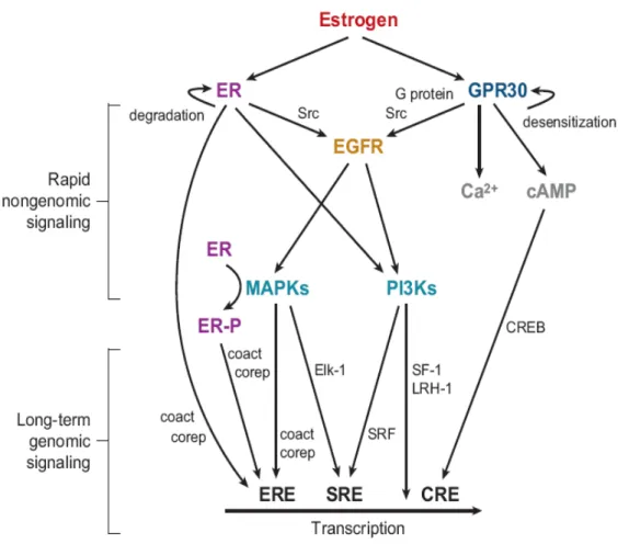
Fig. 2: Genomic (I and II) and nongenomic (III and IV) actions of estrogens
G protein–coupled receptors (GPCRs) represent the largest class of cell surface signaling molecules in the human genome (Venter et al., 2001). GPCRs are coupled to a heterotrimeric signal-transducing guanine nucleotide-binding proteins (G proteins). Ligand binding (Gether and Kobilka, 1998) to these receptors activates their downstream regulatory proteins (Prossnitz et al., 2004) and an effector enzyme to generate an intracellular second messenger. All G protein−coupled receptors (GPCRs) contain seven membrane-spanning regions with their N-terminal segment on the exoplasmic face and their C-terminal segment on the cytosolic face of the plasma membrane. One such receptor, GPR30, was cloned by different groups using highly disparate approaches (Carmeci et al. 1997; O'Dowd et al. 1998; Owman et al., 1996; Takada et al., 1997) in the late 1990s. It was not until 2000 that a possible function for this GPR30 was identified from experiments demonstrating MAP kinase (ERK1/2) activation by estrogen, as well as the pure ER antagonists ICI182,780 and Tamoxifen, which mimics estrogen function in certain tissues but acts as an antagonist in other tissues and are collectively known as SERMs (Selective Estrogen Receptor Modulator). Responses were demonstrated in breast cancer cell lines expressing GPR30 but not in cell lines lacking GPR30 (Filardo et al., 2000). Signaling in response to estrogen could be restored in the latter cell lines by expressing GPR30. They found that estrogen-dependent signaling acted through a pertussis toxin-sensitive pathway: indicating the involvement of G proteins (Filardo et al., 2000).
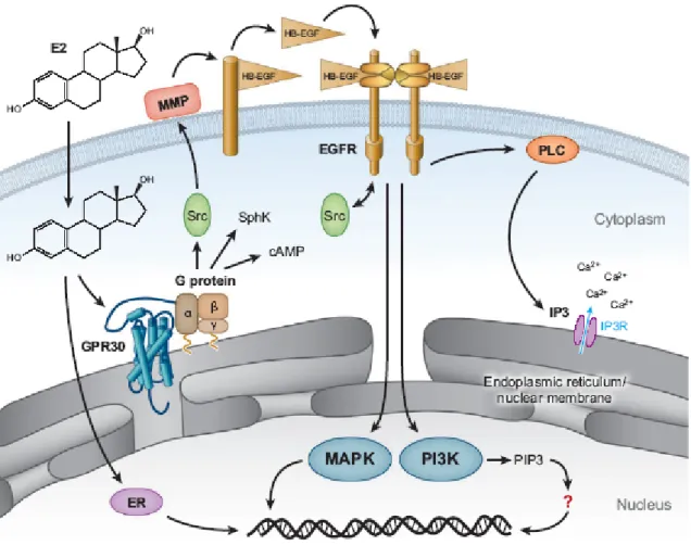
Cellular activation by GPR30 occurred through a mechanism involving transactivation of the epidermal growth factor receptor (EGFR) via a G protein– dependent pathway (Filardo et al., 2000-2002-2008; Maggiolini et al., 2004; Vivacqua et al., 2006a-2006b;). At that time such transactivation pathways from GPCRs to EGFR were still a relatively new concept yet were known to involve metalloproteinase cleavage of proheparin-binding (-bound) epidermal growth factor–like growth factor (pro-HB-EGF) (Daub et al.,1996; Prenzel et al., 1999). A follow-up report described GPR30-mediated elevation of cAMP by estrogen as a mechanism to restore EGF activated ERK1/2 to basal levels through protein kinase A (PKA)-dependent inhibition of Raf-1 activity (Filardo et al., 2002). Furthermore, GPR30-mediated up-regulation of nerve growth factor production in macrophages by induction of c-fos expression has also been demonstrated (Kanda and Watanabe, 2003). The up-regulation of c-fos by estrogen and phytoestrogens has also been shown in breast cancer cells (Maggiolini et al., 2004).
The majority of GPCRs are expressed in the plasma membrane, but some GPCRs may be functionally expressed at intracellular sites (Gobeil et al., 2006). This is particularly true of GPCRs with lipophilic ligands. Where is GPR30 localized? This question is still open, because using subcellular markers, one team showed that GPR30 is expressed in an intracellular compartment, the endoplasmic reticulum but also in the Golgi apparatus and nuclear membrane. In addition, they were unable to detect transfected or endogenously expressed GPR30 on the plasma membrane (Revankar et al., 2005; Revankar et al., 2007).
Recently, other two teams reported expression of GPR30 in the plasma membrane (Thomas et al., 2005; Funakoshi et al., 2006). The proposed role of GPR30 in cellular estrogen responsiveness was, until recently, based on the correlation of receptor expression with estrogen-mediated signaling (Filardo et al., 2000; Kanda and Watanabe, 2003a; Kanda and Watanabe, 2003b; Kanda and Watanabe, 2004; Ylikomi et al., 2004). The affinity of E2 for GPR30 was demonstrated using tritiated estrogen fluorescent E2 derivates (Revankar et al., 2005; Revankar et al., 2007; Thomas et al., 2005). The ER antagonists ICI182,780 and tamoxifen, were also shown to bind GPR30 (Thomas at el., 2005) which is consistent with previous studies showing that these same compounds were agonists for GPR30 (Filardo et al., 2000).
Furthermore it was demonstrated that Tamoxifen activates PI3K through GPR30 but not ERα, suggesting a possible involvement in tamoxifen-resistant breast cancers and/or the increased incidence and severity of endometrial cancers in women treated with tamoxifen. GPR30 has been demonstrated to mediate the proliferative effects of both estrogen and tamoxifen in endometrial cancer cells (Vivacqua et al., 2005).
Fig. 3: Mechanisms of estrogen-mediated signalling through GPR30. Estrogen is freely permeable gaining access to intracellular estrogen receptors, ER alpha and GPR30.
GPR30 transcripts were reported to be widely distributed in normal and malignant human tissues, with high levels of expression found in the heart, lung, liver, intestine, ovary, and brain (O’Dowd et al., 1998), although there were discrepancies in the reported expression levels in some tissues (i.e., the placenta, lung, and liver) (Owman et al., 1996; Takada et al., 1997). Several primary breast cancers (Camerci et al., 1997) and lymphomas (Owman et al., 1996) also expressed GPR30 transcripts, although many others were negative.
A critical question regarding the expression pattern of GPR30 in tumors centers on its co-expression with classical ERs and whether the two receptor types are expressed in an overlapping or an exclusive pattern. That MCF-7 cells express all three estrogen receptors (ERα, ERβ, and GPR30) whereas SKBR3 cells express only GPR30 suggested that all combinations of receptor expression patterns would likely be possible. Approximately two-thirds of all breast carcinomas express ERα. Whereas in these patients ERα antagonists such as Tamoxifen and Raloxifene have represented front-line endocrine therapy, aromatase inhibitors are now expanding in use. Nevertheless, approximately 25% of patients with ER-positive breast carcinomas do not respond to Tamoxifen therapy (EBCTCG 2005).
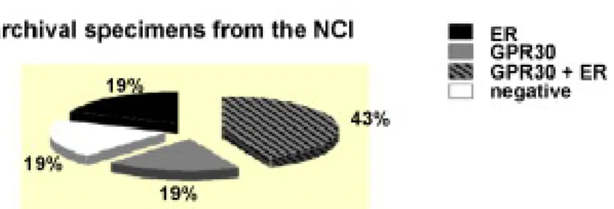
An analysis of 321 cases of primary breast cancer showed that approximately 60% of the breast tumor cases expressed levels of GPR30 similar to that normal breast cancer, while 40% of the breast cancer cases expressed low or undetectable levels of GPR30 protein. Codependency for GPR30 and ER was observed, as roughly 40% of the cases co-expressed each receptor type. Twenty percent of the tumors were doubly negative, failing to express GPR30 and ER, with the remaining 40% expressing either one receptor or the other. Interestingly, half of the 122 ER-negative tumors, scored positively for GPR30, possibly suggesting that an ER-negative tumor that retains GPR30 may remain estrogen responsive by signaling through EGFRs (Filardo et al., 2008).
Fig. 4:Co-expression of GPR30 and ER in primary human breast tumors. (Filardo et al., 2008)
Therefore, the recent identification of the first GPR30-selective ligand G-1 (Bologa et al., 2007) has provided new opportunities to further differentiate between the functions of the ER family member and GPR30 in mediating the multifaceted mechanisms of estrogen action.
A large and compelling body of epidemiologic and experimental data implicates estrogens in the etiology of neoplasias such breast and ovarian carcinoma (Eisen et al., 1998; Barkhem et al., 1998; Williams et al., 1999; Chen et al., 1999; Bai et al., 2000; Rodriguez et al., 2001; Riman et al., 2002; Lacey et al., 2002).
Estrogens can enhance the development of breast cancer by stimulating cell proliferation rate and thereby increasing the number of errors occurring during DNA replication (epigenetic effects), as well as by causing DNA damage via their genotoxic metabolites produced during oxidation reactions (genotoxic effects) (Gadducci et al., 2005).
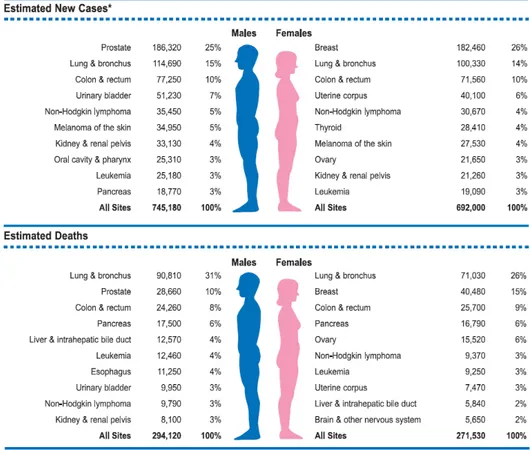
Breast cancer is the most common cancer in women and is estimated to have accounted for 182,460 new cancer diagnoses and 40,480 deaths in 2008 (Jemal et al., 2008). The incidence is highest in highly industrialized countries like North 20
America, Northern Europe, and Australia, where age-adjusted rates are 75-92 per 100,000 women (standardized to year 2000 world population), and lowest in Asia and Africa, where incidence is less than 22 per 100,000 (Parkin et al., 2001). Ovarian cancer is the fourth leading cause of tumor death in Western countries representing the most fatal gynecologic malignancy with the overall 5-year survival rate about 10% to 20% (Boete et al., 1993) and is also estimated to have accounted for 21,650 new cases and 15,520 deaths in 2008 (Jemal et al, 2008).
Fig. 5: Ten Leading Cancer Types for the Estimated New Cancer Cases and Deaths, by Sex, United States, 2008.(Jemal et al., 2008).
Breast and ovarian cancer are common in western countries: environmental factors may play an essential role in hormone-dependent tumor etiology. In fact, estrogenic activity can be found in a large variety of natural and man-made compounds.
Phytoestrogens are natural substances derived from sources such as plants or fungi: they are typically flavonoids or isoflavonoids. For example the phytoestrogens Genistein and Quercetin, copiously present in soyabeans, vegetables and fruits, exert estrogenic activity through direct binding and activation of the estrogen receptor alpha and beta, influencing breast cancer cell proliferation in a dose-dependent manner (Maggiolini et al., 2004). Synthetic estrogenic compounds, called xenoestrogens, environmental estrogens or disruptors, include a variety of pesticides, polychlorinated biphenyls and plasticizers and are almost ubiquitous in our society (Starek 2003; Jacobs and Lewis 2002).
Fig. 5: Data from Budavari (1996),Harris et al. (1997),IARC (1998),Illinois Environmental protection agency(1997), Routledge et al. (1998) Smith and Quinn (1992), Soto et al., and SRI International (1995).
Atrazine, belongs to the 2-chloro-s-triazine family of herbicides and is the most common pesticide contaminant of groundwater and surface water (Fenelon and Moore 1998; Kolpin et al., 1998; Miller et al., 2000). Atrazine is able to interfere with androgen- and estrogen-mediated processes (Cooper et al. 1999, 2000, 2007; Cummings et al., 2000; Friedmann 2002; Narotsky et al., 2001; Stoker et al., 2000). This action occurs without direct agonism or antagonism of the ER or Androgen Receptor (AR) (Roberge et al. 2004). Previous studies have shown
that atrazine reduces androgen synthesis (Babic-Gojmerac et al. 1989; Kniewald et al., 1995) as well as stimulates estrogen production (Heneweer et al., 2004; Keller and McClellan-Green 2004; Sanderson et al., 2002). Epidemiologic studies, also have related long-term exposure to triazine herbicides with increased risk of ovarian cancer in female farm workers in Italy (Donna et al. 1989) and breast cancer in the general population of Kentucky in the United States (Kettles et al. 1997).
Whether E2 acts on the EGFR/ERK transduction pathway only through GPR30 binding or also through ERα binding is less clear, since E2 binds to both receptors although with different affinity. Using the selective GPR30 ligand G-1 in the first study our research group evaluated the ability of GPR30 to mediate proliferative effects in ovarian cancer cells expressing both ERα and GPR30. We demonstrated a cross-talk between the ERα and GPR30 to induce proliferative effects induced by E2 and G-1 in ovarian cancer cells.
In our second study we evaluated whether GPR30 is also implicated in the growth effect induced by the pesticide Atrazine in ovarian cancer cells and also in this case we found that GPR30 and ERα are both involved in this response. Our third question was about the regulation of GPR30 expression. We found that GPR30 expression is modulated by EGF through the MAPK pathway. Our results contribute to provide new insight on the role of GPR30 in transducing estrogenic mechanisms implicated in tumor progression.
G Protein–Coupled Receptor 30 (GPR30) Mediates Gene
Expression Changes and Growth Response to
17β-Estradiol and Selective GPR30 Ligand G-1 in Ovarian
Cancer Cells
Lidia Albanito, Antonio Madeo, Rosamaria Lappano, Adele Vivacqua, Vittoria
Rago, Amalia Carpino, Tudor I. Oprea, Eric R. Prossnitz, Anna Maria Musti, Sebastiano Ando` and Marcello Maggiolini
Cancer Res 2007; 67: (4). February 15, 2007
INTRODUCTION
Ovarian cancer is the fourth leading cause of tumor death in Western countries (Greenle et al., 2000). ERα mediates the mitogenic action of estrogens in breast cancer by inducing a variety of genes involved in cell proliferation. A large body of evidence has identified different mechanisms whereby membrane-initiated signaling involving growth factor receptor or membrane ERs mimics or contributes to the function of nuclear ERs (Levin et al., 2005; Deroo and Korach, 2006). Recent studies have shown that GPR30, mediates the non-genomic signaling of E2 in a variety of estrogen-responsive cancer cells through activation of the EGFR transduction pathway (Bologa et al., 2006; Maggiolini et al., 2004; Revankar et al., 2005; Thomas et al., 2005; Filardo et al., 2002; Vivacqua et al., 2006). Considering that GPR30 binds most ER ligands (Thomas et al., 2005), it remains unclear how ERα contributes to GPR30/EGFR signaling in cancer cells. Therefore, the recent identification of the first GPR30-selective
ligand G-1 (Bologa et al., 2006) has provided new opportunities to further differentiate between the functions of the ER family member and GPR30 in mediating mechanisms of estrogen action. In the present study, we have ascertained the ability of G-1 to induce cell growth of estrogen-sensitive ovarian cancer cells expressing ERα as well as breast cancer cells not expressing ERα. We used BG-1 cancer cells as a model, which derived from a solid tumor tissue of a patient with stage III ovarian adenocarcinoma and express clinically relevant levels of ERα but lack ERβ (Bardin et al., 2004).
RESULTS
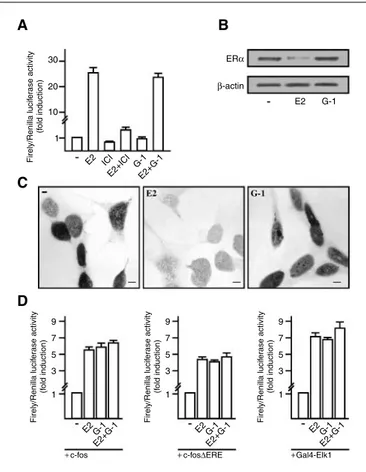
G-1 does not active ERα but induces the transcription of c-fos promoter constructs.
We first transiently transfected an ER reporter gene in BG-1 cancer cells: the exposure to 100nM E2 induced a strong ERα transactivation which was no longer observed in presence of the ER antagonist ICI 182,780. In contrast, treatments with 100nM G-1 failed to induce luciferase expression or to block that observed on addition of E2 (Fig. 1A). Considering that the down-regulation of ERα induced by an agonist has been considered an additional hallmark of receptor activation (Santagati et al. 1997), we analyzed ERα protein levels. As documented by Western Blotting analysis, the levels of ERα were robustly down-regulated only in presence of E2 (Fig. 1B). To confirm the aforementioned observation we did an immunocytochemical staining: the expression of ERα was reduced only by E2 (Fig. 1C). In order to evaluate the role of GPR30 we
evaluated if its specific ligand could activate a transiently transfected full-length human c-fos promoter 2.2kb) and a c-fos mutant lacking the ERE sequences (-1.172bp). As can be seen in fig. 1D, G1 transactivated c-fos similar to E2.
The ternary complex factor member Elk1 is crucial for the ERK-dependent activation of the promoter of the c-fos gene (Karin 1994). G-1 and E2 activated Elk1 in the context of a Gal4 fusion protein; however, the transcriptional response was not substantially enhanced by E2 in combination with G-1 (Fig. 1D), suggesting that E2 and G-1 act through the same signal transduction pathway.
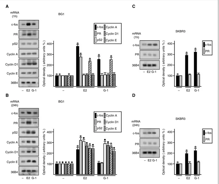
G-1 and E2 induce the mRNA expression of c-fos and other estrogen target genes.
To evaluate whether G-1 and E2 could up-regulate c-fos along with other well-known estrogen target genes in BG-1 cells, we performed semi-quantitative RT-PCR experiments comparing mRNA levels after standardization with a housekeeping gene encoding the ribosomal protein 36B4. Of note, a short treatment (1 h) with 100 nM of E2 and G-1 enhanced c-fos levels, which were still evident after a 24-h exposure to E2 (Fig. 2A and B). The expression of pS2, cyclin A, and cyclin E was stimulated by both E2 and G-1 after 24 h of treatment (Fig. 2A and B), whereas the levels of cyclin D1 increased at both short and prolonged exposure to both compounds (Fig. 2A and B). In contrast, the expression of PR was up-regulated only by E2 at both times of observation, indicating that an E2-activated ERα-dependent mechanism is involved in the regulation of this gene. To further support this finding, we turned to the SKBR3 28
cells, which do not express detectable amounts of ERs. As shown in Fig. 2C and D, E2 failed to regulate PR, whereas both E2 and G-1 retained the ability to induce c-fos expression, which we previously showed to be dependent on GPR30 expression.
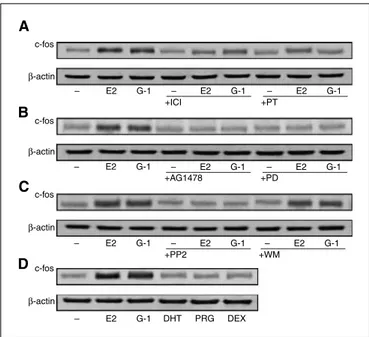
Transduction pathways involved in the up-regulation of c-fos protein levels exerted by G-1 and E2.
Does G-1–dependent activation of c-fos require ERα- and/or GPR30-mediated signaling? As shown in Fig. 3A, either the ER antagonist ICI 182,780 or the GPCR inhibitor pertussis toxin reduced the induction of c-fos obtained after treatment with 100 nmol/L of E2 and G-1 for 2 h, suggesting that both receptors are implicated in the effect of each ligand. Furthermore, the stimulation of c-fos by both G-1 and E2 was equally abrogated by the EGFR kinase inhibitor tyrphostin AG 1478, the mitogen-activated protein kinase (MAPK) inhibitor PD 98059, and the Src family tyrosine kinase inhibitor PP2 but not using the phosphatidylinositol 3-kinase (PI3K) inhibitor wortmannin (Fig. 3B and C), suggesting that both ligands signal through the EGFR/ERK signaling pathway. Moreover, steroids, such as DHT, progesterone, and dexamethasone, did not increase c-fos protein levels (Fig. 3D), revealing that a ligand specificity is required for the regulation of c-fos in ovarian cancer cells. To further assess the role of ERα and GPR30 we analyzed the response of c-fos to both compounds in the absence of either ERα or GPR3O expression. As shown in Fig. 4A, both antisense oligonucleotides turned down the c-fos induction by E2 and G-1, 29
although each oligonucleotide selectively silenced only the expression of the specific oligonucleotide target sequence (Fig. 4B and C). Moreover, the rapid activation of ERK1/2 on addition of 100 nmol/L of E2 and G-1 was abrogated by both antisense oligonucleotides (Fig. 5A). The inhibitors of EGFR signaling, such as AG 1478, PD 98059, and PP2, prevented ERK1/2 activation by E2 and G-1 (Fig. 5B), thus eliciting a repressive action similar to that observed for c-fos up-regulation by ligands.
G-1 and E2 stimulate the proliferation of the ovarian BG-1 and 2008 tumor cells and breast SKBR3 carcinoma cells.
In BG-1 cells, the growth-stimulatory effects induced by G-1 and E2 were abolished by the EGFR inhibitor AG 1478, the MAPK inhibitor PD 98059, and the Src kinase inhibitor PP2 (Fig. 6A), establishing that the EGFR/ERK signaling pathway mediates the stimulatory action of both ligands. Moreover, the abrogation of ERα or GPR30 expression also abrogated the E2-stimulated and G-1–stimulated cell growth (Fig. 6A). Similar results were also obtained using a different ovarian cancer cell line, named 2008 (Fig. 6B), which expresses the same receptor pattern of BG-1 cells (Safei et al., 2005). However, the results shown in Fig. 2C and D show that G-1 is able to up-regulate c-fos in ER-negative SKBR3 cells. It has been previously reported that E2 induces ERK activation in SKBR3 cells (Maggiolini et al., 2004). Therefore, we investigated the ability of ligands to stimulate SKBR3 cell proliferation. As shown in Fig. 6C, 100 nmol/L of E2 and G-1 promoted SKBR3 cell growth, which was abolished by AG 1478, PD 98059, and PP2 or by abrogation of GPR30 expression (Fig. 6C). 30
To rule out the possibility that SKBR3 cells expressed detectable levels of ERs sufficient to signal cell growth, we assessed ligand-induced proliferation in the presence of ERα and ERβ antisense oligonucleotides. As evidenced in Fig. 6D, the transfection of either ERα and ERβ antisense oligonucleotides at a concentration able to abrogate the target receptor expression respectively in MCF7 and MDA-MB-231 breast cancer cells had no effect on SKBR3 cell growth, establishing in this cellular context that GPR30 is sufficient to signal E2-induced proliferation. Cumulatively, these data indicate that, although ERα is required for the G-1/GPR30 signaling pathway in ovarian cancer cells, GPR30 may induce cell growth independently of ERα expression depending on the tumor type.
Taken together, these findings suggest that both ERα and GPR30 are required for proliferation of ovarian cancer cells in response to either E2 or G-1. Because the effect of both ligands on cell growth as well as on c-fos induction was abrogated by inhibition of EGFR kinase activity or its downstream effectors, our results also indicate that both ERα and GPR30 are both necessary to signal proliferation of ovarian cancer cells through the EGFR/ERK transduction pathway.
G Protein-Coupled Receptor 30 and Estrogen Receptor α
are involved in the proliferative effects induced by
Atrazine in ovarian cancer cells.
Lidia Albanito, Rosamaria Lappano, Antonio Madeo, Adele Chimento, Eric R
Prossnitz, Anna Rita Cappello, Vincenza Dolce, Sergio Abonante, Vincenzo Pezzi and Marcello Maggiolini
Environ Health Perspect doi:10.1289/ehp.11297
INTRODUCTION
Atrazine, one of the most common pesticide contaminants, has been shown to up-regulate aromatase activity in certain estrogen-sensitive tumors without binding or activating the ER (Roberge et al. 2004; Tennant et al. 1994a, 1994b). GPR30 which is structurally unrelated to the ER, mediates rapid actions of E2 and environmental estrogens and is able to mediate rapid E2-dependent responses such as gene expression and cancer cell proliferation (Revankar et al., 2005; Thomas et al., 2005; Maggiolini et al., 2004; Vivacqua et al., 2006a; Vivacqua et al., 2006b). Given the ability of atrazine to exert estrogen-like activity in cancer cells, we evaluated whether atrazine could signal through GPR30 in stimulating biological responses in cancer cells.
RESULTS
Atrazine does not activate ERα in cancer cells.
Does atrazine trans-activate ERs? To investigate the potential capability of atrazine to act through the ERs and considering that atrazine increases the
incidence of estrogen-sensitive tumors in different experimental models (Cooper et al. 2007), we transiently transfected an ER reporter gene in estrogen-sensitive ovarian (BG-1), breast (MCF-7) and endometrial (Ishikawa) cancer cells. The exposure to E2 provoked a strong ERα transactivation which was absent in the presence of the ER antagonist ICI in all cell lines used (Fig. 2A, Fig. 2B and Fig. 2C). In contrast, the treatment with atrazine did not stimulate luciferase expression and did not block the induction observed upon addition of E2 (Fig. 2A, Fig. 2B and Fig. 2C). Atrazine is also unable to active an expression vector encoding ERα transiently transfected in ER-negative SKBR3 breast cancer cells (Fig 2D). We then used a heterologous system formed by chimeric proteins consisting of the DNA binding domain (DBD) of the yeast transcription factor Gal4 and the ERα or ERβ hormone binding domain (HBD) which were transiently transfected in SKBR3 cells. Gal4 ERα ERβ were strongly induced by E2 but not upon atrazine treatment (Fig. 2E and Fig 2F).
Atrazine neither regulates ERα expression nor competes with estrogen binding to ERα.
Then we investigated whether atrazine could modulate ERα expression in BG-1 cells. The ERα expression was down-regulated at both mRNA and protein levels by 100 nM E2, whereas 1 μM atrazine did not produce any modulatory effect (Fig 3A and Fig 3B). In agreement with these results atrazine did not show any binding capacity for ERα (Fig. 3C) as already reported (Cooper et al. 2007). Altogether, our findings rule out that the estrogenic action of atrazine occurs through binding and direct activation of ERα.
Aromatase activity is not induced by atrazine.
Precedent studies demonstrated that atrazine is able to up-regulate aromatase expression in different cell contexts (Cooper et al. 2007; Fan et al. 2007a, 2007b; Roberge et al. 2004; Sanderson et al. 2001, 2002).We analyzed aromatase activity through tritiated water release assays in BG-1 cells. 1 μM atrazine did not stimulate aromatase activity in BG-1 cancer cells but in contrast strongly induced in human H295R adrenocorticocarcinoma cells previously used as a model system to assess aromatase catalytic activity (Heneweer et al. 2004; Sanderson et al. 2001). Atrazine resulted neither an ERα activator nor an aromatase regulator in estrogen-sensitive ovarian cancer cells.
ERK phosphorylation is stimulated by atrazine.
In order to evaluate if the potential estrogenic activity of atrazine is exerted through a rapid cellular response, we evaluated its ability to induce ERK phosphorylation in BG-1 cells. Atrazine and E2 stimulated ERK phosphorylation (Fig. 5A; Fig 5B and Fig. 6A). The ERK activation was observed also in 2008 ovarian cancer cells which present a receptor expression pattern similar to BG-1 cells (Safei et al. 2005) (Fig. 6D). Which transduction pathway is required in this activity induced by atrazine? We then investigated ERK phosphorylation co-treating the cells with specific inhibitors. The ER antagonist ICI, the EGFR and ERK inhibitors AG and PD, respectively, prevented ERK activation induced by both E2 and atrazine, whereas GFX, H89 and WM, inhibitors of protein kinase C (PKC), protein kinase A (PKA) and PI3K, respectively, did not (Fig. 6B; Fig. 6C; Fig. 6E and Fig. 6F). A previous study observed that ICI is able to trigger ERK phosphorylation (Filardo et al.
2000). We treated the SKBR3 breast cancer cells to increasing concentrations of ICI but we did not observe any ERK activation.
Atrazine up-regulates the mRNA expression of estrogen target genes.
After having determined that atrazine induces a rapid ERK activation, we evaluated in BG-1 cells its ability to modulate the expression of the early gene c-fos along with other estrogen target genes. We performed semi-quantitative RT-PCR assays comparing mRNA levels after standardization with a housekeeping gene encoding the ribosomal protein 36B4. A 1h treatment with 1 μM atrazine enhanced c-fos and cyclin A levels although to a lesser extent than 100 nM E2, which also stimulated PR, pS2 and cyclin D1 expression (table 1). After a 24 h treatment, atrazine increased PR, pS2 and cyclin A levels while E2 additionally induced the expression of c-fos, cathepsin D, cyclins D1 and E (Table 1).
Transduction pathways involved by atrazine in the up-regulation of c-fos protein levels.
The protein expression of c-fos was used as a molecular sensor of atrazine action at the genomic level. After a short treatment (2h) atrazine and E2 induced up-regulation of c-fos in BG-1 and 2008 cells (Fig. 8). The induction of c-fos level was abolished by the ER antagonist ICI and the EGFR and ERK inhibitors, AG and PD respectively (Fig. 8). GFX, H89 and WM, inhibitors of PKC, PKA and PI3K, respectively, did not interfere with c-fos stimulation (Fig. 8). Thus, in ovarian cancer cells atrazine involves ERα and the EGFR-MAPK pathway to trigger c-fos protein increase. Our previous results demonstrate that c-fos stimulation by E2 occurs through GPR30 and requires ERα and EGFR-mediated signaling in cancer cells expressing both receptors (Albanito et al. 2007;
Maggiolini et al. 2004; Vivacqua et al. 2006a, 2006b). Could atrazine act in a similar manner? The transactivation of c-fos induced by both E2 and atrazine, were no longer observed silencing either ERα or GPR30 in BG-1 and 2008 cells (Fig. 9). Furthermore, could atrazine induce a rapid response in a cell context expressing only GPR30? Using ER-negative SKBR3 breast cancer cells we analyzed ERK phosphorylation and c-fos expression after treatment with atrazine. As shown in (Fig. 10), atrazine was able to induce gene expression which was abolished knocking-down GPR30.
The proliferation of ovarian cancer cells induced by atrazine occurs through GPR30 and requires both ERα and EGFR-MAPK-mediated signaling.
We observed that both E2 and atrazine induced the proliferation of BG-1 and 2008 cells in a concentration-dependent manner (Fig. 11A and Fig. 11E). The proliferative effects elicited by E2 and atrazine were abolished in presence of AG and PD (Fig. 11B and Fig. 11F) or after knocking-down the expression of either GPR30 or ERα (Fig. 11C; Fig11D; Fig. 11G and Fig. 11H). Both receptors and the EGFR/MAPK transduction pathway are involved in the growth effects induced by atrazine in ovarian cancer cells.
Epidermal Growth Factor Induces G Protein-Coupled
Receptor 30 Expression in Estrogen Receptor-Negative
Breast Cancer cells.
Lidia Albanito, Diego Sisci, Saveria Aquila, Elvira Brunelli, Adele Vivacqua,
Antonio Madeo, Rosamaria Lappano, Deo Prakash Pandey, Didier Picard, Loredana Mauro, Sebastiano Ando`, and Marcello Maggiolini.
Endocrinology 149(8): 3799–3808, 2008 INTRODUCTION
Numerous data have suggested that the interaction of EGFR with estrogenic pathways can occur at different levels. E2 can couple various G proteins, thereby triggering nongenotropic effects through the trans-activation of the EGFR (Levin 2003; Keen and Davidson 2003; Roskoski 2004; Razandi et al., 2004). It has been shown that GPR30 is implicated in EGFR transactivation by E2 (Filardo et al., 2000). E2 induces also EGF-like activity in female reproductive tissue (DiAugustine et al., 1988; Nelson et al., 1991) and similarly activates biochemical signals associated with the EGFR transduction pathway (Migliaccio et al., 1996; Martin et al., 2000). GPR30 can act independently from ERs in inducing estrogen-dependent EGFR action. How is GPR30 expression regulated? In this study we used SKBR3 and BT20 breast cancer cells lacking ERs, to evaluate the regulation of GPR30 expression by E2, G-1, IGF-1 and EGF.
RESULTS
EGF transactivates the 5’flanking region of GPR30 through an AP1 site in ER-negative breast cancer cells
To analyze GPR30 expression we cloned a vector coding a 648-bp fragment located at the 5’ flanking region of the human GPR30 gene containing different transcription factor binding sites, such as those for the AP1 and SP1 activating proteins (Fig. 1). Then we transiently transfected the construct in SKBR3 and BT20 breast cancer cells. We evaluated its response induced by E2 and G-1 and the growth factors EGF and IGF-I, both involved in tumor development and progression. Only EGF was able to trans-activate the cloned GPR30 5’flanking region (Fig 2A and Fig 2B). The luciferase induction stimulated by EGF was not observed co-treating the cells with the EGFR and ERK inhibitors AG and PD, whereas the response to EGF was not abrogated by PP2, H89, or LY, inhibitors of the Src family tyrosine kinase, the protein kinase A (PKA), and phosphatidylinositol 3-kinase (PI3K) transduction pathways, respectively (Fig. 2B, Fig 2D). We also cloned two expression vectors mutated in AP1 and SP1 sites which are potentially involved in the responsiveness to EGF (Fig. 3A). In both SKBR3 and BT20 cells, the construct mutated in the AP1 site, -477 to -471 region (GPR30AP1mut) did not respond to EGF, whereas the construct mutated in the SP1 site, -138 to -133 region (GPR30SP1mut) still maintained the EGF responsiveness (Fig. 3, B and C).
EGF up-regulates GPR30 expression
To evaluate GPR30 expression and the transduction pathways involved in its regulation, we performed a semiquantitative RT-PCR assay. A short EGF
treatment (1h) in SKBR3 cells increased GPR30 mRNA expression, and while AG and PD prevented this response, PP2, H89, and LY did not evidence any inhibitory effect (Fig. 4A and Fig 4B). The protein levels of GPR30 were evaluated after a 2h EGF exposure and the regulation was similar to the mRNA regulation (Fig. 4C). Using a different approach, we evaluated the cellular localization of GPR30 after EGF stimulation by confocal microscopy in SKBR3 cells. The treatment with EGF (2h) induced an intracellular GPR30 accumulation, which was no longer observed in presence of AG or PD (Fig. 5A). The specificity of detection in SKBR3 cells was verified by neutralizing the GPR30 antibody by 10-fold molar excess of the antigen peptide (Fig. 5B). The GPR30-negative HEK-293 cells showed no immunodetection of GPR30 (Fig. 5C, upper panels), whereas the nuclei were stained with propidium iodide (Fig. 5C, lower panels).
The EGFR-ERK transduction pathway mediates GPR30 induction by EGF
In SKBR3 cells a rapid ERK1/2 phosphorylation induced by EGF was no longer evident in presence of AG and PD but still persisted using PP2, H89, and LY in combination with EGF (Fig. 6A). In a variety of hormone-sensitive tumor cells (Maggiolini et al.,2004; Vivacqua et al., 2006a; 2006b), EGFR/ERK-mediated pathways led to early induction of c-fos. In line with the results obtained on ERK activation, EGF induced a strong c-fos increase, which was abrogated by AG and PD but by PP2, H89, or LY (Fig. 6B). Is the c-fos up-regulation induced by EGF involved in GPR30 expression? To answer this question we performed a ChIP analysis immunoprecipitating cell chromatin with an anti-c-fos antibody and amplifying the AP1 site located within the GPR30-5’flanking region. The
treatment with EGF strongly recruited c-fos to the AP1 site, which was dependent on EGFR/ERK signaling. In fact, AG and PD inhibited this association whereas PP2, H89, and LY did not elicit any inhibitory activity (Fig. 6C). Using a primer DNA control that does not contain the AP1 site we did not visualize any ethidium bromide staining (Fig. 6C)
The up-regulation of GPR30 by EGF engages E2 to boost the proliferation of breast cancer cells
In SKBR3 and BT20 cells, the growth effects, induced by E2 and EGF alone, further increased in presence of both substances (Fig. 7A and Fig 7E). How does GPR30 contribute to this biological effect? The growth effects of E2 alone or in combination with EGF were prevented transfecting cells with shGPR30, which knocked down GPR30 expression (Fig. 7B and Fig. 7F). We also, transfected the cells with a DN/c-fos expression plasmid, which blocked the AP1-mediated transcriptional activity (Fig. 7C and Fig. 7G), and we did not observe the proliferative effects induced by either mitogen (Fig. 7D and Fig. 7H). Consequently, c-fos/AP1 signaling exerts a key role in the growth stimulation of both mitogens in SKBR3 and BT20 cells. Taken together, the up-regulation of GPR30 after exposure to EGF may represent a molecular mechanism through which EGF engages E2 to boost the proliferative effects elicited in these ER-negative breast cancer cells.
Materials
and
Reagents
17β-estradiol (E2), Epidermal Growth Factor (EGF), Insulin-like Growth Factor-1 (IGF-Factor-1), 2-chloro-4-(ethylamine)-6-(isopropylamine)-s-triazine, (Atrazine), Genistein (G), 4-hydroxytamoxifen (OHT), Cycloheximide (Cx), Pertussis Toxin (PT), 5α-Dihydrotestosterone (DHT), Progesterone (PRG),
Dexamethasone (DEX), H89, LY 294,002 (LY), Wortmannin (WM), and PD98059 (PD) were purchased from Sigma-Aldrich (Milan, Italy). (1-[4-(6-bromobenzo[1,3]diodo-5yl)-3a,4,5,9b-tetrahydro-3H-cyclopenta[c]quinolin -8-yl]-ethanone) (G-1) was kindly provided by ChemDiv,Inc. (San Diego, CA). AG1478 (AG) and AG490 was purchased from Biomol Research Laboratories (DBA, Milan, Italy). ICI 182,780 (ICI) was obtained from Tocris Chemicals (Bristol, United Kingdom) and GF109203X (GFX) from Calbiochem, (VWR International, Milan, Italy). All compounds were solubilized in DMSO, except E2 and PD which were dissolved in ethanol.
Cell Culture
All cell lines were grown in Dulbecco’s Minimal essential medium (DMEM) or in RPMI 1640 medium without phenol red supplemented with L-glutamine (2mM), Penicillin (100U/ml), streptomycin (100U/ml), and 10% foetal bovine serum (FBS). Cells were switched to medium without serum 24hs before transfections or confocal staining and immunocytochemical staining, 48hs before RT-PCR or immunoblot, and 72hs before the evaluation of ERK1/ERK2 phosphorylation.
Plasmids.
The firefly luciferase reporter plasmids used were:
♦ XETL, for ERα and ERβ carrying the firefly luciferase sequences under the control of an estrogen-response element upstream of the thymidine kinase promoter;
♦ HEGO used for the expression of ERα;
♦ GK1, gene reporter for all Gal4 fusion proteins; ♦ Gal4 chimeras Gal-ERα and Gal-ERβ fusion protein;
♦c-fos and the deletion mutant c-fosΔERE ( which lacks the ERE sequence), encoding -2.2-kb and -1172 bp 5’ upstream fragments of the human c-fos respectively;
♦ Gal4-Elk1 fusion protein; ♦ DN/c-fos;
♦ GPR30/AS expression plasmid.
Transfection and Luciferase assays.
Cells were plated into 24-well dishes with 500 ml of regular growth medium per well the day before transfection. The medium was replaced with that lacking serum on the day of transfection , which was performed using Fugene 6 reagent as recommended by the manufacturer (Roche Diagnostics, Milan, Italy) with a mixture containing 0.3 μg of reporter plasmid and 3ng of pRL-TK. Ligands were added to the cells after 8-9hs and incubated for 8-24hs, depending on the different assays. Luciferase activity was measured with the Dual Luciferase kit (Promega, Milan, Italy) according to the manufacturer’s
recommendations. Firefly luciferase activity was normalized to the internal transfection control provided by the Renilla luciferase activity. The normalized relative light unit values (RLU) obtained from untreated cells were set as 1-fold induction on which the activity induced by treatments was calculated.
RT-PCR.
The evaluation of gene expression was done by semiquantitative RT-PCR by using the direct incorporation of digossigenin-11-dUTP(DIG-dUTP) during the amplification of cDNA. After the extraction from cells, RNA was converted in cDNA by the reverse transcriptase enzyme and the genes of interest were amplified by PCR. PCR products were then separated on 1.2% agarose gel and transferred on a nylon membrane, probed with the antibody against digossigenin conjugated to peroxidase and revealed using the ECL system ( GE Healthcare, Italy).
The primers used were:
36B4 forward 5´-CTCAACATCTCCCCCTTCTC-3´ 36B4 reverse 5´-CAAATCCCATATCCTCGTCC-3´ GPR30 forward 5’CTGGGGAGTTTCCTGCTGA-3’ GPR30 reverse 5’-GCTTGGGAAGTCACACCAT-3’ ERα forward 5’-AATTCAGATAATCGACGCCAG-3’ ERα reverse5’-GTGTTTCAACATTCTCCCTCCTC-3’
c-fos forward 5´-AGAAAAGGAGAATCCGAAGGGAAA-3´ c-fos reverse 5´-ATGATGCTGGGACAGGAAGTC-3´
PR forward 5´-ACACCTTGCCTGAAGTTTCG-3´
PR reverse 5´-CTGTCCTTTTCTGGGGGACT-3´ pS2 forward 5´-TTCTATCCTAATACCATCGACG-3´ pS2 reverse 5´-TTTGAGTAGTCAAAGTCAGAGC-3´
Cyclin A forward 5´-GCCATTAGTTTACCTGGACCCAGA-3´ Cyclin A reverse 5´-CACTGACATGGAAGACAGGAACCT-3´ Cyclin D1 forward 5´-TCTAAGATGAAGGAGACCATC-3´ Cyclin D1 reverse 5´-GCGGTAGTAGGACAGGAAGTTGTT-3´ Cyclin E forward 5´-CCTGACTATTGTGTCCTGGC-3´
Cyclin E reverse 5´-CCCGCTGCTCTGCTTCTTAC-3´
Western Blotting
.Cells were grown in 10-cm dishes, exposed to ligands, and then lysed in 500 ml of 50mmol/L NaCl, 1.5 mmol/L MgCl2, 1 mmol/L EGTA, 10% glycerol, 1%
Triton X-100, 1% SDS, a mixture of protease inhibitors containing 1mmol/L aprotinin, 20mmol/L phenylmethylsulfonyl fluoride, and 200 mmol/L
sodium orthovanadate. Protein concentration was determined using Bradford reagent according to the manufacturer’s recommendations (Sigma-Aldrich). Equal amounts of whole protein extract were resolved on a 10%
SDS-polyacrylamide gel electrophoresis in glycine buffer (0.02mM Tris, 0.2mM glycine, 1% SDS). Proteins were transferred to a nitrocellulose membrane (GE Healthcare, Italy) using the above buffer with the addition of 20% methanol, and probed overnight at 4°C with the specific antibodies and then revealed using the ECL system (GE Healthcare, Italy). All the antibodies were
purchased from Santa Cruz Biotechnology, DBA, Milan, Italy. Polyclonal GPR30 was purchased from MBL-Eppendorf, Milan, Italy.
Aromatase assay.
The aromatase activity in subconfluent cell culture mediumwas measured by tritiated water-release assay using 0.5 µM[1ß-3H(N)]androst-4-ene-3,17-dione
(25.3 Ci/mmol;DuPont NEN, Boston, MA, USA) as a substrate.The cells were treated in a 6-well dish in culture medium in the presence of Atrazine or DMSO for 40 hs and then incubated with [1 ß -3
H(N)]androst-4-ene-3,17-dione. Incubations were performed at 37 °C for 6 h undera 95%:5% air/CO2
atmosphere. The results obtained were calculatedas pmol/h, and normalized to milligram of protein (pmol/h permg protein) and expressed as percentages of untreated cells (100%).
ER binding assay.
BG-1 cells were stripped of any estrogen by keeping them in medium without serum for 2 days. Cells were incubated with 1 nM [2,4,6,7-3H]E2 (89 Ci/ mmol;
Amersham Bioscience) and increasing concentrations of nonlabeled E2 or Atrazine for 1 h at 37° C in a humidified atmosphere of 95% air/ 5% CO2. After
removal of the medium, cells were washed with ice-cold PBS/0.1% methylcellulose twice, harvested by scraping and centrifugation, and lysed with 100% ethanol, 500 µl per 60 mm dish, for 10 min at room temperature (Lee et al. 1996). The radioactivity of extracts was measured by liquid scintillation counting.
Chromatin immunoprecipitation (ChIP).
Cells grown in 10 cm plates were shifted for 24 hs to medium lacking serum and then treated for 2 hs with vehicle or 50 ng/ml EGF. The immuno-cleared chromatin was precipitated with anti-c-fos antibody. A 4 μl volume of each sample was used as template to amplify by PCR two fragments located next to the GPR30-5’ flanking region: one fragment of 261 bp containing the AP1 site and the second fragment of 364 bp (from -937 to -1301) not containing the AP1 site. The primer pairs used to amplify the first fragment were: CGTGCCCATACCTTCATTGCTTCC-3’ (forward) and 5’-CCTGGCCGGGTGTCTGTAG-3’ (reverse) while the primer pairs used to amplify the second fragment were: 5'-CCGTGGCCCGCTGCATAGAGAAC-3'
(forward) and 5'-GAGAGGGAGAAGTGGGCTGTC-3' (reverse). The PCR conditions were 45 seconds (s) at 94°C, 40 s at 58°C, and 90 s at 72°C. The amplification products obtained in 25 cycles were analysed in a 2% agarose gel and visualized by ethidium bromide staining. 3 μl of the initial preparations of soluble chromatin were amplified to control input DNA before precipitation.
GPR30 plasmid constructs and GPR30 short hairpin RNA
(shGPR30).
To generate the luciferase expression vector for the GPR30-5’flanking region (GPR30), a 641 bp fragment next to the 5’-flanking region of the GPR30 gene was amplified by PCR using the following primer pairs AACACTGGCTTTCCCTTCCTATCT-3’ (forward) and 5’-CTTGAAGTGAGCCTGGCATTTGTC-3’ (reverse) from genomic DNA which
was extracted from SkBr3 cells by Trizol reagent as suggested by the manufacturer (Invitrogen, Milan, Italy). PCR primer pairs were selected analyzing the 5’-flanking region of GPR30 gene in chromosome 7, location 7p22.3. The PCR amplification was performed using 1.25U GoTaq DNA polymerase according to the manufacturer instructions (Promega, Milan, Italy). PCR conditions were 5 min 95°C, followed by 1 min 94°C, 1 min at 58°C, and 1 min at 72°C for 30 cycles. The fragment was then inserted in the pCR 2.1 plasmid using the TA cloning kit (Invitrogen, Milan, Italy), sequenced and cut with Hind III and Xho I. The insert was cloned in the pGL3 basic vector (Promega, Milan, Italy). Analyses of GPR30-5’ flanking region revealed an AP1 (-471 to -477) and an SP1 (-133 to -138) consensus binding sites. Mutations from position -471 to -477 in the GPR30-5’ flanking sequence corresponding to an AP1 motif and from position -133 to -138 corresponding to the SP1 binding site (Fig 2 A), were generated using QuikChange XL Site-Directed Mutagenesis kit (Stratagene, Milan, Italy). The following pairs of primers were used to generate the AP1 and Sp1 mutants: GPR30AP1mut (forward) 5’-CCCTGCCTGTGGGAGACGCCCACGTCCAGCCTCC-3’ and (reverse) 5’-GGAGGCTGGACGTGGGCGTCTCCCACAGGCAGGG-3’; GPR30SP1mut (forward)
GGACGAGCACGCGGAGATCACTCGCCTCCACGG-3’ and (reverse) 5’-CCGTGGAGGCGAGGTGATCTCCGCGTGCTCGTCC-3’. All plasmids were sequenced before use. Plasmid 3x-FLAG-hGPR30 was constructed using the
HindIII/BamHI sites in pCMV10.3x-ratFLAG. hGPR30 was amplified with
primers CCCCAAGCTTatggatgtgacttcccaag and CAGCGGATCCctacacggcactgctgaac (restriction sites are underlined).
Reference plasmid was Prl-3x-FLAG expressesing an unrelated 26 kDa protein. Short hairpin (sh)RNA constructs against human GPR30 were bought from Openbiosystems (Biocat.de, Heidelberg, Germany) with catalog number RHS4533-M001505. The targeting strand generated from the shRNA vectors sh1, sh2, sh3, sh4 and unrelated control are respectively: CGAGTTAAAGAGGAGAAGGAA, CTCCCTCATTGAGGTGTTCAA, CGCTCCCTGCAAGCAGTCTTT, GCAGTACGTGATCGGCCTGTT, CGACATGAAACCGTCCATGTT. In order to evaluate the effectiveness of the
different shRNA constructs, HEK-293 cells were seeded at about 50% confluency in 6 cm plates. 6-8 hs later, cells were transfected using the calcium-phosphate method with 1 µg of 3x-FLAG-hGPR30, 10 µg of shRNA construct and 2 µg of Prl-3x-FLAG. Prl-3xFLAG was used as a transfection control. 40 hs after transfection, cells were harvested and lysed with 20 mM Tris-HCl [pH 8], 100 mM NaCl, 10% glycerol, 0.1% NP-40, 1 mM monovanadate, 1 mM DTT, and protease inhibitors. DNA was sheared by several passages through a 25-gauge needle. Lysates were cleared by centrifugation, and protein concentrations were determined by the Bradford method. 30 µg of lysates were subjected to western blot analysis with the FLAG antibody M2 (Sigma, Lausanne Switzerland). With a 74% knock-down of 3x-FLAG-hGPR30 expression the shRNA construct sh3 showed the highest efficacy. Hereafter, sh3 is referred to as shGPR30.
Immunocytochemical staining.
Cells were treated as indicated and then fixed in fresh paraformaldehyde (2%for 30 min). After paraformaldehyde removal, hydrogen peroxide (3% in methanol for 30 min) was used to inhibit endogenous peroxidase activity. Cells were then incubated normal horse serum (10% for 30 min)to block the nonspecific binding sites. Immunocytochemical staining was done using as the primary antibody a mouse monoclonal immunoglobulin G (IgG)gener ated against ERα (overnight at 4°C). A biotinylated horse anti-mouse IgG
(1:600 for 60 min at room temperature)was applied as the secondary antibody (Vector Laboratories, Burlingame, CA). Subsequently, the amplification of avidin-biotin-horseradish peroxidase complex (1:100 for 30 min at room temperature; Vector Laboratories)was carried out and 3,3¶-diaminobenzidine tetrachloride dihydrate (Vector Laboratories)was used as a detection system. Cells were rinsed after each step with TBS [0.05 mol/L Tris-HCl plus 0.15 mol/L NaCl (pH 7.6)] containing 0.05% Triton X-100. In control experiments, cells were processed replacing the primary antibody with mouse serum (Dako S.p.A., Milan, Italy)or using a primary antibody preabsorbed (48 h at 4jC)with an excess of purified ERα protein (M-Medical).
Confocal microscopy
.50% confluent cultured cells grown on coverslips were serum deprived for 24hs and then treated as indicated. Then, cells were fixed in 4% paraformaldehyde, permeabilized with 0.2% Triton 100X, washed three times with PBS and
incubated for 1 h with 2 mg/ml primary antibody against GPR30. After incubation with the GPR30 antibody, the slides were washed three times with PBS and incubated with 1 mg/ml rhodamine-conjugated donkey anti-rabbit IgG (Calbiochem, Milan, Italy). The cellular expression and localization of GPR30 was evaluated by confocal microscope with 1000x magnification. The optical sections were taken at the central plane.
Proliferation assay.
For quantitative proliferation assay, 10 000 cells were seeded in 24-well plates in regular growth medium. Cells were washed once they had attached and then incubated in medium containing 2.5% charcoal-stripped FBS with the indicated treatments. Medium was renewed every 2 days (with treatments) and cells were trypsinized and counted in a haemocytometer on day 6. A concentration of 200 ng/L of the indicated shRNA, or GPR30/AS (200 ng) or 200 ng of empty vector was transfected using Fugene 6 Reagent as recommended by the manufacturer the day before treatments, and then renewed every 2 days before counting.