UNIVERSITÀ DEGLI STUDI DI MESSINA
Dipartimento di Scienze Chimiche, Biologiche, Farmaceutiche ed Ambientali
Dottorato di ricerca in Scienze Chimiche
XXIX Ciclo (CHIM/06)
Engineering of Graphene Materials
for Biomedical Applications
From graphite to new functionalized graphene
by top down-approaches
Giulia Neri
Supervisor Coordinator
Prof.
Anna Piperno
Prof.Sebastiano Campagna
Table of Contents
Aim of the work ... 2
Chapter 1: ... 6
1.1 Graphene materials ... 7
1.2 Characterization of G materials ... 13
1.3 Biomedical Applications of G Materials ... 16
Chapter 2: ... 24
2.1 Graphene as platform for drug delivery ... 25
2.2 Graphene nanoplatforms ... 26
2.3 Silibinin-Conjugated Graphene Nanoplatform ... 33
2.4 Reactive Azlactone Graphene Platform (RAGP) ... 42
2.5 Catalytic Properties and Immobilization Studies of Graphene Catalase Bioconjugate (G-Cat) ... 54
2.6 Experimental section ... 60
Chapter 3: ... 74
3.1 Functionalized G nanosheets by a top-down approach from graphite using oxazolone chemistry ... 75
3.2 Computational studies ... 87
3.3 Graphene/Gold Nanocomposites for SERS applications ... 93
3.4 Experimental section ... 105
Chapter 4 ... 110
4.1 Basic concepts about peptide self-assembly ... 111
4.3 SD1: a peptide for gene-delivery ... 119
4.4 Experimental section ... 124
Acknowledgements ... 130
2
Aim of the work
The discovery of graphene (G) and its functionalized derivatives has been accompanied by increasing research attention to explore these new materials for biomedical applications due to their unique structure and excellent mechanical, optical and electrical properties.
Generally, synthetic methods of G are classified in two categories: top-down and bottom-up. The former approach entails exfoliation of a layer of G from graphitic materials. The latter approach involves the building up of G using carbon-based materials. The bottom-up approach is simple, although it produces material with relatively more defects than the top-down approach. Top-down strategies separate the stacked sheets by disrupting the Van der Waals forces that hold the sheets together. Damaging of the sheets during the exfoliation process and re-agglomeration of the separated sheets are some of the disadvantages of the top-down technique.
The biocompatibility of G is the most critical issue for the applications in the biomedical/pharmaceutical field and its chemical modification is the key factor for designing stable and safe drug delivery nanodevices.
It is known thatsize, shape, morphology, thickness, degree of functionalization and dispersibility of G play critical roles in regulating biological behaviours and toxicity. Therefore, the development of adequate G preparation methods represents a key factor to achieve good G materials for biomedical applications.
The thesis describes the synthesis and the characterization by complementary techniques of new functionalized G materials obtained by two different top-down approaches. Moreover, some biomedical applications of newly synthesized G materials have been investigated.
3
The first chapter reports an overview of the main characteristics of G materials and describes the currently available procedures for their fabrication and summarizes the main biological applications.
The chapter II about reports the functionalization of G materials with the terminal alkyne units and their synthetic applications by click assembly with azido-functionalized compounds (Fig. 1).
The alkyne-terminated graphene platform (G-Alk) was obtained in good yield by reduction of graphene oxide (GO) with hydrazine followed by grafting of
p-(2-propynyloxy)-benzene units by reaction with diazonium salts. The click assembly
of G-Alk with the azido flavonoid Silibinin (Sil) provided a new drug delivery nanoplatform (herein called G-Sil). The cytotoxicity of the new platform has been evaluated on human mesenchymal stem cells and the anticancer effects have been studied on human osteosarcoma cell lines. Our G nanoplatform did not show any cytotoxicity even at high concentration (1000 μg/ml) and Sil grafted onto G maintained its antiproliferative activity.
4
The click reaction of G-Alk with the oxazolone (APDMA) containing an azido moiety has been used to build the reactive G platform (RAGP). The RAGP has been employed for the silylation of G surfaces and for the selective conjugation with compounds of biological interest such as glutathione (GSSG) and catalase (Cat).
The chapter III reports the direct delamination and functionalization of graphite into G, exploiting the reactivity of mesoionic compounds in solvent-free conditions (Fig.2). For the first time we have demonstrated that the solvent-free 1,3-DC reaction of mesoionic compounds is an effective tool for the direct functionalization and delamination of graphite flakes into few layers of G nanosheets. The procedure has been tested by employing two differently substituted oxazolones and a high degree of functionalization (2.1–4.6%@700 °C) was obtained for both substrates under mild conditions (70–120 °C). The graphite exfoliation efficiency depends upon the oxazolone substitution pattern. The exfoliation and functionalization was confirmed by micro-Raman and X-ray photoelectron spectroscopies, scanning transmission electron microscopy (STEM) and thermogravimetric analysis (TGA). Moreover computational studies showed that the reaction proceed mainly on the corner and on the edge of graphenic system.
5
These data were confirmed by decoration of the surface G layers with gold nanoparticles (Au NPs). STEM analysis showed that Au NPs are mainly located on edge of G layers. Finally, the properties of G-NH2/Au nanocomposites as SERS
materials have been investigated using Rhodamine 6G (R6G) as a probe.
The final chapter of the thesis (chapter IV) is focused on the development of a coiled-coil peptide for gene-delivery. This section reports the results of the research project carried out at the National Physical Laboratory of London (NPL, UK), within the Biotechnology group, under the supervision of Dr Max Ryadnov and Dr Emiliana De Santis. The chapter reports the characterization studies of peptide (SD1). TEM analysis has been used to confirm the formation of peptide sphere structures by self-assembling process; qualitative tritations have been employed to prove the formation of SD1/siRNA complex. From these studies, SD1 emerged as a good candidate for gene-delivery; actually, SD1/siRNA complex is under biological evaluation.
6
Chapter 1:
Graphene:
Synthesis, Characterization and
Biological Applications
7
This chapter reports an overview on G materials focusing the attention on the preparation methods, the characterisation techniques, that are generally, used to investigate the structure, morphology and chemical composition of G materials. Moreover, the chapter covers recent literature and deals with the basic concepts, the chemistry, the potentialities and the problems due to the toxicity of the G materials proposed for biomedical applications.
1.1 Graphene materials
G is an allotropic form of carbon; it exhibits a bi-dimensional honeycomb lattice structure (Fig. 1.1), where each carbon is hybridized sp2 with an estimated interatomic distance of 0.142 nm and a hexagon center to center distance of 0.246 nm (Fig. 1.2).(1) G exposes a large surface area, rich of π electrons, which determinate its outstanding proprieties. G shows an am-bipolar conductivity with an electron mobility until to 40.000 cm2/Vs (2), a good thermal conductivity (between 4.84 ± 0.44 and 5.30 ± 0.48 103 W/mK at r.t.) (3), an optical transmittance (~ 97.7%) (4); moreover it is a robust material and at the same time flexible and lightweight with a density of 0.77 mg/m2.(5)
8
Figure 1.2 Illustrative figure of the atomic distance in G material.
Since the discovery of G, the research (6) on it has increased exponentially (7) and the family of ‘‘G materials” under consideration has expanded beyond monolayer G to include related materials with significant variations in layer number, lateral dimension, rotational faulting, and chemical modification. This increasing interest caused confusion among researchers in regard to the terms that would have been used to name G materials. Recently, a nomenclature for G derivatives has been proposed to clarify and overcome some questions in this respect (8):
“Graphene – a single-atom-thick sheet of hexagonally arranged, sp2-bonded
carbon atoms that is not an integral part of a carbon material, but is freely suspended or adhered on a foreign substrate. The lateral dimensions of graphene can vary from several nanometers to the macroscale.
Graphene layer – a single-atom-thick sheet of hexagonally arranged, sp2-bonded
carbon atoms occurring within a carbon material structure, regardless of whether that material structure has 3D order (graphitic) or not (turbostratic or rotationally faulted). The ‘‘graphene layer’’ is a conceptual structural unit that has been used for many years to describe the structure and texture of 3D carbon materials with primary sp2-hybridized bonding.
9
Turbostratic carbon – three-dimensional sp2-bonded carbon material in which
there is no defined registry of the layers, meaning there is no spatial relationship between the positions of the carbon atoms in one G layer with those in adjacent layers. This is a common structure in carbon materials prepared at lower temperatures or in ‘‘hard carbons’’ that do not pass through a fluid phase during carbonization and resist the development of 3D crystalline order even upon very high temperature heat treatment.
Bilayer graphene, trilayer graphene – 2D (sheet-like) materials, either as free-standing films or flakes, or as asubstrate-bound coating, consisting of 2 or 3 well-defined, countable, stacked G layers of extended lateral dimension.
Multi-layer graphene (MLG) – a 2D (sheet-like) material, either as a free-standing flake or substrate-bound coating, consisting of a small number (between 2 and about 10) of well-defined, countable, stacked G layers of extended lateral dimension.
Few-layer graphene (FLG) – a subset of multi-layer G (defined as above) with layer numbers from 2 to about 5.
Graphene nanosheet – a single-atom-thick sheet of hexagonally arranged, sp2
-bonded carbon atoms that is not an integral part of a carbon material, but is freely suspended or adhered on a foreign substrate and has a lateral dimension less than 100 nm.
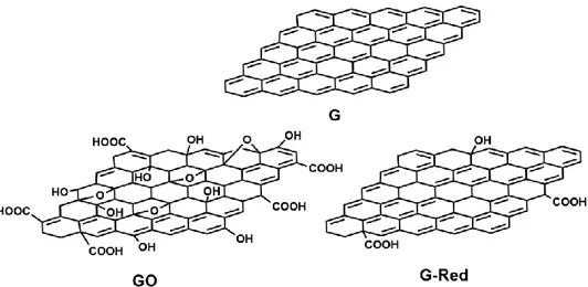
Graphene oxide (GO) – chemically modified G, prepared by oxidation and exfoliation that is accompanied by extensive oxidative modification of the basal plane. GO is a monolayer material with a high oxygen content, typically characterized by C/O atomic ratios less than 3.0 and typically closer to 2.0.
Reduced graphene oxide (rGO herein called G-Red) – GO (as above) that has been reductively processed by chemical, thermal, microwave, photo-chemical, photo-thermal or microbial/bacterial methods to reduce its oxygen content.
10
Graphene materials (also graphene-based materials, graphene nanomaterials, graphene-family nanomaterials) – overarching terms for the collection of 2D materials defined above that contain the word ‘‘graphene’’, including multilayered materials (N less than about 10), chemically modified forms (GO, G-Red), and materials made using G, GO, or another G material as a precursor.”
Moreover, the G derivatives are also named, according to synthetic strategy employed to obtain them. Generally, these synthetic strategies are classified in two big categories: bottom-up and top-down techniques.(9) The “scotch-tape method” has been the first to be applied and it has been proposed by Geim and Novoselov.(10) It is a mechanical exfoliation (top-down approach) and provided particular thin tallies of graphite of high quality, [Highly Ordered Pyrolitic Graphite (HOPG)]. Although. HOPG is a high quality material, the scotch-tape method is not a suitable strategy to apply on large scale.(10)
Another top-down strategy concerns the “liquid exfoliation of G”, by using chemical solvents such as DMF or NMP under strong sonication conditions. However, several studies have demonstrated that the liquid exfoliation is not an efficient method because the G layers obtained show many surface defects.(11)
The main methods of alternative bottom-up strategy are the chemical vapor deposition (CVD) and the epitaxial growth (EG). EG strategy consists in the production of G layers by using monocrystalline silicon carbide, which shows two different surface terminations, Si-terminated and C-terminated. The process consists in a thermal desorption of silicon from monocrystallines SiC; if the G layers are generated by Si-terminated, they show a homogenous surface with high carrier mobility. On the contrary the C-terminated ones show a heterogeneous surface with many defects. In addition, from Si- and C-terminated, FLG and MLG systems are obtained, respectively. EG is an efficient method to obtain G layers of high quality on large scale and the number of G layers depends on the applied
11
temperature. In order to reduce the cost of fabrication, more attention has been paid to find an alternative to the expensive SiC.(12)
In CVD method the G grows on a metal poly/monocrystalline support and acetylene or methane are used as carbon source. The process occurs at high temperature (~ 1000 °C), under hydrogen flow, to keep the respective metal in its catalytic state. Copper and nickel are the most employed in CVD strategy; generally, by using copper, monolayer systems have been obtained, whereas the use of nickel generates few layers. To improve the efficiency of CVD strategy and to reduce the costs, some changes about this method have been proposed.(13) Other efficient methods to produce G of high quality have been developed.(14) In particular, the research focused the efforts on the synthesis of functionalized G materials.(15) It has been demonstrated that the functionalization of G is the key to control its chemical-physical properties.
Both covalent (16) and non-covalent (17) surface modifications of G-based materials can be exploited to convey specific properties to G and to increase its colloidal stability.
Among the covalent approaches the radical reactions are the most applied. Diazonium salts are used to generate in situ aryl radicals, which attack the G surface, forming new C sp3-aryl-bond on the sp2 G network. (18)
Other important approaches to fabricate functionalized G materials are 1,3-dipolar cycloaddition (1,3-DC) (19, 20), the Bingel reaction (21) the Diels–Alder reaction (DA).(22, 23) DA reaction is the most investigated cycloaddition process, since Haddon and co-workers have suggested that G can be used as either a diene or a dienophile for DA reactions.(24)
Other methods for the functionalization of G include the introduction on G surface of single atoms such as fluorine and chlorine by halogenation processes, other halogens are not used due to stability problems. The fluorination is an
12
uncontrollable process due to the fast kinetics; the fluorinated G showed a reduced conductivity with respect to G.(25) Conversely, chlorination is a better controllable
process and the product exhibits a good carrier mobility. G materials can be subjected to hydrogenation reaction on SiO2/Si substrates.(26)
The most employed approaches for the preparation of G materials involve the chemistry of GO obtained from graphite by Hummer or Brodie methods.(27) The oxidation process produces first graphite oxide that after strong exfoliation treatment produces GO (Fig. 1.3), which could be subsequently subjects to chemical reduction to obtain G-Red (Fig. 1.3). The strong oxidation conditions adopted in these processes destroy the G sp2 network, and the reduction process restore only partially the sp2 network. Hence, these G materials cannot be used to produce electrical devices, but they are strongly investigated in laboratory for other applications like those in biomedical field since the residual polar groups increase their biocompatibility.
13
1.2 Characterization of G materials
A very important aspect of G materials research is the definition of their chemical composition, crystal structure, thickness, lateral dimension, by employing a combination of spectroscopy, microscopy and thermogravimetric techniques. The Raman spectroscopy is one of the most extensively, non-destructive optical techniques used to detect the structural features of G materials.(28) Information can be gained about the amount of G network order, its exfoliation degree or in some cases, the number of stacking layers.(28) In a typical G Raman spectrum, two main characteristic bands are present: i) the so called G-band centered at about 1580 cm-1, due to in plane modes vibrations of sp2 network; ii) the so called 2D, or D’, band at about 2680 cm−1 (Fig. 1.4), anovertone of the D one, generally located at about 1330 cm-1, due to different inter-plane vibration modes. The D peak is absent in pristine G due to its crystal symmetries. On the contrary, D band is present in Raman spectra of G derivatives, such as GO or G-Red, as consequence of the inherent chemical treatments which induce imperfections on the carbon basal plane. In this condition, breathing modes of aromatics rings will be activated and D band results evident. Moreover, in the spectrum of G derivatives, other two bands: D´ (1620 cm-1) and D+G (2940 cm-1), can be detected (Fig. 1.5). They are activated by the increased disorder of the sp2 network, due to the defects introduced
by chemical functionalization of G surfaces.
The interaction forces among G layers influences the intensity, the shape and the position of these bands. Therefore, information about the number of layers can be obtained by studying the band modifications. In single-layer G, 2D band shows a narrow peak around 2950 cm-1 while, in presence of overlapped layers, the forces due to the interactions between the layers of the stacked G, determine a splitting of the 2D peak. Thus, some different vibrational modes are activated and the 2D band is characterized by four sub-bands.(28) At the same time, upon increasing the layers
14
number a smaller red shift of the G-band can also observed. Hence, the number of G layers can be indirectly estimated by the I2D/IG band intensity ratio, as well as
analyzing their shapes and their shifts. The IG/ID band intensity ratio is commonly
used to indicate the disorder degree of the G material.(29) In the pristine graphite, a high IG/ID ratio value (3.7) is observed, indicating a highly ordered system while,
in the graphene based materials, a progressive decrease of the graphitization degree is clearly evident.(29)
Figure 1.4 Raman spectra of a monolayer graphene system and graphite bulk.(28)
15
Information about the chemical composition and the kind of chemical bond fraction present on G surfaces (i.e. C-O bonding coordinations) can be obtained by X-ray photoelectron Spectroscopy (XPS).(29) XPS is used to investigate the G functionalization and to study how the chemical surface’s coordination changes during G material synthesis (i.e from graphite to GO and G-Red). In particular, C 1s peak is made up by five different chemical shifted components, assigned to C atoms bonded to various oxygen function groups: 289.2 eV (carboxyl groups), 287.5 eV (carbonyl groups) 286.55 eV (epoxy groups) and 285.86 eV (hydroxyl groups). Another important contribute is related to sp2 carbon atoms in aromatic rings assigned at 284.5 eV. At the same time three characteristic peaks contribute to the O 1s band: 533.43 eV (oxygen bonded to aromatic carbon), 532.03 eV (oxygen single-bonded to aliphatic carbon) and 531.08 eV (oxygen double-bonded to sp2 carbon). Further information about the G functionalization can be obtained by taking into account the signals related to other chemical species, such as N 1s, Si 2p and S 2p.
Indications about the functionalization of materials are obtained carrying out Fourier Transform Infrared Spectroscopy (FT-IR) measurements. The FT-IR spectra of G materials is characterized by a band around 1573 cm−1 due to the presence of C=C stretching in graphitic domains.(30) Moreover, through FT-IR
spectra is possible to monitor of the chemical changes of G surface. The GO spectrum shows several contributes relative to the strong oxidative process such as: the band centered at ~3430 cm−1, due to the O–H stretching of hydroxyl and carboxylic acid moieties, the band at ~1719 cm−1 due to the C=O stretching of carboxylic acids groups and a peak at ~1046 cm−1 attributed to the C–O stretching; in addition, a band at ~2922 cm−1 corresponding toC–H stretching of aliphatic sp3 carbon can be observed.(30) FT-IR spectrum of G-Red shows a clear decrease of
16
treatment; the observed bands are attributable to residual oxygen functional groups.(30)
Several microscopy techniques such as Scanning Electron Microscopy (SEM), Transmission Electron Microscopy (TEM) and Atomic Force Microscopy (AFM) are used to investigate the morphology of G materials.(29) These analyses (Fig. 1.6) give information about the layers distribution and, about the edge of the layers, allowing to estimate their shape and thickness.
Figure 1.6 (a) and (b) Optical images of graphene layers. (a) Single and a few-layers are indicated with yellow arrows. (b) A layer displaying ripples is shown. (c) and (d) Field emission SEM images (FE-SEM): (c) overview of layers deposited onto the substrate. (d)High magnification of the area marked with a circle in (c), showing the porous structure of the layers.(31)
1.3 Biomedical Applications of G Materials
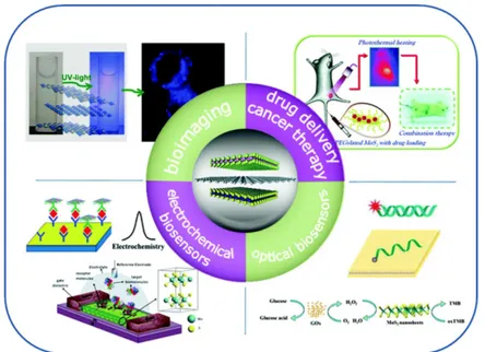
Considering the exceptional properties of G materials, they have been object of extensive research in several fields including the biomedical and pharmaceutical ones.(32, 33) They have been proposed as drug delivery systems for photothermal
17
and photodynamic therapy, as biosensing materials for nano-theranostic applications and as a scaffold in tissue engineering (Fig. 1.7).(34)
At the same time, the most important issue is still the potential long-term toxicity concerning G and its derivatives.(35) By these studies, it was understood that the toxicity of G, like other materials previously employed in biomedicine, is strongly dependent on the functionalization degree of its surface. The toxicity of pristine G has been demonstrated in several works (36), while suitable functionalized G materials shown no toxic effects in the tested dose ranges.(37) Other studies have demonstrated that, a suitable functionalization of G materials, allows to control their behavior in biological systems and to increase their biocompatibility towards human cells.(38) Depending on target application, several synthetic strategies based on covalent or non-covalent binding, have been proposed. Functionalized G materials have been obtained by covalent approach, exploiting the reactive groups like carboxylic acid, hydroxyl and epoxy groups widely present on GO (39), scarcely on G-Red surface. Other approaches are based on hydrophobic interactions, π-π stacking and electrostatic binding.(38)
18
G and its derivatives, due to their high capacity of loading biomolecules on their large surface area, were employed to build several drug delivery systems. The G surface, rich of delocalized π electrons, showed high ability of forming complexes with several aromatic drugs by π-π stacking, like doxorubicin (DOX) (40), camptothecin (CPT) (41) and SN38 (irinotecan, an analogous of CPT).(42) Both DOX and CPT were loaded on GO surfaces, obtaining a synergic cancer cell killing effect, in dose dependent manner.(43)
The preparation of G-Red/DNA complex with a high water solubility was described. During the chemical reduction from GO to G-Red, the DNA has been introduced by π-π stacking interactions, between GO surface and the bases of single-stranded DNA. At the end of chemical reaction, DNA covers the G-Red surface. This method was used to develop several DNA biosensors.(44)
An interesting gene delivery system was prepared by functionalization of GO, a negatively charged material, with poly-ethyleneimine (PEI), a positively charged polymer, via electrostatic interactions. PEI imparts a positive charge to G surface, allowing a subsequently loading of DNA or RNA sequences, negatively charged. The system GO-PEI showed: i) a lower toxicity compared to bare PEI; ii) an improvement of physiological stability compared to GO; iii) high gene delivery transfection efficiency, due to a better protection from action of nuclease enzymes.(45)
Another delivery system, based on G materials, was prepared by covalent conjugation of polyethylen glycol (PEG) and chitosane on GO surfaces. The conjugation was achieved between the amine groups of PEG/chitosane and the carboxyl groups of GO, improving the biocompatibility of carbon material. Moreover, it reduces the non-specific binding to biological molecules and cells, promoting a specific action in anticancer therapy.(46) Other studies have demonstrated that the covalent conjugation with biomolecules increases the
19
biocompatibility of G. For example GO conjugated with dextran (DEX), showed a significant improvement of the stability in physiologic solution with respect to GO. The conjugation of GO with sulphate biomolecules improves the colloidal stability of GO in biological solution.(37)
GO and G-red were also investigated as platforms to prepare systems to be used in theranostic therapy. This is a novel medicine strategy that combines diagnosis and therapy: “all-in-one single platform”.(38)
The increase in efficacy of a therapeutic formulation is directly related to its ability to selectively target the affected tissue, to overcome the biological barriers and to react smartly to the disease environment by releasing the therapeutic agents. It has been demonstrated that G-based materials with the lateral size of several tens of nanometers offer passive targeting toward tumour sites, and that the conjugation with a targeting agent such as folic acid (FA) confers active targeting due to over-expression of FA-receptors on membrane surfaces of cancer cells.(47)
G materials were also explored for photothermal therapy (PTT) and photodynamic therapy (PDT).
PTT is based on a local increase of temperature that kills the cancer cells, due to an agent that generates heat, under controlled light irradiation. G is suitable for this application because it exhibits an important absorption in near infrared region (NIR).(48) An interesting phototermal system based on G materials was obtained
by chemical reduction of nanoGO-PEG to ultra small nanoG-Red-PEG, coating with PEGylation phospholipid and finally bonding to the arginine-glycine-aspartic acid peptide chain. This system showed in vitro a high efficient and selective cancer cells ablation.(49) Other works have demonstrated that, the efficient action of these systems depends on the dimensions of G materials and on their functionalization degree.(50) GO was also used to build photodynamic systems.(51) In PDT a photosensitizer molecule is subject to appropriate light irradiation, which causes the
20
local release of reactive oxygen species (ROS) which kill the cancer cells.(52) For
example, nGO-PEG was conjugated, by noncovalent interactions with zinc phthalocyanine (ZnPc), under Xe light irradiation; this system showed selective cytotoxic effects on tumor cells.(53) Also, GO functionalized with TiO2
nanocomposites has shown an important anticancer activity under visible light irradiation.(54) Another interesting phototermal system consists in GO conjugated with chlorine e6 (Ce6), this system under laser irradiation at 633 nm has showed an important anticancer action.(47)
G and its derivatives were also studied for their antibacterial effects. However, literature data are conflicting and unclear results were obtained by different research groups. It was demonstrated that GO with respect to G-Red and graphite showed the major antibacterial effects on Escherichia coli.(55)
G induces an effective membrane damage in Gram-positive bacteria and at the same time, this effect was not observed on membrane of Gram-negative bacteria.(56) A bactericidal activity of GO, coated with silver nanoparticles, for both Gram-negative and Gram-positive was reported.(57) In contrast with these studies, other works demonstrate that the bacteria grow faster on substrates containing GO with, respect to substrates where is absent.(58)
G surface could be decorated with inorganic nanoparticles: Au, Ag Pt, Ni, TiO2,
ZnO, Fe3O4 etc.(59-61)
GO-TiO2 was used in photodynamic therapy.(54) Nanohybrids obtained by
decoration of GO with iron oxide nanoparticles (IO NPs) showed interesting applications in biomedical field because the drug release can be controlled by using magnetic forces.(62) Moreover, they could be used in magnetic resonance imaging and as contrast agent for cell labeling.(63)
Nanohybrids of G-Red covered with Au NPs, have been used in drug-delivery applications and in bio-imaging.(64)
21
The potential application of G in tissue engineering has been demonstrated during the last years. In particular, it has been proven that the scaffold of chitosan-PVA nanofibers containing G is able to control the distribution and the cell proliferation. Considerable attention was attracted on the design of scaffolds for bone tissue engineering. In particular, it was observed a very good attachment and growing of MC3T3-E1 mouse pre-osteoblast cells on a GO-chitosan scaffold covalently bonded.(65) The authors reported that the cells penetrate the pores of the scaffold and, at the same time, their interconnections were increased. The studies have shown a high quality of the cellular proliferation. Moreover, the combination of chitosan with GO avoids the degradation of the polysaccharide.(66)
In summary, literature data suggest a great potential of G and its derivatives for biomedical applications. These materials exhibited a large surface area where it is possible to graft a wide variety of drugs and biomolecules. Furthermore, these materials have shown interesting physical and optical properties useful in bio-imaging and for the photothermal therapies. It was very important to understand that a suitable functionalization allows to overcome the associated problems of short life toxicity. Currently, studies about the long life toxicity are less advanced. Recently, G materials have been intensively investigated as substrates to be functionalized for SERS (Surface Enhanced Raman Spectroscopy) applications like bio-sensing (67) or for the detection of molecules or tumour cells.(68) SERS is a more
suitable technique comparing to Raman since the latter often has a very small cross-section and generally, the problem of fluorescence arises. SERS, as the name itself suggests, is a surface phenomenon where the intensity of the signals are enhanced by a factor of 1012 – 1014 (69), respect to a common Raman spectrum. SERS phenomenon can be attributed to two mechanisms: the electromagnetic mechanism (EM), due to the strong amplification of the local EM field (70) and the chemical effect (CM) that involves the creation of new electronic states generated by the
22
interaction between the metal and the molecules adsorbed on it.(71). Such new
electronic states allow for resonant Raman scattering processes.
The substrates play a crucial role for an effective surface enhancement. In particular, the control of the distances among the localized surface plasmons (LSPs), on a sub-nanometres scale, is a critical parameter to control the EM and therefore the efficiency of SERS phenomenon. In fact, an increase of the distance among the LSPs causes a decrease of coupling between them and, therefore, a decrease of the enhancement factor.
With respect to other materials, G layers influence positively both the EM and the CM coupling, enhancing the SERS process. Summarizing G shows: i) interesting optical properties, ii) high surface/volume ratio, iii) sub-nanometer scale thickness and iv) a great affinity with noble metals. (72)
24
Chapter 2:
Graphene:
Innovative Material for
Biomedical Applications
25
This chapter deals about the functionalization of G material with terminal alkyne units and their synthetic applications by click assembly with azido-functionalized compounds.
2.1 Graphene as platform for drug delivery
Graphene (G) and its multifunctional derivatives, due to their advantageous properties, have recently emerged as new and interesting drug nanocarriers. In particular, these resulting G systems have showed the potential to be applied for systemic, targeting and local drug delivery.
G materials display many advantages compared with other drug delivery systems: the ability to provide high loading capacity for many different drugs and therapeutic molecules (weight ratio of loaded drug to G nanomaterials could reach 200%) (73);
their flexibility and capability to be engineered into complex multifunctional drug delivery systems, for combined therapies, a new development which is hardly achievable with other nanomaterials;
covalent or non-covalent surface modifications can be used to impart specific biological activity to G materials, as well as to improve their biocompatibility and colloidal stability.
Although preliminary preclinical studies are encouraging, the clinical applications are still far and require addressing some open challenges. The most critical issue for the biomedical applications of these materials is their biocompatibility and toxicity, which are the primary concerns. Most of the current literature data report that the G is a biocompatible material with a low toxicity. In spite of this, some conflicting and unclear results have been reported by different research groups.(74,75) However, an agreement has been gradually reached: the functionalization of G
26
materials significantly improves their biocompatibility; this step is essential for designing stable and safe drug delivery nanocarriers. Other important questions regard the development of nanocarriers with reproducible dimensions and properties and the requirement to achieve a deeper understanding of G’s interaction with living cells (tissues and organs), especially about the cellular uptake mechanism.
2.2 Graphene nanoplatforms
Among the different subtypes of G-based nanomaterials, GO is certainly the more investigated one for drug delivery purposes (e.g., compared to pristine G and G-Red). As-produced GO is highly oxidized with a large number of residual epoxide, hydroxyl and carboxylic acid groups on its surface. The reduction of GO, with reducing agents (i.e. N2H4), restores partially the structure and properties of G. In
comparison with GO, G-Red is known to have higher electrical conductivity and optical absorbance, a fact that makes it a more attractive tool for applications in theranostic nanomedicine.(38) Therefore, a better understanding of G-Red materials behaviour in biological systems is needed to improve their performances inside the nanodevices. It is noteworthy, that both the biocompatibility and the pharmacokinetic profile of G depend on the modality of functionalization and on its conjugation with biological compounds.(76)
G-Red sheets, unlike GO layers, are hydrophobic with limited aqueous dispersibility. In addition, the existence of high π-π stacking and Van der Waals forces, which facilitate the irreversible aggregation of G layers or their restack to graphite, strongly limit the G-Red applications. To avoid these phenomena, a suitable G-Red functionalization is mandatory in order to make the structural, physical, chemical and electronic properties of the material, suitable to the specific applications.(77)
27
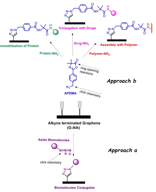
In my ongoing research program, aimed at the discovery of biocompatible drug delivery systems based on carbon nanomaterials, the interest was focused on the decoration of reduced G platform with an alkyne terminated group (G-Alk). The alkyne moieties introduced on the nanoplatform prevent the aggregation of G sheets, improve their dispersibility in organic media and, being reactive structural fragments, they can be further functionalized by click chemistry reactions.
Two different synthetic approaches were exploited (Fig. 2.1): a) the alkyne units were directly coupled with the azide functionality linked to a target biomolecule; b) the alkyne moieties were employed to prepare a reactive azlactone graphene platform (RAGP) on G sheets. Afterwards, RAPG can be very efficiently exposed to ring-opening reactions with functionalized primary amine derivatives. In particular, an aminosilane coupling agent (APTS), a biological fragment (glutation oxide, GSSG) (78) and a natural enzyme such as Catalase (Cat) have been incorporated.
28
29
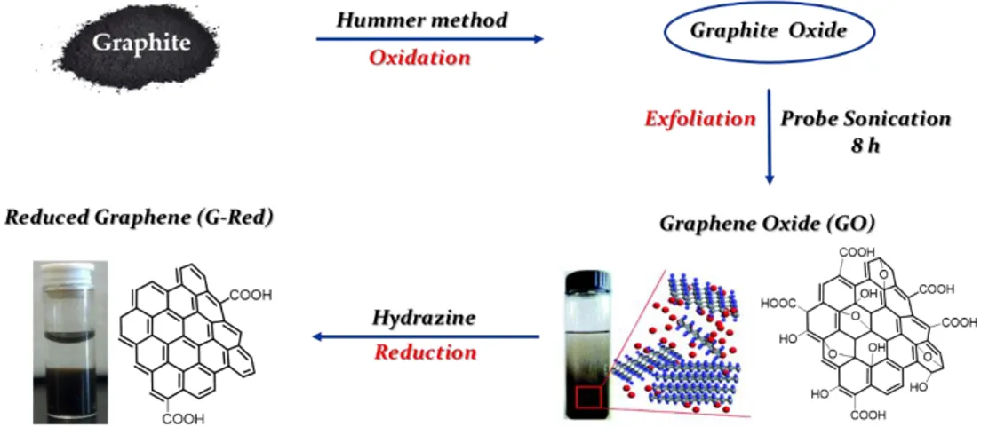
G-Red was prepared by oxidizing natural graphite powders to graphite oxide, using Hummers method (79) and successive exfoliation to GO, by ultrasonication. Then,
GO was reduced with hydrazine, under controlled conditions, according to a literature procedure (80), to obtain reduced G sheets (G-Red) (Fig. 2.2).
Graphite, GO and G-Red have been characterized by TGA, XPS, Raman and TEM analyses. The results are in agreement with literature data.(79; 80)
Figure 2.2 Schematic illustration of the preparation of reduced graphene (G-Red).
Raman spectroscopy was used to probe structural characteristics of the investigated carbon-based materials, providing useful information on the defects (D-band) related to the vibrations of the sp3 hybridized carbons, and located at about 1330 cm-1, and in-plan vibration of sp2 carbon atoms (G-band) centred at about 1580 cm-1. Raman spectra are also characterized by the second-order Raman 2D band
(known as D’), which is a defect-independent contribution. The effect of the reduction process on the Raman spectral profiles points out an increased defect density, showing a progressive reduction of the graphitization degree (i.e., of IG/ID
in Fig. 2.3), confirming the highly disordered structure of the carbon based materials.
30
Figure 2.3 Raman spectra of graphite, graphene oxide (GO), reduced graphene (G-Red).
The XPS spectra of graphite, GO and G-Red (Fig. 2.4), showed the C 1s regions composed by six contributions at about 284.5, 285.8, 286.6, 287.7, 288.9 eV and 291 eV. These contributions are ascribed to C=C/C in aromatic ring, OH, C-O-C, C=O and OH-C=O and π-π bounds, respectively. The contributions of C-OH, C-O-C, C=O and OH-C=O are completely absent in the graphite spectrum as expected. Conversely, they are prevalent in GO spectrum due to the chemical oxidation process. In G-Red the chemical reduction process restore in part the sp2 network, with a decrease of C-OH, C-O-C, C=O and OH-C=O contributes (Tab. 2.1).
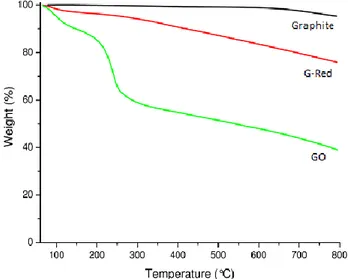
TGA analysis results (Fig. 2.5) agree well with Raman and XPS data. The graphite is stable up to 700 °C, unlike G-Red and GO. The latter shows an overall important weight loss of 51% up to 600 °C, due to the degradation of oxygenated groups, which occur significantly at about ~ 200°C. At variance, G-Red being poor of oxygenated groups shows a weight loss of only 15% up to 600 °C.
31
Figure 2.4 C 1s X-ray photoelectron spectra of:(a) graphite; (b) graphene oxide (GO); (c) reduced graphene
(G-Red).
Table 2.1 Relative percentage of functional groups obtained by fitting the C 1s XPS spectra.
32
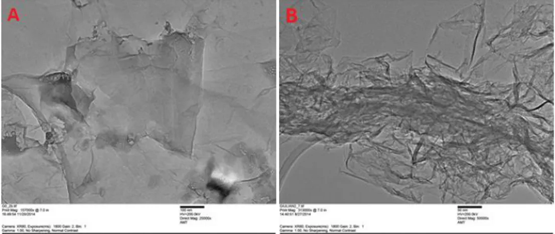
The morphology of GO and G-Red was investigated by TEM analysis. (Fig. 2.6). The layers were estimated to be ~100 nm large and ~1-2 nm thick. The sheets of GO appear partially folded, due to the large area occupied, while the G-Red ones appear homogeneous and quite smooth with little aggregation.
Figure 2.6 TEM images of graphene based materials: GO (A), G-Red (B).
To prevent the aggregation of G sheets, G-Red was treated with diazonium compounds to graft the p-(2-propylnyloxy)-benzene moieties, on the G surfaces. The reaction of G-Red with p-(2-propynyloxy)-benzamine (5), sodium nitrite and hydrochloric acid was performed at 0 °C, under continuous sonication (Scheme 2.7). The functionalization of G-Red surfaces gave alkyne-terminated graphene materials (G-Alk). By TGA analysis it was estimated that G-Alk contains 0.8 mmol/g of alkyne moieties and, as expected, it showed a better dispersibility in water with respect to G-Red (Scheme. 2.7).
33
Scheme 2.7 Synthesis of G-Alk and photograph of G-Red and G-Alk dispersed in water (2 mg/mL) three days after preparation.
2.3 Silibinin-Conjugated Graphene Nanoplatform
Surface modifications of G materials with natural biomolecules provide them specific biological activity and improve their biocompatibility. In order to prepare a biocompatible drug delivery system based on carbon nanomaterials, the interest has been focused on the decoration of G-Alk surface with Silibinin (Sil), a natural flavonoid endowed with several interesting biological properties.(81)
Sil (Fig. 2.8), is the major active constituent of Silymarin, a standardized extract of the milk thistle seeds of Silybum marianum employed mainly as dietary supplements to treat a wide range of aliments related to vital organs such as liver and kidney. Sil exhibits strong antioxidant activity through scavenging hydroxyl radicals. Recently, it has been extensively studied as hepatoprotective and anti-cancer agent. Studies on anti-cancer prevention, using various cell lines, have demonstrated that Sil has a strong inhibitory effect on tumor cell survival and proliferation, efficiently attenuating tumor induced angiogenic response. Currently, Sil is in phase II clinical trials in patients with prostate cancer.(82)
However, the use of Sil in the preparation of pharmaceutical products is restricted due to its low solubility in both hydrophilic and lipophilic medium, which greatly reduces its bioavailability and resorbability after oral administration.
34
Figure 2.8 Silibinin (Sil) structure.
Therefore, several nanotechnological approaches have been proposed to overcome this issue.(81, 83) Innovative aspects of the drug design include the synthesis of
hybrid derivatives (obtained by molecular hybridization or bioconjugation) with improved affinity and efficacy with respect to the parent drugs and the multiple combination strategy involving the co-delivery of agents that synergistically and simultaneously affect different pathways.(84, 85)
Concerning the first design strategy, the development of salinomycin-silibinin hybrid derivatives has been investigated and no superior efficacy compared with the single components has been detected (85), whereas the different combinations of Sil with other anticancer drugs induced relevant synergistic effects.(86, 87)
In this chapter it is described the synthesis of a G-Sil bioconjugate and its characterization by micro-Raman spectroscopy, scanning transmission electron microscopy (STEM), thermogravimetric analysis (TGA), Fourier transform infrared spectroscopy (FT-IR) and UV-Vis spectroscopy. The cytotoxicity of G-Alk and the anti-cancer effects of G-Sil were evaluated in vitro on human mesenchymal stem cells (MSCs) and a human osteosarcoma cell line (MG63), respectively.
Flavonoid Sil was modified with an azide functional group to obtain the reactive fragment for conjugation with G-Alk surfaces (approach a). The selective
35
esterification at 23-OH group was performed according to literature protocol.(83)
The reaction of 4-(azidomethyl)benzoyl chloride (2) and Sil, in presence of (BF3·Et2O) in acetonitrile/dichloromethane gave the azido-silibinin (Sil-N3), as
main product in 48% yield (Scheme 2.9).
The product was purified by flash chromatography and the structure was confirmed with the support of analytical and spectroscopic data.
The click chemistry reaction of G-Alk with Sil-N3, using CuSO4/Na-ascorbate as
metal source, gave the G-Sil bioconjugate (Scheme 2.10).
Scheme 2.9 Synthesis of Azido-Silibinin (SilN3).
36
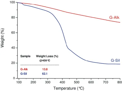
Unambiguous evidence of Sil conjugation on G-Alk sheets was provided via thermal gravimetrical analyses (Fig. 2.11). The TGA profile of G-Alk showed a constant mass loss up to 800 °C. A different behavior can be observed in the G-Sil curve, which consists of a two-step weight loss profile, corresponding to: i) first step (4.8% weight loss <160 °C), removal of solvent and decarboxylation of residue carboxylic acids; ii) second step (68.0% weight loss, range 280-600 °C), decomposition of flavonoid moieties linked by triazole fragments. The curve trend above 600 °C suggests a thermal stability of residual material, due to the complete degradation of grafted organic moieties. The high efficiency of the clicking step provides a wide surface loading in the “clicked” Sil that causes the relevant weight loss detected in the TGA analysis. From TGA data (Fig. 2.11) the amount of Sil grafted on G-Alk surfaces is responsible of the weight loss up to 450 °C, corresponding to ∼0.82 mmol/g. This value suggested an exclusive linkage of Sil to G surface by triazole moieties with a quantitative yield in the click reaction (the estimated amount of grafted alkyne moiety on G-Alk was ∼0.8 mmol/g).
The grafting of Sil to G-Alk surface has been also confirmed by FT-IR and UV-Vis spectroscopy (Fig. 2.12 and Fig. 2.13). The FT-IR spectrum of G-Alk showed the peak at 1641 cm−1, typical of C=C stretching of the G skeleton (88) and the signal of O-H stretching vibrations observed at about 3414 cm-1, due to residual OH and COOH functionalities remaining after hydrazine reduction. The expected weak band at about 2100 cm-1 of alkyne moieties is not recognizable. The spectrum of pure Sil showed a strong and broad band, due to -OH stretching vibration at 3452 cm−1 and peaks at 1636 cm-1(C=O stretching) and at 1535 cm-1 (C=C skeleton vibration of aromatic ring stretching). The FT-IR bands of Sil units resulted clearly observable in the G-Sil nanohybrid, with some absorption bands slightly shifted from the values of pure Sil, probably due to interactions with G nanosheets (formation of hydrogen bonds, conjugation processes, etc.).
37
Figure 2.11 TGA profiles of G-Alk and G-Sil under N2 atmosphere. The table reports the values of weight loss
(%) at 450 °C.
38
The UV–Vis spectrum of pure Sil displayed two characteristic bands: one centred at 286 nm (Band II) and another, less intense (Band I), with a maximum at 324 nm (Fig. 2.13). (89) The Band II of G-Sil bioconjugate is remarkably wider than free Sil and slightly shifted towards longer wavelength. The flattening of this peculiar band is plausibly due to the non covalent interactions of the aromatic rings of Sil (those ones far from the conjugation sites) with the adjacent graphene nanosheet.
The UV–vis spectrum (λ ≥230 nm) of G-Alk in DMF/H2O showed the characteristic
pattern of reduced-graphene based materials, while this feature is hidden by Sil band in the G-Sil spectrum. Moreover, self-recognition phenomena between Sil rings cannot be excluded.
The modifications of G material, from G-Alk to G-Sil, is also confirmed by Raman spectroscopy analysis. In the Raman spectrum of G-Alk sample, G and D bands, assigned to the in-plane E2g zone-center mode and disorder-induced modes, are
centred at 1580 cm-1 and 1332 cm-1, respectively (Fig. 2.14). In G-Sil nanohybrid, D and G bands peak intensity is much weaker and slightly shifted towards lower frequency compared to that of G-Alk (Fig. 2.14). The attenuation of characteristic peaks suggested a conjugation process, due to the interactions between the G-Alk platform and Sil. On the other, no changes due to Sil-grafting was observed in 2D band (not shown), which presents the characteristic features of a multilayer G-Alk. The morphology of functionalized G materials was investigated by STEM analyses. STEM images of G-Alk showed a large number of homogeneous and quite smooth sheets, overlapping each other (Fig. 2.15 A). On the other hand, the presence of amorphous materials is evident in the G-Sil sample (Fig. 2.15 B) and the G sheets appear mainly bundled together
39
Figure 2.13 UV-Vis spectra of Sil, G-Alk and G-Sil. Normalized spectrum of Sil ([Sil]= 400 µM) is 25 times more intense with respect to the reported trace.
40
Figure 2.15 STEM images of G-Alk (A) and G-Sil (B).
Preliminary in vitro studies on G-Alk and G-Sil nanoplatforms were performed in order to evaluate the cytotoxicity of G materials as well as cell-based assays to test the anti-cancer effect of the Sil bioconjugate (G-Sil). G-Alk evaluation showed the total absence of cytotoxicity towards both mesenchymal stem cells (MSCs) and human MG63 osteosarcoma cell line.
For all the tested concentrations, no statistical differences have been recorded between G-Alk and the group of cells alone (Fig. 2.16). No apoptotic cells were detected after 48h of incubation of the cultures with G-Alk (Fig. 2.16). The fact that G-Alk, even at the high concentration of 1000 µg/ml, did not affect the viability of the cells, especially that of MSCs, highlights the cytocompatibility of this material, providing new perspectives for its use in biomedical applications. In order to estimate the likelihood of using this nanoplatform for the preparation of a drug delivery system, the effects of G-Sil on MSCs and MG63 cells were tested in vitro to find out if Sil, bound to the G-Alk platform, maintained its anticancer properties. It is well known that Sil exhibits a strong inhibitory effect on tumor cell survival and proliferation, and efficiently attenuates tumor invasion.(90) Important anticancer effects have been also detected against osteosarcoma, the most common primary malignant tumor of bone.(91) Two different concentrations of Sil, as free compound
41
and as bioconjugate with G materials, were tested. The results showed that 100 µg/ml of free Sil feebly reduces the cell viability of MG63 and MSCs, at variance with that of the cells alone (Fig. 2.17).
Interestingly, Sil maintains its inhibitory effect against MG63 cells also after conjugation with G-Alk and it did not affect the viability of MSCs. It is possible to hypothesize a retarded uptake process, cell type-dependent, which could reduce the G-Sil cytotoxicity on MSCs with respect to free Sil. A similar behaviour was reported for Sil-loaded lipid nanoparticles.(92)
These results are a very important starting point to put down the basis for future characterization of these G based materials in more complex biological systems. Further investigations and exploitations are needed to evaluate the potential of the G-Sil nanohybrid as co-delivery system in the combined anticancer therapy, which nowadays remains the most challenging task to deal with in the field of the nano-oncology.
Figure 2.16 G-Alk cytotoxicity assay. The graphs showed totally absence of G-Alk toxicity on MG63 (A) and MSCs (D) cells after 48h of culture assessed by MTT test. No statistical differences exist at all the tested
G-Alk concentrations compared to cells only. Noapoptotic MG63 cells (B) and MSCs (E) were observed after
the treatment with 100 µg/ml of G-Alk at 48h. Dead MG63 cells (C) and MSCs (F) were observed in positive control. Cells nuclei in blue, apoptotic cells in green (*), nuclear localization of Dox in red. Scale bars: 50 µm.
42
Figure 2.17 Cell viability assay. MG63 cells (A) and MSCs (B) viability assessed by MTT assay after 48h of culture with different concentrations of free Sil and G-Sil bioconjugate.
2.4 Reactive Azlactone Graphene Platform (RAGP)
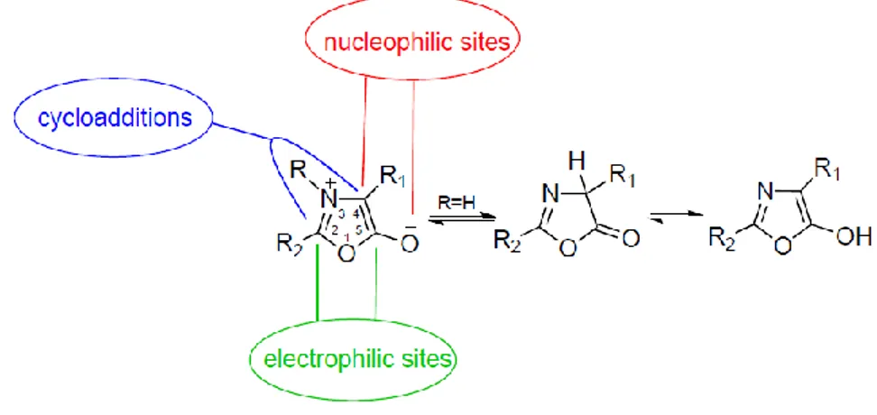
Azlactones (oxazol-5-(4H)-ones) are multifunctional compounds that contain numerous reactive sites (the electrophilic centres at C2 and C5; the nucleophilic site at C4; the azomethine ylide 1,3-dipole), allowing a wide variety of transformations (Fig. 2.18). Recently, they were employed as mesoionic azomethine ylides in the functionalization of carbon nanomaterials such as fullerenes and carbon nanotubes.(93, 94)
The 4,4-dimethyl-5-oxazolone was studied in industry for many years by groups at 3M, Polaroid, Rohm and Haas GmbH; recently, 4,4-disubstituted azlactones have been used as a way to modify polymer surfaces.(95-97) In presence of two
43
substituents at the C4 position, the reactivity as a nucleophile or azomethine ylide is forbidden and the lactone-based functional group displays only high reactivity toward nucleophilic agents, such as primary amines, alcohols and thiols by means of a ring-opening addition reaction.(95-98)
Figure 2.18 Representative scheme of reactive sites of oxazolone
Along my research activity, particular attention was addressed on the development of an efficient and versatile approach to introduce a pendant oxazolone functionality on G-Alk surfaces (Approach b, Fig. 2.1). The integration of the azlactone functionality into G is appealing because, no activation or pre-treatment is necessary for the post-functionalization of G based materials, since they are more stable to hydrolysis than activated esters, and no side reactions are expected during functionalization. Furthermore, it is envisaged that the range of applications for which azlactone-functionalized G materials can be tailored is both broad and diverse. It could include the design of drug delivery systems, or the fabrication of supports for the immobilization of biomolecules and direct implications for the development of novel biosensors, in the context of nanoscale detection of pathogens and other metabolites related to human health issues. Moreover, the reactive
44
azlactone G platform could be suited for the deposition of G films on the surfaces of amine functionalized polymers.
The reactive azlactone graphene platform (RAGP) was built by click scaffold assembly of an alkyne and an azide fragments. The terminal alkyne moiety was grafted on graphene surfaces (G-Alk), while the azide group was introduced in the azlactone fragment (APDMA). The azlactone fragment APDMA was prepared by sequential acylation–cyclodehydration involving a two-step procedure (Scheme 2.19). The first step is a Shoeeten-Baumman reaction, between the
4-(azidomethyl)benzoyl chloride and the 2-amino-2-methylpropanoic acid. The
intermediate was subjected to a ciclodehydration reaction, induced by ethyl chloroformate, to obtain the oxazolane fragment (APDMA). The structure of APDMA was supported by 1HNMR, 13CNMR and FT-IR analysis; in particular the FT-IR spectrum of APDMA showed the strong stretching bands at 2097 cm-1 (v N=N=N), 1818 cm-1 (v C=O), 1652 cm-1 (v C=N) and 1200 cm-1 (v C–O–C) (Fig. 2.20), characteristics of oxazolone structure.
Scheme 2.19 Synthesis of azido-azalactone (APDMA).
Figure 2.20 FT-IR spectrum of APDMA.
45
The click chemistry reaction between APDMA and G-Alk, using CuSO4
/Na-ascorbate as the metal source (99), led to the reactive azlactone graphene platform
(RAGP) (Scheme 2.21).
Scheme 2.21 Synthesis of reactive azalactone graphene nanoplatform (RAPG).
Once the appropriate RAGP structure was obtained, it was important to quantify the ring opening efficiency of azlactone, in order to proceed with further functionalizations of the nanoplatform. A study of the experimental conditions to optimize the efficiency of the ring-opening reaction was carried out by treating, in toluene without catalysts, one equivalent of APDMA with one equivalent of
aminopropyltriethoxysilane (APTS), chosen to act as a nucleophilic agent (Scheme
2.22). FT-IR and 1H NMR analyses of the crude reaction mixture confirmed the
presence of the coupling product, without observable side reactions (Fig. 2.23). The downfield shift of resonance of amino methylene protons (peak c, Fig. 2.23) from 2.45 to 3.31 ppm, the disappearance of characteristic azalactone absorption at 1818 cm-1 and the presence of the carbonyl amide group signal at 1649 cm-1 (Fig. 2.23) indicate the formation of the coupling compound with 100% atom efficiency. Hence, the same procedure was applied for the post functionalization of RAPG using APTS and GSSG (glutathione oxidized), (Scheme 2.24).
46
Figure 2.23 1H NMR of APTS and crude of ring opening reaction. The inset shows the IR spectrum of APDMA
and the crude of ring opening reaction.
47
Scheme 2.24 Schematic illustration of ring opening reactions on RAGP with primary amines to give graphene functionalized materials (GF-APTS and GF-GSSG).
The pristine and functionalized graphene platforms were characterized by TGA, TEM, XPS, FT-IR and Raman spectroscopy.
The thermal gravimetric analyses (TGA) provides a clear evidence of the grafting of organic groups. In fact as reported previously, G-Red shows much higher thermal stability with a constant mass loss up to 800 °C, the TGA curve of G-Alk shows a minor thermal stability, with no significant change in profile (Fig. 2.25). A different behavior can be observed in the RAGP, GF-APTS and GF-GSSG profiles, where the rapid weight loss at about 500 °C can be attributed to the decomposition of the organic component linked by the azlactone fragment.
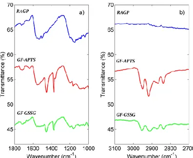
The presence of functional groups on the G surface is confirmed by FT-IR spectra. In Fig. 2.26 are shown selected region of RAPG, GF-APTS and GF-GSSG FT-IR spectra. The C=C stretching band at 1590 cm-1of G is present in all the samples. The signal of C=O amide group at about 1630 cm-1, is indicative of the coupling of amine
48
groups with azlactone moiety. Both in GF-APTS and GF-GSSG spectra the strong bands, located approximately at 2800 and 2900 cm-1 corresponding to sp3 C-H
stretching, are present.
Figure 2.25 TGA profiles of G-Red, G-Alk, RAGP, GF-GSSG, and GF-APTS, under a N2 atmosphere.
The table reports the values, at 500 1C, of weight residue (%) and loading(%).
49
In Raman spectra (Fig. 2.27), since functionalization causes an increase of the structural disorder in the graphitic lattice (e.g. the sp2-defect), the I
G/ID ratio
decreases from 1.08 in the G-Alk sample down to 0.61 in the GF-APTS one. In the GF-APTS and GF-GSSG samples, the D- and G-bands bands slightly shift towards higher wavenumbers (from 1312 cm−1 and 1580 cm−1 in RAPG sample up to 1332-1341 cm−1 and 1588-1608 cm−1 in the GF-APTS and GF-GSSG ones, respectively) as an effect of the electron transfer from graphitic lattice to oxygenated moieties, as confirmed by FT-IR and XPS analyses. Thus, the typology of oxygenated functionalities have a significant impact on the wavenumber position of D and G bands. The addition of APTS and GSSG reflect also on the width (FWHM) of the Raman bands. FWHM slightly decreases, hinting a shrinkage of the distributions of the angles and lengths of C bonds as well as of the strains.
50
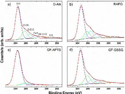
In XPS spectra (Fig. 2.28) the C1s bands were deconvolved using six contributions: a main contribution at 284.5 eV attributed to C=C/C–C in the aromatic ring and four other contributions at higher binding energies corresponding to carbon atoms bonded to oxygen in different surface functionalities (C–OH, C–O–C, C=O and OH–C=O) centered at 285.8, 286.6, 287.7, and 288.9 eV, respectively. The contribution at about 291.0 eV refers to π–π bonds.(100-102) As expected, the percentages corresponding to C–C and carbon oxidized contributions are almost unchanged from G-Alk to RAGP sample (Table 2.2). On the other hand, the addition of APTS induces an evident decrease in the degree of surface carbon oxidation (Tab. 2.2). This behavior is directly related to the symmetrical and shrinked Si 2p and O 1s lineshape features. Particularly, the position of the Si 2p band at 102.0 eV is indicative of silicon–oxygen bonds in silanes (Fig. 2.29 c, Table 2.3).(103)
The GF-GSSG sample is characterized by the presence of S–S bonds at 164.0 eV and, to a lower extent, by oxidized sulphur bonds at 168.0 eV (Fig. 2.30 d). The GF-GSSG and RAGP samples show only slight differences in terms of carbon-oxidized phases. Finally, the N 1s lineshapes showed a similar profile in all the samples, but its intensity decreases in the functionalized samples (GF-APTS and GF-GSSG) (Fig. 2.29 b, Table 2.3).
The morphology of based and functionalized graphene materials was investigated by TEM analyses (Fig. 2.30). The TEM micrograph of GO showed partially folded layers, while the G-Red and G-Alk ones showed the presence of quite homogeneous and smooth sheets, with little aggregation. The TEM images of RAGP clearly show a planar backbone conformation and transparency with ripple lines between the layers. The presence of well-defined edges revealed a large number of thin sheets of G. In the amine functionalized G materials there are very
51
noticeable layers of amorphous materials due to the APTES or GSSG residue bound on both surfaces of G sheets.
Elemental mapping information were obtained by STEM analyses (Fig. 2.30). STEM analyses are in good agreement with XPS results which show that the graphene nanomaterials have different C, O, Si and S contents, as expected on the basis of the chosen functionalization (Fig. 2.30). Three elements as carbon, oxygen and nitrogen are dispersed in the samples. The content of these three elements is similar in RAGP and GF-APTS, respectively, whereas it is reduced in GF-GSSG. The lower amount of peculiar elements as S in GF-GSSG, respect to Si in GF- APTS, suggested an additional grafting of APTS on the G surfaces, probably on residual phenolic and carbonyl groups.
Figure 2.28 C 1s bands of XPS spectra of G-Alk (a), RAGP (b), GF-APTS (c), and GF-GSSG (d).
52
Figure 2.29 O 1s (a), N 1s (b) bands of XPS of, G-Alk, RAGP, GF-APTS , and GF-GSSG. Si 2p (c) and S 2p (d)XPS bands of GF-APTS and GF-GSSG respectively.
Table 2.3 Atomic content percentage and bonding fraction percentage by XPS analysis.
Figure 2.30 TEM (A–C) and STEM (D, F, H) images with linescans and corresponding total element percentages, detected by peculiar emission lines (for colours see ESI,† Fig. S9–S11). (A, D) RAGP; (B, F)
GF-APTS; (C, H) GF-GSSG. Inset of (A–C) SAED patterns of each sample area; (E) % of O in RAGP; (G) % of Si in GF-APTS; (I) % of S in GF-GSSG.
53
From TGA, XPS and STEM analyses the amounts of APTS and GSSG grafted onto G surfaces were estimated to be ∼1 mmol /g and ∼0.3 mmol /g, respectively. These values suggested an exclusive linkage of GSSG to G surfaces by the azlactone moiety in a 1:1 molar ratio, because the estimated amount of the linked oxazolone moiety on RAGP was ∼0.3 mmol/g; for APTS an additional grafting occurred.(104)
Probably, based on experimental results and according to literature data (105), additional reactions between the silyl group of APTS and the phenolic and carboxylic groups occurred, when RAGP was treated with APTS. The structure proposed for GF-APTS product has been reported in Fig. 2.31.
In the case of functionalization with GSSG the residue carboxylic groups cannot react with amino groups of GSSG without a coupling reagent.
In conclusion, it has been demonstrate that azlactone moieties grafted by the click chemistry reaction on reduced functionalized G might be useful as a reactive platform for the rapid engineering of G based materials with functionalized primary amines. The reactive platform shows a high yield of silane grafting with respect to graphene silylation methods provided in the literature. Moreover, it can be employed for the selective conjugation of compounds of pharmaceutical and biological interest such as GSSG.